×

Regional variations in ambulatory care and incidence of cardiovascular events [Research]

BACKGROUND:

Variations in the prevalence of traditional cardiac risk factors only partially account for geographic variations in the incidence of cardiovascular disease. We examined the extent to which preventive ambulatory health care services contribute to geographic variations in cardiovascular event rates.

METHODS:

We conducted a cohort study involving 5.5 million patients aged 40 to 79 years in Ontario, Canada, with no hospital stays for cardiovascular disease as of January 2008, through linkage of multiple population-based health databases. The primary outcome was the occurrence of a major cardiovascular event (myocardial infarction, stroke or cardiovascular-related death) over the following 5 years. We compared patient demographics, cardiac risk factors and ambulatory health care services across the province’s 14 health service regions, known as Local Health Integration Networks (LHINs), and evaluated the contribution of these variables to regional variations in cardiovascular event rates.

RESULTS:

Cardiovascular event rates across LHINs varied from 3.2 to 5.7 events per 1000 person-years. Compared with residents of high-rate LHINs, those of low-rate health regions received physician services more often (e.g., 4.2 v. 3.5 mean annual family physician visits, p value for LHIN-level trend = 0.01) and were screened for risk factors more often. Low-rate LHINs were also more likely to achieve treatment targets for hypercholes-terolemia (51.8% v. 49.6% of patients, p = 0.03) and controlled hypertension (67.4% v. 53.3%, p = 0.04). Differences in patient and health system factors accounted for 74.5% of the variation in events between LHINs, of which 15.5% was attributable to health system factors alone.

INTERPRETATION:

Preventive ambulatory health care services were provided more frequently in health regions with lower cardiovascular event rates. Health system interventions to improve equitable access to preventive care might improve cardiovascular outcomes.

[Seminar] Hypertrophic obstructive cardiomyopathy

Hypertrophic obstructive cardiomyopathy is an inherited myocardial disease defined by cardiac hypertrophy (wall thickness ≥15 mm) that is not explained by abnormal loading conditions, and left ventricular obstruction greater than or equal to 30 mm Hg. Typical symptoms include dyspnoea, chest pain, palpitations, and syncope. The diagnosis is usually suspected on clinical examination and confirmed by imaging. Some patients are at increased risk of sudden cardiac death, heart failure, and atrial fibrillation.

Have You Planned Your Heart Attack?

BOOK REVIEW

Have You Planned Your Heart Attack?

By Dr Warrick Bishop

RRP: $34.99

Reviewed by Chris Johnson


Anyone for a self-help guide to having a heart attack?

Actually, a new book by Hobart-based cardiologist Dr Warrick Bishop is all about discovering what you might need to understand in order to reduce your risk of having one.

Have You Planned Your Heart Attack? is an engaging read, full of cases studies, graphics and easy-flowing chapters explaining the advances in technology that make it possible to determine risk and make informed preventive decisions.

Bishop’s enthusiasm for CT imaging is evident throughout.

“Taking a picture of the coronary arteries using CT to determine their health isn’t new, it just isn’t being done routinely,” he says.

“Yet, by using these technological advances you can be ahead of the game about your cardiovascular health.

“Wouldn’t you want to know if the single biggest killer in the Western world was lurking inside of you?”

According to the Australian Heart Foundation, 55,000 Australians suffer a heart attack each year.

This self-published book poses the questions: But what if we could be forewarned or prepared for a potential problem with our own arteries? What if we were able to put in place preventive measures that may avert a problem?

The book is all about preventive care and is very much directed at patients – the kind of book a doctor might suggest or recommend a patient reads.

While not bogged down in jargon or technical explanations, it is also substantial enough for doctors to gain further insights into cardiovascular disease and particularly cardiac CT imaging.

Bishop is a practising cardiologist with an interest in cardiovascular disease prevention – with a special interest in cardiac CT imaging, lipid management and eating guidelines.

The introduction to the book aptly sets the scene for what follows, with the author describing how he helped resuscitate a 52-year-old man who had collapsed with cardiac arrest on a fun run in 2005.

The man survived and the outcome was so good it made the front page of the local newspaper.

“When I arrived at work on the Monday I felt fairly pleased to have been a contributor to such a positive outcome,” he writes.

“Before I could become too proud, however, one of my staff pointed out that I had seen the very same gentleman two years earlier for an exercise treadmill test.

“The test had been normal and I had reassured him that ‘everything’s okay’. This revelation shocked me!

“Had I done the wrong thing by this man? Had I misinterpreted the test? Were there other factors of which I had not been aware?

“As it turned out, I had done nothing wrong … My original assessment in 2003 had limitations. This book is about how, with today’s technology, we can do better – potentially much better.

“It is about improved dealing with risk through investigation and management.”

High-profile television journalist Charles Wooley, who reveals he is a patient of Dr Bishop, writes an eloquent foreword to the book.

“Warrick Bishop is a lean and determined-looking man whose shaven head and athletic fitness bring to mind Vladimir Putin, without the unhappy associations,” Wooley writes.

“Indeed, what drew me to Dr Bishop was that he specialises in looking inside the working heart. Using non-invasive imaging technology, he sees inside our coronary arteries to determine just how encrusted the pipes have become.

“For you and me, Warrick Bishop’s picture is worth a thousand words.”


Painkillers can increase the risk of heart disease and should be restricted

Medications such as ibuprofen and aspirin, known as non-steroidal anti-inflammatory drugs or NSAIDs, are widely available over the counter from pharmacies and supermarkets. But health providers have known for some time they can be unsafe for people with chronic health problems such as kidney disease, high blood pressure or heart failure. The Conversation

NSAIDs can also have dangerous interactions with other commonly taken medications, notably many types of blood pressure and blood-thinning pills such as warfarin and aspirin.

Two recently published studies have brought back into the spotlight the possible heart-related side effects of NSAIDs. One found an increased risk of heart failure in users of NSAIDs, while another an increased risk of cardiac arrest.

Heart failure is a disease that presents with symptoms such as shortness of breath, fluid retention, leg swelling, and fatigue. This is a result of the heart not being able to pump blood around the body effectively. There are many causes of heart failure, including heart attacks, high blood pressure and excessive alcohol consumption.

A cardiac arrest occurs when the heart stops functioning abruptly and results in complete loss of effective blood flow through the body. The most common cause of a cardiac arrest is a heart attack, where heart muscle is damaged from loss of blood supply due to a blockage in a heart blood vessel. There are many other causes of a cardiac arrest that include structural heart abnormalities and inherited heart diseases of muscle and electrical function.

Heart failure is when the heart isn’t able to pump blood around the body effectively.
from shutterstock.com

The recent studies are an important reminder that over-the-counter medicines are not without risk. This class of anti-inflammatory pain killers should no longer be available for sale in grocery stores, but instead restricted to prescription-only or behind-the-counter status in pharmacies.

How they work

Non-steroidal anti-inflammatory drugs are commonly used to relieve pain. They can be either prescribed by a doctor or purchased by the patient over the counter from a supermarket or pharmacy.

NSAIDs are used in a broad range of health conditions associated with pain and inflammation, including types of arthritis, headaches, musculoskeletal injuries and menstrual cramps. Their easy availability, effectiveness and presumption of safety contribute to their widespread use.

They work by inhibiting enzymes called cyclooxygenase 1 (COX-1) and 2 (COX-2). These are involved in a number of internal pathways that result in production of hormone-like substances called prostaglandins, which promote inflammation and increase pain perception.

Prostaglandins also protect the stomach lining from acid, by decreasing acid production and increasing mucus secretion and its neutralising properties. So inhibiting prostaglandins also reduces their protective functions. This is why frequent users of anti-inflammatories may suffer from gastric ulcers.

NSAIDs can either inhibit both COX-1 and COX-2 (non selective) or inhibit COX-2 only (selective). Drugs like ibuprofen and aspirin are non selective and inhibit both the COX enzymes.

COX-1 mediates gastrointestinal, kidney, and clotting function, while COX-2 is induced primarily in states of inflammation and tissue repair. That’s why blocking the COX-2 pathway reduces the effects of inflammation such as fever, swelling, redness and pain.

Importantly, COX-2 inhibition accounts for the anti-inflammatory drug effects of NSAIDs, while COX-1 inhibition can lead to side effects including gastrointestinal ulcers, prolonged bleeding and impaired kidney function. However, it’s not entirely safe for the drugs to inhibit COX-2 only.

Nurofen (ibuprofen) works by inhibiting both COX enzymes.
from shutterstock.com

Animal studies have shown blocking COX-2 and the subsequent pathway of prostaglandin production may have the unwanted effects of increasing the tendency of blood to clot inside arteries, and a reduced ability of the heart to heal after a heart attack.

In the early 2000s, a number of large studies found a significant association of negative heart events, such as heart attack and stroke, with the use of selective COX-2 inhibitors. This resulted in two of these drugs, Valdecoxib and Rofecoxib or Vioxx, being withdrawn from the market.

In Australia there are only a small number of COX-2 inhibitors available, including Celecoxib and Meloxicam. These are prescription-only medicines and the maximum prescribed dose is at a level at which the heart risks are minimal.

COX-2 inhibitors are used in people who require a non-steroidal anti-inflammatory but have a history of stomach upset or ulcers, or who were thought to be at risk of developing stomach ulcers.

Risk of heart failure

Non-steroidal anti-inflammatory drugs are associated with elevating blood pressure as well as sodium and fluid retention. Both of these effects may unmask previously undiagnosed heart failure, or worsen the symptoms in people known to already have heart failure.

Vioxx was a selective inhibitor and take off the market for its adverse effects on the heart.
Wikimedia Commons

Research published in the British Medical Journal in September 2016 studied 92,163 people admitted to hospital with heart failure, and found NSAID use in the two weeks prior to admission was associated with a 19% increased risk of hospital admission for heart failure. This was compared with people who had not used NSAIDs prior to admission.

The association of NSAIDs with an exacerbation of heart failure was also seen in many older studies. For example, an Australian study in 2000, suggested almost 20% of all heart failure related admissions to hospital may be attributed to recent NSAID use.

Risk of cardiac arrest

Further heart safety concerns with NSAIDs were raised in a recent study from the University of Copenhagen, published in the European Heart Journal.

Data was collected from nearly 30,000 patients who had suffered cardiac arrest between 2001 and 2010. Of these, around 3,500 were found to have been treated with an NSAID within 30 days of having a cardiac arrest.

Use of any NSAID was associated with a 31% increased risk of cardiac arrest. The commonly used non-selective NSAIDs, diclonenac (Voltaren) and ibuprofen were associated with a 50% and 31% increased risk respectively.

A large proportion of cardiac arrest is a result of clot formation in the arteries of the heart and underlying plaque formation which can rupture. NSAIDs may increase the risk of cardiac arrest by raising blood pressure, forming blood clots and blocking the heart’s own blood vessels.

It is important to emphasise that in people with no known heart disease and who don’t have any heart risk factors, short term use of these anti-inflammatories carries a minimal increase in heart-related risk.

These recent studies should not create community panic about the safety of NSAIDs when used for short periods of time and at low dosage.

But the high burden of heart disease and heart disease risk factors, such as high blood pressure, obesity and diabetes (which are often unrecognised), warrant a personalised approach to NSAIDs, which weighs the benefits and risks of their use.

This was recommended in the Therapeutic Goods Administration review of the heart related effects of NSAIDs in 2014. These anti-inflammatories should be available for purchase through prescription by a medical practitioner or behind the counter at the pharmacy.

Michael Stokes, Cardiologist and PhD Candidate, University of Adelaide and Peter Psaltis, Co-director, Vascular Research Centre, South Australian Health & Medical Research Institute

This article was originally published on The Conversation. Read the original article.

Higher risk of heart attack with DOACs

People with atrial fibrillation are more prone to myocardial infarction if they’re on direct-acting oral anticoagulants rather than warfarin, a large UK study shows.

Two DOACs, dabigatran and rivaroxaban, more than doubled the risk of MI compared with warfarin in a population of over 30,000 people with an atrial fibrillation diagnosis and follow-up of up to nine years.

The retrospective study adjusted for a range of potential confounders, including sex, BMI, smoking and alcohol, and history of heart or liver disease along with medication to treat these conditions.

The researchers from the University of Southampton and Maastricht University in the Netherlands also found higher MI risks with aspirin compared with warfarin.

Aspirin has been widely used for low-risk patients with atrial fibrillation, although the latest guidelines no longer recommend it.

Related: Spotlight on anticoagulant antidotes

The study, the first to focus on MI risks with DOACs and warfarin, looks set to reignite a debate around the issue, particularly with dabigatran.

The higher risk of MI with dabigatran was first noticed in the original RE-LY trial that led to the drug’s approval. In that trial, dabigatran users had a 38% higher risk of MI than warfarin, although the numbers were too small to be significant.

However, a meta-analysis of RE-LY and six other randomised trials of dabigatran continued to show an increased risk compared with warfarin, with or without the RE-LY data.

The authors of the current study say the differences in MI risk are probably due to the protective effects of warfarin in the pathology of angina pectoris, rather than DOACs actually increasing adding to the risk.

They say the protective effects of warfarin in MI is relatively well known, but the current findings argue strongly against them being a class effect of anticoagulants in general.

“More research should be ongoing as use of direct acting oral anticoagulants increases in the population,” they write in the British Journal of Clinical Pharmacology.

Read the study here.

Latest news


[Correspondence] Preventing delirium: beyond dexmedetomidine

The incidence of postoperative delirium in elderly patients is very high, particularly in patients admitted to the intensive care unit (82%) and those who have undergone major orthopaedic (51%) or cardiac (46%) surgery.1 Postoperative delirium is associated with increased morbidity, mortality, and health-care costs; however, no preventive pharmacological strategies are available.1 A Lancet study by Xian Su and colleagues (Oct 15, p 1893)2 offers new hope. Treatment with dexmedetomidine in elderly patients admitted to the intensive care unit after non-cardiac surgery reduced the incidence of delirium from 23% to 9%.

[Editorial] Polypills: an essential medicine for cardiovascular disease

Adoption of the notion of a polypill, or fixed-dose combination treatment, to prevent cardiovascular disease has been slow. In today’s Lancet, and ahead of the American College of Cardiology meeting in Washington, DC, USA (March 17–19), we publish a Series of articles that discusses evidence for the use of polypills in the prevention of cardiovascular disease, barriers to acceptance, and ongoing challenges to their widespread use.

Mycobacterium chimaera and cardiac surgery

Recent reports from the United Kingdom, Europe and the United States have described a small number of invasive infections with Mycobacterium chimaera associated with cardiac surgery that have been associated with high mortality. M. chimaera is likely to have been transmitted to patients by aerosols generated from contaminated heater–cooler units used during cardiopulmonary bypass. Here we describe the outbreak and discuss the relevance for Australian clinicians. Our primary objective is to raise awareness locally of this rare but serious health care-associated infection so that patients can be promptly diagnosed and optimally treated.

To formulate an evidence-based overview of the topic, as applied to clinical practice, we conducted a PubMed search of original papers and review articles in the period 2004–2016 using the term “Mycobacterium chimaera”, along with publications released by government agencies, public health bodies and medical device regulatory authorities, and selected conference presentations.

Initial outbreak description

In 2012, clinicians at the University Hospital Zurich, Switzerland, diagnosed two patients with ultimately fatal infections due to M. chimaera, a member of the M. avium complex (MAC) group of slow-growing non-tuberculous mycobacteria.1,2 The patients — one with prosthetic valve endocarditis and the other with bloodstream infection — had no known direct community or inpatient contact with each other, but had both undergone cardiac surgery with insertion of prosthetic material at the same hospital (in 2008 and 2010).1 Typing suggested that the strains from these two cases were genetically related and were, in addition, distinct from M. chimaera strains isolated from respiratory samples from other patients at the same hospital.

Detection of these two cases of an unusual and fatal infection prompted an outbreak investigation for a potential hospital source.3 Given the propensity of MAC organisms to be found in association with water, the investigation focused on points of contact between water and patients undergoing cardiac surgery. M. chimaera was isolated from the water circuit of five heater–cooler units (HCUs).3 These devices are used during cardiopulmonary bypass to regulate temperatures of extracorporeal blood and cardioplegia solution, and do not have direct contact with patients. When contaminated HCUs were in operation, air samples from the operating theatre itself and its exhaust air flow were also positive for M. chimaera. The authors hypothesised that aerosolised M. chimaera from contaminated HCUs was the source of infection. Subsequent experiments performed in a cardiac operating theatre with an ultraclean laminar airflow system confirmed aerosolisation of M. chimaera from a contaminated HCU and demonstrated smoke dispersal from an HCU to the operative field in 23 seconds when the unit’s airflow was directed towards the operating table.4

In parallel, a case detection exercise identified another four patients with invasive M. chimaera infection at the same hospital, with all six having undergone cardiac surgery with insertion of prosthetic material between August 2009 and March 2012. A nationwide investigation involving the 16 Swiss centres that perform cardiac surgery was mandated by the Federal Office of Public Health in 2014, and this identified eight contaminated HCUs but no further patient cases.5 Swiss authorities made a public announcement about the cluster in July 2014.6

International reports and response, including Australia

After the initial reports from Switzerland, invasive M. chimaera infections following cardiac surgery involving cardiopulmonary bypass have been reported in the Netherlands, Germany, the UK and the US.710 At least 50 cases have been diagnosed worldwide (Professor Hugo Sax, University Hospital Zurich, personal communication). Seventeen probable cases identified in the UK had undergone heart valve repair or replacement in ten National Health Service (NHS) trusts since 2007.9 A nationwide prospective case finding investigation, using a mandatory reporting system, performed in Germany from April 2015 until February 2016 identified five patient cases from three cardiac surgery centres.10 All five patients were exposed to HCUs manufactured by a single manufacturer. M. chimaera was also identified in samples from new machines and the environment at the manufacturing site.10 As a result of this investigation, German public health authorities triggered notifications of a suspected common source for the outbreak via the European Early Warning and Response System and under the World Health Organization International Health Regulations framework.10

Public Health England coordinated testing of 24 HCUs (LivaNova [formerly Sorin Group]); also the manufacturer of the devices used in Zurich) at five NHS trusts.9 M. chimaera was culturable from water sampled from 15 of the units, and from surrounding air for about a third of the devices while they were running.9 The manufacturer issued a field safety notice in June 2015 identifying the risk of contamination by bacteria and subsequent aerosolisation during operation (http://www.livanova.sorin.com/products/cardiac-surgery/perfusion/hlm/3t). The notice recommended enhanced disinfection with updated instructions for use including revised recommendations for operation, disinfection and microbial monitoring.

In October 2015, the US Food and Drug Administration (FDA) issued a Safety Communication to raise awareness of the issue of M. chimaera infections associated with HCUs and the Centers for Disease Control and Prevention issued guidance for health care facilities, health care professionals and patients.11,12 In the context of heightened surveillance, in November 2015 the Pennsylvania Department of Health reported clusters of patients with non-tuberculous mycobacterial infections among patients exposed to open heart surgery at two hospitals.13 Whole-genome sequencing of M. chimaera strains from 11 infected patients and five HCUs (Stöckert 3T [LivaNova]) from two US states (Pennsylvania and Iowa) confirmed their genetic relatedness, strongly suggesting “a point-source contamination of Stöckert 3T heater–cooler devices”.14

In Australia, the Therapeutic Goods Administration (TGA) announced an investigation into this issue and released recommendations to health facilities in May 2016.15 In August, the TGA reported one case of possible patient infection with M. chimaera following open cardiac surgery.16 In October, a TGA alert identified the affected product as the Stӧckert 3T HCU, and reported that in Australia “25% of these devices have tested positive for the Mycobacterium chimaera organism or other organisms”, but all positive machines were manufactured before September 2014.17 In keeping with a recent recommendation from the FDA,18 the TGA recommended that health services “consider transitioning away from” 3T devices manufactured before September 2014.17 Ten HCUs from four Western Australian hospitals (of 15 HCUs tested from five hospitals) were colonised by genetically related M. chimaera strains, along with other mycobacteria.19 The M. chimaera strain cultured from a pleural biopsy of a patient who had undergone surgery at one of these hospitals was considered distinct from the HCU strains, suggesting that the HCU was not the source of infection in this case.19 Subsequently, whole-genome sequence comparisons of 43 M. chimaera strains from HCUs in four Australian states and four regions of New Zealand confirmed close genetic relatedness of these strains with those from HCUs in the northern hemisphere.20 Whole-genome sequencing also showed that a patient isolate was identical to an isolate from an HCU used in the facility during the patient’s surgery.20

While there is no clear consensus on how to best manage HCUs to minimise risk, some recommendations have been released.9,12,14,21,22 The Australian Commission on Safety and Quality in Health Care (ACSQHC) released national infection control guidance in September 2016, outlining recommended risk mitigation strategies for Australian health service organisations.23 A simple precautionary step is to ensure that HCU ventilation airflow is directed away from the patient and towards the theatre exhaust whenever operational, although it is not clear whether this eliminates risk entirely.4 Other options, at least until M. chimaera contamination is excluded by microbiological testing, include physical removal of HCUs from the operating theatre (to an adjacent room) — as was nationally implemented in the Netherlands8 — or placement of the unit in a custom built sealed container that directs ventilation outflow out of the operating theatre, as described in Zurich.3 The TGA and ACSQHC note that the latter approaches may not be feasible in all facilities and should be planned in discussion with the Australian sponsor of the device to ensure it will not compromise device function.15,23

Clinical description of HCU-related M. chimaera infection

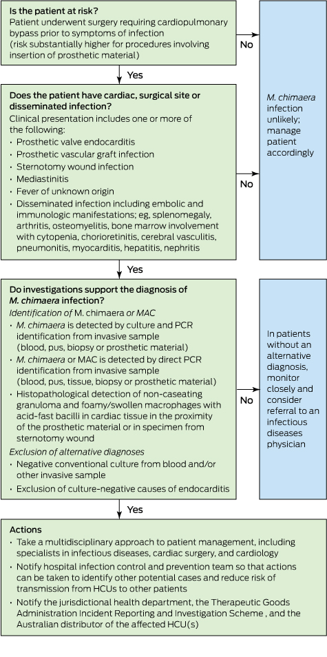
Although the risk of invasive M. chimaera infection following cardiac bypass surgery in Australia is estimated to be very small, the long incubation period and unusual non-specific clinical presentation mean that general practitioners and specialists need to know when to suspect this rare condition and how to diagnose it (Box).

The diagnosis in cases so far has been complicated by long delays between surgery and symptom onset, non-localising clinical presentations, and the need for directed microbiological testing using special tests. The following summary is compiled from existing published reports: six confirmed cases in Switzerland,1,3 which were subsequently reported in combination with three confirmed and one probable case from the Netherlands and Germany,7 five additional confirmed cases in Germany,10 and 17 probable cases in the UK.9

Although the risk of infection appears to relate principally to cardiac bypass procedures involving insertion of prosthetic material, there is one report of infection following coronary artery bypass grafts.10 HCUs are also used during extracorporeal membrane oxygenation, but no cases of infection have been attributed to this exposure.9 Establishing the risk of infection among exposed patients is difficult given incomplete data and the possibility that some cases have either been missed or remain in latent phase, but the risk is very low. Public Health England reported that about 100 000 patients underwent valve repair or replacement between 2007 and 2014, and estimated that the incidence rate of M. chimaera infections among these patients was 0.4 (95% CI, 0.2–0.7) per 10 000 person-years of post-operative follow-up.9

The period during which patients underwent initial cardiac surgery extends from 2007 to 2013.7,9 According to Haller and colleagues,10 the manufacturer involved in their investigation confirmed that HCUs delivered before mid-August 2014 may have been contaminated with M. chimaera. The latent period from cardiac surgery to diagnosis has ranged from 3 months to 5 years (median, 19 months among UK patients).9,10 Among the 15 European patients, median age was 63 (range, neonate to 80 years) and 14 were male.7,10 Patients have not, in general, been significantly immunocompromised.

Patients have presented with endocarditis or other cardiac infection, disseminated or non-cardiac infection, and surgical site infection.3,7,9 Fourteen of 15 European cases had cardiac infection: endocarditis, prosthetic valve infection, paravalvular abscess, graft infection and one case of myocarditis. In many cases, however, initial presentation was with non-cardiac disease: bone infection (osteoarthritis, spondylodiscitis), cholestatic hepatitis, nephritis or surgical site infection. Other manifestations included splenomegaly, ocular disease (panuveitis or chorioretinitis), and mycobacterial saphenous vein donor site infection.7 Of 13 probable cases in the UK with clinical description, six presented as cardiac infection, five as disseminated or non-cardiac infection, and two as surgical site infection. Several patients have been misdiagnosed with sarcoidosis or connective tissue disease, and commenced on corticosteroids or other immunosuppressive agents.1,7,24

Diagnosis of M. chimaera infection requires directed investigations. Public Health England has recommended that patients with endocarditis and/or disseminated infection should undergo mycobacterial culture and molecular testing (16S rRNA gene sequencing) of tissue samples and three sets of mycobacterial blood cultures in addition to routine diagnostics.9 Patients with wound infections unresponsive to routine antibiotic therapy require tissue or bone samples to make the diagnosis rather than swabs.9 To permit harmonised assessment of patients who have previously presented with a syndrome that may be consistent with M. chimaera infection related to HCU exposure, the European Centre for Disease Prevention and Control has produced definitions for retrospective identification of probable and confirmed cases.22

Antimicrobial therapy has centred on combination treatment with clarithromycin, rifabutin and ethambutol, with the addition of fluoroquinolone or amikacin in some cases.3,7 Clinicians involved proposed that initial antimicrobial therapy to reduce the burden of infection should be followed by surgical intervention followed by further antimicrobial therapy.5 Serial fundoscopy to monitor chorioretinitis may be a useful tool to monitor response to therapy.3 Nine of 17 patients in the UK died.9 Six of 15 European patients with confirmed or probable infection died.7,10 In four cases reported by Kohler and colleagues,7 death was attributed to uncontrolled M. chimaera infection despite directed therapy (15–375 days).7 Three patients were being monitored following completion of treatment at the time of reporting.

In Australia, when a patient case of M. chimaera infection is confirmed or an HCU is found to be contaminated, this should be reported to the relevant hospital infection control team, the jurisdictional health department, the TGA Incident Reporting and Investigation Scheme, and the Australian distributor of the affected unit(s).

Summary

There is an evolving international outbreak of M. chimaera infection associated with contaminated HCUs used for cardiac bypass surgery. While the risk to exposed patients is very low, cases are challenging to detect and associated with high mortality. Diagnosis requires an awareness of this possibility among clinicians and microbiologists, particularly when evaluating patients presenting with one or more of sternal wound infection or mediastinitis, prosthetic valve endocarditis, early prosthetic valve failure or systemic inflammatory condition. Further, these complications may present months or years after a bypass procedure. A first case has been identified in Australia, and we must remain vigilant in order to detect and manage any further cases.

Box –
Proposed flow chart for evaluating patients with suspected heater–cooler unit (HCU)-associated Mycobacterium chimaera infection


Adapted from Kohler et al7 and Therapeutic Goods Adminsitration.16 MAC = Mycobacterium avium complex. PCR = polymerase chain reaction.

Streptococcus pyogenes pericarditis in a healthy adult: a common organism in an uncommon site

Clinical record

A 64-year-old man presented with progressive dyspnoea, chest pain and rigors 5 days after returning from the United States. This was preceded by a 3-week history of sore throat, left submandibular lymphadenopathy and non-productive cough and fevers, which began while he was overseas.

The patient’s overseas travel was limited to major cities. His past medical history was unremarkable. He did not smoke, consume excessive alcohol or use illicit drugs. The patient did report a dental root infection in the week before travel, which completely resolved following drainage.

On presentation to our hospital, the patient was alert and afebrile but hypotensive with blood pressure of 80/60 mmHg on a peripheral vasopressor infusion. Vital signs included a heart rate of 106 beats/min, respiratory rate of 36 breaths/min and oxygen saturation of 99% on supplemental oxygen (4 L/min). Heart sounds were muffled with no pericardial rub heard. Jugular venous pressure was elevated and his electrocardiogram showed low voltage QRS complexes and saddle-shaped ST elevation laterally (Figure 1).

Inflammatory markers revealed a white cell count of 26.7 × 10⁹/L (reference interval [RI], 4–11 × 109/L) with predominant neutrophilia and a C-reactive protein level of 207 mg/L (RI, < 5 mg/L).

Chest x-ray showed left lower lobe consolidation with an ipsilateral effusion. Computed tomography pulmonary angiogram (CTPA) was performed to rule out pulmonary embolism and unexpectedly revealed a 1.5 cm circumferential pericardial effusion (Figure 2). A transthoracic echocardiogram then confirmed tamponade physiology (Figure 3) and was used to guide emergency pericardiocentesis and pigtail catheter placement. About 350 mL of purulent straw-coloured fluid was drained, resulting in immediate haemodynamic improvement.

Biochemistry of the pericardial fluid showed an exudate with a glucose level of < 0.5 mmol/L (RI, 5.9–8.8 mmol/L), a lactate dehydrogenase level of 1270 U/L (RI, 276–517) and a protein level of 56 g/L (RI, 28–38 g/L), with a fluid–serum ratio of 0.875 (RIs based on the methods of Ben-Horin and colleagues).1 Microscopy yielded moderate numbers of gram-positive cocci with subsequent culture of Streptococcus pyogenes. Sequencing of the bacterial genome revealed this to be emm-1, sequence type 28. The pericardial drain was left in situ for 48 hours and drained a total of 975 mL. Analysis of pleural fluid showed a reactive, culture-negative transudate.

Serial echocardiograms during admission demonstrated increasing fibrinous organisation of the residual effusion. Despite this, the patient recovered completely after 4 weeks of intravenous benzylpenicillin and 2 weeks of oral amoxicillin. A transthoracic echocardiogram at 2 months showed complete resolution of the pericardial effusion without constriction.

Bacterial causes represent only 5% of infectious pericarditis cases in developed countries, with the vast majority attributed to viral infection or autoreactivity.2,3 Among bacterial pericarditis cases, Staphylococcus aureus is most frequently implicated (22–31%), while Streptococcus pyogenes is exceedingly uncommon.3 In the post-antibiotic era, there is an increasing trend towards haematogenous spread as the primary mechanism of pericardial infection, as opposed to direct extension from respiratory sources.2,3

Advances in molecular techniques have facilitated the identification of over 250 types of S. pyogenes based on sequencing of the emm gene, which encodes the M protein, an important determinant of organism virulence.4 Emm-1 is the most common isolate causing invasive disease in Australia.3 Sequence type 28 in particular is highly virulent and may account for the rare invasive syndrome described here.

Untreated, purulent pericarditis is invariably fatal and, even with appropriate therapy, mortality approaches 40%.3 In a review of eight cases of S. pyogenes pericardial effusion in children aged under 15 years, all progressed to tamponade and one to death.5 Any evidence of tamponade with a history of fever should prompt immediate echocardiography.2 Our patient was initially presumed to be in septic shock, and the diagnosis may have been missed or delayed if not for the incidental finding on CTPA.

Drainage of a pericardial effusion is recommended in the following settings: suspicion of neoplastic or bacterial aetiology; symptomatic effusion refractory to medical therapy; and any case of tamponade.2 Guidelines regarding duration of pericardial drainage are currently lacking, although it is acceptable to remove catheters when less than 30 mL is drained in 24 hours.2 In one case series of 1108 patients, 71% of patients with large effusions undergoing pericardiocentesis eventually required pericardiectomy owing to inadequate drainage.6 Bacterial effusions are particularly prone to rapid re-accumulation and loculation.2 Although pericardiocentesis is often the quickest and simplest method of drainage, a surgical approach via a subxiphoid window remains the gold standard.2

Purulent pericarditis is a rare but potentially fatal condition, which may present in otherwise healthy adults. Attention to early clinical signs of tamponade followed by prompt echocardiography may be lifesaving.

Lessons from practice

  • Streptococcus pyogenes pericarditis can rapidly progress to tamponade and death if left untreated.

  • Purulent pericarditis may be initially misdiagnosed as septic shock, potentially delaying pericardiocentesis.

  • Targeted antimicrobial therapy and adequate drainage of purulent pericardial effusions are key in allowing full clinical recovery.

Figure 1 –
Electrocardiogram showing low voltage QRS complexes and saddle-shaped ST elevation laterally.

Figure 2 –
Computed tomography pulmonary angiogram showing a 1.5 cm circumferential pericardial effusion (arrow).

Figure 3 –
Transthoracic echocardiogram confirming tamponade physiology (arrow).