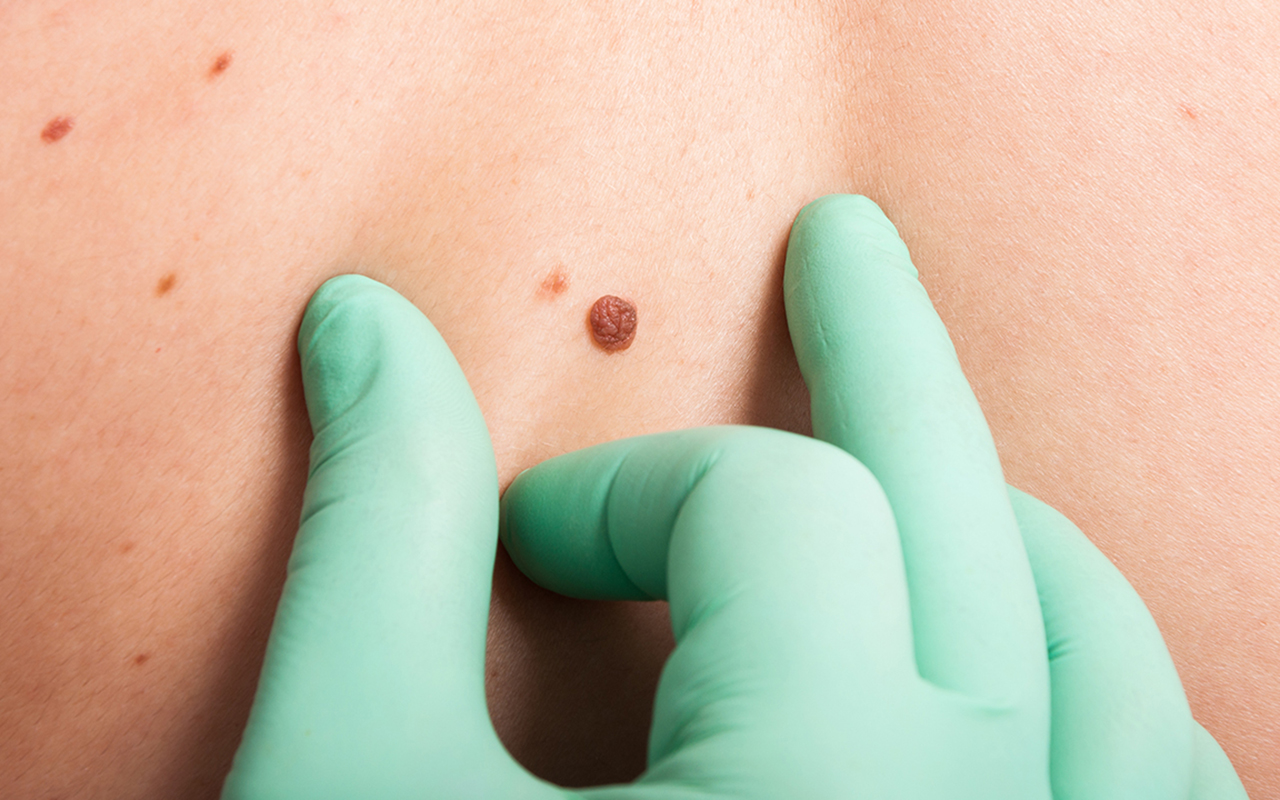
PARTIAL biopsies (including punch and shave biopsies) have been increasingly used for diagnosis of melanomas, with the proportion of shave biopsies rising from 9% of all skin biopsies in 2005 to 20% in 2015; however, researchers warn they are associated with inaccuracies in staging melanomas.
Research published by the MJA studied changes in the choice of skin biopsy technique for assessing invasive melanoma in Victoria, and examined the impact of partial biopsy technique on the accuracy of tumour microstaging.
“The aim of diagnostic skin biopsy is to accurately detect or exclude melanoma and to accurately stage the primary tumour in order to inform therapy planning,” wrote the authors, led by Associate Professor Victoria Mar, Director of the Victorian Melanoma Service. “There are critical differences in the ability of the various methods of complete and partial biopsy to achieve this outcome, and melanoma guidelines in Australia, the United States and Europe advocate complete excisional biopsy.
“Punch biopsies are usually intended to sample and diagnose large, clinically obvious melanomas (high false negative rates make them unsuitable for diagnosing clinically equivocal lesions), whereas the aim with many shave biopsies is to diagnose and completely remove the lesion, thereby allowing the planning of definitive therapy.
“While shave and saucerisation biopsy techniques have been promoted as more accessible and less expensive than excision, they are associated with microstaging inaccuracy,” they wrote.
“Shave biopsy can transect the base of invasive melanomas [which] may lead to inaccurate initial assessment of tumour depth, with negative implications for microstaging, planning of therapy, and determining the prognosis (here and here).”
Mar and colleagues analysed data from 400 patients randomly selected from the Victorian Cancer Registry for invasive melanoma histologically diagnosed in Victoria during 2005, 2010 and 2015, stratified by final tumour thickness: 200 patients with thin melanoma (< 1.0 mm), 100 each with intermediate (1.0–4.0 mm) and thick melanoma (> 4.0 mm).
They found that 833 excisional and 337 partial diagnostic biopsies were undertaken. The proportion of partial biopsies increased from 20% of patients in 2005 to 36% in 2015; the proportion of shave biopsies increased from 9% in 2005 to 20% in 2015, with increasing rates among both GPs and dermatologists; 94 of 175 shave biopsies (54%) transected the tumour base; wide local excision subsequently identified residual melanoma in 65 of these cases (69%); 21 tumours diagnosed by shave biopsy (12%) were T-upstaged. With base-transected shave biopsies, tumour thickness was underestimated by a mean 2.36 mm for thick, 0.48 mm for intermediate, and 0.07 mm for thin melanomas.
“Excisional biopsy remains the most appropriate diagnostic biopsy technique for invasive melanoma,” the authors wrote, citing clinical practice guidelines from Australia, the US and the UK.
“[We] identified a marked increase in the use of shave biopsy in Victoria between 2005 and 2015, associated with a substantial rate of tumour base transection and underestimation of tumour thickness,” they concluded.
“Accurately ascertaining thickness is increasingly important not only for prognosis, but also for decisions about adjuvant therapies and clinical trial opportunities. Where excisional biopsy is readily achievable, it remains the most appropriate diagnostic biopsy technique for assessing invasive melanoma.”
Prof Gerry Milton addressed us students thus:
-I am Gerry Milton and regarded by many as a world authority in Melanoma and arguably am.
However I get it wrong nine times out of ten. This is how I achieve this: if you are not overdiagnosing Melanoma nine to one you are underdiagnosing it. This is the result of underdiagnosing it. First slide please,
By the end of the hour we left shaken.
However, about ten years later the great and the good of my year had forgotten the lesson.
Harvey had long ago described the circulation of the blood. I have described the circulation of the patient.
So they came to me as well and Prof Milton’s words rang often in my ears
Unfortunately shave biopsies to diagnose melanoma in suspicious lesions is still widely taught in the various skin cancer courses and various diplomas/health certificate courses (some given by prestigious local universities)
Re Dr Withnall
Has the profession become so venal as to endanger patients to save a few minutes or a few dollars?
I have always excised for the reasons given above .Colleagues who read this newsletter wouldn’t have a problem .May be this information ,should be given by the insurance company in their newsletter which I hope will reach better to my colleagues
Hi Christopher.
An excisional BIOPSY of a melanoma actually rebates the dollar value of a WIDE LOCAL EXCISION of a melanoma.
I (and many others) have this in writing from DoHA and Medicare.
However, it is interesting that your reading of the schedule is absolutely accurate.
I asked that it be re-worded, as regards the ‘curative intent’ portion as this causes misinterpretation which – unfortunately (and inexcusably, actually) – may potentially be contributing to worse patient outcomes, as you point out.
Please define ‘excision biopsy’- I was always taught that if suspicious of melanoma always do a 1cm wide of lesion removal- turns out to be a clinical mistake on my part and the lesion is benign then I wear the cost
If in doubt, cut it out! Do it properly! Even if it is only just a seb. K as I have seen IEC’s in Seb. K’s .
The increase in shave over excision is possibly linked to the time and remuneration mismatch if a “suspicious” lesion turns out to be benign. Exactly the same excision procedure leads to vastly different remuneration depending on the result of the biopsy.
If you do an excisional biopsy and the lesion turns out to be a SebK = 30071, Dysplastic naevus = benign TCUS item #, Melanoma = 30071 (by my understanding of MBS rules because no intent to cure if not taking adequate margins). Therefore very different remuneration for exactly the same process and time taken, and expertise required. It’s no wonder the quick and easy shave biopsy is becoming the diagnostic method of choice.
Perhaps the highly confusing MBS rules should be updated to reflect this evidence?
Anatomical Pathologists have been highlighting this for many many years. I hope the message gets through.