GPs now have updated guidelines for the diagnosis and management of melanomas, including expanded criteria which will help them recognise atypical presentations of the disease.
Dr Victoria Mar, from the Victorian Melanoma Service and Monash University, and colleagues have published a guideline summary in the MJA outlining the characteristics of melanomas that lack the classical clinical features of asymmetry, border irregularity, colour variegation, and diameter greater than 6 mm (ABCD).
“There is evidence that the rate of early detection of superficial spreading melanomas in Australia has improved, with a corresponding reduction in both the median tumour thickness and in melanoma mortality from this subtype,” Dr Mar and colleagues wrote.
“However, a number of studies in Australia and other countries have shown an increasing or stable incidence rate of thick melanomas … This is in part due to their atypical clinical features.
“Improved diagnostic accuracy of these subtypes can significantly reduce mortality from melanoma.”
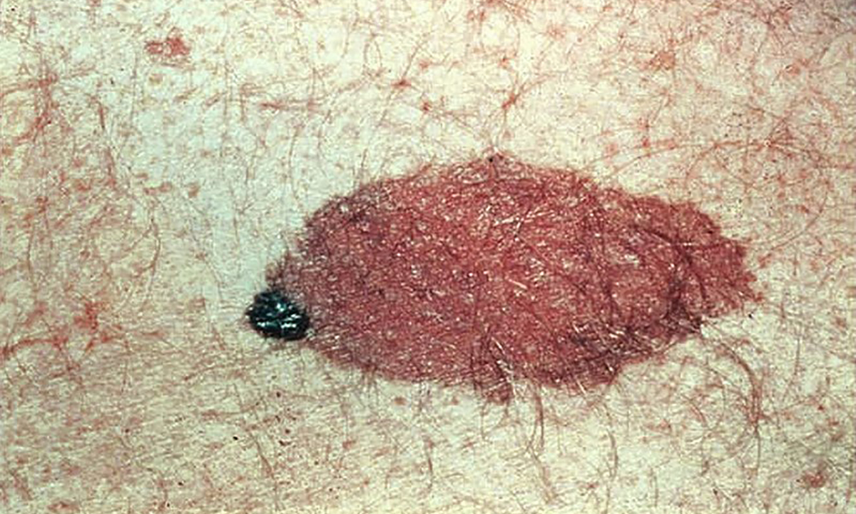
In an exclusive podcast, Dr Mar said that, in contrast to the melanomas doctors and patients are familiar with thanks to strong public health campaigns around the ABCD criteria, atypical melanomas could be quite small, symmetrical, pink or skin-coloured lesions that grow quite quickly.
“Often, we have patients saying to us ‘oh, I didn’t think that looked like a melanoma’ or ‘my doctor didn’t think that looked like a melanoma’, and it surprises both the patient and the doctor that it’s a melanoma, and often quite a thick and aggressive [one],” Dr Mar said.
“They’re the ones we want to make people more aware of and to help them improve their early diagnosis of those types of tumours.”
The widened criteria recommended by the new guidelines add elevated, firm and growing (EFG) to the ABCD mantra.
“Melanomas are generally distinguished from benign lesions by their history of change, and thick melanomas often do not conform to the ABCD rule but usually meet the EFG criteria,” the summary authors wrote.
“Therefore, careful history taking is important, and any lesion that continues to grow or change in size, shape, colour or elevation over a period of more than 1 month should have a biopsy taken and be assessed histologically or referred for expert opinion.
“Suspicious raised lesions should be excised rather than monitored.”
Pigmentation, or a lack of it, is one of the major differences between typical and atypical melanomas, and presents the biggest challenge for GPs examining suspicious lesions.
“Often, people think of melanoma as a dark spot or something that’s changed and become quite black in colour, but that’s certainly not always the case,” Dr Mar told MJA InSight.
“In fact, a lot of the more aggressive melanomas are pink or skin-coloured, and that is quite scary to doctors as well, because we have a lot of [those] on our skin – they might be pimples, they might be inflammatory lesions. But [those things] do tend to come and go within a period of a month.
“Oftentimes, melanomas do have a very subtle pigment, which will be a clue. It might not be [visible] clinically, but if you use dermoscopy … you might see some faint grey dots or blueish areas. You might also see some atypical vessels or dotted vessels within the lesion, which are more common in melanomas.
“So, there are certain clues that we can look for in these pink or seemingly skin-coloured lesions that might help us to be more suspicious of melanomas,” she said.
“The take-home message [for GPs] is, listen to the patient carefully for that history of change. Any changing mole that has been growing for over a month really does need to be either biopsied or referred for a second opinion by a dermatologist.
“Any raised lesion that is suspicious at all should be excised. It shouldn’t be photographed and monitored.
“And to have, in the back of your mind, when you do see a pink or skin-coloured lesion: is there any pigment that I can see and could this be a melanoma?, in which case biopsy shouldn’t be delayed.”
In an MJA editorial to be published on 16 October, Professor Mark Smithers, from the University of Queensland, and colleagues write that “in Australia in 2016, it was estimated that 1770 people would die from melanoma”.
“The focus on early detection must continue through public health and medical education, as well as research into more personalised, technology-driven care that focuses on identifying individuals who have a higher risk of developing the disease.”
To find a doctor, or a job, to use GP Desktop and Doctors Health, book and track your CPD, and buy textbooks and guidelines, visit doctorportal.
Any skin lesion that causes concerns, either by the doctor or when repeatedly expressed by the patient/family, should be excised for histopath—simple as that; it generally takes only ten minutes in the office and resolves the issue. This can be as cost effective as repeatedly bringing the patient back for review, and avoids catastrophic diagnostic mistakes. ( i have seen many over 40 years of surgical practice) .
While the ABCDE and EFG criteria are useful, they should not be relied on exclusively .