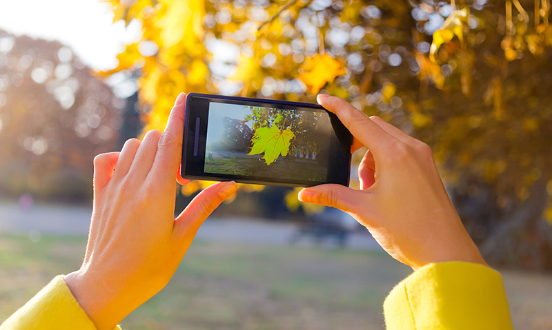
THE use of mobile phone cameras to capture radiological images from computer screens can lead to misleading information being conveyed and is of little use in the diagnostic process, a leading radiologist has warned.
Dr Greg Slater, president of the Royal Australian and New Zealand College of Radiologists, said a single-frame image transmitted by a mobile phone was not nearly as much help in the diagnostic process as it might seem.
“There is a lot of potential harm in this. [A single image] is a very limited part of the study, it’s transmitted in a non-private way, and it’s transmitted in a way that markedly reduces image quality. It can be misleading and can be missing a lot of diagnostic information,” Dr Slater told MJA InSight.
Dr Slater’s comments came as a short report, published in the MJA, outlined a “near miss” after a mobile phone was used to transmit imaging of a post-operative computed tomography (CT) brain scan of a 45-year-old woman. The woman had presented with spontaneous subarachnoid haemorrhage secondary to a ruptured anterior communicating artery aneurysm, which had been managed with a craniotomy and ventricular drain.
- Related: MJA — Pitfalls in photographing radiological images from computer screen
- Related: MJA InSight — James Churchill: Mobile image
- Related: MJA — Legal considerations of consent and privacy in the context of clinical photography in Australian medical practice
Initial serial post-operative CT brain scans showed an evolving infarction in the left middle cerebral artery territory, and elevated intracranial pressure (>40 mmHg) and neurological fluctuation prompted a repeat CT scan.
The intensive care unit consultant took a photograph of the scan from a computer screen using a mobile phone and sent the image to Professor Jeffrey Rosenfeld, the on-call neurosurgeon, who noted extensive bifrontal infarction on the image.
The patient was urgently prepared for decompressive craniectomy; however, on reviewing the scans at a radiology workstation before surgery, Professor Rosenfeld detected the discrepancy and the procedure was cancelled.
The patient recovered and remained neurologically well 6 months after discharge.
Report co-author Professor Rosenfeld, senior neurosurgeon at the Alfred Hospital in Melbourne, said while the technology could enable rapid decision-making, the case highlighted the associated pitfalls.
“We found that just photographing the image can lead to a distorted picture. The shading may not be the same as what you’re seeing on the original screen because of the angle of the mobile phone to the screen that you’re photographing the image on,” he said.
“The angle of the camera and the distance of the camera from the screen is quite important for the quality and shading of the image.”
Professor Rosenfeld and his co-authors wrote that guidelines existed to address the myriad privacy concerns associated with this increasingly common practice, but there were no detailed guidelines regarding the technical issues.
AMA guidelines focus on privacy and consent issues, and generally caution doctors to be aware of the potential for impaired quality.
The authors proposed six steps to avoid problems:
- Use original images wherever possible.
- Compare the photo with the original before sending.
- When photographing computer screens, position the camera perpendicularly to, and at arm’s length from the screen, enlarging the image with digital zoom as required.
- Before making clinical decisions, review the original imaging, including confirming the correct patient details with an observer or peer.
- After photographing, ensure that images are deleted from the phone and any online data storage accounts, and record in writing the image use in the case notes.
- Teach undergraduates as well as practising clinicians the technical aspects of the use of mobile phone images.
Dr Slater said a better practice would be to make use of technology that allows doctors to have remote access to the image provider’s original data archive online. He said the technology was becoming more widely available, and allowed the clinician to view images that were not degraded.
“Seeing the whole study and being able to manipulate that study is of far more diagnostic value than a single image that you just get through messenger, and it can be available extremely rapidly,” Dr Slater said, adding that this password-protected method also ensured patient privacy was not compromised.
However, Professor Rosenfeld said remote access systems may not be the answer.
“There have been some very expensive systems that have been designed to transmit images from a central X-ray facility to a personal computer. A number of hospitals have done that but it’s been unsuccessful because it’s clunky, difficult-to-use technology,” he said.
Professor Rosenfeld said that mobile phone technology, when used properly, provided a simpler solution to remote clinical decision-making in neurosurgical emergencies and many other clinical scenarios.
- Related: MJA InSight — App may solve photo legal risks
- Related: MJA InSight — Legal risks in medical phone photos
- Related: MJA InSight — Privacy concerns over mobile apps

 more_vert
I note the suggestion of zooming on a smartphone. This electronically enlarges the central pixels, by discarding the peripheral pixels. This means a loss of image quality
It is better to optically zoom by bringing the camera of the smartphone closer in so the image fills the screen of the phone. To minimise reflections, turn down the room lights.
This is not such a ‘near miss’ as it sounds. The authors are critical of the diagnostic quality of the image sent to the neurosurgeon. However, neither the radiologist involved in taking the images, nor the intensivist reviewing them, could make the definitive surgical decision, despite having the original images before them. Optimally, the surgeon would have NBN bandwidth to review the images on the multi screen computer in his rooms. Or be in the hospital – but hospitals save millions of dollars a year by having specialists on-call, not on duty 24/7. It will be several years before my Sydney rooms (CBD, opposite State Parliament) get NBN, let alone my phone if I am out shopping or in the car when the hospital calls.
However, the realistic alternative in this scenario, is that the intensivist who does not fully understand the picture, would describe it over the telephone to the surgeon, who would make a prudent decision such as preparing this patient for theatre and coming in to review the imaging and the patient. As a hand surgeon, an SMS photograph of the x-ray and the overlying soft tissue wound, gives me infinitely more information than a verbal description from the casualty doctor. I can plan the likely operation and arrange prostheses, equipment etc over the phone to theatre staff, thereby minimizing delays once I see the actual patient/imaging/wounds.
Xrays and CT or MRI scans frequently fail to show subtle serious pathology like scaphoid fractures or scapholunate tears – this is not a ‘near-miss’, but any test needs interpreting by someone who understands the limitations of diagnostic imaging in the relevant clinical context.
I found taking a short video was always better than a still shot.
Smart phone cameras have great resolution and are convenient. Just remember to delete the images. As for the suggestion that hospitals should implement teleradiology, keep dreaming. We were lucky to have access to a computer lol
This is a clear example of poor implementation of teleradiology.
Taking a single photo of a computer screen does not comply with the RANZCR Teleradiology standards.
The technology for online web access is widely avaiable and hospitals should invest in this as a matter of urgency.