PUBLIC funding of photographic melanoma surveillance in high-risk patients would be money well spent, according to a leading dermatologist, who says the cost of treating advanced disease far outstrips the cost of funding these early detection aids.
Associate Professor John Kelly, head of the Victoria Melanoma Service at Melbourne’s The Alfred Hospital, said funding total body photography (TBP) and sequential digital dermoscopy imaging (SDDI) for the early detection of melanoma in high-risk patients would prevent the expensive treatment of advanced disease.
“The new targeted chemotherapies and the new checkpoint inhibitors that we use for advanced melanomas are very exciting, but very expensive”, he said.
Professor Kelly was commenting on Australian research published in JAMA Dermatology evaluating the effectiveness of TBP and SDDI. (1)
The prospective, 5-year follow-up study of 311 patients assessed as being at extreme high-risk of melanoma detected 75 primary melanomas — 38% using TBP and 39% using SDDI. The median Breslow thickness of postbaseline incident melanomas was in situ (interquartile range [IQR], in situ to 0.6 mm).
Patients in the study had 6-monthly full-body examinations that were compared with baseline TBP images. Patients with equivocal lesions were referred for short-term (3 months) or long-term (≥ 6 months) SDDI, and atypical lesions were excised.
The benign to malignant excision ratio was 1.6:1 for all lesions excised and 4.4:1 for benign melanocytic lesions. By Year 2 of the follow-up period, the cumulative risk of developing a novel primary melanoma was 12.7%. In the next 3 years of follow-up, the incidence of new primary melanoma had halved.
Professor Kelly said the findings added weight to an application by the Australasian College of Dermatologists, of which he is a Fellow, for Medicare funding for dermatologist-provided services for high-risk patients. The application, which is under consideration by the Medical Services Advisory Committee, was submitted in January this year.
Professor Kelly said some patients at the Victorian Melanoma Service, which provided total body photography at a relatively reasonable price of about $200, still shied away from paying for the service.
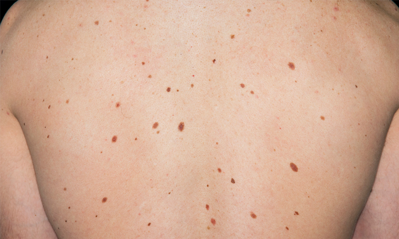
He said some high-risk patients could have up to 500 moles on their body, making it difficult to detect when one mole was changing.
Having a photographic record of the skin provided an important baseline for comparison, and the more detailed the image — such as those captured in SDDI — the more powerful the technique in identifying subtle degrees of change.
Professor Scott Menzies, of the Sydney Melanoma Diagnostic Centre and a coauthor of the Australian study, said the research was the first to show the relative effectiveness of these two well used methods of melanoma detection.
“Both of those techniques need to be used to monitor these high-risk patients”, said Professor Menzies, who is also a professor of medicine (dermatology) at the University of Sydney.
He said although the detection of five melanomas greater than 1 mm in thickness in the study was disappointing, it was pleasing to be able to detect 5 of the 6 nodular melanomas at less than 1 mm.
“That’s very unusual. Most nodular melanomas are thicker than 1 mm because nodular melanomas grow very quickly, so we were extremely happy with that finding”, Professor Menzies said.
Professor Kelly said even though some desmoplastic and nodular melanomas in the study were detected at greater than 1 mm thickness, they were still detected earlier than they would have been without close surveillance.
“Finding those unusual kinds of melanoma earlier will make a big impact on the death rate”, he said.
Mr Terry Slevin, Cancer Council Australia spokesman and former chair of the council’s skin cancer committee, also welcomed the study findings as confirmation of the importance of close surveillance for this very high-risk patient group.
However, he said, care was needed to apply these findings to only the high-risk population. “There’s no doubt that there are lesions which are much harder to detect and the prospect of them coming about in those who have already had a melanoma is significant. The risk in the rest of the population … is probably far less, so I guess the cautionary note is the extent to which we take this evidence and then apply it to the broader community”, he said.
An editorial accompanying the JAMA Dermatology study said it was difficult to know whether this accelerated detection significantly changed mortality, but it was evident that the diagnostic measures had a favourable impact on morbidity. (2)
1. JAMA Dermatol 2014; Online 25 June
2. JAMA Dermatol 2014; Online 25 June
I agree with Dr Kelly, even when offered TBP for less than $100 and a $36 rebate many high risk patients decline.