×

Medical cannabis: time for clear thinking

Australia is behind the times on the medical use of cannabis

The debate about the medical use of cannabis in Australia has become confused with the proposal for a formal clinical trial instead of proceeding to legislation in New South Wales, the Australian Capital Territory and Victoria. Debates about prohibition of cannabis have a long history,1 as has the proposal for medical cannabis in Australia.2 Politicians are nervous about being “soft on drugs”, especially before an election. The clinical trial proposed, if successful, presumes that cannabis would then be approved and regulated as a pharmaceutical substance.

We need to be across the facts and options. Cannabis can never be a pharmaceutical agent in the usual sense for medical prescription, as it contains a variety of components of variable potency and actions, depending on its origin, preparation and route of administration. Consequently, cannabis has variable effects in individuals. It will not be possible to determine universally safe dosage of cannabis for individuals based on a clinical trial.

Extreme views in the debate about any form of cannabis decriminalisation are advanced with almost religious fervour. On the one hand, some assert that cannabis is a dangerous, highly addictive drug which causes schizophrenia, and that any move to relax prohibition would be a disaster. This view defies published evidence. On the other hand are those who have used cannabis for years, swearing it causes no trouble. They see prohibition as a totally inappropriate curb on individual freedom.

Facts about cannabis

The assertion that cannabis is highly addictive ignores firm evidence. The most authoritative review comparing addictiveness of drugs rates physical dependence on a scale of 0–3.3 Heroin is ranked 3; tobacco, barbiturates and benzodiazepines, 1.8; alcohol, 1.6; and cannabis, 0.8. Cannabis may, of course, be a pathway to more addictive drugs if obtained from illegal sources that also offer powerful alternatives.

The view that cannabis carries no risk likewise ignores much published evidence.4 Recent Australian and New Zealand longitudinal studies show significant social, behavioural, educational and mental problems with frequent use of cannabis by young people (aged 15–25 years). Psychosis occurred more frequently following long-term heavy use than among non-users, but no schizophrenia was noted in this study.5 A recent review of the evidence implicating cannabis in the development of schizophrenia found only that it can accelerate its expression at an earlier age and may aggravate existing schizophrenia. Of course, non-users also develop schizophrenia.6 Others have identified heavy cannabis use in the young as a possible factor in later psychosis, without specifying schizophrenia.7

Australians, together with citizens in the United States and New Zealand, are the world’s greatest users of cannabis per head of population.8 Prohibition has failed to prevent widespread use and young people report that they can readily access it.9 Young people need to be strongly dissuaded, on health grounds, from frequent or even regular use of cannabis, but this has little relevance to cannabis used for medical purposes or the debate surrounding it. Potential medical users are often, for example, in the later stage of a battle with painful cancer, finding problems with morphine, other analgesics and nausea with chemotherapy. Others seek relief from painful conditions such as muscle spasm in multiple sclerosis. Cannabis is believed to reduce seizures in Dravet syndrome, a rare genetic myoclonic epileptic encephalopathy beginning in infancy.10 Most parents of affected children (84%) report much lessened frequency or abolition of seizures with medical cannabis. They should have continuing access to it until trials using purified cannabidiol (CBD), believed to be the active component for these children, provide a superior agent.

We are behind the times on medical cannabis. Currently, 23 states in the US have legalised use of cannabis for medical conditions, as has Canada since 2001. Other countries approving it include Israel, Holland and the Czech Republic. Portugal, in 2001, removed penalties for personal possession and use of all illicit drugs, but with rigorous administrative processes to handle problem use. Eliminating prohibition is not a disaster if there are sensible processes to control drug-related harms.11

An Australian and US study found that removal of legal action and possible imprisonment for possession and use makes no difference to the patterns of use of cannabis.12 World Health Organization mental health surveys of 17 countries found that “countries with stringent user-level illegal drug policies did not have lower levels of use than countries with liberal ones”.13 There is no rational basis for the view that weakening prohibition to permit use for medical conditions would lead to a surge in general use.

Cannabis has at least two important active elements: δ-9-tetrahydrocannabinol (THC) and CBD. The former is responsible for the high of intense comfort and pleasure when presented to the brain in sufficient quantum. Its presence is greatly enhanced by heating marijuana above 170°C, as in a bong, converting the inactive precursor THC-A to THC. THC infused at high dose can produce a powerful euphoria but also hallucinations and other psychotic effects in some normal individuals, followed by complete recovery.14 CBD, on the other hand, does not give a high but has other effects including suppression of nausea and pain. It counteracts some of the effects of THC.15 The plant Cannabis sativa has more than 100 alkaloids with potential to influence the cannabis receptors CB1 and CB2, which respond to normal cannabinoids.16

Response to cannabis varies from person to person, partly due to genetic variation among users.17 The content of THC and CBA varies among different strains of marijuana. Some users vary the type of plant they use to benefit from these different effects.

What would a clinical trial entail?

Cannabis as such cannot be subjected to a double-blind clinical trial. Participants would have to agree to be treated with it, hoping to gain relief from distressing pain or nausea. Each would become aware whether they are receiving cannabis or a placebo. Dose would have to be adjusted for each individual. Any trial would use cannabis with multiple active constituents, varying with the source of marijuana used and its preparation.

If a person in the late stages of painful cancer seeks the euphoria of THC, why should they not have it? They must have a right to withdraw from a trial if it does not suit them. Participants in the control group may demand to transfer to the active arm on seeing others feeling better. Cannabis should supplement morphine for pain as necessary, not replace it.

Are there barriers in principles of medical practice?

There may be medicolegal issues if a medical practitioner prescribes a preparation of unquantified potency or with an incomplete description of its constituents and without full knowledge of side effects and their extent. But this has not proved to be a problem in those US states where the patient makes the choice to use cannabis following a medical consultation. A recent readership survey conducted by the New England Journal of Medicine sought comment on a published case report of a cancer patient where a senior psychiatrist and a pain management specialist had both recommended against use of cannabis. Seventy-six per cent of respondents from several countries responded that they would recommend use of cannabis in such a case.18 Medical marijuana is now widely used. A recent US study found that the states with medical cannabis use over 10 years had a lower death rate from opioid overdose than those without.19

Why not go ahead with legislative approval?

The real question is whether a person who is suffering pain and distress can access cannabis on their own initiative, following medical consultation as to their symptoms. They can access other herbal remedies from authorised providers such as health food stores or a pharmacist. If legislation permits sale to people suffering from a condition diagnosed by a doctor and scheduled in legislation, there should be no problem with provision of cannabis by this route without waiting for completion of a clinical trial. This is especially the case with Dravet syndrome patients where a formal clinical trial with a proprietary CBD concentrate20 may take several years to complete.

We should ensure that cannabis is provided only to approved users who should be registered. As there is no legal supplier, users should have permission to grow their own plants — up to 10 at any one time — but be forbidden from selling their product. Any proposal for commercial production should be subject to strict control, with analysis of THC, THC-A and CBD content by a government toxicology laboratory for both cannabis oil and the leaf product. Venues for sale, presumably pharmacies or health food shops, should be registered. People aged between 15 and 25 years should be excluded as recipients, except where it is provided specifically for a cause covered by legislation. The legislation should also make cannabis available for medical research.

In summary, use of cannabis should be decided by the patient, following medical advice about the condition from which they seek relief, with patients being registered under state legislation. If there is to be a nationally approved trial, it should be one of documenting clinical experience from cannabis use under state legislation of the kind foreshadowed by recently elected Victorian Premier Daniel Andrews.21

Death due to intravenous use of α-pyrrolidinopentiophenone

Intoxication with synthetic cathinones (psychoactive designer drugs) can involve cardiovascular, autonomic, neuromuscular and neuropsychiatric features. We report a case of cardiac arrest and subsequent death in a 44-year-old man after intravenous use of one such drug — α-pyrrolidinopentiophenone. We believe this is the first death associated with this drug to be reported in Australia. Currently, no specific antidote exists for cathinone exposure.

Clinical record

A 44-year-old man with a history of substance misuse injected himself with a powder named “Smokin’ Slurry Scrubba” that he and his girlfriend had purchased over the counter from a shop. Soon after injecting the powder, the man stripped off all his clothes, jumped over a barbed-wire fence and smashed a window. He was restrained and pinned to the floor by security staff while emergency services were called, during which time he suffered a cardiac arrest.

Cardiopulmonary resuscitation (CPR) was commenced by bystanders. On arrival of ambulance paramedics, the patient was confirmed to be in asystolic cardiac arrest and the paramedics continued CPR. Spontaneous circulation was restored after a further 3 minutes of advanced cardiac life support protocol. He was intubated at the scene by the paramedics. The total duration of cardiac arrest was estimated to be 16 minutes. His vital signs immediately after the arrest were: Glasgow Coma Scale (GCS) score, 3; blood pressure, 65/20 mmHg; heart rate, 85 beats/min; tympanic temperature, 39.7°C. An adrenaline infusion was started at 30 µg/min to treat persistent systemic hypotension.

On arrival at the emergency department, about 1 hour after the cardiac arrest, the patient’s temperature had climbed to 39.9°C and he was still hypotensive (blood pressure, 83/45 mmHg). On physical examination, he had a GCS score of 3, and his pupils were dilated (right, 5 mm; left, 6 mm) and unreactive to light. A venous blood gas and serum analysis using samples taken on arrival at the emergency department showed a severe metabolic and respiratory acidosis with evidence of hyperkalaemia, rhabdomyolysis, ischaemic hepatitis, elevated lactate level and acute renal failure (Box 1). An electrocardiogram showed a sinus tachycardia of 126 beats/min with normal intervals and no ischaemic changes, and results of a non-contrast computed tomography scan of his brain were reported as normal. A total of 5 litres of cooled crystalloid solution was given intravenously. Within an hour of arrival at the emergency department, the patient’s temperature had normalised to 37.2°C and his blood pressure had stabilised while on the adrenaline infusion. Other initial treatment included intravenous calcium gluconate and sodium bicarbonate for treatment of hyperkalaemia. He began to respond to painful stimuli and required sedation.

On arrival at the intensive care unit, about 6 hours after the cardiac arrest, the patient’s pupils had become equal and reactive at 2 mm diameter. Repeated blood analysis revealed abatement of his lactic acidosis, but worsening of his rhabdomyolysis (Box 1). An echocardiogram taken at this time was normal. Results of a qualitative urine drug screen (Roche Diagnostics immunoassay) using a sample taken on the patient’s arrival at the intensive care unit were positive for benzodiazepines but negative for amphetamines, cannabis, cocaine and opioids. There was also evidence of disseminated intravascular coagulation — an international normalised ratio of 2.5 (reference interval [RI], 0.8–1.1), an activated partial thromboplastin time of 82.2 s (RI, 25.0–37.0 s) and a plasma fibrinogen level of 1.4 g/L (RI, 2.20–4.30 g/L) — but no clinical evidence of bleeding. Due to the increasing creatine kinase levels, the patient was started on continuous venovenous haemodiafiltration in an attempt to mitigate further renal damage.

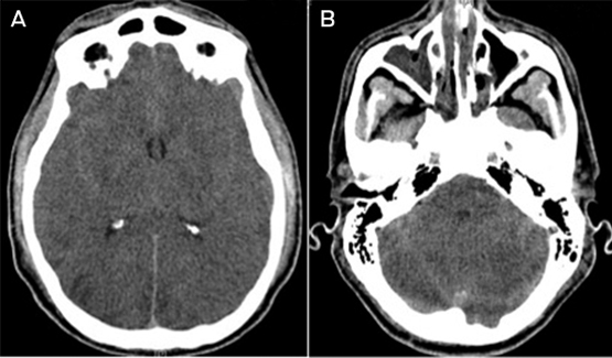
About 24 hours after the cardiac arrest, the patient developed signs of raised intracranial pressure with fixed dilated pupils and haemodynamic instability; he did not respond to noxious stimuli and he had lost brain stem reflexes. A non-contrast computed tomography scan of his brain showed diffuse cerebral oedema with tonsillar herniation and multiple areas of cerebral infarction (Box 2). About 43 hours after the cardiac arrest, the patient was declared brain dead by clinical criteria, and supportive care was withdrawn.

The only physical findings of note on postmortem examination were cerebellar tonsillar herniation and pulmonary oedema. There was no evidence of trauma. Coronial analysis of antemortem and postmortem blood samples (using liquid chromatography mass spectrophotometry) revealed the qualitative presence of α-pyrrolidinopentiophenone (α-PVP). No other common drugs of misuse were detected.

The formal conclusion of a subsequent coronial inquest was that the patient had died from cardiac arrest and cerebral oedema, which were the results of α-PVP toxicity. The possibility of physical restraint and positional asphyxia contributing to his death was raised but these factors were considered not to be the primary cause of death.

Discussion

To our knowledge, this is the first death due to α-PVP use reported in Australia. Assessing all the evidence, our patient died from the expected complications of exposure to α-PVP.

α-PVP is a synthetic cathinone designer drug that is misused for its stimulant and psychoactive effects. It is an analogue of pyrovalerones1 such as 3,4-methylenedioxypyrovalerone (MDPV). Synthetic cathinones such as α-PVP are widely sold on the internet; they are commonly referred to as “bath salts”, “legal highs” or “research chemicals”, and often labelled “not for human consumption” in an effort to evade regulatory control.2 Synthetic cathinones are also structural analogues of cathinone, the naturally occurring β-ketone amphetamine analogue found in the Catha edulis (Khat) plant.

As structural analogues of β-ketone amphetamines, cathinones are expected to have amphetamine-like effects. Many cathinones have been shown to be inhibitors of monoamine transporters, but selectivity of cathinones for serotonin, noradrenaline and dopamine transporters varies considerably in vitro.3 Pyrovalerone-cathinones (eg, MDPV) are potent and selective dopamine and noradrenaline uptake blockers, but are not effective releasers of monoamines. The potency of these drugs on the noradrenaline and dopamine transporters is associated with their stimulant and psychoactive effects.

Synthetic cathinones are most commonly nasally insufflated (snorted) or ingested, but rectal insertion, inhalation, and intravenous and intramuscular injection have also been described.2,4 Desirable effects of cathinones reported by users include increases in energy, empathy and libido.

The effects of synthetic cathinones (as a group) are said to begin to occur within 15–45 minutes of exposure and the desired effects last from 2 to 7 hours.4 However, the undesirable effects can last from hours to days. Clinical features of synthetic cathinone intoxication include cardiovascular, autonomic, neuromuscular and neuropsychiatric symptoms and signs (Box 3).2,4,5

No specific antidote exists for cathinone exposure, and there are limited published data on specific management strategies. Current practice is based on experience treating patients intoxicated with other sympathomimetic drugs, for which supportive care is the mainstay of treatment.4 Benzodiazepines are recommended for control of agitation and seizures, and for treatment of hypertension and tachycardia, and passive or active cooling is recommended for treatment for hyperthermia that is not resolved with anxiolysis and sedation.4,5 In Australia, poisons information centres can be contacted for further advice.

While there have been a few media reports of violence, aggression and deaths related to use of α-PVP, few cases have been reported in peer-reviewed journals. One article describes the deaths of five patients for whom α-PVP was detected in postmortem blood, but this does not necessarily mean that α-PVP intoxication was the cause of death or that α-PVP was the only drug present.6 Another article describes a case of parenteral MDPV overdose, confirmed by a quantitative assay of MDPV in the patient’s blood, in which the clinical course was very similar to that of our patient.7

Our patient probably died as a consequence of the complications of α-PVP exposure. The time of death and clinical course fit with the features of autonomic hyperarousal: psychomotor agitation, delirium, tachycardia and hyperthermia, and subsequent cardiac arrest, rhabdomyolysis, renal failure, hepatic injury, anoxic brain injury and death. These features have also been described as “excited delirium syndrome”.8 Excited delirium syndrome as a diagnostic entity is controversial and widely debated in the medical literature. The role of excessive stimulation of the sympathetic nervous system in subsequent cardiac death after drug exposure has also been widely debated. It has been postulated that overstimulation of the heart by catecholamines leads to increased contractility, blood pressure and heart rate, resulting in increased oxygen demand, followed by myocardial ischaemia, fatal cardiac arrhythmias and sudden death.

The single biggest factor influencing death from α-PVP intoxication as the outcome in this case was the route of exposure to the drug — that is, intravenous injection. From toxicokinetic first principles, this results in faster and higher blood concentrations of drugs compared with ingestion and nasal insufflation as routes of exposure.

Synthetic cathinones have been present in notable quantities in Australia since the mid 2000s. While the exact prevalence of use in Australia is difficult to determine, the availability and popularity of synthetic cathinones has increased over the past decade, as evidenced by surveys of regular drug users, wastewater analyses and drug seizures by the Australian Federal Police.9

Our patient tested negative for amphetamines when a urine drug screen was performed. Testing for α-PVP was only available through coronial investigation. This highlights that medical professionals in Australia need to be aware of the limitations of commonly available drug screens, which might not detect new psychoactive substances.

1 Results of a venous blood gas analysis for a 44-year-old man after he had a cardiac arrest due to α-pyrrolidinopentiophenone toxicity

Component

1 h after arrest

6 h after arrest

Reference interval


pH

6.62

7.21

7.35–7.43

Partial pressure of carbon dioxide (mmHg)

90

42

32–45

Bicarbonate (mmol/L)

8.8

16.1

22–32

Lactate (mmol/L)

29.0

4.9

< 2.0

Potassium (mmol/L)

6.2

3.3

3.6–5.1

Urea (mmol/L)

13.4

12.4

2.9–7.1

Creatinine (µmol/L)

201

169

60–110

Alanine transaminase (U/L)

1 976

1 913

< 45

Aspartate transaminase (U/L)

1 818

2 457

< 45

Creatine kinase (U/L)

2 763

24 660

< 200

Myoglobin (µg/L)

43 190

10–92


2 Non-contrast computed tomography scans of the brain of a 44-year-old man after he had a cardiac arrest due to α-pyrrolidinopentiophenone toxicity


Scans taken 24 hours after cardiac arrest showing diffuse cerebral oedema with tonsillar herniation and multiple areas of cerebral infarction (A, level of the basal ganglia; B, level of the mid posterior fossa).

3 Clinical features of synthetic cathinone intoxication

System

Effects


Cardiovascular

Tachycardia, vasoconstriction, systemic hypertension, arrhythmias, cardiovascular collapse, myocardial infarction,

Autonomic

Sympathetic hyperstimulation (autonomic hyperarousal), mydriasis, hypertension, hyperthermia

Neuromuscular

Seizures, stroke, tremors, muscle spasm, cerebral oedema

Neuropsychiatric

Agitation, hallucinations, panic attacks, paranoia, anxiety, insomnia, anorexia, depression, suicidal ideation, violence (self-mutilation, suicide and homicidal activity)

Think carbon monoxide

Gas heater use increases the risk of carbon monoxide poisoning in the home

During the cooler months, we need to be aware of the increased risk of accidental carbon monoxide (CO) poisoning from domestic gas heaters. Specific causes include faulty installation, inappropriate use, inadequate maintenance, blocked flues and fires.

The signs and symptoms of CO poisoning are non-specific. Headaches, malaise, nausea and dizziness are common, and gradual cognitive deterioration and reduction in functional capacity have been reported in low-grade chronic exposure.1 Further questioning should elucidate whether the symptoms are occurring in other members of the household (including pets), and whether the patient feels better when outside the house. Failure of health professionals to recognise the symptoms and signs of CO poisoning can result in the discharge of a patient back into a potentially fatal environment. At highest risk are older people, patients with comorbidities, children, pregnant women and their unborn babies.

CO is a colourless, odourless and tasteless gas resulting from the incomplete combustion of hydrocarbon fuels. It binds strongly with haemoglobin and cytochromes, preventing oxygen transport and its use in the tissues. This can result in end-organ damage.2

The diagnosis of CO poisoning is based on history and examination, in conjunction with an elevated carboxyhaemoglobin level determined using pulse CO-oximetry, arterial blood gas analysis or CO breath testing. The half-life of CO bound to haemoglobin is 4 hours when the patient is breathing room air. This poses a diagnostic challenge, as the level may have fallen significantly by the time of testing. Additionally, high-flow oxygen may be administered by ambulance officers,3 which further reduces the half-life.

The treatment of CO poisoning involves immediately removing the patient from the source and instituting high-flow oxygen. The patient should be transferred to an emergency department for full investigation and for consideration of hyperbaric oxygen therapy.4 Such patients are also susceptible to ongoing health issues, including delayed neuropathy and myocardial damage, and require follow-up.5

What should we tell our patients about prevention of CO poisoning in the home?

  • Use accredited professionals to install gas appliances and service them every 2 years to ensure that no gas can leak.
  • Never tamper with air vents on gas heaters.
  • Never use external gas heaters in enclosed areas.
  • Install a CO alarm in the house.
  • Learn how to recognise CO poisoning.

By increasing awareness of CO poisoning, its prevention and treatment, we can reduce the incidence of this dangerous condition.

The power of systems thinking in medicine

The convergence of seemingly small events accruing over time can have severe consequences. This is
a central message of many aircraft accident investigations. For instance, an attempt to streamline maintenance procedures for an engine mount created the conditions for the United States’ deadliest aeroplane crash
in 1979 (http://www.airdisaster.com/reports/ntsb/AAR79-17.pdf). The investigation found a constellation of interacting factors — design deficiencies, faulty maintenance practice, failures of regulatory oversight and flawed aviation industry economics. As noted in relation
to a later aeroplane crash (http://www.theatlantic.com/magazine/archive/1998/03/the-lessons-of-valujet-592/306534), it was a “system accident”. The complexity of aviation systems creates conditions for small changes to interact with other system elements across technical, organisational and cultural domains to produce significant outcomes that are hard to predict and control.

All clinicians recognise the complexity of health care delivery. The system accident idea has been adopted enthusiastically by some exponents of ways to improve clinical safety, despite more recent reservations about its applicability (Health Serv Res 2006; 41: 1654-1676). Nevertheless, the assessment of clinical mishaps and adverse events requires a systems approach (not only technical, but also organisational, social and cultural).

As a starting point, registries are powerful tools for systematically detecting and monitoring clinical problems and adverse events, and for informing interventions. The study by Roxanas and colleagues of Australia and New Zealand Dialysis and Transplant Registry data (doi: 10.5694/mja13.10435) shows that the incidence of end-stage renal failure due to lithium therapy, although small, is growing. They express concern that accepted doses of lithium over a long time may result in irreversible renal impairment and end-stage disease, reinforcing the need for regular and frequent monitoring of renal function. Registry data analysis is the monitoring system providing the backbone for reducing risks for those receiving lithium therapy.

There is also a need for systems to oversee and analyse incidents in whole areas of health care. Cunningham
and colleagues (doi: 10.5694/mja13.11347) point out that, in the case of chiropractic practice, there is little in place for monitoring
for adverse incidents. Without such a system, proper investigation of incidents in chiropractic care cannot occur.

Significantly greater challenges exist in assessing
health impacts of activities with complex influences from societal and cultural practices in the community. Clenbuterol — a β2-adrenergic agonist with anabolic as well as bronchodilating properties, registered only for veterinary use and banned in sport — is now illicitly used in the community to aid bodybuilding and weight loss. Brett and colleagues (doi: 10.5694/mja13.10982) report a case series of clenbuterol toxicity reported to the NSW Poisons Information Centre. Details of cases suggest that it is also being used for deliberate self-harm,
and that accidental ingestion has occurred. While the authors acknowledge that the study presents an incomplete picture
of actual use in the community, would a deeper engagement with ideas from complexity science help in understanding the complexity of substance misuse? What additional systems need to be put in place for us to know and perhaps anticipate changing patterns of use?

No matter how complex the health problem, data registries will always have a central role in disease and health care monitoring and practice. Presently, patients often need to explicitly consent to their information being added to a registry. Olver (doi: 10.5694/mja13.10695) examines the ethical dimensions of opt-out consent, where patient data are automatically added unless consent is expressly refused. He argues that this approach is acceptable in the context of low-risk research and for improving clinical quality. Although not called a registry, the concept has been used for decades in civil aviation for mandatorily collecting flight data and operational feedback from aviation personnel. Only relatively recently have similar approaches taken hold across a broad range of health care activities.

Decades of research and application of safety assurance and improvement systems in aviation and other industries have resulted in a critical respect for the complexity of many human endeavours — the importance of monitoring outcomes and processes, understanding why and how incidents happen, and appreciating the multifaceted nature of the solutions. There are certainly limitations to mapping approaches to aviation safety to health care systems. But the analogy provides a useful starting point and source of ideas. Preventing adverse health outcomes and health care incidents matters enormously to the community. Just as a systems approach has led to civil aviation being appreciably safer, it should also be pursued by those wanting well founded solutions to complex, multidimensional problems in health.

Clenbuterol toxicity: a NSW Poisons Information Centre experience

Clenbuterol is a β2-adrenergic agonist with a long half-life of around 25–40 hours and high bioavailability,1 currently registered in Australia as a Schedule 4 medicine for veterinary use.2 It is marketed in tablet, gel and injectable forms as an equine bronchodilator and a bovine tocolytic agent. Its anabolic properties have seen it used in food-producing animals to increase lean meat yield.3 However, concerns about toxicity to humans from contaminated meat led to its use for this purpose being banned in the United States in 1991 and the European Union in 1996. Since then, there have been outbreaks of clenbuterol toxicity from contaminated meat across Europe and China,46 and outbreaks of clenbuterol contamination of heroin.7

The spectrum of toxicity in humans includes sympathomimetic effects such as restlessness, tachycardia and tachyarrhythmias,8 gastrointestinal disturbances and rhabdomyolysis, and metabolic disturbances such as hyperglycaemia and hypokalaemia. There have also been case reports of myocardial ischaemia in otherwise healthy people,9,10 and of respiratory distress in adults after nasal insufflation.11

In the past 10 years, a number of elite athletes have used clenbuterol as a performance-enhancing drug, leading to its ban by the International Olympic Committee and World Anti-Doping Agency.12,13

In humans, clenbuterol has been used in tablet form as a bronchodilator at doses of 20–40 μg daily,14 but it is not registered with the Australian Therapeutic Goods Administration. More recently, the use of clenbuterol as a slimming and anabolic agent has been publicised in the media in stories of use by celebrities.15 There is concern about the increasing unprescribed use of clenbuterol by the general public and the ease of online purchase of this drug. Although poisons centres in the US and France have published observational reports on the level of use and spectrum of toxicity of clenbuterol,14,16 the situation in Australia remains unknown.

Here, we describe NSW Poisons Information Centre (NSWPIC) reports of clenbuterol exposures.

Methods

The NSWPIC receives around 110 000 calls annually — 50% of all poisoning-related calls in Australia. The NSWPIC handles calls from New South Wales, Tasmania and the Australian Capital Territory from 6 am to midnight, and provides overnight cover for the whole nation for 7 of 14 days per fortnight.

We retrospectively reviewed the NSWPIC database and the database of the NSWPIC toxicologist for the 9 years from 1 January 2004 to 31 December 2012 using the search terms “clenbuterol”, “ventipulmin”, “oralject”, “planipart”, “broncopulmin” and “claire”.

We manually reviewed all cases from this search for inclusion; those included were categorised by reported source of call, formulation type, reason for use and clinical features. Other variables we examined were age, sex, hospitalisation status and geographical location. Exposure was categorised as unique episodes rather than calls received. Calls referred to toxicologists are also recorded in a separate database and include more clinical details; we present significant toxicities recorded in this database.

We used the median and range to describe data and performed statistical analyses with Microsoft Excel (Microsoft Corporation).

Ethics approval for this study was granted by the human research ethics committee of the Sydney Children’s Hospitals Network.

Results

NSWPIC database

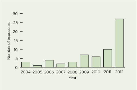
There were 63 unique episodes related to clenbuterol exposure over the 9-year study period, with a worrying increase from three episodes in 2004 to 27 episodes in 2012 and a particularly notable increase in the last year (Box 1). Most of the calls (46) were from NSW, but 17 were from interstate.

Twenty-five patients were female, 33 were male and sex was not recorded in five exposures. Sixty calls about exposures were for adults and three for children (up to 16 years). Exposures among children comprised an intentional ingestion by a 16-year-old and unintentional ingestions by a 1-year-old and a 2-year-old. The median age for all patients with clenbuterol exposure was 21 years (range, 1–83 years). Calls were taken from both metropolitan and regional areas and the numbers of calls appeared to be proportional to population size in those areas.

Clenbuterol was mostly taken in the veterinary liquid or gel form (24 exposures) at a median of 2 mL (range, 0.5–20 mL) or two “scoops” (range, 1–2 scoops). The actual dose is highly variable between liquid formulations. Two people took tablets (one and three tablets, respectively) and of the eight episodes in which the dose was known, the median dose was 0.8 mg (range, 0.08–5000 mg).

Thirty-six calls were from hospitals, two from paramedics, one from a general practice and 21 directly from the public. Caller background was not recorded on four occasions. Of the calls from the public, 14 patients were referred to hospital, three were referred to a general practitioner and four were advised to stay at home.

When recorded, the reasons for use included slimming (12 exposures) and bodybuilding (four), but also self-harm (two). There were three unintentional exposures.

At least 53 patients (84%) required hospitalisation. In only five calls about exposures were patients asymptomatic. The commonest clinical features in those with symptoms were tachycardia (24 patients), gastrointestinal disturbances including nausea, vomiting and diarrhoea (16), and tremor (11). There were a range of other features (Box 2), most notably hypokalaemia (four patients), chest pain (three), ST-segment changes on electrocardiogram (ECG; three), an increase in troponin level (two) and cardiac arrest (one). In the 14 exposures for which the time of ingestion was recorded, most calls were made after exposure in the preceding 6–24 hours. Eight people were recorded as taking clenbuterol over a period of weeks to months.

Case reports

Analysis of NSWPIC toxicologist records revealed three noteworthy cases. The first was an elderly man who drank an unknown amount of clenbuterol from an unlabelled bottle, thinking it was water. He had a history of ischaemic heart disease and diabetes, and presented to the emergency department soon after the ingestion with hypotension (blood pressure, 90/70 mmHg) and sinus tachycardia on ECG (heart rate, 140 beats/min). His hypotension resolved with fluid resuscitation. An esmolol infusion was started, but 8 hours later the hospital supply of esmolol ran out. Given that his heart rate was still high (128 beats/min), he was given metoprolol, orally, with good effect. His troponin level (high sensitivity) at 12 hours had risen to 403 ng/L (reference interval [RI], < 14 ng/L).

The second case was that of a dosing error by a young bodybuilder who took 7 mL instead of seven drops of clenbuterol solution. His heart rate at presentation was 235 beats/min and, after an ECG, he was thought to have a supraventricular tachycardia, although his blood pressure was 133/82 mmHg. His potassium level was 2.2 mmol/L (RI, 3.5–5 mmol/L) and he was agitated, so was given diazepam. An esmolol infusion was started, but after 12 hours the hospital supplies had again run out, so his therapy was changed to metoprolol with good effect.

The third case was that of a young man who had been using clenbuterol for the preceding 8 weeks for bodybuilding, and may have increased his dose in the few days before the call to the NSWPIC. He had a cardiac arrest while playing sport and was immediately treated with cardiopulmonary resuscitation by a bystander, with return of spontaneous circulation 9 minutes later. He subsequently made a good recovery.

Discussion

The number of calls to the NSWPIC about clenbuterol toxicity is increasing. Calls indicate that clenbuterol is predominantly being used for slimming and bodybuilding, and that it is the veterinary product that is being used. However, it was not possible to determine where this product was being sourced.

The misuse of this drug poses the secondary risk of accidental poisoning of children. It is often taken in doses far exceeding the safe therapeutic doses in humans, and order-of-magnitude dosing errors occur. Exposure calls did not show clustering in urban or rural areas.

Clenbuterol use produced a range of sympathomimetic toxicities, which were often prolonged (as calls were most often made 6 to 24 hours after exposure) and occasionally associated with cardiac damage. The case of cardiac arrest is particularly alarming, although we do not know whether this individual had any underlying undiagnosed structural heart disease or cardiac channelopathy.

Although we did not record concomitant medication use in our study, a descriptive US study found that clenbuterol is often taken with thyroxine, anabolic steroids and products containing caffeine and stimulants, which appears to exacerbate toxicity.14

Limitations of our descriptive study include non-standardised methods of telephone enquiry and coding which is likely to have led to underrecording of clenbuterol exposures. Our study presents exposures reported to the NSWPIC, and so is likely to significantly underestimate total clenbuterol exposures. There was also a lack of outcome data from hospital admissions due to the limited information provided by phone, and follow-up not being routinely performed by Australian poisons information centres. This is predominantly a NSW experience with limited data from the rest of the country (resulting from the on-call structure) and so our findings are not generalisable to the whole country.

Clenbuterol exposure should be considered in patients presenting with prolonged sympathomimetic effects after taking bodybuilding or slimming products. Treatments described in other studies include charcoal, benzodiazepines, electrolyte replacement and intravenous fluids. β-Blockers are often used, but as our case descriptions illustrate, hospital supplies of esmolol are often exhausted and treatment with metoprolol appears to be sufficient. However, if β-blocker therapy is being considered, care should be taken to differentiate clenbuterol toxicity from other sympathomimetic overdoses with unknown ingestants because there is a theoretical risk of hypertensive crisis with unopposed α-adrenergic agonism.

Given the increasing use of non-prescribed clenbuterol and the associated toxicity, it has been argued that sales and use should be restricted by attaching an appendix D or B to the Schedule 4 registration of this agent, thus putting it into the same category as benzodiazepines and anabolic steroids. Diversion of veterinary products for misuse in humans is a longstanding issue17 that also should be tackled.

1 Calls to the NSW Poisons Information Centre about 63 unique episodes of clenbuterol exposure from 1 January 2004 to 31 December 2012

2 Clinical features relating to 63 unique episodes of clenbuterol exposure reported by callers to the NSW Poisons Information Centre from 1 January 2004 to 31 December 2012

Clinical features

Number


No symptoms

5

Tachycardia and/or palpitations

24

Gastrointestinal disturbances
(nausea, vomiting, diarrhoea)

16

Tremor

11

Anxiety and/or agitation

10

Diaphoresis

7

Headaches

5

Hypertension

4

Hypokalaemia

4

Abdominal pain

3

Chest pain

3

ST-segment changes on electrocardiogram

3

Hypotension

2

Hyperglycaemia

2

Elevated troponin level

2

Cardiac arrest

1

Food contaminants capable of causing cancer, pulmonary hypertension and cirrhosis

Pyrrolizidine alkaloids in food could be a cause of chronic disease

Pyrrolizidine alkaloids (PAs) are natural substances with known toxicity that occur in a number of weeds found in agricultural production systems worldwide.1,2 Bread made from PA-contaminated grain
is a recurring cause of large regional outbreaks of a food poisoning-like syndrome characterised by rapid liver failure and death or development of hepatic sinusoidal obstruction syndrome (HSOS) and cirrhosis.1,2 Similar liver damage can also result from the consumption of herbal medicines, teas, salads and spices containing PAs.14 PAs have been detected worldwide as contaminants in milk, eggs, meat and honey at levels that are too low to cause rapid liver failure or HSOS but perhaps sufficient to initiate chronic diseases, including a range of cancers and pulmonary arterial hypertension (PAH) leading to right-sided heart failure.2,5

Mechanism of PA toxicity

PAs are converted by liver enzymes to extremely reactive alkylating agents, which rapidly form adducts with sulfhydryl, hydroxyl and amine groups on DNA and proteins and other vital molecules in hepatocytes and adjacent sinusoids.1,2 The metabolites alkylate and rapidly deplete sinusoidal glutathione, leading to increased activity of matrix metalloproteinases, degradation of the extracellular matrix and release of sinusoidal endothelial cells, which aggregate with blood cells and adherent monocytes to obstruct sinusoidal blood flow and cause HSOS.6 They also react spontaneously with water to produce (±)-6,7-dihydro-7-hydroxy-1-hydroxymethyl-5H-pyrrolizine (DHP).1,2 DHP is a less reactive alkylating agent than the initial metabolites, and it enters the circulation and forms complexes with DNA and proteins in a wide range of tissues.1,2 Circulating DHP and protein–DHP adducts formed in the liver are considered to be responsible for the long-lasting and slowly developing effects of PA exposure.1,2

Alkylation of DNA by PA metabolites leads to accumulation of somatic mutations, and cancers of
the liver, lung, kidney, skin, intestines, bladder, brain
and spinal cord, pancreas, adrenal gland, muscle (rhabdomyosarcoma) and blood (leukaemia) have
been produced in experimental animals.1,7

Exposure to levels of PAs lower than those causing acute toxicity (liver failure and HSOS) leads to PAH and right-sided heart failure in a high proportion of treated animals, and PAs (in particular, monocrotaline and its metabolites) have been used experimentally to produce an animal model of these conditions.8 This model is widely used to study somatic mutations leading to vascular remodelling in the pathogenesis of PAH, and also to assess the efficacy of drugs. Intermittent low-level dietary exposure to PAs could therefore be a cause of PAH, especially in susceptible people, such as individuals already carrying bone morphogenetic protein receptor type 2 mutations or other predisposing factors that are associated with primary PAH.9

Risk assessment

Genotoxic carcinogens have no safe level of exposure. A tolerable or acceptable level is therefore set for the purpose of food regulation, but a range of opinions currently exist on the acceptable level of PAs in consumed products. For example, a German Federal Pharmaceutical Ordinance has, since 1992, banned the sale of all but a few traditional herbal medicines containing PAs that must be shown to contain no more than 1 μg of PAs per daily dose or 0.1 μg if the herbal product is taken daily for more than 6 weeks per year.2,10 Pregnant and lactating women are advised not to consume these medications, and warning labels must appear on the product. The German Federal Institute for Risk Assessment has more recently expressed the view that a target of zero PA exposure is justified;11 however, this would be impractical to enforce. Therefore, it has suggested that a daily intake of 0.007 μg of PAs/kg bodyweight “should possibly not be exceeded”.12 The United Kingdom Committee on Toxicity of Chemicals in Food, Consumer Products and the Environment has suggested that a PA exposure of 0.1 μg/kg bodyweight/day is unlikely to cause HSOS, and that cancer is unlikely if consumers are exposed to less than 0.007 μg/kg bodyweight/day.13 Dutch authorities have determined a virtual safe level of 0.00043 μg/kg bodyweight/day, leading to at most one person in a million developing cancer, and have proposed a limit of 1 μg/kg of product.10 Food Standards Australia New Zealand (FSANZ) suggests that dietary exposure to 1 μg/kg bodyweight/day is unlikely to cause HSOS and that, despite their undoubted genotoxicity, cancer is probably an unlikely outcome because no cancers in humans have been attributed to PAs.14 FSANZ is currently reconsidering this position in light of new data and recent international risk assessments, and is planning to determine the relative toxicity of PAs in Australian and New Zealand plants before changing its current assessment.15

Dietary exposure in Australia

PA plants of concern in Australia are widespread and include Paterson’s curse (Salvation Jane, Echium plantagineum), fireweed (Senecio madagascariensis), common heliotrope (Heliotropium europaeum) and rattlepods (Crotalaria spp). They can contaminate Australian grain14 and especially fodder and grain fed to livestock.14 Australian livestock are regularly exposed to and poisoned by PAs. Animal products, including milk, eggs and meat, are likely to be occasional sources of low-level dietary exposure.2

Honey from Echium species such as Paterson’s curse
may contain over 2500 μg of PAs/kg.16 Some Australian eucalypt honeys have been found to contain as much as 800 μg/kg, and blended honeys labelled “pure Australian honey” have been found to contain 250 μg/kg.16

Pure Paterson’s curse honey clearly contains hazardous levels of PAs. FSANZ therefore recommends that Australian honey producers not sell pure Paterson’s curse honey and that it should be blended with honeys from other sources to reduce the level of PAs to levels that are unlikely to cause HSOS. The Australian honey industry is endeavouring to comply with this suggestion, but it is still possible to buy honey labelled Paterson’s curse or Salvation Jane in Australia.

Overall, 10%–15% of Australian honey derives from Paterson’s curse, with higher proportions in some states such as South Australia, Western Australia and New South Wales. If 10%–15% of Australian honey contains 2500 μg of PAs/kg and if all of this honey is efficiently blended with other Australian honeys (assumed to contain zero PAs),
a product containing 250–375 μg/kg will be produced. A daily serving of 25 g of this honey is equivalent to eating 6.2–9.4 μg of PAs/day, which is well above the maximum 0.1 μg/day specified by the German herbal medicine regulations.2,10 For a person weighing 60 kg, this level of consumption would be equivalent to eating 0.1–0.16 μg/kg bodyweight/day, which is well above the 0.007 μg/kg bodyweight/day that would ensure that cancer is unlikely.12,13 At the other extreme, if a pregnant (or lactating) woman bought 1 kg of pure Paterson’s curse honey and consumed a serving of 25 g/day, she could be exposed to 62.5 μg of PAs/day for 40 days. If she weighed more than 62.5 kg, she would be within the tolerable limit set by FSANZ14 and, accordingly, unlikely to suffer HSOS; however, the fetus (or breastfeeding infant) may be exposed to a risk of disease. There is a case report of a pregnant woman in Germany whose daily use of a cooking spice containing 25 μg of PAs led to HSOS, liver failure and death of her fetus.4

Future directions

More local monitoring and reporting of PAs in all potential food sources is required, to enable clinicians to assess the level of dietary exposure to PAs and to consider possible health consequences. Such data would also provide important information that consumers (especially pregnant or lactating women) could use to guide food-buying decisions. Clinical and epidemiological studies are also needed, to ascertain whether some cases of liver disease, cancer and PAH are being caused by dietary PAs. Methods for measuring DNA–DHP and protein–DHP adducts in vivo are now available — these indicate current dietary exposure to PAs in specific populations and allow comparison with the incidence of chronic diseases. An appropriate history from patients with cancer, PAH or chronic liver disease of unknown aetiology about the foods that they consume will also assist in establishing or refuting PAs as a significant cause.

Cane toads and bush tucker: starvation ketoacidosis in a bushwalker

We describe the case of a 35-year-old woman who presented to a remote Northern Territory hospital with severe metabolic acidosis after a 10-day solo bush walk, during which she survived on various specimens of “bush tucker” including a cane toad.

Clinical record

An otherwise fit and healthy 35-year-old woman presented to our emergency department at a remote Northern Territory hospital with vomiting and abdominal pain. An independent traveller from a southern Australian city, she had embarked 10 days earlier on a solo bushwalk into an isolated national park with the intention of living on “bush tucker” — wild plants, berries and native animals that she would find along the way. She had not sought any advice from authorities or local people before commencing her bushwalk. She followed the path of a river from which she drank fresh water throughout her journey. By Day 4 she had completely consumed her small supply of food and began eating berries that she identified using a popular Australian pictorial guide.1 On Day 6, being extremely hungry, she ate a frog that leapt into her tent, which she subsequently grilled over a spirit flame. She immediately developed nausea and vomiting and became acutely aware of the distance to medical care and her serious predicament. Still vomiting intermittently and without any food, she embarked on a 4-day march that we estimate to be around 100 kilometres to a campsite with a public telephone, where she called an ambulance.

On arrival at the hospital she was diaphoretic, nauseated and anxious. She had sunburn on exposed areas and had multiple insect bites and abrasions. Apart from a respiratory rate of 26 breaths/min, her vital signs were normal and the only abnormality on physical examination was mild epigastric tenderness. Initial venous blood gas examination showed severe metabolic acidosis with respiratory compensation (Box 5). Electrolytes, renal function, liver function and creatine kinase were all within normal limits, as was an electrocardiogram. She received 2 L of intravenous normal saline and a sandwich, and her symptoms rapidly improved.

She was admitted and given breakfast, lunch and dinner. Eight hours later, her pH had normalised and the following day she was discharged.

Discussion

What had caused this patient’s metabolic acidosis? The patient herself believed that toxic fumes from the spirit flame were responsible; however, this did not fit the clinical picture, and inspection of her camp stove showed that it used an ethanol-based fuel. The anion gap was raised at 37 mEq/L (reference interval, 8–16 mEq/L), indicating the presence of unmeasured anions, as would be the case in ketoacidosis, but it was of course possible that the patient had consumed unidentified acids given her ingestion of various types of “bush tucker”. Only capillary ketones were measured, which perhaps highlights the reliance of remote health services on point-of-care or “bedside” tests in the acute setting. Formal assays of ketoacids, if available, would have been informative.

Was her cane toad meal to blame? When shown an image of a cane toad (Bufo marinus), the patient was confident that this was what she had consumed, recognisable by its larger size compared with most frogs found in the region and distinctive dry and warty appearance. The cane toad has parotid glands filled with venom containing an array of toxic compounds, including large quantities of cardiac glycosides. Poisoning can result in abdominal pain and vomiting, paralysis, seizures and digitalis toxicity-like effects.2,3 No deaths have been recorded in Australia, though it is toxic to other reptiles and is implicated in plummeting populations of the Australian death adder and other native wildlife in northern Australia. An assessment of serum digoxin levels could potentially have confirmed the toxic source, as bufatoxins from the cane toad cross-react with cardiac glycosides, however it was decided not to pursue this avenue as the duration from time of exposure to presentation was 4 days. It did seem reasonable that the toad may have caused vomiting; however, this would be expected to lead to a metabolic alkalosis. What about the red berries? A search through the patient’s bush tucker book did not result in their identification, and their appearance did not match any known toxic plant found in the region.4

There are myriad dangers to be found in remote outback Australia, including a wide variety of poisonous plants and animals;5 however, none could explain this presentation. Starvation is a not uncommon cause of ketoacidosis that has been reported in a variety of settings including gastric banding,6 extreme diets and eating disorders,7 pregnancy,8 hospitalisation9 and in combination with vomiting10 and exercise.11 The acidosis produced is generally mild,12 but there are case reports of a more severe acidosis when starvation is combined with other forms of physiological stress, such as infection and dehydration in a postpartum woman13 and in a paediatric patient with hypoxic brain injury.9

Ketoacidosis is the result of production of ketones by the liver, a clinical picture recognised most commonly in the setting of insulin deficiency and hyperglycaemia of diabetes. In that situation, hypoinsulinaemia promotes lipolysis of peripheral fat stores and stimulates the production of ketones, which are derived from the partial oxidation of free fatty acids in the liver.14 Ketone bodies are mildly acidic, and acidosis develops when production is greater than consumption.

Although ketogenesis is pathological in the setting of diabetes, the physiological mechanism of ketone production has evolved to protect vital organs by providing them with an alternative energy source in the fasting state. When glycogen stores diminish through fasting or vigorous exercise, and an alternative energy substrate is needed for the brain and peripheral tissues, the decreasing serum glucose and insulin levels trigger release of glucagon, adrenaline, cortisol and growth hormone, which initiate breakdown of peripheral fat stores, thus sparing muscle proteins from catabolism in the earlier stages of starvation. Once lipid stores are depleted in the later stages of starvation, metabolic demands are met by catabolism of muscle and other proteins.10

Ketone bodies are produced even in the early phase of starvation (12–24 hours), but at this stage they only provide about 2%–3% of total body energy requirements. After 3 or more days of fasting, however, glycogen stores are largely depleted and the ketone bodies acetoacetic acid and β-hydroxybutyrate account for 30%–40% of total body energy requirements.12 It has been hypothesised that exercise combined with starvation is synergistic in driving ketogenic metabolism, which can lead to severe metabolic derangement, such as that seen in our patient.

There are a variety of preparatory activities that anyone planning an extended bushwalk in the Australian outback should undertake. They are: consult local people with detailed knowledge of the environment and its possible dangers; observe the rules and recommendations set out by the Parks and Wildlife Commission NT or other relevant authority and apply for the appropriate permits before setting out; take into account extreme weather variables, the dangers posed by native wildlife, food and water requirements and other unforeseen events for which a satellite phone or emergency position-indicating radio beacon could be lifesaving; and finally, exercise caution in eating bush tucker as there are many highly toxic plants and animals in Australia, and the popular pictorial bush tucker guides are no alternative to the deep understanding and knowledge that allows traditional Indigenous peoples to live safely off the country.

Venous blood gas biochemistry results at admission, showing severe metabolic acidosis with respiratory compensation

Physiological parameter

Result

Reference interval


pH

7.05

7.32–7.43

PCO2

12.6 mmHg

41–50 mmHg

HCO3

11.6 mmol/L

23–27 mmol/L

Lactate

2.0 mmol/L

0.5–2.2 mmol/L

Glucose

6.9 mmol/L

4.0–6.4 mmol/L

Base excess

18.5 mmol/L

2 to 2 mmol/L

Capillary ketones

7.1 mmol/L

< 0.6 mmol/L

Mercury poisoning from home gold amalgam extraction

This is the first Australian report of confirmed minimal change disease with nephrotic syndrome, which occurred in a 62-year-old man who inhaled mercury vapour in his home. This case highlights the immediate and delayed effects of such poisoning on multiple organs. Prompt and sometimes prolonged treatment may prevent long-term damage.

Clinical record

A 62-year-old man first presented to his general practitioner complaining of a cough, dyspnoea and lethargy for which he was prescribed oseltamivir phosphate, as his symptoms were presumed to indicate influenza. He re-presented 2 days later with worsening dyspnoea, at which point he revealed a history of mercury exposure. He had attempted to extract gold from an amalgam containing mercury by heating the amalgam in an aluminium pan inside his home. He was exposed for 3 hours — initially, to fumes, and subsequently, through direct skin contact as he attempted to clean up some spilt liquid amalgam. The windows were open and he used a tea towel covering his nose as protection from the fumes. A chest x-ray showed extensive alveolar shadowing consistent with pneumonitis. He was admitted to a regional hospital, and was given supplemental oxygen and antibiotics. His blood mercury level was 5933 nmol/L (level of concern, > 70 nmol/L). On the advice of a toxicologist, he was transferred to a tertiary hospital for chelation therapy.

On arrival at the tertiary hospital, his oxygen saturation on room air was 92% and signs on examination and repeat chest x-ray results were consistent with severe pneumonitis. His other vital signs were stable. Findings of other clinical examinations, including neurological examination, were unremarkable. Laboratory investigations showed he had an increased white cell count of 16.3 × 109/L (reference interval [RI], 4.0–11.0 × 109/L), a platelet count of 617 × 109/L (RI, 150–400 × 109/L), a serum albumin level of 21 g/L (RI, 34–48 g/L), and mild abnormalities in liver function test results (total protein, 60 g/L [RI, 65–85 g/L]; globulin, 39 g/L [RI, 21–41 g/L]; total bilirubin, 15 µmol/L [RI, 2–24 µmol/L); γ-glutamyl transpeptidase, 274 U/L [RI, < 60 U/L]; alkaline phosphatase, 228 U/L [RI, 30–110 U/L]; alanine aminotransaminase, 92 U/L [RI, < 55 U/L]; aspartate aminotransferase, 102 U/L [RI, < 45 U/L]; and lactate dehydrogenase, 294 U/L [RI, 110–230 U/L]). In a spot urine sample, the mercury concentration was 7556 nmol/L and the mercury : creatinine ratio was 2519 nmol/mmol (level of concern, > 5.8 nmol/mmol). Pulmonary function testing was suboptimal owing to the patient’s inability to suppress coughing on inspiration, but the results suggested a restrictive deficit. Analysis of a spot urine sample showed a protein concentration of 60 mg/L (RI, < 150 mg/L) and a protein : creatinine ratio of 18 mg/mmol (RI, < 12 mg/mmol). The patient’s serum creatinine level was 82 µmol/L (RI, 50–120 µmol/L) and his estimated glomerular filtration rate was > 60 mL/min/1.73 m2.

The patient was treated with dimercaptosuccinic acid (DMSA) chelation therapy, 800 mg three times a day for 7 days, after which the dose was reduced to 800 mg twice daily for a further 14 days. He was also treated with prednisolone 50 mg daily, gradually tapering the dose to zero over 3 weeks. Box 1 shows the 24-hour urinary mercury excretion in relation to exposure to mercury vapour, serum albumin concentrations and treatment with chelation therapy.

During this initial hospital stay, the patient’s dyspnoea decreased markedly, and subsequent chest x-rays showed resolution of interstitial shadowing. His liver function test results also normalised, and his serum albumin level rose to 33 g/L. The mild vertigo he had reported, with no other neurological symptoms or deficits, resolved spontaneously.

He was discharged on DMSA 800 mg twice daily and prednisolone 15 mg daily (tapering dose), and it was planned to repeat the assessment of his mercury levels after completion of the initial 3-week course of DMSA. This reassessment showed ongoing elevated levels (blood mercury, 418 nmol/L; urinary mercury, 1019 nmol/24 h), so the patient was rechallenged with DMSA at 800 mg three times a day for 34 days, commencing on Day 66 after exposure. Improved clearance resulted, as evidenced by a subsequent increase in urinary mercury excretion (from 1762 nmol/24 h on Day 62 to a maximum of 5790 nmol/24 h on Day 67). Consequently, treatment with DMSA at 800 mg twice daily was resumed, with the intention of giving a prolonged course, and his blood mercury levels and renal mercury clearance were assessed periodically. His serum albumin level had normalised by this point, and his prednisolone course had been completed.

Approaching 1 month into this prolonged course of DMSA, he re-presented with a history of fatigue, increasing peripheral oedema, nausea and vomiting. Physical examination revealed significant peripheral oedema with intravascular volume depletion. Biochemical analysis showed a serum albumin level of 6 mg/L (nadir, 3 g/L), urinary protein level of 13.4 g/24 h (RI, < 150 mg/24 h), serum creatinine level of 133 µmol/L (peak, 220 µmol/L) and serum cholesterol level of 13 mmol/L, indicating severe nephrotic syndrome. A renal ultrasound scan was unremarkable. In a renal biopsy specimen, light microscopy showed glomeruli of normal appearance and electron microscopy showed podocyte effacement consistent with minimal change disease (Box 2). He was treated with prednisolone, regular infusions of concentrated albumin, and diuretics, and was restricted to 1.2 L of fluids daily. Treatment with an HMG-CoA (3-hydroxy-3-methylglutaryl-coenzyme A) reductase inhibitor and prophylactic warfarin therapy were also commenced. His nephrotic syndrome abated and renal function gradually normalised over the course of 4 months.

The patient experienced significant nausea, which necessitated cessation of chelation therapy after a total of 7 weeks. At this point, his blood mercury level was 112 nmo/L; within 1 month it fell below the level of concern. Three months after cessation of treatment with DMSA, his urinary mercury level was also normal. When last reviewed, his serum albumin level was 35 g/L and urinary protein excretion was 0.25 g/24 h. Resolution of his respiratory injury was almost complete, without evidence of developing neurotoxicity.

Discussion

We present a case of prolonged exposure to mercury vapour with characteristic “fume fever” illness followed by pneumonitis and nephrotic syndrome, and an associated body mercury burden requiring prolonged chelation therapy.

Heating of mercury forms mercury vapour, which is actively absorbed in the lungs. About 80% of mercury vapour formed from amalgams is absorbed through inhalation.1 Once absorbed, metallic mercury is rapidly oxidised to mercurous and mercuric ions,2 and distributes in a variety of tissues including the brain, kidneys, liver, testes, thyroid gland and oral mucosa. A small amount of elemental mercury remains in the blood and can easily pass through the blood–brain barrier and the placental barrier.

The symptoms and signs of mercury inhalation vary according to the concentration of mercury to which the patient is exposed and the duration of exposure. Acute exposure to high levels primarily causes respiratory symptoms such as dyspnoea, chest pain, tightness, and dry cough, secondary to chemical pneumonitis. Absorption at the alveolar and bronchiolar levels causes capillary damage, pulmonary oedema, and desquamation and proliferation of airway lining cells, leading to the obliteration of air spaces.3 Airway obstruction and capillary leakage may cause alveolar dilatation, pneumothorax and, in severe cases, acute respiratory distress syndrome.4 Systemically, mercurous and mercuric ions can bind with sulfhydryl groups, leading to inactivation of sulfhydryl-containing enzyme systems and structural proteins and alteration of cell-membrane permeability.5

The evolution of clinical symptoms after mercury vapour inhalation may be described in three phases.6 The initial phase is typically an influenza-like illness occurring 1 to 3 days after exposure. The intermediate phase is dominated by severe pulmonary toxicity and may involve renal, hepatic, haematological, and dermatological dysfunction. Our patient had hypoalbuminaemia and a mild and transitory abnormality in liver enzyme levels, followed by a delayed onset of minimal change disease with severe nephrotic syndrome. It is possible that DMSA chelation may have contributed to the nephrotic syndrome by subjecting nephrons to a high load of chelated mercury. Both his initial hypoalbuminaemia and subsequent nephrotic syndrome appeared to respond to steroid therapy. The most commonly reported histological abnormality in the kidney associated with mercury exposure is membranous glomerulopathy;7 however, minimal change disease, with negative findings on light microscopy and confirmed by characteristic findings from immunofluorescence and electron microscopy, has been previously described.8 The late phase is characterised by gingivostomatitis, tremor and erethism, which we have not seen in our patient.

Aggressive supportive care including continuous cardiac monitoring and pulse oximetry, supplemental oxygen therapy and mechanical ventilation4 remains the cornerstone of therapy after acute inhalational mercury poisoning. Several chelating agents bind mercury, increasing its water solubility and augmenting its renal elimination. Historically, dimercaprol, D-penicillamine and N-acetyl-penicillamine have been used.5 Recently, DMSA has proven to be more effective and less toxic. It is unclear to what extent chelation therapy reduces mercury tissue burden or prevents long-term neurological injury.9 Awareness among clinicians of the immediate and the delayed consequences of mercury poisoning, with prompt treatment, may help to avoid long-term organ damage.

1 Twenty-four-hour urinary mercury excretion in relation to exposure to mercury vapour, serum albumin concentrations and treatment with chelation therapy


DMSA = dimercaptosuccinic acid; shading indicates periods of therapy.

2 Micrographs of the patient’s renal biopsy specimen, showing features consistent with minimal change disease


A: Light micrograph showing glomeruli of normal appearance (haematoxylin and eosin stain; original magnification, × 200). B: Electron micrograph showing flattening of podocyte foot processes (arrows).

Toxicity from bodybuilding supplements and recreational use of products containing 1,3-dimethylamylamine

To the Editor: 1,3-Dimethylamylamine (DMAA) is a substance with amphetamine-like effects found in bodybuilding and weight-loss products and recreational drugs (also marketed as methylhexaneamine, dimethylpentylamine and geranium).1 DMAA was recently banned in Australia and other countries owing to toxicity, lack of health benefits, and concerns about long-term safety and potential for misuse.2 However, published evidence for safety concerns has been remarkably limited; just a few cases report cerebral haemorrhage and cardiomyopathy.1

Many adverse effects from products containing DMAA have been reported by members of the public and health professionals to the NSW Poisons Information Centre (NSWPIC). We searched product and ingredient names obtained from a Human Performance Resource Center list3 by free-text searching on the NSWPIC database, as described previously, from 1 January 2004 to 12 October 2012.4 J A B performed the data extraction and coding, and N A B assisted where notes from telephone calls were unclear.

Over the last 3.5 years of the study period, 50 calls reported adverse effects in adults from recreational (16) or “therapeutic” (33) use of DMAA-containing products (many of which contain other ingredients, such as caffeine); intent of use was undetermined in one case. Calls increased over this time: five in 2009, four in 2010, 12 in 2011 and 29 in 2012. The median recorded age of adults exposed to DMAA was 20 years (range, 15–40; n = 17), and among those for whom sex was recorded there were more men (28) than women (17). In addition, five children aged 1–3 years accidentally ingested DMAA. A 2-year-old who ingested AmphetaLean required overnight hospitalisation and midazolam for agitation and tachycardia. The range of symptoms reported for adults is shown in the Box. Symptoms persisted for more than 12 hours in at least six people.

The number of calls reporting adverse effects from ingestion of products previously classified as “supplementary sports foods” is remarkable. PICs have the potential to provide toxicovigilance data, particularly for products that fall outside conventional medicine and chemical regulations and patterns of use.5 In just 3 years, our PIC alone accumulated more cases than the number used by the United States Food and Drug Administration to support action against DMAA.6 The NSWPIC only receives half of Australia’s PIC calls. Earlier signal detection through prospective follow-up of cases by an Australian network of PICs is feasible. Only further monitoring will determine whether problems from the illicit use of DMAA or other multi-ingredient sports supplements promoting “legal highs” have been solved by regulatory action.

Adverse effects and hospital presentations after recreational
or therapeutic use of DMAA-containing products among adults (n = 50)*

No. of people


Presented to hospital

36

Referred to hospital

8

Symptom

Nausea or vomiting

25

Palpitations or tachycardia

21

Headache

8

Anxiety or agitation

7

Chest pain

2

Sweating

3

Dizziness

4

Tremor or shakiness

3

Hypertension

4

Flushing

2

Insomnia

2

CNS depression

2

Acute coronary syndrome

1

Anaphylactic reaction

1

Seizure

1

Suicidal behaviour

1

Apnoea

1

Muscle spasm

1

Visual disturbances

1

Diarrhoea

1

Lethargy

1

Urinary hesitancy

1

Tinnitus

1


DMAA = 1,3-dimethylamylamine. CNS = central nervous system. * Four cases involved co-ingestants. Follow-up information was not available for most cases, so it is possible there were subsequent, more serious outcomes. Branded products used were: Jack3d (8), OxyElite (5), Diablo (4), Mesomorph (4), Napalm (4), Thermofuse (3), Hummer Energy Pills (3), Hemo-Rage (2), Giggle (2), Limitless (2), Phen 375 (1), Launch Energy Powder (1), Lean Impact Supplement (1), White Lightning (1), RoxyLean (1), Hypnotic State of Trance (1), Space Trips (1), and Exotic Herbal High (1). DMAA was taken as a pure substance on six occasions.