×

Drug tests put on trial following tragedy

Safety protocols for human drug trials are coming under renewed scrutiny after one man died and three may have suffered permanent brain damage in a French test that went badly wrong.

In one of the most serious reported incidents in years, six men taking part in the Phase 1 clinical trial of an experimental molecule developed by the Portuguese pharmaceutical firm Bial were rushed to hospital in Rennes, north-west France after suffering severe adverse reactions to the drug.

One man who was declared brain dead has subsequently died, and University Hospital of Rennes Chief Neuroscientist Dr Pierre-Giles Edan said MRIs conducted on three other volunteers, who were all healthy males aged between 28 and 40 years, showed deep cerebral haemorrhage and necrosis. Dr Edan warned the damage “might be irreversible”.

A fifth man was suffering neurological problems and a sixth was being monitored but was in a stable condition.

French Health Minister Marisol Touraine described the situation as “an accident of exceptional gravity”, and pledged to “get to the bottom” of it.

Three separate investigations, including one by the French prosecutor, have been launched to determine whether the tragedy was caused by the drug itself or by an error in the way the trial, being conducted by the private Rennes-based firm Biotrial for Bial, was conducted.

The trial was the first time the Bial compound BIA 10-2474 had been used on humans following a series of tests involving laboratory animals.

It had begun in July, and the company said 108 people had taken the drug without experiencing any “moderate or serious” adverse reaction before the six men received an oral dose on 7 January. Phase 1 trials are to test for the safety of novel treatments, and typically involve taking escalating doses. Experts suspect the men received a higher dose than previous recipients.

One of the issues likely to be the focus of investigation is why the six men received the dose at the same time, rather than staggered over time.

Bial designed the drug to provide relief from pain and anxiety by acting on cannabinoid receptors in the brain.

The six volunteers each received 1900 euros ($A3000) to take part in the trial.

In a statement, Bial said that as soon as the five volunteers showed severe symptoms, “they were immediately transferred [to hospital]”.

“Our main concerns at this time are with the monitoring of the trial participants, in particular the five hospitalised volunteers,” the company said.

Bial said the trial had been approved by French regulatory authorities, and had been conducted “since the beginning in accordance with all the good international practices [sic] guidelines”.

It said results, including the completion of toxicology tests in the pre-trial phase, had “permitted the start of the clinical trials in humans”.

The company said it was committed to ensuring the well-being of participants and to determine “thoroughly and exhaustively the causes which are at the origin of this situation”.

The Daily Mail has reported that French authorities have registered only 10 drug trial incidents since 2000, all with consequences far less serious than those in Rennes.

The incident has drawn parallels with a drug trial in Britain in 2006, in which six volunteers developed serious health problems and two were left in a critical condition. One, Ryan Wilson, spent 147 days in hospital after suffering multiple organ failure, and had to have all his toes and several fingers amputated. The German company that made the drug collapsed.

The accident reinforced calls for doses to be staggered over time, so that an adverse reaction in one subject could be detected before exposing others to the same risk.

Adrian Rollins

Studying the Thenar Eminence of Amateur cooKs (STEAK) study: a double-blinded, cross-sectional study

Steak browning is the result of the protein myoglobin being denatured by heat, and is strongly correlated with heterocyclic amine formation.1 Heterocyclic amines are suspected to be a risk factor in colorectal cancer because of their association with oxidative stress, so that overcooked meats may be carcinogenic.2 On the other hand, the levels of potentially toxic bacteria, including Campylobacter jejuni, Escherichia coli O157:H7, Salmonella spp. and Listeria monocytogenes, rapidly decline the more a steak is cooked.3 At the same time, lean beef has been found to have positive cardiovascular health benefits in that it reduces low-density lipoprotein-cholesterol levels, and should thus not be excluded from a balanced diet.4 As a result, the importance of determining the doneness of a steak is not limited to the fancy of gastronomes, but is an important health question.

Several methods have been developed to assess the doneness of a steak, including the invasive techniques of internal steak temperature monitoring and visual assessment.5 A third, non-invasive technique is the “finger test”, using the thenar eminence of the human hand. The thenar eminence is made up of the abductor pollicis brevis, flexor pollicis brevis and opponens pollicis muscles. This method for determining steak doneness compares the tension of the surface of the steak with that of the thenar eminence while the hand is in different positions. The objective of this study was to determine the accuracy of the finger test.

Methods

This was a double-blinded, cross-sectional study. Ethics approval was obtained from the Monash University Human Research Ethics Committee (MUHREC, approval CF15/441 – 2015000216). Our reporting of this research conforms with the STROBE statement on cross-sectional studies.6

Participant selection

The researchers advertised the research sessions by word of mouth in Melbourne, Australia. Participants were included in the study if they were over 18 years of age, were not a professional cook or enrolled in a course leading to a qualification as a professional cook, and were able to attend a research session during a 14-week study period.

All participants provided written consent before participating in the study. Participant sex and age were recorded, as well as data on how often the participant cooked a hot meal each week and their self-rated steak-cooking ability (on a scale of 0, unable to cook steak, to 10, a master steak cook).

Participants were then provided with written instructions and photographs, and a demonstration of how to conduct the finger test to determine the doneness of the sample steaks. A steak was considered rare if it had the same tenseness as the thenar muscles during a gentle pinch between the thumb and index finger. In a similar manner, a medium-rare, medium or well-cooked steak has the same tenseness as the thenar muscles during a gentle pinch between the thumb and the middle, ring or little fingers respectively (Box 1).

Steak preparation

We used Australian beef porterhouse steaks, purchased from Aldi in packs of four and stored at 4°C. How well the steak was to be cooked was determined by a computer-based random number generator. Each participant in a research session tested the same six steaks in the same order. We collected data on steak weight, cooking time and internal temperature immediately before and after cooking.

All steaks were cooked by one of the authors (TV) in a Crofton non-stick cooking pan (Aldi). The stoves used included gas burner and induction-heated models. Oil and seasoning were not used during cooking. Steak doneness was monitored by assessing the internal steak temperature during cooking, recorded by a wireless grilling thermometer (Bar B Chek, model ET-2213AU, Maverick Industries) with the temperature skewer passing through the long axis of the steak. Steaks were cooked to 40°C and then turned onto the uncooked side. After the steak reached the predetermined temperature, it was removed from the pan. A rare steak was cooked to 53°C, a medium-rare steak to 58°C, a medium steak to 63°C, and well-done steak to 75°C. All steaks rested for at least 2 minutes before being tested by a participant.

Data collection

Participants were isolated before being asked to sequentially estimate the doneness of three steaks using the finger test. After each participant had rated the first three steaks (pre-results), all participants were provided with written feedback on how well each steak had been cooked and how this compared with the participant’s estimates. Participants were then isolated again and asked to sequentially rate three more steaks using the finger test (post-results). Participants were given one steak at a time by the supervising researcher, and were not allowed to alter their response after moving on to the next steak. The supervising researcher and participant were both blinded as to how well the steak had been cooked. All steaks were presented with the first cooked surface face-up to minimise visual cues that may have confounded results.

Outcomes

Our primary aim was to determine whether participants could estimate, better than chance, how well a steak had been cooked, and whether their estimates improved with experience.

Our secondary aims were to determine whether a participant’s estimates were correlated with their age, sex, cooking experience or self-rated steak-cooking ability. We also examined steak-related variables, including weight and total cooking time, and whether participants generally over- or underestimated how well the steaks had been cooked.

Statistical methods

For our primary outcome, we used a χ2 goodness-of-fit test to assess whether participants successfully estimated steak doneness more frequently than would be expected by chance (25%). We compared pre-result and post-result outcomes using the McNemar test. We assumed a Gaussian distribution for our participants’ demographic characteristic and the steak-related variables, and therefore used Pearson correlation coefficients to quantify the correlation between these variables and the proportion of successful estimates.

After collecting the data, we decided to also evaluate whether participants repeatedly under- or overestimated the doneness of the tested steaks. To do this, we assigned values of −3 to +3 to each estimate, expressing the relationship of the estimated doneness with our internal control (eg, an estimate of well-done for a steak that was cooked medium was scored as +1). We then used a one-sample t test to compare pre- and post-results with a theoretical mean of 0 (ie, no difference), and compared pre-result and post-result differences with unpaired t tests.

All analyses were performed with GraphPad PRISM (version 6.0g, GraphPad Software); P < 0.05 (two-tailed) was defined as statistically significant.

Results

Participants

We recruited 27 participants, but one was unable to commence data collection and was excluded from the final analysis. Each participant assessed the doneness of six steaks, resulting in 156 data points. Of our 26 participants, 10 were men (38%) and the median age was 26 years (range, 24–79 years); they each cooked a median of three hot meals per week (range, 0–7) and their median self-rated steak-cooking ability was 5 out of 10 (range, 0–10).

Primary outcomes

Participant accuracy in determining steak doneness is summarised in Box 2. For the pre-result assessments, participants did not correctly estimate the doneness of steaks more frequently than by chance (χ2[1, n = 78] = 2.07; P = 0.15), but were able to estimate doneness better than chance in the post-result stage (χ2[1, n = 78] = 9.04; P < 0.01); the same applied to the overall results (χ2[1, n = 156) = 9.88, P < 0.01). The McNemar test indicated there was no significant improvement between pre- and post-result assessments (P = 0.14).

Secondary outcomes

Correlations of the accuracy in determining steak doneness using the finger test with sex, age, how many times a week a person prepared a hot meal, and the participant’s self-rated steak-cooking ability were not statistically significant (Box 3). The steak’s initial weight and cooked doneness (rare v medium-rare v medium v well-done) were also not statistically correlated with the participant’s probability of correctly estimating doneness (Box 3).

Participants underestimated the doneness of pre-result steaks by an average of 0.56 points (95% CI, −0.85 to −0.28 points; P < 0.001) and post-result steaks by 0.08 points (95% CI, −0.36 to +0.21 points; P = 0.60). The pre-result versus post-result finger test difference was +0.49 points (95% CI, +0.08 to +0.89 points; P < 0.05).

Discussion

Key results

Participants in our study were able to use the finger test to determine, better than chance, how well a steak had been cooked. There was a trend to improvement with practice, as shown by the difference between the pre- and post-result assessments, but this difference was not statistically significant. We did not identify any participant demographic characteristics or steak variables that were correlated with greater accuracy in using the finger test.

Although participants underestimated the doneness of the steaks by 0.56 points during the pre-result stage, this difference did not translate practically into a difference from actual doneness by a whole number interval. The 95% CI for this calculation did not include −1.00, so that it is unlikely that participants were routinely underestimating steak doneness during this stage.

While participants were able to use the finger test to improve the probability that they could determine how well their steak had been cooked, an overall accuracy of only 36% (56 of 156 assessments) shows that its practical application is likely to be limited. In particular, we recommend against readers using the finger test to determine the doneness of steaks for the purpose of returning the steak to the cook for further preparation. In such cases, the reader might find their steak returned, the degree of doneness unchanged, but the steak newly marinated in excess juices from the cook’s anger-provoked sialorrhea.

Limitations

Most of our participants (23 of 26, 88%) were 30 years old or younger. A broader range of ages, particularly staggered towards older participants, might find a different outcome, as older participants are likely to have cooked, on average, many more steaks in their lifetime, a factor that would not have been captured with significance in our analysis.

Many participants in our study commented that it was difficult to determine how well the steak had been cooked because they felt different degrees of doneness in different parts of the steak. Participants were uniformly asked to provide their best estimate using the finger test, but uncontrolled variables, particularly fat content and its distribution, may have reduced the sensitivity of the finger test.

Recommendations

The finger test has shown a small benefit for amateur cooks, and future research should look at its applicability to other types of meat (eg, pork or lamb) and cooking techniques (eg, boiling or grilling).

Given the results of our study, we suggest that amateur cooks and those wishing to reduce their risk of acute food poisoning or potential carcinogen intake continue to use the invasive tests (ie, internal steak temperature or visual assessment) to determine steak doneness.

Box 1 –
Hand positions for determining the doneness of steak using the thenar eminence: A, raw; B, rare; C, medium-rare; D, medium; E, well-done

Box 2 –
Accuracy of steak doneness assessment by the 26 participants

Result of assessment

Pre-test

Post-test

Overall


Incorrect

53

47

100 (64%)

Correct

25

31

56 (36%)

Total

78

78

156 (100%)


Box 3 –
Correlation of demographic characteristics of participants with the their proportions of correct estimates of steak doneness

Pearson r

P


Participant demographics

Sex

0.28

0.16

Age

0.02

0.94

Hot meals per week

0.02

0.93

Self-rated steak-cooking ability

0.03

0.90

Steak variables

Steak weight

0.21

0.32

Steak doneness

−0.30

0.15


Chronic Ayurvedic medicine use associated with major and fatal congenital abnormalities

Although there are potential associations between lead exposure during pregnancy and adverse pregnancy outcomes, there are limited data on whether in-utero lead exposure is associated with major congenital abnormalities. We describe a case of a major congenital renal abnormality, which resulted in severe pulmonary hypoplasia and neonatal death, that was potentially associated with maternal chronic lead ingestion via an Ayurvedic medicine throughout pregnancy.

Clinical record

A 28-year-old primigravida was referred to a maternal–fetal specialist because a fetal ultrasound at 20 weeks showed oligohydramnios, absence of the right kidney and a small echogenic left kidney with poor corticomedullary differentiation, and intrauterine growth restriction. She had a history of lethargy throughout her pregnancy and a normocytic anaemia with a haemoglobin level of 95 g/L (reference interval [RI], 97–148 g/L) at 24 weeks’ gestation and 88 g/L (RI, 95–150 g/L) at 30 weeks’ gestation. Her white cell count and platelet count were normal at 24 weeks’ and 30 weeks’ gestation. Liver and renal function test results, and serum vitamin B12, folate and ferritin levels were also within normal limits at both 24 weeks’ and 30 weeks’ gestation. Chorionic venous sampling performed at 24 weeks did not detect any significant genomic abnormalities. At 30 weeks’ gestation, the maternal blood film showed basophilic stippling and her blood lead level (BLL) was high at 3.2 μmol/L (RI, < 0.48 μmol/L) or 67 μg/dL (RI, < 10 μg/dL). The toxicology unit was consulted and chelation was commenced, including oral dimercaptosuccinic acid (DMSA) at a dose of 10 mg/kg three times a day for 5 days followed by 10 mg/kg twice daily for 14 days. Three weeks after chelation, her BLL was 0.7 μmol/L (14.4 μg/dL).

For the previous 6 months, the patient had been taking two tablets per day of an Ayurvedic medicine, prescribed by a practitioner in India, that she was self-importing to Australia. Analysis of these tablets (performed by DTS Food Laboratories, a National Association of Testing Authorities-accredited laboratory) using inductively coupled plasma mass spectrometry, a process used to detect metals and non-metals, showed a lead content of 47%, small amounts of mercury (1.7%), and arsenic (< 0.01%). Urine arsenic and mercury levels were within normal limits. No other sources of lead exposure or ingestion were found.

Fetal growth improved after chelation therapy commenced; however, further ultrasound scans of the fetus showed diminished left renal tissue with progression to anhydramnios by 30 weeks’ gestation. By 34 weeks’ + 5 days’ gestation, there was no fetal renal tissue visible on ultrasound. The family received extensive counselling from the neonatal and renal specialists and the paediatric toxicologist, with consideration as to whether to dialyse the baby if chelation was needed and if the expected related respiratory failure was considered survivable postnatally.

Three days before an elective caesarean section at 39 weeks, the mother received intravenous calcium disodium edetate 40 mg/kg twice daily to decrease her lead concentrations before delivery. She gave birth to a 3.14 kg baby who had severe hypoxic and hypercapnic respiratory failure and a small right pneumothorax as a result of pulmonary hypoplasia, and severe contractures with bilateral dislocated hips, each resultant of the antenatal anhydramnios. The baby did not have typical facies of Potter sequences or other congenital abnormalities associated with Potter syndrome. Renal ultrasound showed no left renal tissue and a severely dysplastic right kidney of 1 cm diameter. The cord BLL at delivery was low at 0.37 μmol/L (8.0 μg/dL). Given the low BLL, chelation was not initiated. The baby died 2 days later from severe respiratory failure owing to poor antenatal pulmonary development, a result of the severe renal dysgenesis. At autopsy, the BLL was 0.55 μmol/L (11.5 μg/dL) and there was severely hypoplastic and multicystic renal tissue bilaterally (right kidney, 15 g; left kidney, 16 g), with normal ureters and bladder. Three weeks’ postpartum, the mother’s BLL was 0.8 μmol/L (17.3 μg/dL).

Discussion

Anaemia during pregnancy is common and iron deficiency is a common cause. However, normocytic anaemia in the absence of other causative factors requires further investigation using blood film analysis. Basophilic stippling on blood film is associated with lead toxicity, arsenic toxicity, thalassaemia, sideroblastic anaemia, thrombotic thrombocytopenic purpura and hereditary pyrimidine 5′-nucleotidase deficiency.1 The combination of basophilic stippling with unexplained anaemia needs further investigation, and questions regarding potential sources of lead or arsenic exposure need to be asked. Other common sources of lead include occupational exposure, fishing sinkers, old paint (before 1960), retained bullets and pottery.

During pregnancy, chronic lead toxicity can present with anaemia and lethargy, peripheral neuropathy, hypertension, liver and renal dysfunction, abdominal pain and, in severe cases, encephalopathy.2 Long-term effects on maternal and fetal IQ are also a concern.

Lead crosses the placenta at as early as 12–14 weeks’ gestation and is mobilised from maternal bone during the formation of the fetal skeleton. Studies have shown that 85% of cord BLLs come from maternal bone stores.2 Although there are potential associations between lead exposure during pregnancy and adverse pregnancy outcomes, including spontaneous abortion, pregnancy-related hypertension, low birth weight and neurobehavioural development, the literature is less clear on whether there is an association between major congenital abnormalities and lead toxicity during pregnancy.2,3

One case report describes an asymptomatic female worker with occupational lead exposure who had a BLL of 62 μg/dL at 8 weeks’ gestation and was removed from exposure with a reduction of BLL to 5 μg/dL at 14 weeks’ gestation. The infant developed VACTERL association (vertebral anomalies, anal atresia, cardiac defects, tracheo-esophageal fistula and/or esophageal atresia, renal and radial anomalies, and limb defects); it was not possible to determine whether this was caused by the lead toxicity.4 Another case involved a mother with a BLL of 31 μg/dL at 21 weeks’ gestation from a retained bullet. There were neurological, cardiovascular and pulmonary abnormalities at birth but no functional abnormalities at 10 years of age.5

In our case, the mother had been taking Ayurvedic medication for 6 months spanning the period before pregnancy and during the first trimester during organogenesis. The baby had renal abnormalities (agenesis of one and absence of the other kidney), along with pulmonary hypoplasia and anhydramnios. The risk of nephrotoxicity increases proportionally with increasing BLL, and effects on glomerular filtration have been reported at a BLL < 20 μg/dL.6 Although it is not possible to be certain that these abnormalities were caused by the lead toxicity, a high BLL was present on investigation.

Evidence for chelation therapy for lead toxicity during pregnancy is limited to animal data and case studies.2 Removal from ongoing exposure is key to the treatment of any patient with heavy metal toxicity. DMSA was administered in this case and there have been conflicting animal studies on whether DMSA increases the risk of fetal toxicity secondary to lead or whether it significantly reduces the lead concentration without harm. Chelation during the first trimester is controversial because this is the period of organogenesis; therefore, the potential benefit of chelation has to be balanced against the unknown risks of the chelation agents on fetal outcome. Generally, unless the mother is encephalopathic or has other markers of severe lead toxicity, chelation would be deferred until at least the second trimester.7 In our case, consideration was given as to which chelating agent, if needed, could be used for the baby. Given the fetus and baby had minimal kidney tissue, an intravenous chelating agent may have caused nephrotoxicity through deposits of chelation complexes. Oral succimer via a nasogastric tube was to be the chelating agent of choice, as it would not have those same concerns of nephrotoxicity.

The popularity of traditional medicines has been increasing in Asia, North America and Australasia.8 Ayurveda is the most widely practiced traditional medicine system on the Indian subcontinent. The addition of heavy metals into Ayurvedic preparations may either be purposeful or a result of contamination. During pregnancy, there is an increased recommended iron intake of 50% compared with that for women of childbearing age. The number of women substituting Ayurvedic medicine for iron supplementation during pregnancy and to what extent is unknown. In a study from the United States, an analysis of 70 Ayurvedic products showed that 14 (20%) contained enough heavy metals to be over the recommended maximum amounts.9 There is existing legislation to help regulate the provision of Ayurvedic and herbal medicines in the United Kingdom and the US. In Australia, the Therapeutic Goods Administration (TGA) allows listing of complementary medicines if the names of the ingredients are provided to the TGA; however, testing of content (eg, for safety) is not required for such listing.10 In addition, the purchase of these medications for personal use from overseas and their availability over the internet can prove challenging to monitor.

Health care practitioners and consumers need to be aware of the potential for heavy metal toxicity to be associated with the use of Ayurvedic medications. A focused drug history including traditional remedies should be undertaken in all patients. Pregnant patients need to know the side effects of medications prescribed throughout pregnancy and the risks associated with taking complementary medicine, especially given the “loopholes” in content testing by regulatory agencies not only in some countries overseas but also in Australia.

Surviving accidental paraquat ingestion: a limited evidence zone

A 17-year-old youth accidentally ingested paraquat, a herbicide which is an uncommon but potentially highly lethal cause of poisoning. He was initially managed at a regional hospital where, upon advice from a Victorian Poisons Information Centre toxicologist, he was commenced on dexamethasone, continuous venovenous haemodiafiltration (CVVHDF), and N-acetyl cysteine (NAC) and sodium salicylate infusions. He was transferred to a transplant centre for consideration of lung transplantation where he was maintained on CVVHDF, dexamethasone and NAC for 2 weeks. Fortunately, despite ingestion of a potentially lethal dose of paraquat at 36.48 mg/kg, he recovered with mild restrictive respiratory deficits, steroid side effects, and oropharyngeal burns as sequelae.

Paraquat toxicity primarily damages the respiratory and renal systems. In the alveolar epithelium, absorbed paraquat concentrations can be up to 10–20 times the serum paraquat levels.1 Here it undergoes oxidation to form superoxide radicals leading to progressive pulmonary fibrosis. In the kidneys, paraquat induces acute tubular necrosis, which impairs its elimination and further increases lung concentrations. The ingestion of greater than 20 mg/kg of paraquat, which represents only 10–20 mL of a 20% w/w solution typically leads to acute kidney injury (AKI) and progressive pulmonary fibrosis causing death in 1–3 weeks.2

The diagnosis of paraquat poisoning may be made urgently by the semiquantitative urinary dithionite spot test. Knowing the serum paraquat concentration is useful for prognostication purposes, and five nomograms predicting outcome from 4 to 200 hours postingestion are available for use.3 However, none of these as yet alter clinical management.

The optimal management of paraquat poisoning, as with most pesticide poisonings, remains unclear. The patient in this report developed rapid AKI with creatinine levels increasing from a baseline of 90 μmol/L to 239 μmol/L within 28 hours of ingestion. For this, he was maintained on CVVHDF for 2 weeks, although the benefit of this treatment is unclear. Haemoperfusion, as opposed to continuous haemofiltration, is more efficacious at removing paraquat from the serum, and despite widespread clinical use the evidence for its use is mostly limited to animal studies.4 Paraquat induces an intense inflammatory reaction, so other treatment modalities have focused on immunosuppressive and antioxidant therapies. Small, underpowered studies have previously used pulsed methylprednisolone and cyclophosphamide followed by 2 weeks of dexamethasone, although cyclophosphamide has been shown to have little efficacy in small randomised controlled trials.5 Sodium salicylate and NAC both act in an anti-inflammatory manner, although the data are again mostly limited to animal studies.3

Paraquat toxicity remains an uncommon event in Australia. When paraquat poisoning is suspected, the involvement of a specialist toxicology service accessible through avenues such as the Poisons Information Australia provides an indispensable avenue for expertise to manage this limited evidence zone.

Beware of blotting paper hallucinogens: severe toxicity with NBOMes

Clinical record

16-year-old male presented to the emergency department after ingesting what he believed to be LSD (lysergic acid diethylamide) on red blotting paper while camping with friends in rural New South Wales in late 2014. He had no past medical or mental health history, and was taking no regular medications. He had three seizures before arriving in the ED, where his Glasgow coma scale score was 9. He had a fourth seizure about 1 hour after presenting, and was given 5 mg midazolam intravenously. His initial venous blood gas parameters were: pH 6.93 (reference range [RR], 7.35–7.45); PCO2, 120 mmHg (RR, 35–48 mmHg); and base excess, −7 (RR, 0.5–1.6). He was then intubated, ventilated, paralysed with rocuronium, and sedated with morphine/midazolam for transfer to a tertiary intensive care unit. His heart rate was 70 bpm, his blood pressure 130/60 mmHg, and he was afebrile after intubation. Over the next 3 hours and before medical retrieval, his blood gases normalised with improved ventilation (pH 7.4; PCO2, 29.6 mmHg).

He had no further seizures after his transfer to the tertiary intensive care unit. His overnight urine output was initially reduced; this improved with increased fluid replacement. On arrival at the intensive care unit, his blood parameters were: white cell count, 16.3 × 10⁹/L (RR, 4–11 × 109/L); neutrophils 12.1 × 10⁹/L (RR, 1.7–8.8 × 10⁹/L); haemoglobin, 136 g/L (RR, 130–180 g/L); platelets, 198 × 109/L (RR, 150–400 × 109/L); sodium, 142 mM (RR, 134–145 mM); potassium, 3.9 mM (RR, 3.5–5.0 mM); and creatinine, 108 mM (64–104 mM). He remained haemodynamically stable and was extubated the following day. He was transferred to the paediatric ward, and on Day 2 his creatinine and creatine kinase levels were rising, with normal urine output (Figure). Except for some initial nausea that lasted for 24 hours after extubation, he had no other symptoms over the next 3 days, and experienced no hallucinations or agitation. His creatinine levels peaked at 246 mM [RR, 64–104 mM] 37.5 hours after ingestion, and his creatine kinase levels peaked at 34 778 U/L (RR, 1–370 U/L) 90 hours after ingestion. He was discharged well on Day 5 without complications.

NBOMe assays are not currently part of routine emergency toxicology testing; worldwide, only a few forensic and commercial laboratories offer qualitative NBOMe testing in blood or urine. Blood specimens from the patient were sent to the Department of Pathology at Virginia Commonwealth University (USA) for NBOMe detection and quantification. The specimens were tested by previously validated high-performance liquid chromatography/mass spectrometry assays.1,2 25B-NBOMe was detected in the blood specimen at a concentration of 0.089 μg/L, 22 hours after ingestion.

Dimethoxyphenyl-N-[(2-methoxyphenyl)methyl]ethanamine derivatives (NBOMes) are a novel class of potent synthetic hallucinogens originally developed as 5-HT2 receptor agonists for research purposes, but which have become available as recreational drugs in the past few years.3 They are available under a number of street names, including “N-bombs”, and are often sold as “acid” or “LSD” on blotting paper, as a powder, or as blue tablets (“blue batman”). They have been increasingly associated over the past 2 years with deaths and severe toxicity in North America and Europe.3–5 Most reports have concerned 25I-NBOMe intoxication, and there is much less information on the 25B- and 25C-NBOMe derivatives.2,6,7 While difficult to assess because of the sparse number of reports, 25B-NBOMe may be more toxic than the more commonly reported 25I-NBOMe.3,4 Our case is consistent with previous reports of severe NBOMe toxicity, with agitation, tachycardia and mild hypertension, seizures, rhabdomyolysis and acute kidney injury.3

There have been few reports of NBOMe poisoning in Australia, and only one report of a fatality.8 Most reports in Australia have been in the popular media, describing the presence of NBOMes in this country. There is limited information available to health care professionals about their potential toxicity. An international online survey in 2012 found that NBOMes were being used in Australia, although not as commonly as in the United States.5 NBOMes are reported to be relatively inexpensive, and are usually purchased over the internet. For this reason, as in our case, intoxicated NBOMe users may present to rural and smaller regional hospitals. As in other reports, our patient believed he had taken “acid” or LSD. The one reported death in Western Australia involved a woman who had inhaled a white powder she thought to be “synthetic LSD”; she began behaving oddly, before collapsing and dying.8 In comparison with the dramatic systemic effects seen in our case and those described in the literature, LSD is not associated with such severe medical complications.9

NBOMe toxicity is characterised by hallucinations and acute behavioural disturbance, with seizures, rhabdomyolysis and acute kidney injury in more severe cases.3,4,6,9,10 Our patient was postictal when he presented, and required immediate sedation and intubation, after which he was reported to have a normal heart rate and blood pressure. Rising creatinine and creatine kinase levels were recognised on the medical ward after the patient had been extubated.

Previous reviews3,4 suggested that there are two different presentation types of NBOMe toxicity: one form dominated by hallucinations and agitation, and another involving more severe medical complications. Patients presenting with the first type should be managed in a similar manner to other patients with acute behavioural disturbance, including verbal de-escalation and oral or parenteral sedation as required.11 In many cases, these patients will present with undifferentiated behavioural disturbance, and only the persistence of hallucinations or agitation and the history given by the patient will suggest the diagnosis. In patients with more severe medical complications, directed supportive care is appropriate, including intubation and ventilation for coma, and fluid replacement for rhabdomyolysis and acute renal impairment. Serial electrolyte, creatinine and creatine kinase measurements should be made in all cases to identify these complications and to monitor the progress of the patient. Further, such investigations may potentially play a role in identifying NBOMe as a cause in patients who present with undifferentiated agitation and hallucinations lasting 24 hours or more.

Lessons from practice

  • Dimethoxyphenyl-N-[(2-methoxyphenyl)methyl]ethanamine derivatives (NBOMes) are hallucinogenic substances that have become available as drugs of misuse in the past few years.

  • NBOMe toxicity can cause acute behavioural disturbance, and in severe cases can cause seizures, rhabdomyolysis and acute kidney injury.

  • NBOMes may be distributed as lysergic acid diethylamide (LSD) or “acid” on blotting paper.

  • Treatment is supportive, including sedation for agitation and intravenous fluid therapy for rhabdomyolysis and acute renal failure.

Clinicians need to be aware that newer synthetic hallucinogens, such as NBOMes, are available in Australia, and that patients may believe them to be “acid” or LSD. NBOMes cause prolonged agitation and hallucinations and, in more severe cases, seizures, rhabdomyolysis and acute kidney injury.

Figure 


Serial measurements of creatinine and creatine kinase levels in our patient after ingesting NBOMe.

Summary statement: new guidelines for the management of paracetamol poisoning in Australia and New Zealand

A large proportion of accidental paediatric exposures and deliberate self-poisoning incidents involve paracetamol; it is the leading pharmaceutical agent responsible for calls to Poisons Information Centres in Australia and New Zealand. Management of paracetamol poisoning has altered since the previous guidelines were published in 2008, so that they do not reflect current practice by clinical toxicologists. The key changes from the previous guidelines concern the indications for administration of activated charcoal; the management of patients taking large or massive overdoses; modified-release and supratherapeutic ingestions; and paediatric liquid paracetamol ingestion.

Main recommendations

The management of patients with paracetamol overdose is usually straightforward. Acute deliberate self-poisoning, accidental paediatric exposure and inadvertent repeated supratherapeutic ingestions all require specific approaches to risk assessment and management.

Each initially involves a risk assessment (Box 1). The key factors to consider in paracetamol poisoning are the ingested dose and serum paracetamol concentration (early), or clinical and laboratory features suggesting liver damage (late). Serum paracetamol concentration should be used to assess the need for acetylcysteine administration in all patients presenting with deliberate self-poisoning with paracetamol, regardless of the stated dose. The management of acute paracetamol exposure with known time of ingestion is summarised in a management flow chart (Box 2) and the management of supratherapeutic ingestion is shown in Box 3.

It is important to note that the paracetamol treatment nomogram has not changed, and that the acetylcysteine regimen remains the same as in the previous guidelines.

Changes in management

Gastric decontamination

It was previously recommended that activated charcoal be administered within 1 hour of paracetamol ingestion. The current guideline advises that 50 g activated charcoal should be administered to a cooperative, awake adult within 2 hours of ingestion of a toxic dose of immediate-release paracetamol, and within 4 hours of modified-release paracetamol ingestion (Box 2).

For immediate-release paracetamol overdoses of greater than 30 g, activated charcoal should be administered up to 4 hours after ingestion. For massive modified-release paracetamol overdoses, absorption may continue until 24 hours after ingestion, and patients may still benefit from activated charcoal treatment after more than 4 hours.

Modified-release paracetamol

As per the previous guideline, acetylcysteine treatment should be started immediately if more than 200 mg/kg or 10 g (whichever is lower) has been ingested. Two assessments of serum paracetamol concentration 4 hours apart are required, the first at least 4 hours after ingestion.

The recommendation about when to discontinue acetylcysteine infusion has changed. Serial paracetamol concentrations, measured 4 hours apart, must be below the nomogram line and decreasing. Further, near the completion of acetylcysteine infusion (ie, 2 hours before completion of infusion), serum alanine aminotransferase (ALT) and paracetamol concentrations should be measured. Acetylcysteine infusion should be continued if the ALT level is increasing (greater than 50 U/L) or the paracetamol concentration is greater than 10 mg/L (66 μmol/L).

Large or massive paracetamol overdoses

Patients who ingest large or massive doses of paracetamol were not discussed in previous versions of the guidelines. Most patients ingest less than 30 g of paracetamol, with only a small percentage of overdoses having a paracetamol concentration greater than double the nomogram line. Those who ingest greater doses may have decreased paracetamol clearance and increased risk of hepatotoxicity despite treatment, and may benefit from modifying the standard paracetamol management. Patients considered at high risk of hepatotoxicity are those with high initial paracetamol concentrations.

Although no randomised control trials have investigated optimum acetylcysteine dosage in these patients, it is the practice of many clinical toxicologists to adjust the dose in large paracetamol overdoses. Who might benefit from an increase in acetylcysteine dose and the optimum dose have not yet been determined. One approach, in patients with a paracetamol concentration more than double the nomogram line, is to double the concentration of the 16-hour infusion of acetylcysteine from 100 mg/kg (current standard acetylcysteine third-bag infusion) to 200 mg/kg intravenous acetylcysteine. Serum ALT and paracetamol levels should be checked near the completion of acetylcysteine infusion. Acetylcysteine should be continued if the ALT level is increasing (greater than50 U/L) or the paracetamol concentration is greater than 10 mg/L (66 μmol/L). The Poisons Information Centre or a clinical toxicologist may be consulted for the most current advice on managing these patients, including the optimal acetylcysteine regimen.

Liquid paracetamol ingestion by children under 6 years of age

No recommendations were made in the previous guideline. When ingestion of more than 200 mg/kg of liquid paracetamol by a child under 6 years of age is suspected (in obese children, this should be based on an ideal body weight), serum paracetamol concentration should be measured at least 2 hours after ingestion. If the concentration 2 to 4 hours after ingestion is less than 150 mg/L (1000 μmol/L), acetylcysteine is not required. If the 2-hour paracetamol concentration is greater than 150 mg/L (1000 μmol/L), it should be measured again 4 hours after ingestion, and acetylcysteine infusion commenced if the value is still greater than 150 mg/L (1000 μmol/L), as per the paracetamol nomogram.

The 2-hour paracetamol concentration should only be used to guide management in a healthy child less than 6 years of age, after an isolated liquid paracetamol ingestion. In all other cases, such as children who present later than 4 hours after ingestion, and children who are older than 6 years of age, treatment is the same as that for acute paracetamol exposure in adults.

Repeated supratherapeutic ingestion

Patients should have serum paracetamol and ALT concentrations measured if they meet the criteria for supratherapeutic ingestion (Box 1). The main changes in the guidelines concern the criteria for assessment in those who have ingested more than 100 mg/kg/day or 4 g/day (whichever is lower) per 24-hour period for longer than 48 hours. Patients only require assessment if they have symptoms such as abdominal pain or nausea or vomiting. Management is outlined in Box 3.

Acetylcysteine dosing recommendation

The guideline essentially remains unchanged, except that dosing should be based on actual body weight rounded up to the nearest 10 kg, with a ceiling weight of 110 kg.

Hepatotoxicity

This was not discussed in detail in the previous guidelines. Acetylcysteine should be continued until the patient is clinically improving, ALT levels are decreasing, the international normalised ratio (INR) is improving and less than 2, and the paracetamol concentration is less than 10 mg/L (66 μmol/L). Regular clinical review and 12-hourly (or more frequent) blood tests are recommended if there is clinical deterioration.

A liver transplant unit should be consulted if any of the following criteria are met:

  • INR >3.0 at 48 hours or >4.5 at any time;

  • oliguria or creatinine >200 μmol/L;

  • persistent acidosis (pH < 7.3) or arterial lactate >3 mmol/L;

  • systolic hypotension with blood pressure less than 80 mmHg, despite resuscitation;

  • hypoglycaemia;

  • severe thrombocytopenia;

  • encephalopathy of any degree, or any alteration of consciousness (Glasgow coma scale <15) not associated with co-ingestion of sedatives.

Conclusions

This is a summary of the updated guidelines for the management of paracetamol poisoning in Australia and New Zealand. The full guidelines are available on the website of the Medical Journal of Australia (www.mja.com.au/sites/default/files/issues/203_05/Guidelines_paracetamol_Aus_NZ_2015.pdf).

Where there are any concerns regarding the management of paracetamol ingestion, advice can always be sought from a clinical toxicologist or the Poisons Information Centre (telephone: 13 1126 in Australia, 0800 764 766 [0800 POISON] in New Zealand).

Box 1 
Paracetamol dosing that may be associated with hepatic injury

Adults and children >6 years of age

Children (aged 0–6 years)


Acute single ingestion

>200 mg/kg or 10 g (whichever is lower) over a period of <8 hours

>200 mg/kg over a period of <8 hours

Repeated supratherapeutic ingestion

>200 mg/kg or 10 g (whichever is lower) over a single 24-hour period

>200 mg/kg over a single 24-hour period

>150 mg/kg or 6 g (whichever is lower) per 24-hour period for the preceding 48 hours

>150 mg/kg per 24-hour period for the preceding 48 hours

>100 mg/day or 4 g/day (whichever is lower) per 24-hour period, for more than 48 hours in those who also have symptoms indicating possible liver injury (eg, abdominal pain, nausea or vomiting)

>100 mg/kg per 24-hour period for more than 48 hours


∗For obese children, the body weight used for calculations should be an ideal body weight.

Box 2 
Management flow chart for acute paracetamol exposure with known time of ingestion


ALT &equals; serum alanine aminotransferase.

Box 3 
Management flow chart for repeated supratherapeutic paracetamol ingestion


ALT &equals; serum alanine aminotransferase.

Alcohol-based hand sanitiser: a potentially fatal toy

We present a case of acute ethanol toxicity in a preschool child who developed depressed mental status and hypotension and required inotrope support as a result of ingesting an alcohol-based hand sanitiser

Clinical record

In September 2014, a 3-year-old girl was brought by her parents to the emergency department (ED) with an acutely altered level of consciousness. The history from the parents stated that the young girl had been playing with an alcohol-based hand sanitiser (ABHS; 70% ethanol, 375 mL [Figure]) with her 1-year-old sibling while the parent was in the room. Ingestion of the ABHS had not been witnessed.

On assessment she had a patent airway, a respiratory rate of 18 breaths/min, an oxygen saturation level of 96% on room air, heart rate of 97 beats per minute, blood pressure of 95/54 mmHg and a temperature of 35.5°C. Her Glasgow coma scale (GCS) score was 10/15 (motor response, 5; verbal response, 3; eye response, 2). Her eyes showed alternating disconjugate gaze, with pupils equally constricted at 2 mm. Her pupils dilated in response to noxious stimuli (eg, trapezius squeeze test). The patient’s medical history included normal development, no infectious contacts, and she was fully vaccinated and had no surgical history. No odour of ethanol was appreciated on her breath or her clothing. Her 1-year-old sibling was behaving normally.

A blood glucose test at the bedside gave a concentration of 4.6 mmol/L (reference interval [RI], 4.4–6.1 mmol/L), and a venous gas analysis showed a pH of 7.34 (RI, 7.33–7.44), a normal Pco2 of 44 mmHg, a bicarbonate level of 23 mmol/L (RI, 24–28 mmol/L)and a base deficit of − 3 mmol/L (RI, − 2 to 2) with an increased anion gap of 17 mEq/L (RI, 4–12 mEq/L).

Her measured serum osmolality was abnormal at 357 mmol/L (RI, 265–295 mmol/L). She had a normal haemoglobin concentration, platelet count and white cell count with no evidence of left shift. Results of an electrolyte assay included an abnormal serum sodium concentration of 151 mmol/L (RI, 135–145 mmol/L) and a chloride concentration of 111 mmol/L (RI, 98–106 mmol/L), with normal concentrations of potassium, urea, creatinine and C-reactive peptide (7 mg/L; RI, < 8 mg/L). Liver enzymes were not analysed. An electrocardiogram was normal. A computed tomography scan of her brain without contrast showed no acute intracranial injury. Within an hour of ED admission, the patient developed hypotension (70/22 mmHg) which was treated with two fluid boluses of normal saline (20 mL/kg each) and peripheral adrenaline infusion (0.05 µg/kg/h).

Treating physicians deemed that she was not clinically septic, dehydrated or severely injured based on her history and serial examinations paired with investigations. However, they had a high suspicion of intoxication. Subsequently, her serum ethanol concentration just before adrenaline administration was reported as 260 mg/dL (legal blood ethanol level for drivers in the state of Victoria, < 50mg/dL).

She was admitted to the paediatric intensive care unit (PICU) without intubation. No additional blood ethanol concentration tests were performed. A peripheral blood culture was negative, and serial blood gas measurements showed a normal venous lactate level and gradual normalisation of pH and sodium and chloride concentrations over the following 15 hours. The patient’s condition improved (at 5 hours after her PICU admission, her GCS score was 15/15, she was normotensive without inotrope support and was mobilising) and she was discharged within 24 hours.

Hand hygiene with alcohol-based hand sanitiser (ABHS) is encouraged in health, education and day-care facilities, workplaces and the home.1,2 The active ingredient of ABHS is ethanol or isopropanol at a concentration of 60% to 95%. The increasing intentional ingestion of these products by teenagers, and consequences, have been well reported.3 The Victorian Poisons Information Centre received a total of 15 729 calls in 2013 relating to children aged under 5 years, and reported that topical antiseptics/hand sanitisers was the fifth most frequent source of poison to which this age group was exposed.4

Our unique case shows the toxic effects of ABHS. The patient had altered Glasgow coma scale scores, associated hypotension, hypernatraemia, hyperchloraemia and one of the highest serum ethanol concentrations yet reported in this age group. A surprising feature was the ingestion of an ABHS that comprised 70% ethanol despite parental supervision of play. The hyperchloraemia and hypernatraemia might be explained by the suppression of antidiuretic hormone by ethanol, resulting in water diuresis, with loss of more water than sodium or chloride and the lack of further fluid intake.5 Strict fluid output was not recorded to confirm this.

A published review of Ovid MEDLINE, EMBASE and CINAHL databases shows that ingestion of household products containing ethanol by children is an increasingly common occurrence, and that there is a paucity of studies from outside North America.6 This review includes two retrospective studies from North American poison centres that report a lack of significant effects from unintended paediatric ingestion of ABHS.6 A previously published case reports mild hypothermia, hypokalaemia and the necessity for mechanical ventilation for airway protection during transport to another facility after ABHS ingestion.7

We used a modified Widmark formula to estimate the minimum weight of ethanol needed to be ingested as follows:

Weight of alcohol ingested in grams = (blood ethanol concentration in mg/dL × total body water in litres)/the percentage of water in blood (which is 80.65%).

The patient’s height was 95 cm (50th centile) and her weight was 16 kg, giving an estimated total body water volume of 9.4 L. To achieve a blood ethanol concentration of 260 mg/dL, she would have needed to ingest 30.3 g of ethanol ([260 mg/dL × 9.4 L]/80.65 = 30.3 g).8,9 This equates to a consumption of a minimum 55 mL of an ABHS comprising 70% alcohol (55.3 g ethanol per 100 mL).8

Currently, the only highlighted label on the ABHS indicates that the liquid is flammable (Figure). This may be insufficient to convey the toxic potential of ABHSs to parents, carers and children.

Lessons from practice

  • Contrary to perceptions, preschool children are able to ingest enough alcohol-based hand sanitiser to develop severe ethanol toxicity.
  • There should be an increased awareness of the hazards associated with alcohol sanitiser ingestion.

A bolt out of the blue: the night of the blue pills

Clinical record

A cluster of 10 patients presented during the night of 31 December 2013 to the emergency department of Royal Perth Hospital with states of agitated delirium or exhibiting unusual behaviour. Eight of the patients had attended an open-air dance party in the city close to the hospital, and nine had arrived by ambulance. All except one admitted to taking non-prescription drugs in tablet form, most believing they were consuming ecstasy (3,4-methylenedioxymethamphetamine, MDMA) in the form of blue or grey pills, in several cases imprinted with a lightning bolt. Media warnings had already been issued in response to similar cases involving acute psychosis reported by another metropolitan emergency department (Fremantle Hospital).1,2

The median age of the patients in our cluster was 20 years (interquartile range [IQR], 18–22 years). The median initial heart rate was 115 beats per minute (IQR, 84–155 beats per minute). Four patients were febrile (temperature ≥ 37°C) but only one had a temperature greater than 38°C. All patients had dilated pupils (median width, 6 mm [IQR, 5–7 mm]). Five patients required intravenous sedation, and in two cases more than 50 mg diazepam was required.

The patients had posed a significant risk to themselves before attending the emergency department: one had been found collapsed on the dance floor, another had wandered through vehicular traffic, and a third had fallen after climbing an 11 metre-high lighting rig.

The clinical syndrome included a state of agitated delirium, with labile mood, tachycardia, dilated pupils, sweating and, in several patients, involuntary movements. Clonus was present in only one case. One patient tried several times to hit staff members, while another spat at them. The most severely affected patient developed status epilepticus, and required intubation and admission to the intensive care unit. After recovery, he stated it was only the second time he had used non-prescription drugs.

The cluster of patients had a significant impact on emergency department resources. They comprised 10 of the 83 patients who presented to the department in the 7-hour period between 19:55 and 02:55. Many required intensive nursing care and intravenous sedation. One patient flipped over the safety railing of his trolley and landed on his head, but was not significantly injured. The median hospital length of stay was 5.4 hours (IQR, 3.0–11.9 hours).

Emergency treatment of the patients followed standard procedures for a sympathomimetic syndrome,3 and included oral or intravenous administration of benzodiazepines and fluids, observation and, in one case, intubation and cooling for status epilepticus. In patients for whom benzodiazepines were indicated, unusually large doses were needed to achieve adequate sedation.

Blood samples were taken from nine of the patients when intravenous cannulae were inserted as part of routine clinical care. Retrospective analysis of stored plasma samples using liquid chromatography–mass spectrometry was undertaken 40 days later by ChemCentre forensic laboratories (Perth, WA) to attempt to identify the substances responsible for the patients’ symptoms. Results were compared with a large library of conventional and novel recreational drugs.

No novel synthetic agents were identified, but methamphetamine was detected in samples from two patients. The clinical syndrome observed and the absence of evidence for conventional drugs of misuse in all but two of the samples aroused suspicions of unidentified synthetic drugs. As analysis of drugs recently seized by police indicated that many “ecstasy tablets” contained high amounts of caffeine, caffeine levels were assessed in our samples, but were found to be uniformly low. Most of the tablets taken by the patients had been marketed as ecstasy, but no MDMA was detected in any of the plasma samples. Interestingly, lactate levels were elevated in all patients (median concentration, 3.1 mmol/L; IQR, 2.5–3.8 mmol/L), and all samples but one contained high levels of ethanol (median concentration, 180 mg/100 mL; IQR, 140–220 mg/100 mL).

Discussion

The continued emergence of novel synthetic recreational drugs is a growing problem in many countries, and the short- and long-term effects of these compounds are poorly understood. There have been recent deaths in Australia linked with such substances.4 Little reliable information is readily available to inform either users or clinicians.

There are several possible technical reasons why new synthetic drugs were not detected in our patients’ plasma samples. These could include adsorption of the drug by gel in the collecting tube5 and instability of the drug at room temperature or when refrigerated at 4°C.67 These problems may have been compounded by the 40-day delay between collection and analysis.

Ongoing research into new synthetic drugs is needed to identify which harmful substances are currently circulating in the community and to inform potential users of their harms. Public warnings about clusters of cases, if deemed appropriate, should be issued on the basis of clinical presentations rather than of definitive analyses, given the time delay involved in performing these. The optimal treatment of patients is unknown and will vary according to the compound ingested. Future research should also consider the most appropriate methods for collecting samples to optimise analysis outcomes, including the temperature at which samples should be stored to preserve the chemicals of interest.

Public health warnings about a dangerous batch of “ecstasy” tablets had been issued to the media earlier in the day on which our patients had taken their pills,1,2 but the effectiveness of these messages is unknown. Most of our patients erroneously thought they had taken MDMA, and all had consumed only a small number of tablets. Media reports often mention the dangers of an “overdose”, implying the consumption of many tablets, which could mislead users into believing that one or two tablets (of an unknown substance) are safe. Public health messages should consider the need to communicate risk effectively, but there may also be unintended adverse consequences. These include encouraging experimentation by alerting naive or non-consumers to potential new drugs.

Lessons from practice

  • The use of novel synthetic drugs is an increasing problem.
  • There is little reliable information to inform users or clinicians about these drugs.
  • The optimal use of the media to warn potential users is yet to be defined.
  • Future storage and analysis of substances should take into account their potential instability and low plasma concentrations.

Severe carbon monoxide poisoning from waterpipe smoking: a public health concern

We believe this is the first Australian report of severe carbon monoxide poisoning caused by waterpipe use. Carbon monoxide poisoning causes neurological dysfunction and myocardial toxicity, effects that can be irreversible. Despite a widespread misconception that waterpipes are safer than cigarettes, the recognised risks of tobacco products also apply to waterpipe use.

Clinical record

A 20-year-old woman was brought by ambulance to the emergency department of a district hospital after an episode of presyncope. She described symptoms of severe light-headedness, mild headache and nausea, but denied experiencing weakness or sensory disturbances. Seizure activity was not reported. She had used a waterpipe for 1 hour before the onset of symptoms. Although this was her first hospital presentation with these symptoms, she had frequently experienced queasiness and light-headedness after using a waterpipe, which she did on most days of the week, each session lasting about 45 to 60 minutes. She denied depression, suicidal ideation, and recent alcohol or drug use.

The patient appeared lethargic. Her vital signs were within normal limits: blood pressure, 115/70 mmHg; pulse rate, 75 beats/min; temperature, 36.7°C; Spo2, 98%; and Glasgow Coma Scale score, 15. Her pupils were dilated (5 mm), equal and reactive to light. Her cranial nerve, upper limb and lower limb functions were normal. There was no nystagmus, nor did she have any cerebellar symptoms. Her chest was clear and her heart sounds were normal, with no murmurs. Full blood count and biochemistry and urinalysis data were normal. Severe carbon monoxide (CO) poisoning was diagnosed on the basis of the initial venous and arterial blood gas levels on room air (Box 1). The results of a urinary drug screen and blood alcohol testing were negative.

High-flow oxygen was administered to the patient, and she was transferred to our institution. The results of serial troponin T assessment 3 and 14 hours after presentation were negative (< 3 ng/L). Her initial electrocardiogram (ECG) showed a normal sinus rhythm. T wave inversion was noted in the anteroseptal leads 5 hours after presentation, but had normalised 9 hours after presentation (Appendix 2). Transthoracic echocardiography showed that left and right ventricular size and systolic function were normal, with normal left ventricular wall thickness, no regional wall motion abnormalities, and normal valve function. Computed tomography coronary angiography was performed because of dynamic T wave changes referable to the left anterior descending artery, but showed that the coronary arteries were normal. The patient was observed for 24 hours and discharged the next day, and was advised to abstain from waterpipe use in the future.

Discussion

We believe this is the first report in Australia of severe CO poisoning caused by waterpipe use. A waterpipe, also known as a hookah, narghile, shisha or goza, is an apparatus for smoking organic material (often flavoured or non-flavoured tobacco, although non-tobacco herbal preparations are also available). Smoke formed by the heating of organic material is siphoned through water before being inhaled. Variants of the waterpipe have been used in the Middle East, Africa and Asia since the 16th century, and were also popular in Victorian England. Alice encountered the Caterpillar smoking a hookah on a mushroom in Alice’s adventures in Wonderland (Box 2).1 The portrayal by Lewis Carroll of the lethargic, irritable and forgetful Caterpillar in this classic children’s tale afforded an astonishingly accurate depiction of the dangerous physiological effects of waterpipe use.

Although longstanding public health initiatives have reduced overall rates of cigarette smoking, waterpipe use, especially by young adults, poses an important public health threat. Despite concerns about the rapid increase in waterpipe use across the world (prevalence of 6%–34% among Middle Eastern adolescents and 5%–17% among American adolescents),2 its prevalence in Australia is unclear. The only available study, a survey of 1102 Arabic-speaking residents in south-west Sydney, found that 11.4% reported current use.3

Two main factors have contributed to its popularity: the pleasant experience of social pipe smoking, and the ongoing misconception that the passage of smoke through water before inhalation filters out most toxic substances. This erroneous perception of reduced harm when compared with cigarette smoking is reinforced by the availability of herbal (tobacco-free) preparations. Waterpipe users, however, typically inhale greater amounts of smoke than cigarette smokers. One session of waterpipe use exposes the user to the same amount of smoke as 50–100 cigarettes.4,5 As a result, waterpipe users are subject to similar risks of cancer, heart disease, respiratory illness, pregnancy complications and other smoking-related health problems.

CO poisoning is also a genuine risk for users, with CO exposure during waterpipe use being almost nine times higher than for cigarette smoking, and peak carboxyhaemoglobin levels three times higher.5 Passive exposure to CO also occurs in hookah lounges, and should be of concern to pregnant women, as fetuses are particularly sensitive to low levels of CO. During waterpipe use, CO is formed from incomplete combustion of charcoal and organic material owing to the reduced oxygen content within the waterpipe apparatus and the relatively low heating temperature. CO binds to haemoglobin with high affinity (more than 200 times that of oxygen) to form carboxyhaemoglobin, which shifts the oxyhaemoglobin dissociation curve markedly to the left, impairing oxygen delivery throughout the body.6 Due to the high affinity of CO for haemoglobin, significant carboxyhaemoglobinaemia can develop during even relatively low-level CO exposure. This reduced oxygen-carrying capacity is not apparent, however, in the reported values for oxygen saturation of haemoglobin (Appendix 1, as they are based on the haemoglobin fraction that can carry oxygen; the carboxyhaemoglobin fraction is thus excluded from the calculation of oxygen saturation.

CO poisoning causes myocardial toxicity. Transient ECG abnormalities, suggestive of myocardial ischaemia, are relatively common, and are evident in 30% of patients with moderate to severe CO poisoning.7 This occurs even in the absence of significant coronary artery disease, and probably reflects impaired cellular respiration. CO also acts as an important signalling molecule, capable of altering the function of various cellular ion channels important to the cardiac action potential, as well as of proteins that regulate intracellular calcium levels in cardiomyocytes.8 CO binding to myoglobin may also account for impaired left ventricular systolic function,9 with the degree of dysfunction correlated with carboxyhaemoglobin levels and duration of CO exposure.10 Left ventricular dysfunction is usually transient, with recovery occurring within 24 hours of CO poisoning. This may explain the normal left ventricular function seen in this patient’s ECG, performed 19 hours after presentation. Myocardial fibrosis can develop, however, after a single episode of severe CO poisoning.11 Neurological symptoms of CO toxicity include headache, dizziness, weakness, nausea, confusion and anterograde amnesia. In the longer term, CO poisoning can also cause lesions in the basal ganglia. Treatment of CO poisoning is largely supportive and involves administration of high-flow oxygen to hasten the elimination of CO. In severe cases, and where facilities are available, hyperbaric oxygen therapy can be considered.

The World Health Organization Framework Convention on Tobacco Control (2005) recommended better regulation of waterpipe use, including health warnings on packaging, prohibiting claims of harm reduction and safety, and banning it in public places, similar to measures currently in place for cigarette smoking.4 In many countries, including Australia, waterpipe smoking has previously escaped the strict regulation imposed on other tobacco products, particularly as tobacco is not always the major constituent of hookah preparations. Although harm minimisation techniques, such as improving ventilation in hookah lounges and limiting the duration of smoking sessions, may reduce the risk of severe CO poisoning, public health measures should focus on encouraging changes to existing legislation, and on discouraging waterpipe use, especially among young adults, by highlighting the health risks.

1 Initial venous and arterial blood gas levels of the patient while breathing room air after using a waterpipe for 1 hour*


Venous blood

(at presentation)


Arterial blood

(20 minutes after presentation)


Parameter

Level

RI

Level

RI


pH

7.37

7.32–7.43

7.43

7.35–7.45

po2 (mmHg)

19

25–40

82.3

75–105

pco2 (mmHg)

53

41–50

43

32–34

Oxyhaemoglobin

35.3%

25%–40%

73.6%

95%–99%

Carboxyhaemoglobin

25.4%

0–1.5%

23.9%

0–1.5%


RI = reference interval. * Further details in Appendix 1.

2 Alice meets the Caterpillar


From Alice’s adventures in Wonderland by Lewis Carroll. Illustration by Sir John Tenniel, 1865.

A prospective cohort study of trends in self-poisoning, Newcastle, Australia, 1987–2012: plus ça change, plus c’est la même chose

Intentional poisoning is a major public health problem and generally occurs in the context of deliberate self-harm and drug misuse. There are 60 International Statistical Classification of Diseases and Related Health Problems, 10th revision (ICD-10) codes for drug-related deaths. In 2009, these codes together accounted for 6.4% of male and 5.5% of female total years of potential life lost in Australia.1 Most deaths are in young people, and drug-related deaths account for a large proportion of lost years of life — causing about 25% of completed suicides.1 The estimated Australian rate of people hospitalised for self-poisoning of 119 per 100 000 population per year2 substantially underestimates total numbers as many patients are not admitted or do not present.3 Most poisonings are in young adults and are impulsive or unplanned. Morbidity and mortality from poisoning has proved surprisingly responsive to targeted public health interventions to reduce the availability of means to poison oneself accidently or deliberately.4,5 Identification of drugs causing disproportionate numbers of poisonings, morbidity or deaths is thus a key aspect of an effective toxicovigilance system.

We aimed to examine inhospital morbidity and mortality associated with poisoning in the greater Newcastle region over 26 years and to broadly determine what factors have (and have not) changed over this time, during which there have been substantial changes in medication use. In particular, the use of psychotropic drugs has changed and there have been large increases in antidepressant prescribing in Australia over the past three decades.6,7 Favourable and unfavourable effects on population suicide rates have been postulated for antidepressants.8,9 Thus we assessed the effect of the increase in antidepressant prescribing on total and antidepressant self-poisoning, and examined changes in prescribing of and self-poisoning with several drugs that have previously been identified as having higher relative toxicity in overdose: short-acting barbiturates, dextropropoxyphene, chloral hydrate, dothiepin, thioridazine, pheniramine, temazepam, amisulpride, alprazolam, venlafaxine and citalopram.1015

Methods

This is a cohort study of patients presenting consecutively after self-poisoning to the Hunter Area Toxicology Service (HATS) between January 1987 and December 2012. Since 1987, HATS has provided a comprehensive 24 hours/day toxicology treatment service for a population of about 500 000. From 1992, ambulances diverted all poisoning presentations in the lower Hunter to this service. HATS currently has direct clinical responsibility for all poisoned adult patients in all hospitals in the greater Newcastle region and provides a tertiary referral service to Maitland and the Hunter Valley. HATS routinely records data on patients who present to hospital (even if the poisoning is uncomplicated).16 Previous studies on poisoning in Newcastle17,18 have shown that no patients were treated exclusively in private hospitals or by their family doctor, indicating that most presentations to medical care facilities are recorded. This cohort does not comprehensively cover unintentional childhood (age < 14 years) poisonings. The local human research ethics committee has previously granted an exemption regarding use of the database and patient information for research.

A structured data collection form is used by HATS to prospectively capture information on patient demographics (age, sex, postcode), drugs ingested (including doses), co-ingested substances, regular medications and management and complications of poisoning.19 At discharge, further information is collected (eg, hospital length of stay [LOS], psychiatric and substance misuse diagnoses). Data are routinely entered into a fully relational Microsoft Access database separate to the hospital’s main medical record system. Data on all patients aged ≥ 14 years who presented following self-poisoning were analysed.

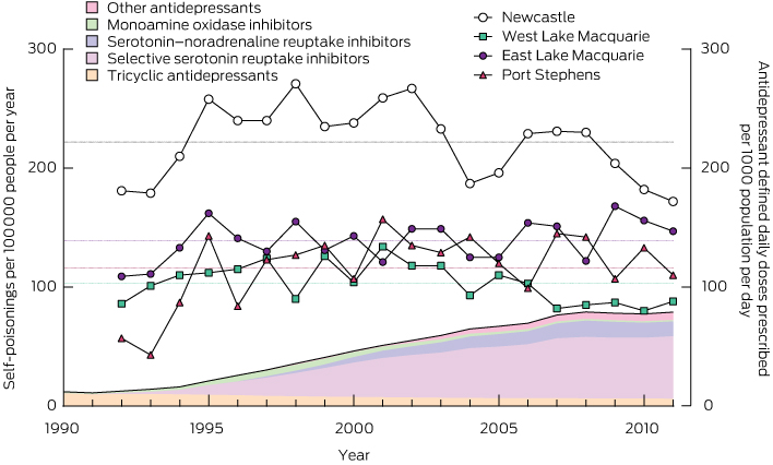
Analyses of population-referenced data (ie, rates) were restricted to postcodes that predominantly cover Newcastle, Lake Macquarie and Port Stephens. Changes in total self-poisoning rates in the four statistical subdivisions in this area were examined between 1991 and 2011. Changes in rates of self-poisoning using the main antidepressant drug classes (tricyclic antidepressants [TCAs], selective serotonin reuptake inhibitors [SSRIs], serotonin–noradrenaline reuptake inhibitors [SNRIs], monoamine oxidase inhibitors [MAOIs] and other) were also examined. Data on rates of antidepressant drug use in these drug classes (standardised by the defined daily dose [DDD]) in Australia from 1991 to 2011 were taken from Australian government publications. We have previously shown that these data agreed within two significant digits with Newcastle-specific data for a range of medications.11

Results

Over the study period, there were 17 266 admissions of patients who had self-poisoned and 11 049 individual patients; the median number of admissions per patient was one (range, 1–115). The number of admissions increased over the first 8 years, but since 1995 has been quite stable (HATS became well established in 1994) (Appendix 1).

Of the total admissions, 15 327 (88.8%) were attempts at self-harm and the remainder were a mixture of unintentional, iatrogenic and recreational self-poisonings. (Data are generally presented for admissions, and may thus include the same patient with different poisonings.) The median age of admitted patients was 32 years (range, 14–97 years) and the female : male ratio was 1.6 : 1 (10 514 female, 6711 male and 39 transgender patient admissions). The median LOS was 16 hours (interquartile range, 9.3–25.7 hours; total time spent in hospital, 15 688 hours). Of the total admissions, 2101 involved admission of the patient to an intensive care unit (ICU) (12.2%) and 1281 involved ventilation of the patient (7.4%). There were 78 inpatient deaths (0.45% of admissions).

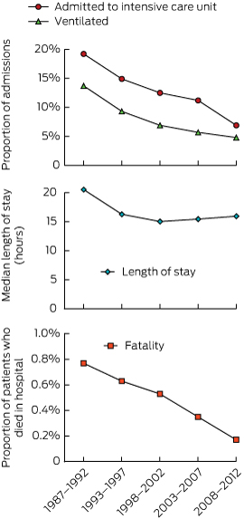
We investigated the changes in morbidity and mortality over the study period by dividing admissions into those in the first 6 years, reported previously,20 and four subsequent 5-year periods (Box 1). Over this period, the rate of admission to ICU dropped from 19.2% (376/1955) to 6.9% (280/4060) and rate of mechanical ventilation from 13.7% (268/1955) to 4.8% (193/4060). The fatality rate dropped from 0.77% (15/1955) to 0.17% (7/4060). The median LOS decreased from 20.5 hours in the first 6 years to about 16 hours in all subsequent 5-year periods.

In the 17 266 admissions, a previous history of psychiatric illness (9692, 56.1%), previous admission for a psychiatric episode (6426, 37.2%), previous suicide attempt (9665, 56.0%) and history of alcohol or drug misuse (8466, 49.0%) were commonly recorded. Few admitted patients were in full-time paid work (2421, 14.0%); some were unemployed (3622, 21.0%), pensioners or retired (4163, 24.1%), students (1058, 6.1%), doing home duties (920, 5.3%) or other (703, 4.1%); data were missing for the remainder (4379, 25.4%). Only 23.8% (4104) were married; others were single (9567, 55.4%), separated (1325, 7.7%), divorced (1302, 7.5%), widowed (409, 2.4%), in de facto relationships (79, 0.5%), other (13, 0.1%); and data were missing for the remainder (467, 2.7%). These demographic, social and psychiatric factors remained stable, except for a slight rise in the proportion of patients reporting previous self-harm (Appendix 2).

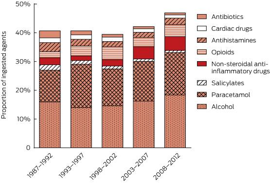
Including co-ingested alcohol, 34 342 substances were involved (mean, 1.99 per patient; range, 1–18 per patient). The major groups of agents involved in self-poisonings and ICU admissions are shown in Appendix 3 (drugs usually available on prescription) and Appendix 4 (non-prescription drugs and other substances). The most commonly ingested substances were benzodiazepines (5470, 15.9%), alcohol (5461, 15.9%), paracetamol (4619, 13.5%), antidepressants (4477, 13.0%), antipsychotics (3180, 9.3%), anticonvulsants (1514, 4.4%), opioids (1232, 3.6%), non-steroidal anti-inflammatory drugs (1104, 3.2%) and antihistamines (743, 2.2%). Prescription items accounted for 18 950 agents (55.2%), of which 14 445 (76.2%) were known to have been prescribed for the patient.

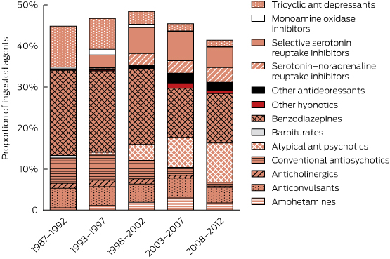
There were major changes over time in the patterns of drugs ingested (Box 2, Box 3), especially for psychotropic drugs and sedatives. Psychotropic drugs consistently accounted for about 50% of all drugs ingested but newer antidepressants and atypical antipsychotics have largely replaced the older drugs (TCAs and conventional antipsychotics). Several of the drug classes for which frequency of ingestion declined (eg, barbiturates, theophylline, TCAs) have disproportionately high toxicity (Appendix 3). In some drug classes, there were larger declines in individual drugs identified as having greater toxicity in the mid 1990s1013 (Appendix 5). This is only partly explained by falling prescriptions for these agents (Appendix 6).

There was a more than sixfold increase in antidepressant DDDs per 1000 people per day between 1991 and 2010 (from 12 to 77). However, the increase in the proportion of poisonings due to antidepressants was very modest (about 1.34-fold). There were no corresponding changes in the population rates of self-poisoning (Box 4, Appendix 6), which fluctuated around the long-term mean in each district. There was thus a large decrease in the rate of self-poisoning per DDD prescribed per 1000 population per day for antidepressants (Appendix 7). In Box 4, the increase in total antidepressant prescriptions is illustrated by the total shaded area and changes in individual classes are illustrated by the coloured shading.

Discussion

The rates of self-poisonings in Newcastle were stable over the past two decades, and the features of the population presenting with self-poisoning were constant. This suggests a long-term ongoing and reasonably predictable need for clinical toxicology treatment and ancillary psychiatric and drug and alcohol support services. Despite large increases in prescriptions for drugs used to treat psychiatric illness (and a range of other major mental health interventions), there appears to have been no positive result in terms of reducing episodes of self-harm.

Interestingly, there was a more than sixfold increase in the use of antidepressants, and while the agents taken in overdose changed substantially, there were only small changes in rates of antidepressant overdoses. Interpreting this surprising finding is not straightforward. It probably indicates that antidepressants are increasingly being prescribed for patients who have minimal risk of self-harm. Reassuringly, there is no evidence in our population to support concerns about pro-suicidal effects of new antidepressant prescriptions. The lack of any change in overall self-harm rates also suggests that increased antidepressant use for depression is not an effective public health strategy to reduce rates of self-harm. The only strategy to prevent fatal poisoning with consistent supporting evidence is restricting the availability of high-lethality methods.4,5

Identification of high toxicity in overdose is a problem that can only be studied after approval for marketing is granted. Postmarketing surveillance by pharmaceutical companies of toxicity in overdose is not a requirement for drug registration in Australia or any other country. There is little incentive for voluntary surveillance. Although most companies record case reports of overdoses of their drugs, this does not facilitate comparisons. Reporting biases mean that such cases may be atypical of the usual clinical picture.

The many new psychiatric medications coming onto the market should mandate coordinated collection of timely information on self-poisoning and suicide. Ideally, this should be done at three levels. First, a national coronial register of drug-related deaths is essential to enable an analysis of relative mortality (as done in the United Kingdom21). Second, data on poisonings reported to poison centres are essential, particularly for childhood poisonings that rarely require admission and for assessing the effect of primary prevention measures. Poison centre data have limitations due to referral bias, lack of uniformity of assessment and lack of clinical information.22 Third, for these reasons, systematic use of clinical databases to record hospital admissions for cases of poisoning is needed to measure relative clinical toxicity.

The HATS clinical database has identified disproportionate effects in overdoses with many drugs. Translation of HATS data into clinical risk assessment and guidelines has occurred, but with lengthy delays. For example, the risk of QT prolongation and torsades de pointes with thioridazine, citalopram and escitalopram10,14,23 were detected in the database 5–10 years after these drugs became available. However, to reduce the time to identify toxicological problems to 1–2 years, collaborating centres in Australia and overseas will be needed to accelerate collection of data on self-poisoning.

Most deaths due to poisoning occur outside hospital. Any significant decrease in mortality from self-poisoning will result from primary or secondary prevention. Efforts at decreasing morbidity and mortality from self-poisoning should continue to target drugs that are frequently taken or are lethal in overdose.

The falls in prescriptions and poisonings with several drugs with greater relative toxicity occurred several years after the problems relating to overdoses with these drugs were identified in the HATS database around 1994–1995 (Appendix 6, Appendix 8). For example, large reductions were due to withdrawal of drug subsidies (those for dextropropoxyphene and thioridazine were withdrawn in 2000) and removal of a formulation that was misused (temazepam gel-filled capsules were withdrawn in 2001). Publication of toxicity data alone had limited (if any) effect in terms of reducing prescriptions of the more toxic drugs. No drugs were banned in Australia on the basis of HATS data, although manufacturers did voluntarily withdraw some of the highlighted drugs (barbiturates and chloral hydrate were withdrawn in 1994–1995, thioridazine was withdrawn in 2009). Further, there has been no drop yet in prescriptions of or poisoning with drugs identified as having greater toxicity in the mid 2000s14,15 (data not shown).

Our data show large drops in rates of poisoning with some of the more lethal drugs, such as TCAs and barbiturates (Box 2, Box 4). The introduction of less toxic antidepressants and sedatives dramatically changed prescribing, which in turn changed the types of drugs taken in self-poisoning. This trend has presumably also been reflected in changes in drug-related deaths. However, the nature of coding in official death statistics means that there are no published Australian data to support this contention. For example, poisoning by “antiepileptic, sedative-hypnotic, antiparkinsonism and psychotropic drugs” are all lumped together under one code in Australia.1 Most fatal poisonings are classified as due to unspecified or multiple agents. Some improvement in coding of drug-related deaths should not be difficult. Much finer detail is provided in other ICD-10 codes. For example, the Australian Bureau of Statistics records deaths from crocodile and rat bites (three and zero, respectively, between 1999 and 2008) separately from those caused by other animal bites.1 The development of the National Coronial Information System, launched in 2000, may make fatal poisoning comparisons possible in future, but only if data are accurately and consistently coded. Assessing the impact that clinical toxicology has on direct patient management and on public health is hindered by the lack of reliable epidemiological and clinical data.

Our data have inherent limitations. There is likely to be selection bias against less severe poisonings (the types of cases where patients might not present for medical attention) and rapidly lethal poisonings (cases in which patients die outside hospital). In surveys of self-harm and anecdotal reports from patients, it has been estimated that a significant proportion of people who have self-poisoned (5%–15%) don’t present for medical care.3 Also, although there is a prospective data collection form, retrospective review of medical records is often required to complement prospectively collected data. Data on the ingested drugs were based on patient history, including corroborating history obtained from ambulance officers and accompanying people and from information on drug containers. Drug concentrations were not measured in most patients, although previous research on specific drugs has found the patient history of drugs ingested to generally be confirmed by an appropriate assay.24

The key strengths of our study are its long duration and the consistent core data fields. There are few similar attempts to gather data longitudinally on self-poisoning over prolonged periods. Many have retrospective identification of cases from hospital coding and thus rely entirely on the completeness of medical records.25 Others have not been collected continuously26 or have focused on psychiatric factors and treatment (rather than drugs ingested and toxicity)27 or were conducted in developing countries where agents ingested differ substantially from those used in Australia.28

However, the uniqueness of the HATS database also highlights a weakness of our study. As there are no comparable current datasets, it is difficult to determine the extent to which the Hunter experience represents that of the developed world. Expansion of the database could be facilitated by database systems integrated with electronic medical records.

We identified interesting and important patterns relating to drug prescriptions, epidemiology of overdose patients and importance of relative toxicity. A massive increase in antidepressant prescriptions has had little impact on rates of self-harm or antidepressant poisoning. Changes in antidepressant classes (generally from more to less toxic) have had significant effects on morbidity and mortality from antidepressant poisoning, and therefore from all poisonings. We were also able to generate information regarding relative toxicity and patient management. However, for many rare poisons, gaining sufficient numbers of patients to generate reliable information about management and prognosis will take decades from one centre. We believe that, in Australia and overseas, there is a need for a coordinated approach to address the toxicity of drugs in overdose. The public health benefits would greatly outweigh the modest costs of enhancing postmarketing surveillance through more widespread systematic collection of poisoning and overdose data.

1 Morbidity and mortality due to self-poisoning, 1987–2012, measured by need for intensive care and ventilation, length of stay and fatality*


* Data were divided into five periods with roughly equal patient numbers.

2 Use of sedatives and psychotropic drug classes in self-poisoning, 1987–2012

3 Use of alcohol, analgesics and selected other drug classes in self-poisoning, 1987–2012


4 Rates of antidepressant prescribing (1990–2011, shaded areas) and total rates of self-poisoning (1992–2011, solid lines and symbols)*


* Total numbers of admissions for self-poisoning before 1993 are slightly lower as the Hunter Area Toxicology Service was just being established. Dotted lines indicate 20-year mean for each district.