×

The efficacy of bariatric surgery performed in the public sector for obese patients with comorbid conditions

Obesity (body mass index [BMI] ≥ 30 kg/m2) is a growing health problem and is recognised as one of the largest contributors to the chronic burden of disease. Currently, 28% of Australians are obese, placing our nation second for men and fifth for women among OECD (Organisation for Economic Co-operation and Development) countries ranked by prevalence of obesity.1,2 In Australia, an inverse relationship exists between high obesity prevalence and low socioeconomic status; incidence is almost double for areas indexed as the most disadvantaged compared with areas within the highest strata.3 Those living more remotely also have higher obesity rates — 59% versus 32%.2 Among Indigenous Australians, the prevalence of obesity is almost double (34% versus 18%) and that of type 2 diabetes mellitus (T2DM) is triple that of non-Indigenous adults, resulting in sevenfold greater mortality due to diabetes.2,4

Bariatric surgery is an effective treatment of severe obesity (class III [BMI ≥ 40 kg/m2] or class II [BMI ≥ 35 kg/m2] with comorbid conditions) and is purportedly cost-effective, compared with conservative measures.57 The evidence supports sustained postoperative weight loss, ameliorating obesity-related comorbid conditions. The Swedish Obesity Study showed a marked reduction in hypertriglyceridaemia, T2DM and hyperuricaemia with surgery after 2 and 10 years.6 The resultant postsurgical weight loss substantially reduces, resolves and even prevents the metabolic complications associated with increasing central adiposity, with 73%–95% T2DM remission rates by 2 years, depending on the type of surgery.8,9

The heavy economic burden of obesity and its comorbid conditions may be alleviated in the long term by surgical management, despite upfront resource costs.10 Severely obese individuals incur twofold higher mean annual health care costs ($2788 v $1472) and use double the number of medications annually (11.4 v 5.3 per person) compared with the general population.7,11 Weight loss surgery can reduce the number of medications required and lower individual health care costs by 26%, a direct saving of $506 per person.7,12 With evidence of reduced mortality and an acceptably low complication profile (estimated mean 30-day mortality < 0.3%), bariatric procedures are supported by national and international health bodies.1315

The National Health and Medical Research Council (NHMRC) 2013 guidelines recommend bariatric surgery as the most beneficial and cost-effective management for motivated individuals with severe obesity.16 Motivation is an essential factor in considering whether an individual with obesity and comorbid conditions is a suitable candidate for surgery. Surgery is not the final step in the clinical pathway of severe obesity management; postoperative commitment to lifestyle change and regular follow-up are requisite for successful weight loss and continued improvement in health.16 Most individuals are motivated to have surgery for greater control of medical ailments, yet it is unknown whether this applies to those whose procedure is fully funded.17

Since 1992, Medicare has reimbursed the cost of bariatric surgery in the private sector. As most surgery is carried out in private hospitals with large out-of-pocket expenses for those without private health insurance, a significant inequity in obesity management exists.18 Paradoxically, this surgery is least accessible to those who are likely to be in greatest need. Since October 2009, a pilot program has been underway in the Sydney and South Western Sydney Local Health Districts in which bariatric surgery has been publicly funded for a limited number of patients meeting strict inclusion criteria. There are no Australian studies to date that have identified if publicly funded surgical intervention for severe obesity confers the same health benefits seen in private health care. This study aims to assess the efficacy of bariatric surgery for such patients in the Australian public health system.

Methods

Study design

Sixty-eight moderately to severely obese participants with comorbid conditions were deemed eligible for bariatric surgery, and inclusion in our study. Participants were attendees at an ongoing collaborative pilot program run by one of three obesity clinics based within the Sydney and South Western Sydney Local Health Districts. All participants received conservative management for their obesity and related health conditions delivered by a multidisciplinary team trained in obesity management. Participants were seen 6- to 12-weekly for dietary advice, behavioural modification, advice on physical activity and, where appropriate, very low energy diets (VLEDs) and pharmacotherapy. Participants with substantial obesity-related comorbid conditions who had not achieved adequate weight reduction with conservative intervention, and who were interested in surgical management, were assessed by their multidisciplinary team to determine suitability for bariatric surgery. Longitudinal data were collected on those individuals who underwent bariatric surgery for management of resistant obesity and associated comorbid conditions.

The inclusion criteria for bariatric surgery were: age 18–75 years, minimum class II obesity (BMI ≥ 35 kg/m2) with comorbid conditions, completion of at least 1 year of medical intervention during which the participant had demonstrated commitment to lifestyle change, and surgery before 31 May 2013 (enabling a minimum follow-up period of 3 months). Exclusion criteria were: inability to consent, irreversible endocrine causes of obesity and significant comorbid conditions that were expected to result in poor outcomes from surgery, such as unstable cardiovascular disease and uncontrolled psychiatric illness. Participants underwent 2 weeks of VLED before the surgery to reduce abdominal adiposity and liver volume in particular, as liver size can complicate surgical access.19 They were also instructed about the necessary commitment to postoperative care, specifically the importance of nutritional input. Two of us (C J T and D J M) performed either laparoscopic sleeve gastrectomy (LSG) or laparoscopic adjustable gastric banding (LAGB) at a single public hospital. The type of surgery performed was decided by the participant in conjunction with the surgeon, after being briefed on the techniques, risks and expected benefits of each procedure. All participants were screened for nutritional deficiencies before and after surgery.

Ethics approval was obtained from four Human Research Ethics Committees within the Sydney and South Western Sydney Local Health Districts. All participants gave written consent for use of their records for research purposes.

Clincial and biochemical assessments

Surgeries were performed on an as-needed basis from 1 October 2009 to 31 May 2013. Pre- and postoperative results were analysed at seven time points until 1 September 2013. The baseline (preoperative) measurements were recorded at the start of the VLED, at least 2 weeks before surgery (time zero). Postoperative data were recorded from follow-up appointments at 3, 6, 12, 18, 24 and 36 months. The primary end points analysed were weight, BMI and waist circumference, and secondary end points were four common obesity-related comorbid conditions: T2DM, dyslipidaemia, hypertension (HTN), and obstructive sleep apnoea (OSA). Improvement was defined by normalisation of laboratory markers (T2DM and dyslipidaemia), blood pressure sphygmomanometry (HTN) and polysomnography results (OSA). Comorbid conditions were considered “partially resolved” when participants’ measurements fell within normal limits, the number of medications was reduced or the use of the continuous positive airway pressure (CPAP) device was discontinued. Comorbid conditions were deemed “fully resolved” when normal measurements remained after all relevant medications were discontinued.

Definition of comorbid conditions

T2DM was defined as fasting blood glucose levels ≥ 7 mmol/L, glycated haemoglobin level ≥ 6.5% and/or requirement of at least one oral hypoglycaemic agent. HTN was defined as systolic blood pressure ≥ 140 mmHg, diastolic blood pressure ≥ 90 mmHg and/or requirement of at least one antihypertensive agent. Dyslipidaemia was defined as total cholesterol level ≥ 5.0 mmol/L, low-density lipoprotein level ≥ 3.5 mmol/L, triglyceride level ≥ 2.0 mmol/L, or high-density lipoprotein level ≤ 1.01 mmol/L for men and ≤ 1.3 mmol/L for women, and/or requirement of at least one lipid-lowering medication.20,21 OSA was defined according to symptoms (snoring, observed apnoea, daytime somnolence), treated HTN and the apnoea–hypopnoea index record from polysomnography.

Statistical analysis

The results at the seven time points were reported as mean and SD. The differences were tested for significance using paired t tests for continuous variables and the McNemar test for paired categorical variables. The t test was used to evaluate between-surgery differences in mean weight at baseline and subsequent time points, to assess the validity of combining the two surgery types in analyses. Mixed-model regression was used to determine if significant change in weight occurred over time at all seven time points, to account for reductions in sample size with time. All statistical analysis was performed using SAS version 9.3 for Windows (SAS Institute).

Results

Participant characteristics

Of the 68 participants offered surgery, two declined and one moved interstate. Sixty-five participants (41 women and 24 men) with a mean age of 51.5 years (SD, 11.8 years; range, 21–73 years) underwent bariatric surgery between 1 October 2009 and 31 May 2013. The level of comorbid conditions in the group at baseline was high, with a mean of eight conditions per patient and class III (severe) obesity. From baseline to 18 months, only three participants were lost to follow-up at each time point; however, numbers are lower with time as operations were done at different times (ie, only 10 patients had surgery 36 months ago whereas 65 had surgery 3 months ago, 58 had surgery 6 months ago, etc [see Box 1]). Data on all participants who completed follow-up at each respective time point according to surgery date are presented in Box 1 and Box 2.

Intraoperative outcomes

All 65 surgeries were performed laparoscopically and none required conversion to open surgery. The most commonly performed procedure was LSG (57 patients; versus eight patients who had LAGB). Despite it appearing that greater weight change occurred in the LSG group, the difference was not significant at 3 months (P = 0.58), 12 months (P = 0.25) or 24 months (P = 0.17). There were no significant intraoperative complications (one participant had a haematoma that resolved with immediate evacuation) and length of hospital stay was within expected times for all patients (LSG, three nights; LAGB, one night).

Short-term postsurgical outcomes (3–6 months)

There was a reduction in all primary end points in the early postoperative period. By 3 months there was a mean weight reduction of 22.6 kg (SD, 9.5 kg) (Box 3) with a 7.4 kg/m2 mean reduction in BMI and 14.5 cm mean reduction in waist circumference (Box 1). All comorbid conditions showed partial resolution from 3 months (Box 2). Of the numbers of participants who had each comorbid condition at baseline and who had data available, there was full resolution by 6 months in 20/45 with T2DM, 14/43 with HTN, 7/47 with dyslipidaemia and 17/41 with OSA (Box 2). The requirement for all relevant medications also reduced accordingly (Box 4).

Medium-term postsurgical outcomes (12–18 months)

By 1 year, there was a significant reduction in all anthropometric measures. The mean weight loss was 34.2 kg (SD, 20.1 kg), and the mean BMI was reduced by 12 kg/m2 to 36.2 kg/m2 (SD, 7.7 kg/m2). By 18 months, of those who reported the respective comorbid conditions at baseline, almost half of the group had full remission of T2DM and three-quarters had resolution of OSA. Modest resolution of dyslipidaemia was seen with both a significant rise in high-density lipoprotein and reduction in triglycerides occurring transiently at 6–12 months in 7 of 47 patients who had dyslipidaemia at baseline (< 0.001).

Long-term postsurgical outcomes (24–36 months)

The trend for continued weight loss and partial or full resolution of comorbid conditions appears maximal at 12–24 months. While the smaller group sizes at the 24- and 36-month follow-ups provided less power for statistical analysis, primary and secondary end points remained stable (P < 0.001). The maximal amount of mean weight loss occurred at 24 months (39.9 kg; SD, 31.4 kg). Three-quarters of the cohort had reduced the number of medications taken for HTN and T2DM by 18–24 months, and by 36 months, all had reduced the number of antihypertensive medications (Box 4).

Postoperative complications

Transient nutritional deficiencies were found in three of 65 participants, improving within 3 months with replacement treatment. Of the eight patients who had LAGB, one underwent conversion, due to gastric band erosion, to a Roux-en-Y bypass at 18 months. Of the participants who had LSG, one had a small bowel obstruction secondary to adhesions and required laparoscopic division at 2 years. Three participants required endoscopic stomach dilatation and a further three continued to experience nausea, dyspepsia and/or vomiting at 3 months, but this resolved by 6 months for two and at 12 months for one participant.

Weight change over time

A total of 288 observations for weight from 65 participants were available for linear mixed-model analysis. Significant change in mean weight was observed over time (P < 0.001). Mean weight adjusted for baseline weight decreased from 136.5 kg at baseline to 101.1 kg at 12 months. Stratifying the analysis by sex showed that men had a higher mean baseline weight, 147.5 kg (SD, 36.9 kg) compared with women, 130.1 kg (SD, 23.8 kg), but no difference was observed after 1 year (Box 3).

Discussion

In our study, the significant weight lost by obese participants who underwent bariatric surgery occurred early and was sustained over the first 3 years. The mean maximal weight loss was almost one-third of participants’ preoperative weight and was achieved by 24 months. Despite decreasing sample sizes, our results showed sustained weight loss at 36 months.

There was full or partial resolution of all comorbid conditions tested, except dyslipidaemia, in most participants by 2 years. Different rates of resolution of comorbid conditions occurred, with remission of T2DM being the earliest. By 24 months, there was an associated resolution of T2DM, HTN and OSA, or the need for pharmacotherapy or devices was reduced in most patients, yet dyslipidaemia showed inconclusive results. These findings parallel other bariatric studies.22,23 The Swedish Obesity Study found isolated improvements in hypertriglyceridaemia postoperatively (including after LAGB), although the most consistent improvements in lipid profiles have been seen after Roux-en-Y bypass surgery.6,24

Most LAGB procedures were performed in the first year of the study, until LSG became a preferred option due to its greater weight loss profile with minimal additional complications and the lower need for surgical follow-up. However, throughout the study, participants had autonomy of procedure choice (on surgeon recommendation). While this may suggest an avenue for bias (the participants chose their “intervention”), bariatric surgery is an elective procedure, and it was deemed necessary to replicate the protocol in the private sector. The limitations of our study include its small size, that it is a case series, that it is non-blinded, possible bias due to participants choosing their surgery type, and the use of a clinical database with the possibility of missing or inaccurate values imputed by health professionals. The low rates of adverse events observed in our study are consistent with those reported in the literature.

The direct operative costs of performing these surgeries in the public sector in our study were estimated to be $7000–$9000 (LSG incurred greater up-front theatre costs than LAGB). Perioperative costs, including 2 years of postsurgical visits, may approach $2000 per person, taking the total cost to $9000–$11 000 per person. A 2005 paper reported the annual cost of managing an individual with T2DM as $9095–$15 850.25 Thus, if an obese person with T2DM has bariatric surgery, the operation would pay for itself after about 1 year.

The participants’ baseline characteristics are representative of a typical demographic seen in the public sector, supporting extrapolation of our results to a wider population. The high attendance rate at 2 years shows that the cohort demonstrated adequate motivation to justify surgical intervention. The health potential from bariatric surgery ranges from improved quality of life and amelioration of comorbid conditions to full resolution of complications and reduced mortality for all individuals, paying or not. Strategies to prioritise access are therefore recommended to reduce the apparent inequality that exists. Limited access to surgery discriminates against those who cannot afford the out-of-pocket costs, yet it is likely that this subgroup would benefit most. In conclusion, we hope that our study provides an evidence base for the surgical treatment of obesity in the public health system and, in turn, that consideration will be given to increasing the supply of publicly funded bariatric surgery in Australia.

1 Patient anthropometric data before and after bariatric surgery

Measurement

Baseline (2 weeks before surgery)

Postoperative month 3

Postoperative month 6

Postoperative month 12

Postoperative month 18

Postoperative month 24

Postoperative month 36


N (n)

65 (65)

65 (65)

55 (58)

49 (52)

30 (33)

17 (23)

7 (10)

BMI (SD), kg/m2

48.2 (9.5)

40.8 (8.3)*

38.9 (7.9)*

36.2 (7.7)*

38.2 (12.1)*

35.7 (7.7)*

38.7 (9.4)

Weight (SD), kg

136.5 (30.3)

113.9 (25.2)*

108.3 (24.8)*

101.1 (22.4)*

99.3 (20.8)*

97.6 (21.9)*

108.6 (25.6)

Waist circumference (SD), cm

132.2 (16.5)

117.7 (15.2)*

114.9 (14.7)*

109.4 (13.7)*

110.2 (12.3)*

108.4 (15.8)*

114.9 (14.6)*

% weight loss from baseline

0

17%

21%

26%

27%

29%

21%


BMI = body mass index. N = total number of participants who attended follow-up. n = total number of participants including those lost to follow-up. * Comparison between baseline and follow-up; P < 0.001. † Comparison between baseline and follow-up; P < 0.05.

2 Proportions of patients* who continued to have conditions found at baseline

Comorbid condition

Baseline (2 weeks before surgery)

Postoperative month 3

Postoperative month 6

Postoperative month 12

Postoperative month 18

Postoperative month 24

Postoperative month 36


T2DM

53

36/52

25/45

21/42

12/23

8/17

3/6

HTN

51

45/51

29/43

18/40

10/27

4/12

2/6

Dyslipidaemia

58

45/58

40/47

33/42

13/24

8/15

4/6

OSA

41

37/41

24/41

15/41

6/24

4/17

2/5

OSA requiring CPAP

27

18/27

14/23

8/22

4/13

2/8

1/3


CPAP = continuous positive airway pressure. HTN = hypertension. OSA = obstructive sleep apnoea. T2DM = type 2 diabetes mellitus. * Numerators are number of participants who had that condition at that time point. Denominators are total number of participants who had that condition at baseline and had data at that time point. † Comparison between baseline and follow-up; P < 0.001. ‡ Comparison between baseline and follow-up; P < 0.05.

3 Patients’ weight change over time after bariatric surgery*


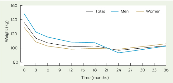
* Weight change at all seven time points is significant (P < 0.001). Despite differences in weight lost for men and women, there were no significant between-sex differences.

4 Number of patients with a reduction in medications used at each time point after bariatric surgery


Antihypertensive medications


Glucose-lowering medications


Lipid-lowering medications


Postoperative month

Patients at
each time point

Patients with reduced use of medication

Patients at each time point

Patients with reduced use
of medication

Patients at each time point

Patients with reduced use
of medication


3

65

31

60

28

64

6

6

54

34

54

29

53

10

12

48

37

47

25

44

12

18

29

22

23

17

24

12

24

16

12

16

15

13

3

36

6

6

6

4

4

1

Dysphagia, regurgitation and weight loss in an elderly man

An 84-year-old man reported unintentional weight loss of 15 kg over 18 months and dysphagia and regurgitation of undigested food over 3 months. Barium swallow showed a large pharyngo-oesophageal (Zenker) diverticulum. These are caused by elevated intraluminal pressure at the Killian dehiscence, an area of hypopharyngeal weakness between the cricopharyngeus and inferior pharyngeal constrictors.

Zenker diverticuli are rare, but can cause significant morbidity and mortality. A barium swallow should precede gastroscopy, as there is a risk of perforation and mediastinitis. We performed endoscopic stapling, as it is less invasive than open repair.

Partial foot amputations may not always be worth the risk of complications

To the Editor: In their recent article, Dillon and colleagues cite reports that the incidence of partial foot amputations is rising and that the functional outcome of these amputations is poor.1 They express difficulty in understanding “why the high risk of complications and secondary amputations associated with partial foot amputation do not weigh more heavily in decisions about amputation surgery”. Finally, they propose that more transtibial than partial foot amputations should be considered to minimise the risk of complications and further amputation.

The authors’ argument is misleading as they combine two very different entities, toe amputation and major forefoot amputation, as the single entity of “partial foot amputation”. As their own work has shown,2 toe amputations make up 90% of all “partial foot amputations” and it is mainly toe amputations that are becoming more common. The evidence they cite about complications and poor outcomes in “partial foot amputations” is based exclusively on (less commonly performed) transmetatarsal and midfoot amputations, and not on toe amputations. While the early morbidity of toe (±metatarsal head) amputation is not insignificant, the late functional outcomes are likely to be more favourable than transmetatarsal or midtarsal amputation.

Our interpretation of the available evidence is that the rising incidence of “partial foot amputations” is driven by more toe amputations in the increasing number of people with type 2 diabetes. This preserves acceptable function and may be contributing to the decline in major amputations. We acknowledge that further research is needed to clarify the relationship between these diverging trends.

Partial foot amputations may not always be worth the risk of complications

To the Editor: Dillon and colleagues present a challenging perspective on the evidence comparing partial foot amputation (PFA) and below-knee amputation (BKA) outcomes.1,2 Australia’s diabetes-related major amputation rates have only recently reduced to international levels3 and we fear that any oversimplistic perspectives may be detrimental to these improved rates and, importantly, to our patients. Thus, we believe these articles1,2 should be read cognisant of some important points.

First, the authors correctly identify that 75% of Australia’s amputations are PFAs (toe, ray or transmetatarsal amputations [TMAs]),4 yet base their PFA arguments nearly entirely on TMA literature.1,2 TMAs are complex procedures and make up just 5%–10% of PFAs.4 Thus, we believe the article should be primarily read as a comparison between TMA and BKA outcomes.

Second, the authors correctly report higher ipsilateral reamputation risks for TMAs compared with BKAs;1,2 however, they omit lower contralateral amputation risks5 and somewhat dismiss well reported lower mortality risks.1,2,5

Last, the authors conclude “very similar functional outcomes” from data comparing methodologically different TMA studies with BKA studies or data within heterogeneous and underpowered studies.1,2 These definitive interpretations may be misleading, especially given the statistically significant functional benefits of TMAs compared with BKAs, as reported in the higher impact studies the authors cite.2,6

We thank Dillon et al for highlighting such a large, yet silent, burden of disease and hope this letter will provide more balance to this crucial life-changing decision facing 8000 Australians and their clinicians this year.4

Partial foot amputations may not always be worth the risk of complications

In reply: We appreciate the opportunity to reply to two letters submitted in response to our article.1

Both letters agree that our perspective piece was mainly based on evidence about the outcomes for people with transmetatarsal amputation, noting that most people undergo amputation of the toe(s) or toes and metatarsals. As highlighted in our supporting work,2 there are comparatively few investigations focusing on outcomes for people with digital, ray, tarsometatarsal and transtarsal amputation. Despite this, we stand by our interpretation that the rates of complications and reamputation seem very similar across levels of partial foot amputation.1,2 To illustrate, a study that stratified large numbers of people by level of partial foot amputation found that the rates of ipsilateral reamputation were not statistically different in groups with either toe, ray or mid-foot (ie, transmetatarsal, Lisfranc and Chopart) amputation.3 Given these data, we argue that our synthesis of published outcomes on the rates of reamputation and other complications for people with different levels of partial foot amputation was reasonable. Our article should not be considered a comparison between the outcomes of transmetatarsal and transtibial amputation.

We do not believe that current data show that toe amputations are becoming more common in Australia. The age-standardised incidence of toe amputation remained stable between 2000 and 2010, while the incidence of partial foot amputation at the toe and metatarsal level and transmetatarsal level increased.4

We are not advocating that more transtibial amputations should be considered to minimise the risk of complications and further amputations. Rather, clinicians should consider the emerging evidence when communicating the perceived benefits to patients; particularly given that this evidence challenges long-held beliefs.2

We are grateful for the opportunity to promote discussion and highlight awareness of the need for further research into outcomes for people facing difficult decisions about limb loss.

Guidelines fall short on bariatric surgery

Appropriate guidance is lacking in long-term nutritional monitoring and support

The National Health and Medical Research Council (NHMRC) Clinical practice guidelines for the management of overweight and obesity were released in early June 2013, replacing the 2003 version, intended for use by specialists and general practitioners.1 A multidisciplinary committee oversaw the guideline development process, aiming to systematically identify and evaluate evidence. In my view, this process has failed with regard to the section dealing with bariatric surgery, thereby failing severely obese Australians and those caring for them.

My greatest concern is the section about nutrition and supplementation after bariatric surgery. Nutritional issues are a critical downside to bariatric surgery and should be front of mind whenever doctors, nurses, dietitians or any other health professionals interact with a patient after surgery. High-quality guidance is required in the care of the thousands of patients who have had bariatric surgery. After bariatric surgery, energy intake is markedly reduced, food choices and diet quality often change, absorption of micronutrients such as iron, calcium, vitamin D and vitamin B12 can be impaired with some procedures and, more rarely, protein malnutrition and more complex deficiencies occur. All bariatric procedures require excellent nutritional support, monitoring and supplementation.

The guidelines’ suggestion that nutritional issues should be assessed through clinically manifest disease, including neuropathy, weakness and muscle wasting, bone pain and oral lesions, is dangerous, reflecting an “after the horse has bolted” mentality. Progressive nerve damage related to nutritional deficiency can be catastrophic and only partially reversible;2 metabolic bone disease related to nutritional deficiency is symptomatic only when it is generally too late to take any effective preventive action; and it is definitely too late when a pregnant woman’s baby is diagnosed with a neural tube defect. Anaemia, metabolic bone disease, and neuropathy are reported at much higher levels after bariatric surgery than in the community. Nutritional issues are predictable, and also preventable with appropriate monitoring.

These nutritional guidelines contrast starkly with the broader literature and the recently released United States guidelines for pre- and post-bariatric surgical care developed conjointly by the American Association of Clinical Endocrinologists, the Obesity Society, and the American Society for Metabolic and Bariatric Surgery.3 Their detailed literature review comes to very different conclusions. However, the recent NHMRC document1 does not detail any critical appraisal of the literature about nutritional support, monitoring or supplementation after bariatric surgery. What was the evidence base for the guidance provided, given its inconsistency with known nutritional deficiencies and the available literature on prevention? As soon as I became aware of the guidelines, I alerted the NHMRC and colleagues who were members of the relevant committees to the specific issues that worried me.

The greatest concern is the process that led to this unsatisfactory section of the guidelines. The review process is described as rigorous and transparent, yet the result, in my view, is not evidence-based, and is potentially dangerous. Rigour and transparency were used when looking at the weight loss extent and duration, the changes in obesity-related comorbid conditions and the overall mortality advantage after bariatric surgery. However, the guidelines are intended to provide practical assistance to health professionals managing chronic disease in patients with severe complex obesity who have undergone bariatric surgery. Surgery does not cure obesity or obesity-related comorbid conditions. Lifelong follow-up and support are required.

I recommended that the NHMRC consider revising the section on nutrition and nutritional supplementation after bariatric surgery. In addition, the NHMRC should review its aims and processes before conducting evidence-based reviews to ensure that guidelines provide the most relevant practical information for the target audience rather than a detailed formal review with limited practical relevance to patient selection and care. The NHMRC has recently confirmed that the section of concern will be revised. Meanwhile, I urge bariatric surgeons and their multidisciplinary teams to follow high-quality guidelines,3 provide individualised advice to patients and their key health care providers. Additional support for practitioners is available in two recent reviews detailing the nutritional aspects of bariatric surgery.4,5

Lack of appropriate imaging before breast augmentation can have serious patient consequences

Advice on preoperative imaging for different groups of women is now available

Breast augmentation has become increasingly popular in recent years, with 316 848 procedures reported in the United States in 2011.1 While there is a lack of accessible data about breast augmentation procedures in Australia, the breast surgical oncology community has ongoing concerns about the potential for breast implants to deter women from participating in appropriate symptom assessment or routine screening and to impair early detection of breast cancer.2

The situation is complicated by a lack of high-level evidence about health outcomes for women with implants who subsequently develop breast cancer. In an observational study of 129 women who had undergone breast augmentation and 3953 women who had not, those who had undergone augmentation presented more frequently with palpable lesions, invasive tumours and axillary nodal metastases, and were more likely to have a false-negative mammogram result. However, overall there was no significant difference in stage of disease, tumour size, recurrence rates or survival between the two groups.3 It was postulated that breast implants may facilitate palpation of tumours and make the breast easier to examine, explaining why lesions of the same size were more frequently palpable in patients with implants.3 This explanation fits with our clinical experience, but there are other possible reasons for the higher rates of invasive tumours and axillary nodal metastases in this group. These explanations relate to the higher likelihood of a false-negative mammogram result in women with implants who do have regular screening, and to the lower rate of screening in women with implants because of reluctance to have mammograms for fear of damaging the implants.

The discrepancy between the higher rates of invasive tumours and axillary node metastases among women who have had breast augmentation on the one hand, and a lack of significant differences in cancer staging between women with and without breast implants on the other, may be explained, in part, by methodological limitations. These include use of the χ2 test, which may not have enough power to detect trends, and lack of consideration of confounding factors, such as age. According to a systematic review of observational studies, women with breast implants who develop breast cancer have later-stage tumours at diagnosis, while meta-analysis of these studies suggested that these women may also have an increased risk of non-localised breast tumours and lower rates of breast cancer-specific survival.4 The authors noted the need for further research to clarify the impact of breast augmentation on cancer detection and prognosis.

What is clear is that patients with breast implants present specific challenges for radiologists involved in breast imaging. Breast implants are radio-opaque and restrict the visualisation of breast tissue with mammography.4 Implants can also compress breast tissue, which can limit detection of subtle findings such as architectural distortion and microcalcifications. Twenty-six experts from five countries reviewed the best available evidence and concluded in a consensus statement that preoperative breast imaging, with mammography and/or ultrasound, should be considered essential, except in rare circumstances, before any breast augmentation procedure.5 Appropriate imaging may vary depending on the woman’s age, risk factors for breast cancer, breast density and clinical examination findings, but would usually involve mammography, possibly ultrasound and sometimes magnetic resonance imaging.

Failure to provide appropriate imaging not only has serious implications for the woman herself, but also for the health system, which is left paying for expensive surgery and adjuvant therapies that may have been avoided had the cancer been detected earlier, as a recent case from our practice demonstrates (Box 1). This case highlights the serious consequences of a probable missed opportunity to diagnose breast cancer 6 months earlier, when the disease most likely would have been entirely in situ. There are two implications for clinical practice. First, thorough clinical examination and risk assessment, as well as appropriate imaging, should become a routine component of the preoperative work-up for women who undergo breast augmentation. Second, if a general practitioner is aware of a patient’s intent to undergo breast augmentation, in Australia or overseas (given the increase in medical tourism), he or she should inform the patient of the importance of preoperative screening and arrange the appropriate imaging.

As a result of such cases, Breast Surgeons of Australia and New Zealand has developed a position statement on preoperative assessment of women who undergo breast surgery, which outlines the consensus view of what imaging is appropriate for different categories of women.2 The recommendations are summarised in
Box 2.

1 Case of breast cancer in a patient with breast implants

A 46-year-old woman presented with odd sensations in her right breast 6 months after undergoing bilateral breast augmentation surgery without preoperative breast imaging.
A mammogram and ultrasound revealed a 6.5 cm area of malignant calcifications. Analysis of a core biopsy specimen showed ductal carcinoma in situ (DCIS) and the patient underwent right mastectomy and sentinel node biopsy. The final pathology report described 9 cm of high-grade DCIS with multiple small foci of oestrogen receptor-negative and human epidermal growth factor receptor 2-positive invasive breast cancer. The sentinel nodes were free of disease. After multidisciplinary consultation, we recommended chemotherapy and trastuzumab treatment, which the patient agreed to.

2 Appropriate preoperative investigations for women undergoing breast augmentation or reduction, according to age and risk status*

Clinical
examination

Mammography

Ultrasound

Magnetic resonance imaging


Age < 40 years

Average risk

Yes

Consider

Consider

No

Increased risk§

Yes

Yes

Yes

Consider

Difficult to clinically assess

Yes

Consider

Yes

No

Age ≥ 40 years

Average risk

Yes

Yes

Consider

No

Increased risk§

Yes

Yes

Yes

Consider

Difficult to clinically assess

Yes

Yes

Yes

Consider


* Reproduced with permission from Breast Surgeons of Australia and New Zealand.2 Significant clinical findings, including abnormal results of clinical examination or abnormal symptoms, require full assessment by a breast physician or breast surgeon and may require a biopsy depending on the nature of the problem. Magnetic resonance imaging is specifically indicated only if recommended by a breast specialist. It can only be claimed on Medicare for patients in Cancer Australia’s risk category 3. § Defined as significant family history (Cancer Australia risk category 2 or 3) or previous clinical history that indicates high risk (eg, prior biopsy specimens that show proliferative breast disease such as lobular carcinoma in situ, atypical ductal hyperplasia, atypical lobular hyperplasia or multiple papillomatosis). Defined as suspicious or inconclusive findings, or situations where assessment is difficult due to anatomical features such as dense breasts.

First aid for burns: too little, too late and often wrong

To the Editor: The Australian and New Zealand Burns Association (ANZBA) defines adequate first aid for acute burns as 20 minutes of cold running water within the first 3 hours of a burn injury.1 Despite first aid campaigns, inappropriate and inadequate first aid treatment for burns continues to occur.2

Following Sydney Children’s Hospitals Network Human Research Ethics Committee approval, we performed a retrospective analysis of the first aid received by 4368 children who presented to the Burns Unit at The Children’s Hospital at Westmead between 1 January 2008 and 31 December 2012.

Nearly a third of children (34% of inpatients and 30% of outpatients) received inadequate, inappropriate or no first aid, irrespective of the size of
the burn. Inadequate first aid included cold compresses or wet wraps in 414 children (9.5%) and Burnaid (Rye Pharmaceuticals) in 238 (5.4%). Inappropriate first aid included ice in 227 children (5.2%), with a wide range of bathroom products, foods, creams and oils used in most of the remaining patients. In 144 children (3.3%), the first aid received was not documented.

Products prescribed by general practitioners, ambulance officers or obtained from pharmacies, such as silver sulfadiazine cream, antiseptics and antibiotic ointments, were often used (455 children; 10.4%). Of the 70 children (1.6%) who were treated with bathroom products, toothpaste was the most common. Of the 49 children (1.1%) who had food applied, dairy products, such as yoghurt, were the most widespread. Of the 19 children (0.4%) who received oils, plant oils, such as tea tree oil, were the most frequent. Alternative treatment from a Chinese medicine practitioner or a “witch doctor” was used in 13 children (0.3%) (Box).

Twenty minutes of cool running water has been proven to be
the most effective in reducing progression of burn depth and time
to re-epithelisation.3 Unfortunately, products such as ice and toothpaste, which may have adverse effects, continue to be used on acute burns.4 While most children in our study eventually received appropriate first aid, 31.1% did not. There remains a need to educate health practitioners and the wider community about appropriate first aid for burns.

Summary of products used as first aid for burns in 4368 children presenting to the Burns Unit at The Children’s Hospital at Westmead

First aid type

No. of children (% of total)


Adequacy of first aid

Adequate

2866 (65.6%)

Inadequate, inappropriate or no first aid

1358 (31.1%)

Not documented

144 (3.3%)

Type of inappropriate first aid

Cold compresses, ice and other

Cool compresses or wet wraps

414 (9.5%)

Ice

227 (5.2%)

Other

39 (0.9%)

Total

680 (15.6%)

Creams, gels or ointments

Burnaid

238 (5.4%)

Antiseptic or antibiotic

98 (2.2%)

Other cream, gel or ointment

52 (1.2%)

Plant-based cream, gel or ointment

52 (1.2%)

Nappy cream

5 (0.1%)

Petroleum-based ointment

4 (0.1%)

Steroid cream

4 (0.1%)

Animal-based cream, gel or ointment

2 (0.0%)

Total

455 (10.4%)

Bathroom products

Toothpaste

67 (1.5%)

Talc powder

2 (0.0%)

After shave

1 (0.0%)

Total

70 (1.6%)

Foods

Dairy products

22 (0.5%)

Egg

6 (0.1%)

Fruit or vegetables

5 (0.1%)

Honey

5 (0.1%)

Spices, salt or sugar

4 (0.1%)

Soy sauce

3 (0.1%)

Drinks (non-water)

2 (0.0%)

Flour

2 (0.0%)

Total

49 (1.1%)

Oils

Plant oils

14 (0.3%)

Oil (other)

4 (0.1%)

Animal oils

1 (0.0%)

Total

19 (0.4%)

Alternative medicine

Chinese medicine

12 (0.3%)

“Witch doctor”

1 (0.0%)

Total

13 (0.3%)

Harriet’s hats

Sometimes a special patient brings colour to clinic

Loaded down with four volumes of medical records, I struggled into the clinic room to see the next patient. Beaming from under a bowler hat covered with silver glitter, a plump, dishevelled woman greets me enthusiastically: “Hello, I’m Harriet! Are you my new neurosurgeon?” There is clear fluid dripping from the tip of her nose, and throughout the consultation she continues to wipe the recurrent drops of leaking cerebrospinal fluid (CSF). I listen in increasing despair as she stoically recounts 12 years of neurosurgical management, a litany of operations like a Michelin tour of the central nervous system. Flummoxed, I tell her I will have to seek advice. Afterwards I learn that she has delivered chocolates to all the staff she knows: the receptionist, the typist, the nurse unit manager, the physio and the surgeon who did her last operation.

At the next visit, I too am on the recipient list for chocolates. Harriet is wearing a hat of purple artificial velvet with appliqué trains. Still CSF drips from her nose. She wipes the drops with white tissues and drops the crumpled blooms to join the growing pile in her lap. I send her away with a bundle of coloured forms for further tests and scans.

Returning months later with her test results, Harriet is clutching her bus ticket and a broken sandal in one hand, and the ever present pile of tissues in the other. Yet more exotic headgear covers her scarred scalp. Is it possible she never wears the same hat twice? I suggest that we do yet another operation, and ask her if she thinks it’s worth the risk and bother. She wipes away a drop: “Definitely doctor. This dripping is awful!”

The operation is painful and leaves her with a bruised, purple face and swollen eyes, but still she smiles. Her concern and consideration for the staff make it difficult to tell who is looking after who. She is reduced to a rainbow knitted cap as she potters about the ward cheering and encouraging everyone.

At the post-op visit she is wearing a large floppy hat decorated with a plastic flower and a painted clown’s face. The clown’s smile is echoed underneath by Harriet’s grin as she reports that she no longer drips! I share her delight and tick the form to indicate discharge from clinic.

Moments later I hear a commotion at reception. Harriet is almost in tears. In spite of everything she has been through, this is the first time I have seen her upset. “But I always have another appointment”, she wails. The receptionist catches my eye and shrugs helplessly.

There are 1000 patients on the waiting list and outpatients is booked out for weeks. But for half her adult life, Harriet’s existence has been punctuated by visits to neurosurgery. I give in and overbook her for clinic in 6 months.

Next time I see Harriet, she is up on the ward, wearing a Santa hat and enormous red and green earrings. The faces of the ward-weary staff light up when they see her, as she hands out chocolates and jokes. She now comes to an appointment twice a year. We tell her she is doing well, and she reminds us that, in the end, medicine is about making people’s lives happier, and that doesn’t always take a fancy operation.

Disclaimer: Although this piece has been inspired by people and places I have known, the resulting story is entirely a figment of my imagination and is not intended to portray any real persons, alive or dead, hatted or bare headed.

The utility of genetics in inherited cancer

Clinical genetics is a small but important component of patient care

Actress Angelina Jolie’s recent public disclosure of
her BRCA1 gene mutation1 has highlighted the
role of genetic testing in cancer prevention and management. Her endorsement of the genetic counselling and BRCA1 predictive testing process as helping her to be “empowered” and enabling “informed choices” to pursue preventive surgery has energised many people to actively participate in clinical decision making.

Over the past two decades, genetic services have increasingly used cancer predisposition genetic data to deliver benefits to patients. But any benefit depends on two factors. First, any gene implicated in pathogenesis needs to be validated as a significant and reproducible component of heritability. Second, penetrance — the likelihood that the carrier of the gene mutation will develop cancer — is pivotal for disease risk analysis. This affects counselling, surveillance for disease and the surgical options available.

Cancer genetic services can deliver significant benefits
to both patients and families. For patients, it provides optimised management of both the sentinel cancer and future cancer risks. For instance, a colon cancer can be analysed for the expression of protein products of the mismatch repair genes that cause Lynch syndrome. Absent staining leads to expedited genetic testing, and the option of subtotal colectomy for mutation carriers to remove the high risk of a second cancer,2 and, for women, the option of risk-reducing surgery for gynaecological cancers. Similarly, detection of BRCA1 and BRCA2, TP53 or PTEN mutations in a breast cancer triggers risk management for a second cancer. Cancer predisposition gene testing in patients and their relatives has been the standard of care for many years in a number of other cancers: familial adenomatous polyposis (APC gene),3 hereditary retinoblastoma (RB1 gene), multiple endocrine neoplasia type 1 (MEN1 gene) and type 2 (RET gene), and von Hippel–Lindau syndrome (VHL gene). Testing is also standard for bowel cancer predisposition genes (APC, MLH1, MSH2, MSH6, PMS2), renal cancer predisposition genes (VHL, BHD, SDHB, FH, MET) and genes associated with paraganglioma-phaeochromocytoma syndrome (genes for SDH subunit A, B, C and D).

Detecting mutation carriers among the patient’s relatives enables disease risk management. For instance, risk-reducing salpingo-oophorectomy alone increases absolute survival in BRCA1 carriers by 15%, and by a further 6% with the addition of breast imaging.4 Finding an APC mutation in a patient with multiple colonic polyps allows preventive strategies for mutation-carrying relatives, saving lives and sparing non-carriers unnecessary burden and cost.3 Reproductive options including pre-implantation genetic diagnosis are discussed where appropriate; combined with in-vitro fertilisation, at-risk couples have the option of ensuring their offspring do not carry the family-specific mutation.

Understanding the biology of the genetic component of neoplastic processes can lead to appropriate disease surveillance in both sentinel cases and relatives carrying the mutation. For instance, the interval between colonoscopies in people with Lynch syndrome needs to be shorter than in the general population because of the associated accelerated malignant transformation of polyps.

In the absence of a significant known family history — an issue in Australia with its high proportion of immigrant families — certain histopathological characteristics of tumours can indicate mutation carriage.5 Immuno-histochemical analysis of colorectal cancers in patients under 50 years and of endometrial cancers in younger women frequently shows loss of expression of the proteins encoded by mismatch repair genes. It is now standard practice to perform BRCA1 and BRCA2 gene mutation analysis in women under 40 years with oestrogen receptor-, progestogen receptor- and human epidermal growth factor receptor 2-negative breast cancers, especially in the presence of high-grade tumours.

Clinical presentation alone is enough to necessitate genetic analysis in some cases, such as in patients under 40 years with central nervous system haemangiomas, those with bilateral or multiple schwannomas, and in patients under 50 years with phaeochromocytoma or paraganglioma.

In the future, whole genome testing of both the tumour and germline DNA in affected individuals may determine cellular pathways that are potentially targetable by therapeutic agents, improving outcomes. However, the advent of testing for panels of genes and whole genome sequencing raises new ethical and social dilemmas. These include unexpected mutations in genes unrelated to the cancer being investigated, and cases where cancer predisposition gene changes are identified “incidentally” while investigating other disorders. Awareness of and preparedness for addressing these issues is essential in this expanding area of investigating tumour and germline mutations for risk assessment, risk management and tailored treatment.