×

Ethical challenges for doctors working in immigration detention

To the Editor: Sanggaran and colleagues starkly illustrate the ethical dilemmas of doctors contracted to an organisation delivering substandard medical care to asylum seekers.1 They pose the question of whether doctors should boycott the system.

The same ethical dilemma sometimes faces doctors working in the limited-resource environment of public hospitals in Australia. The following example illustrates how boycotting the system can achieve results.

In May 2003, medical administrators at Sir Charles Gairdner Hospital (a tertiary referral public hospital in Perth) were alerted to looming problems with provision of prostate biopsies, including failing equipment and unacceptable waiting times for urology patients. In April 2006, amid ongoing administrative inaction despite repeated meetings and correspondence, a patient was diagnosed with metastatic prostate cancer while still on a waitlist for a prostate biopsy.2 Four of five urologists consequently resigned, arguing that they could no longer be part of a system that presided over this sort of substandard care. Their en-masse resignations were widely reported in the media, prompting the direct intervention of the then Western Australian Minister for Health. Only by boycotting the system were their concerns properly addressed.

The American Medical Association Code of medical ethics advocates use of ethically appropriate criteria when allocating limited medical resources.3 Most importantly, the treating physician must remain an advocate for patients.

When we find ourselves involved in organisations delivering substandard medical care, all of us must take the lead of Sanggaran et al and continue to speak out — and sometimes boycott the system — to effectively advocate for our patients.

The importance of surgeons teaching anatomy, especially by whole-body dissection

To the Editor: The reduction in anatomy teaching by whole-body dissection in medical education is a critical matter that has received substantial attention in the medical education literature.1,2 Where anatomy teaching by whole-body dissection has remained, there has been a marked move away from the tradition of such courses being taught by surgeons. A recent review of anatomy education in Australia and New Zealand showed that teaching of gross anatomy is now predominantly undertaken by non-clinical staff, including medical students, science graduates, physiotherapists and technical staff.2 Speculation has arisen that the teaching of anatomy by non-clinical staff may lead to a lack of depth in understanding of topographical clinical anatomy among medical graduates.2

The importance of providing clinical relevance to medical teaching is frequently highlighted. In fact, the importance of being taught by clinicians and surgeons in the anatomy dissection courses is perhaps more relevant to the modern medical curricula, which have limited time for imparting essential clinical anatomy. Anatomical knowledge is still important to safe clinical practice; the range of possible surgery has increased dramatically; and sophisticated technological advances such as modern imaging require a sophisticated knowledge of topographical anatomy.3

The reintroduction of both undergraduate and postgraduate courses in anatomy by whole-body dissection at Sydney Medical School has re-established the tradition of anatomy dissection taught by surgeons. Senior surgeons, both currently working and retired, provide guidance in their area of expertise, and are able to contribute their anecdotes and experiences to provide a relevant clinical perspective.4 Having surgeons from different specialties present when their areas of interest are being dissected provides a propitious environment for acquisition of students’ knowledge and skills.4

In recent years, the demands of the health care system have placed increased strains on clinicians’ commitments to teaching. The amalgamation of basic science departments into medical faculties has affected the design of curricula, resulting in non-medical, non-clinical personnel teaching widely within medical schools.5 With their wealth of clinical experience, surgeons who teach anatomy dissection offer a valuable, rare resource, essential to the provision of a clinical context to students.3 Recruitment mechanisms that attract surgeons to teach anatomy would ensure a high-quality anatomical learning experience for medical students.

Comparing non-sterile to sterile gloves for minor surgery: a prospective randomised controlled non-inferiority trial

Minor surgery is an important aspect of general practice. This is particularly the case in Australia, where the incidence of skin cancer is reported to be the highest in the world,1 and where general practitioners perform most surgical excisions for skin cancer.2

When the use of gloves for surgery was first implemented by William Stewart Halsted in 1890, it was in an attempt to protect his surgical scrub nurse from dermatitis as a result of contact with mercuric chloride — which was used for sterilisation processes — rather than to prevent infection.3 Nowadays, several guidelines exist in Australia and internationally, which recommend that GPs use sterile gloves for small procedures such as minor surgery in general practice.46 However, these guidelines are based on expert opinion rather than on medical evidence.

Before our study, about half of the participating GPs used non-sterile clean boxed gloves when conducting minor skin excisions in general practice, while the other half used sterile gloves. A comprehensive Medline search found few studies relating to the use of sterile versus non-sterile gloves (Appendix). Randomised trials looking at lacerations in an emergency department,7 wisdom tooth extraction in an outpatient setting8 and Mohs micrographic surgery9 all showed no significant difference between infection rates. However, these studies looked for superiority of the sterile gloves rather than non-inferiority of the non-sterile gloves, resulting in negative trials, and the latter two studies were statistically underpowered. An observational study in a private dermatology setting showed no difference in infection rate for minor procedures; however, sterile gloves were shown to result in a significantly lower infection rate than non-sterile gloves for a subgroup of more complicated reconstructive procedures, which comprised flaps and skin grafts.10 Another observational study of Mohs surgery showed no statistical difference in infection rates.11 The only study conducted in a general practice setting was an audit of 126 patients where non-sterile gloves had been used for minor surgery, which showed an infection rate of 2.4%.12

Prior studies of wound infection after minor surgery involving GPs in Mackay, Queensland, showed overall incidences of wound infection of 8.6% and 8.9%.1316 This incidence was higher than expected based on published results of a similar Australian general practice cohort (1.9%),17 a skin cancer clinic cohort (1.5%)18 and a European dermatology clinic cohort (2%).19 A suggested acceptable rate of infection after clean minor surgery is less than 5%.20 The reason for our high infection rate is unclear, but may be related to the hot, humid environment, or to patient behaviour in our rural setting. A low risk of infection after clean surgery means that studies of more than 1000 procedures (sometimes many more) are required, under normal circumstances, to detect a clinically relevant difference in infection from an intervention with statistical confidence.21 Because of the high incidence of infection in our patient cohort, and the high minor surgery workload,22 we decided to use this capacity to investigate the effect of gloves on infection rates. Our trial sought to establish whether non-sterile clean boxed gloves were non-inferior to sterile gloves with regard to surgical site infection after minor skin excisions.

Methods

Study design

We carried out a randomised controlled single-centre trial with patients presenting for minor skin excisions. The study was approved by the James Cook University Human Research Ethics Committee (approval number H4572). The trial was registered in the Australian New Zealand Clinical Trials Registry ACTRN12612000698875.

Setting and participants

The study was conducted in a single private general practice in Mackay, Queensland between 30 June 2012 and 28 March 2013. Six doctors recruited between one and 100 patients. The GPs and practice were purposively selected as they had previously successfully participated in wound management projects.12,15 Consecutive patients presenting for minor skin excisions were invited to take part in the trial. Practice nurses were responsible for recruiting patients and collecting data. Demographic information was collected from all patients, as well as clinical information about diabetes or any other important pre-existing medical conditions. A body site map was used to define excision site. At the end of the study, practice nurses were asked to re-examine computer records in order to fill in any missing data. Two of us (C H and S S) visited participating GPs and practice nurses to provide training and ensure that recording was standardised.

Eligibility criteria

All patients presenting to a participating GP for “minor skin excision” from any body site were eligible to participate in the study. Two-layer procedures were recorded and included. Patients who were already taking oral antibiotics or immunosuppressive drugs were excluded from the study. Other exclusion criteria were skin flaps, excision of a sebaceous cyst and history of allergy to latex.

Surgical wound management protocol

We conducted a workshop for participating GPs to develop guidelines that would ensure that excisions were managed in a standardised manner. The following excision protocol was agreed on:

  • skin preparation with chlorhexidine solution;
  • usual sterile technique (standard precautions);
  • World Health Organization Hand Hygiene Technique with Soap and Water;23
  • local anaesthesia — subcutaneous injection of excision with 1% lignocaine;
  • excision closure with nylon sutures using simple interrupted sutures;
  • dressing application — application of non-woven polyester fabric with acrylic adhesive and non-woven absorptive pads;
  • no application of antibiotics, either topical or oral. No topical antiseptics such as betadine or alcohol. No antiseptic washes or medicated soaps;
  • patient wound advice — provision of written and verbal advice about wound care and time of return for suture removal; and
  • removal of sutures according to body site: head and neck, 7–10 days; torso, 12–14 days; upper limb, 14 days; lower limbs, 12–16 days.

Recruitment, randomisation and blinding

All patients provided written informed consent before enrolling in the study. After agreeing to participate, patients were randomly allocated to the intervention or control groups using computer-generated random numbers. Allocation information was placed in opaque sealed envelopes. The practice nurse enrolled patients and assigned participants to their groups. Patients were not blinded to their group allocation. The assessing practice nurses and doctors were blinded to the allocation of intervention and control groups. All participating patients received written instructions on postoperative wound care. Both groups were asked to take their dressing off after 24 hours and avoid using antiseptics.

Clinical outcomes

Incidence of wound infection was our primary outcome measure, and incidence of other adverse effects was our secondary outcome measure. Wounds were assessed for infection by the practice nurse or the GP on the agreed day of removal of sutures or sooner if the patient re-presented with a perceived infection. Our definition of wound infection was adapted from standardised surveillance criteria for defining superficial surgical site infections developed by the United States Centers for Disease Control and Prevention’s National Nosocomial Infection Surveillance System (Box 1).24 All participating doctors and nurses were briefed regarding the definition of infection and were also given written information. Practice nurses were asked to swab any discharging infections to investigate any pattern of antimicrobial resistance.

Sample size

Sample size was calculated on the basis of our previous study, which showed an infection rate of 8.6%.14 Based on a projected infection rate of 8%, we decided that an absolute increase in incidence of infection of 7% would be clinically significant. Thus differences in infection rates between non-sterile and sterile gloves of up to 7% were considered clinically unimportant, and based on our anticipated infection rate of 8% for sterile gloves, an infection rate of up to 15% for non-sterile gloves was considered non-inferior. This margin was decided by the investigating GPs, based on what they felt would be relevant to their clinical practice, and this margin was prespecified. To detect this non-inferiority margin of 7% with a power in excess of 80%, and a two-sided 95% confidence interval, a total of 186 patients were required in the intervention group and 186 patients in the control group. Based on our previous results in a similar setting, the design effect of investigating GPs, who were the primary sampling unit and were considered to form “clusters”, was estimated to be 1.21, and the required sample size was adjusted to at least 225 patients per group.16

Statistical analysis

All analyses were based on the intention-to-treat principle. Per-protocol analyses were conducted to cross-validate the intention-to-treat results.25,26 Depending on the distribution, numerical data were described as mean, SD; or median, interquartile range (IQR). Percentages were presented with 95% confidence intervals. A two-sided 95% CI for the difference in infection rate was used to assess non-inferiority. In addition, a per-protocol analysis was conducted, which excluded patients with protocol violations. Further, a sensitivity analysis was performed, including patients lost to follow-up: once as treatment successes (no wound infection) and once as treatment failures (with wound infection). Results were adjusted for their cluster effects. P less than 0.05 were considered statistically significant. Data were analysed using IBM SPSS version 21, Stata version 12.1 (StataCorp) and Power Analysis and Sample Size Software (NCSS).

Results

Practice and study characteristics

Of the 576 patients who attended for skin excisions during the collection period, 83 were excluded (Box 2).

Of the remaining 493 patients, 250 were randomly assigned to the intervention group (non-sterile gloves) and 243 to the normal treatment control group (sterile gloves). Fifteen patients were eventually lost to follow-up because they had their sutures removed elsewhere (13 patients) or they were not assessed for infection at the time of removal of sutures (two patients). There was one protocol violation where a patient in the intervention group was given an antibiotic for another infection in the follow-up period. This patient did not have a wound infection and was analysed in the intervention group on an intention-to-treat basis. Follow-up was completed in 478 (97.0%) of randomised patients (Box 3).

Comparisons at baseline

There were no large differences at baseline between the intervention and control groups (Box 4).

Incidence of infection

Infection occurred in 43 of the 478 excisions (9.0%). The incidence of infection in the non-sterile gloves group (8.7%; 95% CI, 4.9%–12.6%) was significantly non-inferior compared with the incidence in the control group (9.3%; 95% CI, 7.4%–11.1%). The two-sided 95% CI for the difference in infection rate (− 0.6%) was − 4.0% to 2.9%, and did not reach the predetermined margin of 7%, which was required for non-inferiority.

A further sensitivity analysis was performed on the 15 patients lost to follow-up. If all of these patients were assumed to have an infection, or if all patients were assumed not to have an infection, the results were still significantly non-inferior (Box 5). There were no adverse events.

Discussion

The results of our study suggest that the use of non-sterile clean boxed gloves was not inferior to that of sterile gloves in relation to the incidence of infection. This was both clinically and statistically significant, as the difference in the incidence of infection did not reach our predetermined margin of 7%, considered significant for non-inferiority. The upper limit of our 95% CI was 2.9%, which was well below our predetermined non-inferiority margin of 7.0%.

Comparison with other studies

Our study produced a similar outcome to existing studies.79 This was an adequately powered, positive randomised controlled trial that tested for non-inferiority of the non-sterile gloves rather than for a significant difference in infection rates. We believe this was the first study of its type to be conducted in a general practice setting.

Limitations of study

Our study did have some limitations. Various characteristics influence infections, and although information on as many variables as possible was recorded, it proved difficult to ensure that baseline data were comparable. For example, there were inadequate data recorded on suture size and patient occupation, and consequently, these factors could not be compared. In addition, the prevalence of diabetes and other medically important conditions was probably underrecorded, and power to analyse these subgroups was limited. Surgical training and technique of the GPs involved is a potential confounder that would be difficult to quantify and was not recorded; however, the procedures performed by individual GPs were equally balanced in the baseline data. Our predetermined margin of 7% for non-inferiority may be considered high, and some clinicians may consider a smaller margin to be clinically meaningful. Although our actual difference in infection was − 0.6%, a larger sample size would be required for the study to be adequately powered to detect smaller differences in infection rate.

Although the diagnosis of infection followed guidelines, it is still subjective and there may be inter- and intraobserver variation.27 The definition we used is the most widely implemented standard definition of wound infection.24,27 We have no evidence to support intra- and interpractice reproducibility of measurement and recording procedures.27

Our sterile gloves were powdered, while our non-sterile gloves were non-powdered. However, we have no reason to believe that powder would affect infection rates.

Generalisability

There are some limits to generalising these findings. The population of Mackay is slightly older and has a lower median household income than the general Australian population.28 Mackay is a provincial town in tropical north Queensland. The climate is hot and humid, with the mean daily maximum temperature ranging between 24.2°C and 30°C during the summer months, and a relative humidity of 75% to 79%.29 We have already discussed that our incidence of wound infection is high compared with similar cohorts of patients in temperate climates; however, we have no reason to believe that the effect of sterile gloves would be less non-inferior, that is, any worse, in similar cohorts of patients with lower incidence of surgical site infection.

We did not include skin flaps in our trial, and previous evidence has shown sterile gloves to be superior for more reconstructive dermatological procedures;10 therefore, we do not recommend extrapolating our findings to more complicated procedures such as skin flaps. However, the findings could be extrapolated to less complicated procedures in primary care, such as contraceptive implant insertion and minor procedures involving class 2 wounds such as suturing of lacerations.

Choice of gloves

There are other considerations that might affect doctors’ choice of gloves. Sterile gloves come in several different sizes, while non-sterile gloves are generally only available in small, medium and large. Latex and powder allergy, as well as preference for and availability of powdered or non-powdered gloves, may also affect choice. A recent study showed high bacterial counts on boxed gloves left open for longer than 3 days,30 although the clinical significance of these bacterial counts is unclear. Another study showed no bacterial growth on clean examination gloves after opening a new box.31

Cost saving

There is some cost benefit in the use of non-sterile versus sterile gloves, with about $1 saved per pair of gloves used. We calculated that a single pair of non-sterile gloves costs $0.153 compared with $1.203 for sterile gloves, saving $1.050 per pair of gloves used for each procedure. The cost saving benefit of using non-sterile gloves — without increasing infection rates — may be of particular relevance to developing countries with limited health care resources.

1 Definition of surgical site infection (SSI)

  • infection must be within 30 days of excision;
  • the infection involves ONLY skin or subcutaneous tissue of the incision, AND at least one of the following:
    • purulent discharge;
    • pain or tenderness;
    • localised swelling;
    • redness or heat at site;
    • diagnosis of SSI by general practitioner; and
  • stitch abscess must not be counted as an infection.

2 Reasons for exclusion from study

Reasons for exclusion from study

Patients (n = 83)


Patient declined to participate

38

Patient was taking oral antibiotics

23

Excision of sebaceous cyst

15

Shave biopsy conducted

3

Patient did not plan to return for removal of sutures

2

No sutures required

1

Flap required

1

3 Flowchart of enrolment, randomisation and follow-up of patients


* There was one protocol violation where a patient in the intervention group was given an antibiotic for another infection in the follow-up period. This patient did not have a wound infection and was analysed on an intention-to-treat basis.

4 Baseline comparison of intervention group (non-sterile gloves) and control group (sterile gloves)

Patient characteristics

Intervention group (non-sterile gloves) (n = 241)

Control group (sterile gloves) (n = 237)


Mean age (SD), years

64.9 (15.8)

65.7 (15.3)

Male

58.9%

60.30%

Smoking status


Never smoked

57.7%

52.7%

Ex-smoker

30.7%

35.9%

Current smoker

11.6%

11.4%

Diabetes mellitus

10.0%

12.7%

Other medical conditions*

38.1%

35.9%

Medications


Warfarin

4.1%

5.1%

Clopidogrel or aspirin

28.6%

27.0%

Steroids, oral or inhaled

6.3%

8.1%

Lesion characteristics


Body site


Neck and face

35.3%

31.2%

Upper extremities

26.9%

30.4%

Trunk

19.1%

19.8%

Lower limb above knee

4.6%

1.6%

Lower limb below knee

14.5%

16.9%

Histology


Naevus or seborrhoeic keratosis

15.3%

13.0%

Skin cancer and precursor

66.4%

70.5%

Other

18.3%

16.5%

Skin integrity


Normal

75.9%

74.7%

Ulcerated

19.1%

19.0%

Procedure characteristics


Mean length of excision (SD), mm

20.0 (14.0–27.0)

20.0 (13.5–27.0)

Median number of days until removal of sutures (IQR)

8 (7–10)

9 (7–10)

Two-level procedure

0

0.8%


IQR = interquartile range. * Medical conditions recorded were: chronic obstructive pulmonary disease (n = 18; 3.8%), hypertension (n = 119; 24.9%), ischaemic heart disease (n = 38; 7.9%), peripheral vascular disease (0) and current cancer (n = 7; 1.5%). † Skin cancers were: melanoma, squamous cell carcinoma and basal cell carcinoma. Precursors were: solar keratosis and intra-epithelial carcinoma. ‡ “Other” included: re-excisions of melanoma and basal cell carcinoma, sebaceous cyst, epidermal cyst, wart and dermatitis.

5 Comparisons as intention-to-treat and per-protocol and sensitivity analyses*

Analysis

Intervention group

Control group

Difference
(95% CI)


Intention-to-treat

21/241 (8.7%)

22/237 (9.3%)

− 0.6%
(− 4.0% to 2.9%)

Per-protocol

21/240 (8.8%)

22/237 (9.3%)

− 0.5%
(− 4.0% to 2.9%)

Sensitivity analysis: lost to follow-up; assumed without infection

21/250 (8.4%)

22/243 (9.1%)

− 0.7%
(− 4.0% to 2.7%)

Sensitivity analysis: lost to follow-up; assumed with infection

30/250 (12.0%)

28/243 (11.5%)

0.5%
(− 3.7% to 4.6%)


* Differences between control and intervention groups are presented with two-sided 95% confidence intervals. Results were adjusted for the clustering effects of treating doctors.

The Australian medical response to Typhoon Haiyan

Our well equipped civilian professionals made a rapid and valuable contribution to internationally coordinated aid

On the morning of 8 November 2013, category 5 Typhoon Haiyan (known locally as Typhoon Yolanda) made first landfall over Eastern Samar province in the Philippines. Sustained, damaging winds of 235 km/h gusting to 275 km/h were accompanied by a tidal storm surge and subsequent inundation. The official number of fatalities stands at 6190, with 28 626 injuries attributed to the event, and over 16 million people affected.1

On 9 November, as reports indicated the scale of the disaster, the government of the Philippines officially requested international humanitarian assistance. Eastern Samar and Leyte provinces, including the major population centre of Tacloban (population 220 000) sustained catastrophic damage.

As part of a $40 million assistance package, the Australian Government deployed a field hospital and a fully self-sustaining civilian medical team with a mandate to assist the Philippines Department of Health in immediate postdisaster medical care. The first Australian medical assistance team (AUSMAT) of 37 medical, nursing, paramedical and logistics professionals deployed on 13 November with over 28 tonnes of equipment. They were relieved on 27 November by a second team of 37.

At the direction of the Philippines Department of Health, a field hospital with 35 inpatient beds, two operating tables, an outpatient clinic and a resuscitation room was deployed to Tacloban, the most critically affected population centre. Clinical activity commenced 7 days after Typhoon Haiyan made landfall — one of the fastest deployments of a foreign field hospital to a sudden-onset disaster.2 The field hospital was registered as a Type 2 facility under the new World Health Organization guidelines for foreign medical teams in sudden-onset disasters.3 This was the first occasion on which a host government was able to use the WHO guidelines to assess the contribution of foreign medical teams.

The AUSMAT field hospital rapidly became a critical adjunct to the overall medical response in Tacloban, providing surgical and trauma care while the major local referral centre gradually restored its own surgical services. The surgical casemix, reflecting the nature of the disaster, comprised a high proportion of traumatic injuries from high-velocity debris. As the deployment continued, individuals with minor to moderate injuries, but who had not yet sought medical care, presented with wound infections that were frequently exacerbated by intercurrent type 2 diabetes. Many of these patients had either been searching for lost family or attempting to rebuild their homes and livelihoods, but not attending to their own need for health care.

During a 23-day operational period, 238 surgical procedures were performed, of which 90 were considered major. A total of 2734 patients were seen. Based on average numbers of outpatients, this meant that, for the time it was operational, the AUSMAT field hospital was as busy as the Royal Darwin Hospital emergency department. In addition to surgical patients, our clinicians treated patients with a variety of acute and chronic medical conditions, ranging from respiratory tract infections and diarrhoeal illness through to uncontrolled hypertension.

Operation Philippines Assist marked two critical points in the evolution of Australia’s capacity to provide professional, emergent medical relief after sudden-onset disasters. It was the first occasion on which a clinical team comprising members from each state and territory was deployed (it also included an orthopaedic surgeon and logistician from the New Zealand Medical Assistance Team). This was also the AUSMAT field hospital’s first deployment overseas as part of an Australian response.

Historical perspective on AUSMAT

Previous responses funded by the Australian Government to regional natural disasters such as those in Aceh, Yogyakarta, Samoa and Christchurch were managed through the state-based disaster medical assistance teams model, with involvement of some multijurisdictional teams. Since 2010, the AUSMAT concept, derived from the global movement towards professional, trained medical disaster-relief teams, has become the national model for medical disaster response. AUSMAT training and deployment is primarily coordinated via the Darwin-based National Critical Care and Trauma Response Centre under the auspices of the Australian Government Department of Health. Each state and territory has a coordination focal point linking local health departments to the national team.

Since 2010, over 400 health professionals and medical logisticians have undergone specific and tailored training to deliver care in typical austere, resource-poor environments. The team member training course focuses on safety and security, cultural awareness, team dynamics in the field and familiarisation with equipment. Its centrepiece is a high-fidelity 36-hour simulated deployment to a fictitious nation where each key competency is tested in field conditions. Specific courses for surgeons and anaesthetists, team leaders and medical logisticians have also been developed.

AUSMAT also has a nationally agreed set of standards governing all aspects of deployment including vaccination and predeparture health checks, in-country codes of conduct and postdeployment psychological debriefing. These standards, documented in the national AUSMAT manual,4 have been endorsed by the Australian Health Protection Principal Committee and ensure that the Australian Government maintains a consistent and predictable medical response to regional disasters.

The need for standards in disaster response

Sudden-onset disasters attract a wide variety of responders, from clinicians trained specifically in humanitarian and disaster response to well meaning but untrained individuals or teams. As seen over a number of natural disasters in the 20th and 21st centuries, significant harm to a disaster-affected population can be caused by foreign medical teams who are either untrained in disaster medicine or poorly resourced and not self-sufficient.5

Analysis of responses to the 2010 Haiti earthquake provided clear evidence of the effects of underprepared and underresourced teams. A review of the surgical response in Haiti found that amputation rates varied considerably between foreign surgical teams, from 1% of surgical procedures to over 45%. The lowest rates occurred among specialised orthoplastic teams experienced in limb salvage.6

One account of a trauma team’s experience in Haiti documents the rapid overwhelming of the team by the scale of the disaster, forcing them to self-evacuate. The authors suggest that individual and institutional medical responders partner with experienced disaster-relief organisations to “facilitate the personnel from the more developed countries to learn how to live and work under unfamiliar austere circumstances”.7

Typhoon Haiyan was a typical natural disaster in that it attracted responders with varied training and differing levels of self-sufficiency, ranging from skilled government teams from Australia, Japan, Korea and Belgium, and well known non-government organisations (NGOs) such as the International Committee of the Red Cross and Médecins Sans Frontières, through to individuals who were essentially “disaster tourists”. In between were many small NGOs and philanthropic organisations. Frequently, the AUSMAT team was asked to supply medications or other supplies to teams that had arrived in the country inadequately equipped to provide effective care. Typically, these teams were not participants in the WHO and Philippines Department of Health global health cluster coordination process.

An extensive body of literature points to the key competencies required by medical disaster responders. Clinical medicine, public health and disaster incident management are the core disciplines practised by disaster health professionals.8

Similarly, the AUSMAT concept is firmly rooted in the philosophy that disaster health professionals must have key clinical and humanitarian competencies. First, they must be registered to practise in their stated profession. Too often, clinicians, under the pretext of saving lives at all costs, extend themselves far beyond their scope of practice without following the fundamental principle of medical practice — first do no harm.

Second, health professionals must be able to perform their clinical specialty in a disaster context. It is outdated practice to pluck individuals from their clinical practice in developed-world tertiary hospitals and deposit them in a disaster zone, expecting them to be able to function in austere circumstances with limited resources. Not only does poor patient care result, but it may cause psychological and professional distress for the clinician. Fortunately, in Australia, the clinical experience of many doctors and nurses in rural and remote settings means they are ideally suited to the demands of practice in an austere environment.

Finally, to appreciate the context in which they work, health professionals must have a set of core humanitarian competencies. These range from an understanding of international humanitarian norms through to self-management skills in the field and an ability to operate safely and securely in difficult circumstances.

AUSMAT’s efficient and timely deployment to Tacloban demonstrated the importance of preparedness and consistency. A repository of well trained and prepared clinicians and support staff with a suite of appropriate skills meant an effective response could be mounted. While training is obviously required for preparedness, the importance of an agreed consistency in disaster training is less obvious. The AUSMAT response to Typhoon Haiyan showed well the advantages of both.

The need for qualified and capable medical professionals to be deployed to assist disaster-affected populations will continue into the future. It is the responsibility of organisations rendering assistance to ensure that personnel are trained in the nuances of humanitarian and disaster medicine and to adhere to the new international standards for deployment of foreign medical teams.


The devastation of Tacloban in the wake of Typhoon Haiyan was mirrored across the Philippine provinces of Leyte and Eastern Samar


Surgeons Vaughan Poutawera (New Zealand) and Cea-Cea Moller (South Australia) complete a skin graft for a diabetic patient with typhoon-related injuries


The Australian field hospital in Tacloban, with outpatient tents in the foreground and wards and operating theatre behind (blue-and-white tents)

Inequalities of access to bariatric surgery in Australia

Bariatric surgery for obesity complicated by severe comorbid conditions should be accessible to all Australians

Severely obese people in Australia can undergo weight loss surgery in the private sector with little difficulty, but publicly insured patients are blocked from equivalent access. A recently published study in the Journal reports that weight loss, improvement in metabolic indices, and clinic attendance after bariatric surgery in public patients compared favourably with that in patients who were privately insured.1 With their findings, the authors’ call for increasing access to bariatric surgery in public patients is an important regional contribution to the national discussion of this vexed question. It is noteworthy that Australia’s world-class contribution to the vast literature on metabolic surgery, which includes randomised controlled trials and authoritative systematic reviews, comes predominantly from work carried out in the private sector.

Bariatric surgery has long been established as the only treatment for the morbidly obese that durably addresses the mechanical effects of obesity, such as sleep apnoea and joint disease, while producing profound metabolic changes including resolution of type 2 diabetes mellitus (T2DM), hypertension and dyslipidaemia. Yet, unlike for other chronic illnesses such as asthma, diabetes and joint disease, Australia still has no framework within which obesity treatment of any kind, including surgery, can be offered to all. While surgery is available for public patients in some Australian states, the services are poorly funded and oversubscribed or, as is the case in New South Wales and Queensland, almost completely absent.

There is solid evidence that the expense of surgery will be offset by reduced costs of managing comorbid conditions, yielding improved quality and length of life, as well as by reduced costs of medication and food.24 For those with T2DM, the savings from eliminating the substantial annual direct and indirect costs of medical treatment can pay for surgery in a year or so.

While the scientific and business cases for bariatric surgery might be strong, in the absence of a coherent funding model, savings accrued in the care of the associated chronic illnesses (largely a federal expense) are not directly available to fund surgical care (a state expense). There is no doubt that persisting attitudes among the medical profession, health administrators and politicians have led to an inadequate will for and understanding of how obesity surgery can be successfully translated into a public hospital setting on a wide enough scale. The prospect of uncontrolled bariatric surgery in the public sector raises the spectre of swelling waiting lists and budgetary overruns from hospital readmissions for revisional surgery and complication management.5,6

While the female preponderance of patients in reported series14,7,8 may fuel the prejudice that bariatric surgery is merely cosmetic, studies examining mortality rates and comorbid conditions9 show that the population of patients ideally targeted in a public bariatric clinic will manifest clear health improvements after surgery. In obese patients with severe comorbid conditions, health is as impaired as in those with malignancy or cardiac ischaemia.10 When faced with an overwhelming demand for a finite resource, a rational approach to cost concerns is to focus on surgery to treat sickness rather than fatness. Body mass index alone is not as good a selection criterion as the presence of serious obesity-associated conditions that are inadequately responsive to standard medical therapies. In this setting, bariatric surgery is best practice.

Treatment-adherent public patients need to be referred for bariatric surgery by a clinician treating their refractory comorbid conditions (eg, tablet-controlled diabetes now requiring insulin). Given the resources being used, the final recommendation for surgery should be made in a multidisciplinary team-like structure. These measures will help assure cost-effective surgery and patient adherence. Equally, performing surgery on patients who are non-adherent with their medical therapies risks their non-adherence to nutritional and supplement advice afterwards.

Innovative public–private collaborations between bariatric surgeons and local health district-based bariatric centres can furnish the case and personnel volume (in excess of 50–100 cases annually per team) needed for a meaningful impact on patient numbers and mandatory to maintain skill levels and clinical coverage for well performed, safe surgery.

The establishment of a definitive dataset for both public and private sectors is now feasible with the development of the Monash University-based Bariatric Surgery Registry.7 Lifelong collection of data vital to the understanding of metabolic disease could be facilitated by the creative use of the national e-health record, with input perhaps from the new generation of wearable computers to track physical activity and metabolic indices. The challenge will be to achieve buy-in by bean counters and bariatric surgeons with the acknowledgement that life-threatening complications of severe obesity merit best-practice treatment.

Sentinel lymph node biopsy for melanoma: an important risk-stratification tool

This test should be routinely considered for patients with intermediate thickness melanoma

Most patients with a new diagnosis of melanoma present with localised disease without clinical evidence of metastasis and are cured by surgical resection. However, a small proportion will harbour occult metastatic disease in the regional lymph nodes. The presence of nodal involvement is the most significant prognostic factor in patients without clinical evidence of distant metastasis.1 The most sensitive test to identify these patients at presentation is sentinel lymph node biopsy (SLNB).1,2 The sentinel lymph node is the first draining lymph node from the area of skin where the primary tumour arose. The technique was first described over 20 years ago.3 Careful histological examination of the sentinel lymph node by a combination of routine histological and immunohistochemistry tests can accurately identify small-volume metastatic disease.

On the basis of initial reports, SLNB has rapidly become the standard of care in major melanoma centres around the world for patients with melanoma greater than 1 mm in thickness (stage T2 and above; Box 1) and incorporated into the American Joint Committee on Cancer staging system.1 Considerable controversy about the routine use of sentinel node biopsy has focused on the lack of a survival benefit from the procedure.4,5 In this review, we seek to put the rationale for SLNB into context in light of both advances in the management of patients with advanced melanoma and the recent publication of the final report of the Multicenter Selective Lymphadenectomy Trial (MSLT-I).2

The era before sentinel lymph node biopsy

Before the introduction of SLNB, a number of randomised controlled trials had compared elective lymph node dissection (ELND) with nodal observation; none of these trials showed an overall survival benefit for ELND.6 The Intergroup study was the most recent of these trials, and incorporated lymphoscintigraphy to identify the draining nodal basin. This study identified a number of subgroups which appeared to benefit from ELND, including patients with melanoma of Breslow thickness between 1 and 2 mm and patients aged less than 60 years.7 SLNB offered the benefit over ELND of identifying those patients who might benefit from early lymph node dissection while minimising the morbidity to patients without nodal involvement.

Prognostic impact of sentinel lymph node biopsy

The MSLT-I study randomly allocated patients to wide local excision (WLE) with SLNB, and immediate lymphadenectomy for nodal metastases, versus WLE and nodal observation with lymphadenectomy only at nodal relapse (Box 2). The primary end point of the study was disease-specific survival (DSS). The study finished accruing patients in 2002, and the final analysis was recently published.2

The MSLT-I study confirmed that lymph node status is the strongest predictor of DSS for patients with intermediate thickness melanoma (hazard ratio, 2.4; Box 3). Note that intermediate thickness in the MSLT-I study was Breslow thickness 1.2–3.5 mm; this was the thickness of melanomas in the primary analysis. There were too few patients (and events) with thin melanoma (Breslow thickness, < 1.2 mm), so no analysis of this subgroup has been provided to date. Other non-randomised studies have demonstrated the prognostic impact of SLNB in certain patients with thin melanoma (0.75–1.0 mm),9 although SLNB in this group of patients remains controversial.

Therapeutic benefit

The major area of controversy about SLNB is its therapeutic impact on DSS. MSLT-I did not show a significant difference in DSS between the two arms. The rate of sentinel lymph node positivity in the SLNB arm was only 16%, and this is the only population who might benefit from the procedure. MSLT-I was therefore likely underpowered to demonstrate an effect on DSS for the entire group.

A planned post-hoc analysis of node-positive patients was performed, comparing the outcomes of patients with nodal disease diagnosed at SLNB (biopsy-based detection) with the outcomes of those presenting with nodal relapse in the observation group. For patients with intermediate thickness (1.2–3.5 mm) melanoma with nodal involvement, this analysis showed a significant improvement in DSS, with 62.5% 10-year survival in the SLNB arm compared with 41.5% in the observation arm (P = 0.006). This difference remained significant even when patients with a false-negative sentinel node were included in the analysis (intention to treat, P = 0.04). For patients with thick (> 3.5 mm) melanoma, there was no survival benefit from early detection of nodal involvement, which likely reflects the high rate of distant metastatic disease in this less common, high-risk subgroup.

The validity of this post-hoc analysis has been questioned, as critics have suggested that SLNB identifies subclinical micrometastatic disease which is not destined to progress to palpable lymphadenopathy.10 Unlike other tumour types, such as breast cancer and differentiated thyroid cancer, where it is clear that micrometastatic disease can lie dormant without clinical significance, the results of MSLT-I and other non-randomised studies suggest that virtually all patients with microscopic disease will inevitably progress to clinical lymphadenopathy (10-year node-positive rate [mean ± SE], 21.9% ± 1.5% in the SLNB group compared with 19.5% ± 1.9% in the observation group).2 Furthermore, the mean number of lymph nodes harbouring metastatic melanoma in the SLNB arm was 1.4 compared with 3.3 for patients undergoing lymphadenectomy at nodal relapse in the observation arm (P < 0.001), suggesting progression during the period of observation.

Risks of sentinel lymph node biopsy

The major disadvantages of SLNB include cost and morbidity. For some patients, the use of SLNB converts their treatment from one deliverable under local anaesthesia to one requiring a general anaesthetic. Furthermore, SLNB requires preoperative lymphoscintigraphy. However, taking these factors into account, a cost-effectiveness analysis of SLNB in the Australian health care setting showed a significant improvement in quality-adjusted life-years with only a marginal increase in costs.11

Surgical complications from SLNB are seen in 5%–10% of patients, and are mostly minor in severity.8,12 The most common of these are early complications, including seroma and infection, and resolve over time. This is in stark contrast with lymphadenectomy where complication rates are significantly higher, particularly the serious long-term morbidity of lymphoedema.

Some have argued that ultrasound could replace SLNB as a non-invasive means of identifying lymph nodes harbouring metastatic disease.10 However, ultrasound is an operator-dependent technique and published results are variable.13,14 While ultrasound may select patients for immediate lymph node dissection, SLNB in these patients allows a thorough pathological assessment of nodal burden. The volume of disease, as well as the location of the metastatic deposit within the sentinel lymph node, provide independent prognostic information with regard to risk of further lymph node involvement as well as DSS.15,16

What to do with the information

The risk of a positive SLNB is associated with increasing Breslow thickness, presence of ulceration and mitotic count. The current clinical practice guidelines for managing melanoma in Australia and New Zealand recommend that “Patients with a melanoma greater than 1.0 mm in thickness be given the opportunity to discuss sentinel lymph node biopsy to provide staging and prognostic information”.17 At our institution, SLNB is recommended for patients with an expected rate of nodal involvement of greater than 5%. This includes all patients with melanoma greater than 1.0 mm thickness and patients with melanoma between 0.75 mm and 1.0 mm with ulceration or an increased mitotic count. For patients with significant comorbid conditions, the impact of the additional prognostic information provided by SLNB needs to be carefully weighed up against the added morbidity associated with the procedure.

The current standard of care for patients with a positive result on SLNB is completion lymph node dissection (CLND). The question of immediate CLND versus observation for patients with a positive SLNB result will be answered by the MSLT-II trial which will report in the next few years. In the interim, the risk of further lymph node involvement beyond the sentinel lymph node can be estimated from various characteristics of the sentinel lymph node including the size and the location of the tumour deposit within the lymph node.15,16,18 Patients with small deposits, particularly if less than 0.1 mm in size, have a very low risk of further lymph node involvement; this needs to be weighed up against the morbidity of the lymph node dissection as well as the risk of metastatic disease. On the flipside, CLND is associated with lower complication rates than delayed lymphadenectomy at the time of clinical progression,19 so if the risk of further nodal involvement is significant, early intervention may be preferable. Thus, patients with a positive SLNB result require a nuanced discussion in a melanoma multidisciplinary meeting where the individualised pros and cons of CLND are considered.

The past 5 years have seen a dramatic change in the landscape of systemic therapy options for patients with metastatic melanoma. As these therapeutic agents filter into the adjuvant setting, one of the most important roles of SLNB is to select patients for inclusion in these trials. Currently, adjuvant trials for clinically localised melanoma (many of which are recruiting in Australia) are confined to the higher risk subsets of patients, particularly those with a positive SLNB result. Future developments in molecular oncology may identify markers to supersede SLNB. However, currently, SLNB is the best stratification tool available. We strongly recommend that patients with a positive SLNB result should be offered inclusion in these trials with a hope of seeing these new therapeutic agents reach the clinic for routine adjuvant use.

Conclusion

Sentinel lymph node biopsy is the most important prognostic test for patients with intermediate thickness melanoma. It identifies patients at high risk of relapse and allows them to be offered involvement in trials of adjuvant therapy. Data from the MSLT-I study suggest that, for patients with intermediate thickness melanoma with spread to regional lymph nodes, biopsy-based detection of nodal disease using SLNB is associated with a significant improvement in DSS. For these reasons a discussion about SLNB should be standard of care for patients with melanoma greater than 1.0 mm thick.17

1 Primary melanoma staging and corresponding Breslow thicknesses

Melanoma T stage

Breslow thickness*

Description


T1

< 1.0 mm

Thin melanoma

T2

1.0–2.0 mm

Intermediate thickness melanoma

T3

2.0–4.0 mm

Intermediate thickness melanoma

T4

> 4.0 mm

Thick melanoma


* Tumour thickness measured between the upper layer of the epidermis and the deepest point of tumour penetration.

2 Design for the Multicenter Selective Lymphadenectomy Trial (MSLT-I)


Stratification was by Breslow thickness (1.2–1.8 mm v 1.8–3.5 mm) and primary tumour site (limb v other site).8

3 Predictors of disease-specific survival2

Prognostic indicator

Hazard ratio

P


Sentinel node status (positive v negative result)

2.40

< 0.001

Ulceration (present v absent)

1.79

0.002

Breslow thickness* (per 1 mm increase)

1.59

< 0.001

Sex (male v female)

1.22

0.32

Clark level (IV or V v III)

1.07

0.73


* Tumour thickness measured between the upper layer of the epidermis and the deepest point of the tumour; see Box 1. † Measure of tumour penetration into the layers of the skin, defined as follows: Level l, confined to the epidermis; Level II, invasion of the papillary dermis; Level III, filling of the papillary dermis, but no extension in to the reticular dermis; Level IV, invasion of the reticular dermis; Level V, invasion of subcutaneous tissue.

Survival, mortality and morbidity outcomes after oesophagogastric cancer surgery in New South Wales, 2001–2008

In reply: International evidence of high-volume institutions having better outcomes for complex cancer surgery is strong.1,2 Our analyses confirm the direction and magnitude of this relationship in New South Wales. Should we ignore the international evidence?

Our analyses showed improved 5-year survival for people with oesophagogastric cancer who received surgery in a higher-volume hospital. The difference in survival was not explained by the age, comorbidity, extent of disease or urgency of admission. We used hospital volume as a measure of hospital experience in the surgical and non-surgical management of oesophagogastric cancer patients. More accurate staging and more effective delivery of adjuvant therapy may be part of the reason for the volume–outcome relationships observed. Patient outcomes are determined by more than what happens on the operating table. This is not about the surgeon but about performing complex procedures frequently enough in institutions able to provide the range of diagnostics, perioperative support services, multidisciplinary care and expertise that surgeons require and patients need for great outcomes.3,4 Can anyone defend institutions performing these procedures at a low volume?

Equivalence of outcomes for rural and metropolitan patients with metastatic colorectal cancer in South Australia

Metastatic colorectal cancer (mCRC) is the fourth most common cause of cancer death in Australia.1 The past 15 years have seen improved outcomes in patients with mCRC, largely due to increased chemotherapeutic and biological treatment options and widespread adoption of liver resection for liver-limited mCRC.2 These improvements have led to an increase in reported median survival from 12 to 24 months since 1995. Despite these advances, patients with unresectable mCRC usually die from the disease, with 5-year overall survival of about 15%.2 Initial treatment for mCRC involves combination chemotherapy or single-agent therapy. Survival is improved in patients who ultimately receive all three active chemotherapy drugs (oxaliplatin, irinotecan and a fluoropyrimidine)3 and have access to biological agents, such as bevacizumab.2

Australia’s geographical challenges (large land area and low population density) contribute to difficulties in service provision and disparity of cancer outcomes.4 Some authors have suggested the observed higher death rate among Australia’s rural population is the result of a double disadvantage: higher exposure to health hazards and poorer access to health services.5,6 There is a complex interplay between remoteness of residence and other known causes of poor cancer outcome, including unequal exposure to environmental risk factors,5 less participation in cancer screening programs,79 delayed diagnosis,10 socioeconomic disadvantage,4,11 and higher proportions of disadvantaged groups such as Indigenous Australians.12 Despite these factors, an Australian study of patients with rectal cancer found that increasing distance between place of residence and a radiotherapy centre was independently associated with inferior survival.6 A recent analysis of cancer outcomes using population mortality data found that reductions in the cancer death rate between 2001 and 2010 were largely confined to the metropolitan population, with an estimated 8878 excess cancer deaths in regional and remote Australia, including 750 CRC deaths.13

Remoteness poses practical difficulties that may lead patients with cancer and their clinicians to make choices based on the need for travel, or because of perceived toxicity risks of different regimens. Population studies have shown that rural patients have reduced rates of radical surgery,9 less adjuvant radiotherapy,14 delays in commencing adjuvant chemotherapy15 and reduced clinical trial participation.16 Rural cancer patients can also face a significant financial and travel burden.17

Rural patients in South Australia have historically had limited access to regional oncology services, as population numbers outside metropolitan Adelaide are insufficient to support onsite oncologists. Until recently, this has meant that most chemotherapy is delivered in Adelaide, reflecting a more centralised service than in Australia’s eastern states. An effort is currently being made to shift to more rural chemotherapy delivery and an expanded visiting oncology service.18

In this study, we used the South Australian Clinical Registry for Metastatic Colorectal Cancer (SA mCRC registry) to investigate disparity in outcomes and treatment delivery for rural patients with mCRC compared with their metropolitan counterparts.

Methods

The SA mCRC registry is a state-wide population-based database of all patients diagnosed with synchronous or metachronous mCRC since February 2006. Previous registry-based analyses have led to the description of important associations of patient subgroups and outcomes.1921 Core data include age, sex, demographics, tumour site, histological type, differentiation and metastatic sites. Treatment data consist of surgical procedures, chemotherapy (including targeted therapy), radiotherapy, radiofrequency ablation, and selective internal radiation therapy. The date and cause of death for each patient in the registry is obtained through medical records review and electronic linkage with state death records. Approval for this study was granted by the SA Health Human Research Ethics Committee.

For this study, we included data collected between 2 February 2006 and 28 May 2012. We compared the oncological and surgical management (primarily metastasectomy) and survival of metropolitan versus rural patients. Based on the accepted registry definitions, patients residing in metropolitan Adelaide (postcodes 5000–5174) were designated the “city” cohort, with all other patients (postcodes 5201–5799) in the “rural” cohort. Patient characteristics, use of chemotherapy across first, second and third lines of treatment, choice of first-line chemotherapy, hepatic resection rates and survival were analysed and compared between the city and rural patient cohorts.

All analyses were undertaken using Stata version 11 (StataCorp). Overall survival (OS) analysis was done using conventional Kaplan–Meier methods. Survival was calculated from the date of diagnosis of stage IV disease to the date of death, with a final censoring date of 28 May 2012. The log-rank test of equality was used for comparisons. OS was used as the end point because this outcome measure was available in the registry data and to avoid misclassification of cause of death in disease-specific survival.

Results

Patient characteristics

Data from 2289 patients, including 624 rural patients (27.3%), were available for analysis (Box 1). There was a higher proportion of male patients in the rural than the city cohort (62.7% v 53.6%; P < 0.001). The colon was the primary site of malignancy in a higher proportion of city than rural patients (75.7% v 71.5%; P = 0.04). Kirsten rat sarcoma viral oncogene homolog (KRAS) mutation testing was performed in around 14% of patients in both cohorts, and the proportion of KRAS exon 2 wild-type tumours was not significantly different between rural and city cohorts (59.8% v 59.7%; P = 0.96). Clinical trial participation did not differ significantly between the cohorts (7.1% v 9.2%; P = 0.10).

Treatment

Chemotherapy

First-line chemotherapy was administered in 58.3% of rural patients, compared with 56.0% of city patients (P = 0.32) (Box 2). As a percentage of patients who received any chemotherapy, rates of second-line (22.5% v 23.3%; P = 0.78) and third-line (9.3% v 10.1%; P = 0.69) chemotherapy administration were also similar between rural and city cohorts. There were differences between the cohorts in the type of first-line treatment: rural patients had less use of combination chemotherapy (59.9% v 67.4%; P = 0.01) and biological agents (16.8% v 23.7%; P = 0.007) than city patients, though numerically these differences were small. When an oxaliplatin combination was prescribed, the oral prodrug of 5-fluorouracil, capecitabine, was used more frequently in rural patients than city patients (22.9% v 8.4%; P < 0.001). Only 21 rural patients (5.8%), and no city patients, received their first dose of first-line chemotherapy in a rural chemotherapy centre.

Non-chemotherapy

Adoption of any of the non-chemotherapy treatment modalities did not differ significantly by place of residence (Box 3). Of note, there was no significant difference in rates of hepatic metastasectomy between city and rural cohorts (13.7% v 11.5%; P = 0.17). Pulmonary metastasectomy rates were higher in city patients (3.2% v 2.1%; P = 0.10), but total numbers were small.

Survival

Among all patients, the median OS was 14.6 months for city patients and 14.9 months for rural patients (P = 0.18) (Box 4, A). Among patients receiving chemotherapy (with or without metastasectomy), the median OS was 21.5 months for city patients and 22.0 months for rural patients (= 0.48) (Box 4, B). For patients undergoing liver metastasectomy, the median OS was 67.3 months for city patients and was not reached in rural patients (P = 0.61) (Box 4, C).

Discussion

Our results demonstrate that rural patients with mCRC in SA receive comparable treatment and have equivalent survival to their metropolitan counterparts. In particular, patients in rural areas are treated with equivalent rates of potentially curative metastasectomy and chemotherapy, two key determinants of length of survival. These are the first Australian data specifically analysing rates of chemotherapy in rural patients with mCRC, and they suggest the excess colon cancer mortality seen in rural patients relates to factors other than access to treatment in the metastatic setting.

While there were no significant differences between the cohorts in rates of patients receiving chemotherapy across all lines of treatment, rural patients received less first-line combination chemotherapy, increased use of capecitabine and reduced use of biological agents in the first line than city patients.

First-line combination chemotherapy with intravenous infusional 5-fluorouracil, folinic acid and oxaliplatin (FOLFOX) has equivalent efficacy to oral capecitabine and oxaliplatin (XELOX).22 The choice between the two regimens is based on differing toxicities and practical considerations. FOLFOX requires a central venous catheter (CVC) and a second visit to a chemotherapy day centre every fortnight for ambulatory pump disconnection. XELOX has the advantages of single 3-weekly clinic visits and no CVC, but compliance with twice-daily chemotherapy tablets and potentially higher rates of symptomatic toxicity (hand–foot syndrome and diarrhoea) are limitations. The higher use of XELOX among rural patients reflects the relative practical benefits of this regimen where travel distances and access to nursing staff trained in CVC management are important considerations. The potential for toxicity of XELOX requires careful patient education and system approaches to enable early recognition and intervention in the event of severe toxicity among rural, often isolated, patients. Early follow-up telephone calls by a nurse practitioner or telemedicine consultations are potential strategies to provide this important aspect of care to rural patients.23,24

We observed a small but significant reduction in the rate of biological agents used in first-line therapy for rural patients, mostly due to reduced bevacizumab prescribing. It is possible clinicians were reluctant to “intensify” therapy in rural patients due to a lack of supervision or access to health care, particularly given risks of haemorrhage. It is also possible this small difference reflects a chance finding. The pattern of bevacizumab prescribing has evolved over the period captured in the registry, and an updated analysis of patients diagnosed since 2010 may provide further insights.

The equivalent rate of attempted curative metastasectomy in rural mCRC patients compared with city patients is reassuring, given this approach provides the only option for long-term survival in mCRC. The survival curves of patients undergoing liver metastasectomy showed a survival plateau at 5 years of 50% or greater for both city and rural patients (Box 4, C). This compares favourably with other modern surgical case series, with reported 5-year survival of 32%–47% after liver resection.25

Delivery of specialised health care services for rural Australians requires policymakers to carefully balance the merits of a centralised versus a decentralised system, with unique consideration for each region. For example, no regional centres in SA have a population sufficient to support a full-time resident medical oncologist and are instead serviced by a visiting (fly-in fly-out) oncologist. Limited infrastructure and staff training have also largely prevented widespread administration of chemotherapy in regional centres. Highlighting this point, we found that only 5.8% of rural patients receiving chemotherapy received their first cycle in a rural treatment centre. The SA Statewide Cancer Control Plan 2011–2015 lists the establishment of regional cancer services and chemotherapy centres as a key future direction to optimise care for rural cancer patients.18 Unfortunately, no publications have assessed outcomes of rural patients with mCRC treated in other regions of Australia, particularly in the eastern states where regional oncology services are common. While our analysis supports equivalent survival outcomes for rural patients treated within SA’s largely centralised service, the practical, social and economic advantages of regional cancer centres remain an important consideration not captured in our study. Given this, we consider that our findings highlight the positive outcomes achieved through high-quality, specialised care, rather than suggest that current regional services in Australia should also adopt a centralised approach.

As our analysis dichotomised patients into city and rural cohorts, it does not provide outcome information based on the degree of remoteness. Despite this limitation, chemotherapy and surgical treatment were almost entirely delivered in Adelaide, and thus our analysis appropriately distinguishes those patients who had to travel to access oncological care. The possibility of inadequate registry ascertainment of rural cases of mCRC also poses a possible limitation. However, we are confident this is not a source of bias, as the registry collects information from all histopathology reports in SA, which are processed centrally in Adelaide. An important limitation of our study is that we report only on mCRC, and stage I–III disease is not included. The impact of treatment differences in early-stage CRC (eg, quality and timeliness of surgery, use of adjuvant chemotherapy) on overall survival of patients with mCRC cannot be determined in this analysis. Reassuringly, however, about two-thirds of mCRC cases in both cohorts were synchronous (ie, no prior early-stage disease), suggesting this is unlikely to limit our conclusions. Further, the equivalent rates of synchronous diagnosis in rural and urban patients may suggest there was no major delay in diagnosis of rural patients.

Although higher cancer incidence and poorer outcomes have been consistently demonstrated for rural cancer patients in Australia, we found equivalent treatment patterns and survival for rural patients diagnosed with mCRC in SA since 2006 compared with their metropolitan counterparts. This confirms optimal treatment of rural patients results in equivalent outcomes to metropolitan patients, irrespective of disadvantage. Further, it suggests previously demonstrated disparate outcomes may be due to factors such as higher incidence of CRC as a result of burden of risk factors and potentially reduced screening participation, rather than treatment factors once mCRC has been diagnosed. Targeting these factors is likely to provide the greatest impact on reducing the excess cancer burden for rural Australians.

1 Patient characteristics, by city versus rural residence (n = 2289)*

Characteristic

City

Rural

P


No. (%) of patients

1665 (72.7%)

624 (27.3%)

Median age (range), years

73 (17–105)

72 (31–100)

0.11

Sex


Male

893 (53.6%)

391 (62.7%)

< 0.001

Female

772 (46.4%)

233 (37.3%)


Primary site


Colon

1260 (75.7%)

446 (71.5%)

0.04

Rectum

405 (24.3%)

178 (28.5%)


Synchronous disease

1070 (64.3%)

407 (65.2%)

0.67

Site of metastasis


Liver only

665 (39.9%)

226 (36.2%)

0.10

Lung only

128 (7.7%)

45 (7.2%)

0.70

Liver and lung only

178 (10.7%)

65 (10.4%)

0.85

All other sites

694 (41.7%)

290 (46.5%)

0.13

> 3 metastatic sites

138 (8.2%)

54 (8.7%)

0.38

KRAS testing

243 (14.6%)

87 (13.9%)

0.77

KRAS wild-type

145 (59.7%)

52 (59.8%)

0.96

Clinical trial participation

154 (9.2%)

44 (7.1%)

0.10


KRAS = Kirsten rat sarcoma viral oncogene homolog. * Data are number (%) of patients unless otherwise indicated. † P values calculated using χ2 tests.

2 Frequency of first-line, second-line and third-line chemotherapy, and regimens, by city versus rural residence


First-line treatment


Second-line treatment


Third-line treatment


Regimen

City

Rural

P

City

Rural

P

City

Rural

P


Total

933 (56.0%)

364 (58.3%)

0.32

217 (23.3%)*

82 (22.5%)*

0.78

94 (10.1%)*

34 (9.3%)*

0.69

Single-agent chemotherapy

271 (29.0%)

118 (32.4%)

0.23

58 (26.7%)

18 (22.0%)

0.40

21 (22.3%)

7 (20.6%)

0.83

Capecitabine

202

82

0.30

24

4


8

0


5-FU

58

31

0.29

3

3


4

1


Irinotecan

11

3


31

11


9

6


Oxaliplatin

0

2


Combination chemotherapy

629 (67.4%)

218 (59.9%)

0.01

115 (53.0%)

44 (53.7%)

0.92

49 (52.1%)

17 (50.0%)

0.83

FOLFOX

491

146

0.001

21

8


14

2


XELOX

53

50

< 0.001

15

4


8

2


FOLFIRI

76

18

0.12

62

26


15

7


XELIRI

1

0


1

2


2

2


MMC–5-FU or capecitabine

8

4


16

4


10

4


Other

33 (3.5%)

28 (7.7%)


44 (20.3%)

20 (24.4%)


24 (25.5%)

10 (29.4%)


Biological agent

221 (23.7%)

61 (16.8%)

0.007

97 (44.7%)

30 (36.6%)

0.21

72 (76.6%)

34 (100%)

0.003

Bevacizumab

185

52


60

22


16

14


EGFR mAb

15

5


26

8


52

19


Other

21

4


11

1


4

1


5-FU = 5-fluorouracil. FOLFOX = folinic acid–5-FU–oxaliplatin. XELOX = capecitabine–oxaliplatin. FOLFIRI = folinic acid–5-FU–irinotecan. XELIRI = capecitabine–irinotecan. MMC = mitomycin C. EGFR mAB = epidermal growth factor receptor monoclonal antibody. * Total rates of second-line and third-line chemotherapy use are expressed as a percentage of patients who received any chemotherapy. † Includes use of raltitrexed and MMC (as single agent and combination).

3 Frequency of non-chemotherapy treatments, by city versus rural residence

Treatment

City (n = 1665)

Rural (n = 624)

P


Lung resection

53 (3.2%)

13 (2.1%)

0.10

Hepatic resection

228 (13.7%)

72 (11.5%)

0.17

Surgery*

858 (51.5%)

345 (55.3%)

0.11

Ablation

12 (0.7%)

3 (0.5%)

0.53

Selective internal radiation therapy

10 (0.6%)

8 (1.3%)

0.10

Radiotherapy

299 (18.0%)

132 (21.2%)

0.08


* Includes resection of colorectal primary cancer.

4 Overall survival (OS) in city versus rural patients

What factors are predictive of surgical resection and survival from localised non-small cell lung cancer?

Lung cancer is the leading cause of cancer death, accounting annually for about 8100 deaths in Australia1 and 1.4 million deaths worldwide.2 Around 90% of lung cancers among men and 65% of lung cancers among women in Australia are attributed to tobacco smoking, which underlines the importance of tobacco control.3

Trends in age-standardised lung cancer mortality since the mid 1980s have shown an approximate 30% increase among women and a 40% reduction among men, reflecting contrasting trends in tobacco smoking around three decades earlier.1,4 While risk of death among lung cancer patients has decreased, about 72% of Australian patients die of this disease within 1 year of diagnosis, and about 86% within 5 years.5

New South Wales, the study site, includes 35% of the Australian population. A comparison of patient outcomes in NSW, Norway, Denmark, the United Kingdom, Canada and Sweden found differences in 1-year stage-specific survivals, prompting questions about treatment differences.6 For localised cases, the 1-year death rate for NSW was higher than for comparison populations.6 Although it is suspected that coding differences accounted for this finding, this is uncertain. It is important to explore underlying differences in death rates and determine whether opportunities for improving outcomes exist.

Unique for Australia, extent of disease is recorded routinely by the NSW Central Cancer Registry (NSWCCR). In this study, we explore clinical and non-clinical predictors of death among patients with localised non-small cell lung cancer (NSCLC), focusing on surgical resection. Cancer Council Australia/National Health and Medical Research Council and other guidelines indicate that surgical resection is the treatment of choice for localised NSCLC.710 This is based on observational studies that showed higher survival rates after treatment by resection.1013

Localised cases comprise around 30% of all NSCLC cases of known stage in NSW.6

In this study, we aimed to explore predictors of lung cancer death in localised NSCLC cases, resection rates, predictors of resection, and differences in death rates between resected and non-resected cases. The null hypothesis is that there is no difference in probability of death between NSCLC patients having and not having a resection.

Methods

Cumulative probabilities of lung cancer death were estimated for NSCLC cases recorded by the NSWCCR and diagnosed at a localised stage between 1 January 2003 and 31 December 2007 (n = 3040). Information on dates and causes of death was obtained from the NSW Registry of Births, Deaths and Marriages, and the National Death Index at the Australian Institute of Health and Welfare for the period to 31 December 2008.5 Probability of death was compared by sociodemographic, patient and cancer characteristics and according to treatment by resection within 6 months of diagnosis.

Resection data were obtained through data linkage between the NSWCCR and the NSW Admitted Patient Data Collection (covering public and private hospitals). Overall, 2987 cases were linked successfully. Of the 53 cases not linked, 24 patients had been admitted to nursing homes or similar facilities where resections were not performed. The remaining 29 patients were assumed not to have had a resection for the purposes of this study.

Subset analyses of survival were undertaken for patients with NSCLC considered to be at low surgical risk based on age (< 70 years at diagnosis), absence of comorbidity, cell type (adenocarcinoma) and location (tumour confined to upper, middle or lower lobe), to determine the resection rate and case outcomes under “ideal circumstances”.

Data sources and items

The NSWCCR is a population-based registry receiving mandated cancer notifications. Operational details are described in the Appendix and previous reports.14 Registry data for localised lung cancer (International Classification of Diseases for Oncology, 3rd edition [ICD-O-3]; C34) were used to classify cases by: (1) demographic characteristic — age at diagnosis, sex, country of birth,15,16 geographic remoteness of residence,17 Australian Bureau of Statistics Socio-Economic Indexes for Areas (SEIFA) Index of Relative Socio-Economic Disadvantage for residential area,18 and local health district (LHD) of residence (there are eight metropolitan and seven rural and regional LHDs in NSW responsible for providing health services); and (2) histology type — adenocarcinoma, squamous cell carcinoma, large cell carcinoma, and other types, using standard ICD-O-3 morphology codes (Appendix).15

Only localised cases diagnosed in 2003–2007 were included (ie, cancers confined to the lung, equating with TNM [tumour-node-metastases] stage 1A and 1B19). Hospital data were used to classify cases by: (1) comorbidity status using combined Charlson Comorbidity Index scores for up to 5 years before cancer diagnosis;20 (2) resection type defined as wedge, segmental, lobectomy or pneumonectomy;16 and (3) payment status categorised as public or private (or Veterans’ Affairs), using the first payment for the lung cancer management.

Data linkage

NSWCCR and hospital patient data were linked by the Centre for Health Record Linkage,21,22 and de-identified linked data files were forwarded to the data analyst. The linking protocols are described elsewhere.21,22

Approval for the study was obtained from the NSW Population and Health Services Research Ethics Committee (reference no. 2009/04/150).

Statistical analysis

Cross-tabulations were used initially to explore associations of resection with sociodemographic, patient and cancer characteristics, and comorbidity status. Multiple logistic regression analysis was then used to determine statistically significant (set at P < 0.05) predictor variables for resection. In addition, relative odds (ie, odds ratios) with 95% confidence intervals were calculated to explore associations of individual variable categories with resection.23

Competing risk regression modelling indicated subhazard ratios (SHRs) for lung cancer death from time of diagnosis to death (in months) or to 31 December 2008, whichever occurred first, in the presence of non-lung cancer death as the competing event.24 SHRs were determined for resection types and patients not having a resection, adjusting for demographic, histological and comorbidity characteristics. The competing risk regression analysis was used to determine statistically significant predictor variables for lung cancer death. In addition, SHRs with 95% confidence intervals were used to explore associations of individual variable categories with lung cancer death. For resection cases, duration (months) from diagnosis to resection and annual resection volume of hospital were investigated as an outcome predictor.25

Collinearity assumptions were tested and log-normal plots examined to test proportionality assumptions in competing risk regression models.23,24 Missing values of predictor variables were coded as dummy variables. The stability of SHR estimates was tested in repeated modelling by restricting the data to cases without missing values. Sensitivity analysis was undertaken using all-cause mortality rather than lung cancer death as the outcome variable. Adjusted cumulative probabilities of lung cancer death were derived from the competing risk regression base model for periods of up to 5 years from diagnosis.24 Stata release 12 (StataCorp) was used for all analyses.

Results

Comparisons of resected versus non-resected cases

Of all 3040 cases, 1168 (38.4%) were treated by resection (Box 1). The peak rate by LHD was 59.1% (65/110). If data for six LHDs with interstate borders were excluded due to potential underrecording because of treatment outside NSW, the resection rate was 42.6% (1072/2517). Of all 1168 resections, 102 were wedge (8.7%), 271 were segmental (23.2%), 738 were lobectomies (63.2%), and 57 were pneumonectomies (4.9%).

Multiple logistic regression analysis indicated a lower probability of resection among older patients compared with the reference age group of under 60 years; 38% lower for 70–79 years and 80% lower for ≥ 80 years in the main analysis (Box 1). Odds of resection were higher for privately insured patients (or those with Veterans’ Affairs coverage), and lower for lower socioeconomic quintiles 3–5 compared with quintile 1, cancers in the main bronchus than those in other sites, squamous and large cell carcinomas than adenocarcinomas, and cases with comorbidity. One LHD presented an elevated probability of resection (relative odds, 2.45; 95% CI, 1.36–4.41) compared with the reference LHD.

When the six border LHDs were excluded, similar results presented, including lower odds of resection for socioeconomic quintiles 3–5 compared with quintile 1, although statistical significance was not reached for quintiles 4–5 (relative odds, 0.67; 95% CI, 0.44–1.03 for quintile 4; and 0.65; 95% CI, 0.42–1.01 for quintile 5).

Probability of lung cancer death

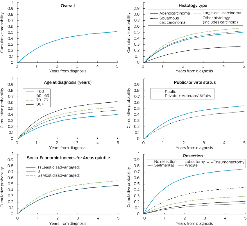
The probability of lung cancer death by period from diagnosis, as indicated by competing risk regression, was 24.7% at 12 months, 37.8% at 24 months, 45.1% at 36 months, 48.8% at 48 months and 52.3% at 60 months.

The competing risk model, adjusted for other variables, indicated the following statistically significant differences: a higher probability of lung cancer death for older ages, for large cell than adenocarcinomas, and compared with lobectomy, for cases having no resection, a wedge resection or pneumonectomy (Box 2; Box 3). The model predicted an adjusted 5-year probability of lung cancer death of 76% in the absence of resection, 30% for wedge resection, 18% for segmental resection, 22% for lobectomy and 45% for pneumonectomy. One LHD had a lower death probability (SHR, 0.61; 95% CI, 0.44–0.86) compared with the reference LHD. A lower death probability was also evident for the privately insured (or with Veterans’ Affairs coverage) and patients born outside Australia. While lower socioeconomic status was not associated with higher death probability after adjusting for insurance status (Box 2), the lowest socioeconomic status was predictive if insurance status was removed from the model (SHR for socioeconomic quintile 5 compared with quintile 1, 1.31; 95% CI, 1.02–1.67).

When the six border LHDs were excluded, similar results presented, although statistical significance was not quite reached for patients born outside Australia in English-speaking countries compared with Australian-born patients (SHR, 0.85; 95% CI, 0.70–1.01).

Unadjusted analyses showed significantly higher probabilities of lung cancer death (P < 0.05) for men, outer regional areas, the most disadvantaged socioeconomic quintiles of 4 and 5, and cases with comorbidity, but statistically significant associations did not apply in the adjusted analysis (Box 2).

When the adjusted analysis was repeated for cases having a resection, hospital resection volume did not approach statistical significance as a predictor of lung cancer death (P = 0.524), although there was some suggestion of a decrease in SHRs with increase in annual volume to 0.94 (95% CI, 0.65–1.37) for 9–36 resections and 0.80 (95% CI, 0.54–1.18) for 37 or more resections.

Low-risk patients

Similar SHR patterns applied as in the full analysis to the 255 patients classified as “low risk”, 181 (71.0%) of whom had a resection. For these low-risk cases, compared with lobectomy cases, both adjusted and unadjusted analyses showed a higher probability of lung cancer death for patients not receiving a resection (unadjusted SHR, 6.19; 95% CI, 3.81–10.06; adjusted SHR, 14.12; 95% CI, 7.23–27.54).

Discussion

Results indicate that between 38% and 43% of localised NSCLC cases diagnosed in NSW residents in 2003–2007 were treated by resection, depending on whether LHDs regarded as susceptible to underrecording from cross-border treatments are included. The resection rate peaked at 59% by LHD for all cases and 71% for NSW as a whole for cases considered at low surgical risk.

Multiple logistic regression analysis indicated that lower resection rates applied to patients older than 70 years. Although the possibility of undertreatment of older patients exists, it is likely that lower resection rates were influenced by higher surgical risk from increased frailty, especially in patients older than 80 years. Predictably, the resection rate was lower among patients with recorded comorbidity, many of whom would have been considered to have a higher surgical risk, but our measure of comorbidity related to morbid conditions and there was no statistical adjustment for less overt frailty.

Patients without private health insurance or Veterans’ Affairs coverage and those from lower socioeconomic areas had significantly lower resection rates in multivariate analysis. Financial barriers may have contributed, along with educational differences and other correlates that affected service use.

Cancers located in the main bronchus had lower resection rates than those in pulmonary lobes. This has been reported elsewhere, and reflects the need for more extensive resection for central lesions for which the patient may not be operable due to inadequate cardiopulmonary reserve.25,26 Squamous cell carcinomas also had lower resection rates than adenocarcinomas, which may be due to their more central locations.25,26 Large cell cancers had a low resection rate, although these are often located peripherally,26 which may be due to less favourable prospects for curative resection, given their propensity for rapid growth and spread.26

Men had a lower resection rate, but a statistically significant difference did not remain after adjusting for age, histology type and comorbidity. Women with lung cancers have a higher proportion of adenocarcinomas, which have higher resection rates. This suggests that the difference in resection rate by sex partly reflected biological variations rather than choice. Residents of regional areas also had lower unadjusted resection rates that were not evident in the multivariate analysis, after adjusting for socioeconomic status and LHD, which may be due to lower socioeconomic status and some undercounting due to cross-border treatment.

Financial barriers, variations in access, and other potentially modifiable barriers need to be addressed to optimise cure rates from localised NSCLC. The highest resection rate by LHD was 59%, which could be used as an overall NSW target in the first instance.

Resection was associated with an approximate 70% reduction in deaths in the 5 years after diagnosis. If the low overall resection rate of 38%–43% were increased to equate with the highest LHD resection rate of 59%, the proportion of all NSCLC patients dying of NSCLC in the 5 years after diagnosis would be reduced by about 10%, based on the differences in probabilities of death by resection estimated in our study.

These data, and research evidence supporting clinical guidelines for resection, could be discussed at workshops of clinicians and made available through web-based communications, to promote heightened awareness of this evidence when planning treatment.710 It is also possible that deaths could be prevented by an increased use of lobectomies in preference to wedge resections, given the elevated risk for wedge resection. This evidence should also be discussed with clinicians.

Similar results applied when parallel analyses were undertaken of all-cause mortality as the outcome, using proportional hazards regression instead of a competing risk analysis, and when restricting cases in the analyses to those with complete data.23 While strenuous attempts were made to control for confounding, the study design was observational and it is acknowledged that potential for confounding would exist from unmeasured factors (eg, frailty, patient choice) and factors inexactly measured (eg, comorbidity). Results therefore should be regarded as a guide and their statistical precision not over-interpreted.

Population-based radiotherapy data were not available to us, but are being assembled for future analysis. In the absence of randomised trial data, it would be useful to compare outcomes of resections, curative radiotherapy and combined treatments for localised NSCLC.

In the meantime, the results of this study are consistent with guideline recommendations that resection be used for localised NSCLC.710 Results indicate marked differences in probability of lung cancer death in the first 5 years from diagnosis of localised NSCLC in NSW depending on whether resection is undertaken. Potential exists to reduce numbers of deaths from lung cancer in NSCLC cases by increasing resection rates.

1 Resection rate and adjusted relative odds of resection for localised non-small cell lung cancers diagnosed in New South Wales, 2003–2007*

Characteristic

No.

Resection
rate, %

P for test (unadjusted)

Adjusted relative odds of resection (95% CI)


Age at diagnosis (years)


< 60

490

52.7%

< 0.001

1.00 (reference)

60–69

832

48.3%


0.93 (0.71–1.23)

70–79

1091

37.2%


0.62 (0.48–0.82)

≥ 80

627

16.3%


0.20 (0.14–0.28)

Sex


Male

1901

36.9%

0.024

1.00 (reference)

Female

1139

41.0%


1.05 (0.87–1.27)

Public/private payment status


Public

1918

33.5%

< 0.001

1.00 (reference)

Private (+ Veterans’ Affairs)

1082

48.2%


2.08 (1.70–2.54)

Unknown

40

[7.5%]


0.09 (0.02–0.35)

Remoteness (residence)


Major city

1747

42.7%

< 0.001

1.00 (reference)

Inner regional

834

35.6%


1.00 (0.76–1.32)

Outer regional

411

26.0%


1.00 (0.64–1.56)

Remote/very remote

48

37.5%


1.47 (0.62–3.50)

Socio-Economic Indexes for Areas quintile


1 (least disadvantaged)

508

45.5%

< 0.001

1.00 (reference)

2

502

44.0%


0.74 (0.52–1.06)

3

624

32.9%


0.58 (0.39–0.87)

4

744

35.5%


0.63 (0.41–0.95)

5 (most disadvantaged)

662

37.3%


0.65 (0.43–0.99)

Country of birth


Australia

1996

36.5%

0.110

1.00 (reference)

Other, English speaking

352

38.4%


1.03 (0.77–1.38)

Other, not English speaking

605

41.2%


1.07 (0.84–1.36)

Unknown

87

[64.4%]


3.27 (1.77–6.03)

Lung location


C340 (main bronchus)

223

8.5%

< 0.001

1.00 (reference)

C341 (upper lobe)

1366

44.5%


9.26 (5.51–15.56)

C342 (middle lobe)

172

43.0%


7.56 (4.08–14.01)

C343 (lower lobe)

841

49.0%


11.82 (6.96–20.09)

C348 (overlapping)

31

45.2%


10.26 (4.05–25.99)

C349 (not specified)

407

[10.1%]


1.59 (0.86–2.94)

Histology type


Adenocarcinoma

1014

56.4%

< 0.001

1.00 (reference)

Squamous cell carcinoma

890

38.7%


0.60 (0.48–0.75)

Large cell carcinoma

908

15.1%


0.15 (0.12–0.20)

Other

228

50.4%


1.16 (0.80–1.67)

Comorbidity status


Absent

1270

45.2%

< 0.001

1.00 (reference)

Present

1518

31.2%


0.74 (0.61–0.90)

Unknown

252

[47.6%]


1.32 (0.93–1.88)


Percentages in brackets and associated raw data were excluded when deriving P values. * Data source: NSW Central Cancer Registry. † Derived using Mann–Whitney U test for ordinal variables and Pearson χ2 for nominal variables.23 ‡ Adjusted for other variables in multiple logistic regression model, including local health districts (expressed as dummy variables).

2 SHRs for cumulative probability of death from localised non-small cell lung cancer diagnosed in New South Wales, 2003–2007, according to patient and cancer characteristics, and treatment by resection*

Characteristic

No. of patients/lung cancer deaths

Adjusted SHR (95% CI)

Adjusted P for test


Age at diagnosis (years)


< 60

490/167

1.00 (reference)

< 0.001

60–69

832/366

1.25 (1.05–1.49)


70–79

1091/585

1.46 (1.23–1.73)


≥ 80

627/420

1.86 (1.54–2.24)


Sex


Male

1901/1009

1.00 (reference)

0.068

Female

1139/529

0.90 (0.81–1.01)


Public/private payment status


Public

1918/1064

1.00 (reference)

< 0.001

Private (+ Veterans’ Affairs)

1082/461

0.76 (0.68–0.86)


Unknown

40/13

0.52 (0.27–0.98)


Remoteness (residence)


Major city

1747/860

1.00 (reference)

0.169

Inner regional

834/431

0.92 (0.78–1.07)


Outer regional

411/227

0.79 (0.63–1.01)


Remote/very remote

48/20

0.62 (0.36–1.07)


Socio-Economic Indexes for Areas quintile

1 (least disadvantaged)

508/237

1.00 (reference)

0.078

2

502/229

1.02 (0.82–1.28)


3

624/321

1.00 (0.79–1.27)


4

744/397

1.19 (0.94–1.52)


5 (most disadvantaged)

662/354

1.24 (0.97–1.59)


Country of birth


Australia

1996/1051

1.00 (reference)

< 0.001

Other, English speaking

352/178

0.84 (0.71–0.99)


Other, not English speaking

605/298

0.84 (0.73–0.97)


Unknown

87/11

0.27 (0.15–0.52)


Lung location


C340 (main bronchus)

223/155

1.00 (reference)

0.005

C341 (upper lobe)

1366/643

0.88 (0.74–1.05)


C342 (middle lobe)

172/74

0.90 (0.68–1.19)


C343 (lower lobe)

841/372

0.85 (0.71–1.02)


C348 (overlapping)

31/15

1.08 (0.65–1.80)


C349 (not specified)

407/279

1.19 (0.97–1.46)


Histology type


Adenocarcinoma

1014/386

1.00 (reference)

< 0.001

Squamous cell carcinoma

890/474

1.06 (0.93–1.22)


Large cell carcinoma

908/627

1.23 (1.08–1.41)


Other (includes carcinoid)

228/51

0.46 (0.33–0.64)


Comorbidity status


Absent

1270/588

1.00 (reference)

0.277

Present

1518/842

1.02 (0.91–1.14)


Unknown

252/108

1.17 (0.96–1.42)


Resection


None

1872/1325

5.80 (4.81–6.99)

< 0.001

Segmental

271/38

0.80 (0.56–1.14)


Wedge

102/26

1.47 (1.00–2.17)


Lobectomy

738/124

1.00 (reference)


Pneumonectomy

57/25

2.47 (1.68–3.61)


* Data source: NSW Central Cancer Registry. † Adjusted for other variables in the model, including local health districts expressed as dummy variables. SHR = subhazard ratio.

3 Cumulative probability of death from lung cancer: localised non-small cell lung cancer cases diagnosed in New South Wales, 2003–2007*


* Derived from competing risk regression including age, sex, public/private status, remoteness, socioeconomic status, local health district, country of birth, lung location of tumours, histology type, comorbidity score, and resection type as predictors of lung cancer death.

Time to end the ban on HIV-positive proceduralists and dentists

To the Editor: This year, the ban on HIV-positive surgeons and dentists practising in the United Kingdom was removed on the provision that they are clinically well, are being treated, and have an undetectable viral load.1 This development aligns the UK with over 20 other countries. Australia is lagging behind other more progressive countries on this issue.2

During the height of the HIV epidemic in the early 1990s, there were no HIV transmissions among 22 171 patients exposed to pre-antiretroviral (ART)-era HIV-positive doctors and dentists during invasive operations.3

There have been only 10 published case reports indicating probable transmission from a proceduralist to patients since HIV was first reported.4 The current risk estimate is 1 in 1 672 000.2 No cases of inadvertent transmission have been reported in the literature from countries that allow HIV-positive proceduralists.2

ART is vital for reducing the risk of transmission. Extrapolating from the ongoing PARTNER study, there have been no transmissions between serodiscordant couples who have regular, unprotected, high-risk sex if the positive partner is receiving ART and virally suppressed 2 years into the study.5 The risk during procedures by HIV-positive virally suppressed proceduralists, with the use of standard universal precautions (such as gloves and sterile equipment), would likely be extremely low.

There are two major problems with a blanket ban on HIV-positive proceduralists. First, those facing a career-ending outcome of a test are likely to avoid being tested after an initial negative test result. Second, the wrong message is sent to the public that people with HIV are highly infectious and are somehow dangerous (despite treatment). Lifting the ban would lead to greater incentives for proceduralists to be regularly tested for HIV, leading to better outcomes for them and their patients. This is important as many infected individuals transmit the virus when they are seroconverting and are unaware of their diagnosis.6 Furthermore, postexposure prophylaxis could also be available to patients after possible exposure during a procedure by following occupational exposure guidelines.

It is time for Australia to align with other progressive nations and end theabsolute ban on HIV-positive proceduralists. The risk of transmission from them, when treated for HIV and using standard precautions, is likely to be negligible.