×

Autoimmune rheumatic diseases: recent advances and current challenges

Biological disease-modifying agents are transforming the treatment of autoimmune rheumatic diseases

Over the past decade, the advent of biological disease-modifying agents has led to transformational changes in the management of inflammatory joint diseases. This has been most obvious with advances in treatments for rheumatoid arthritis (RA). However, steady progress is also being made in treating lupus and other autoimmune rheumatic conditions. In this issue of the MJA, several articles discuss these developments along with current challenges.

For RA, we now have a broad suite of effective therapies with a number of new products and biosimilars in the pipeline. Jones and colleagues1 review the significant advances in our understanding of important cytokine pathways in RA, many of which are now targeted therapeutically. There is evidence for a window of opportunity in the first 6 months of disease, when therapies are both more effective and have a long term effect on disease, independent of subsequent treatment. Achieving this requires shared care between the general practitioner and rheumatologist. The expanded therapeutic options and new approaches have made disease remission a realistic goal. Current challenges in the management of RA include the lack of methods for efficiently targeting therapies to those most likely to benefit, as well as the optimal way to introduce biosimilars into clinical practice.

In contrast to RA, despite significant inroads, management of lupus and ankylosing spondylitis lags behind that for RA. Systemic lupus erythematosus (SLE) is a chronic multisystem autoimmune disease. Uncontrolled disease activity leads to irreversible end organ damage, which in turn increases the risk of premature death. Golder and Hoi2 note that although early and sustained control of disease activity can usually be achieved by the use of conventional immunosuppressants, belimumab is the only targeted therapy approved by the Therapeutic Goods Administration. Significant work is currently underway in improving classification criteria for SLE, thereby improving diagnostic certainty, especially in the previously undifferentiated group. “Treat to target” concepts are changing trial design and clinical practice with evidence-based definition of response criteria for remission and low disease activity on the horizon. As discussed by O’Neill and colleagues,3 while new targeted therapies are awaited, research into quality of care in SLE is a focus of research efforts through activities such as the Australian Lupus Registry and Biobank. This will shed light on areas of deficiency in clinical practice in a real world setting, with findings that have the potential to lead to system improvement based on evidence.

The past decade has seen major advances in the diagnosis and management of ankylosing spondylitis (AS) and in research into its pathogenesis. Brown and Bradbury4 point out that, in common with most chronic diseases, management of AS is best performed by multidisciplinary teams, including medical practitioners and allied health professionals. The role of the physiotherapist is important, as exercise is central to the management of AS. Although biological disease-modifying agents are in use in ankylosing spondylitis, no current treatment leads to disease remission or halts the progression of bony ankylosis, the cause of significant morbidity. Nonetheless, improved diagnostic methods and management have led to major benefits for patients, with marked improvements in quality of life with reduced treatment-associated side effects.

Prevention is important in the overall care of patients with inflammatory rheumatological diseases and requires co-management of patients between the rheumatologist and GP. Smoking is a strong risk factor for disease progression in inflammatory arthritis, as highlighted by Jones et al1 for RA and Brown and Bradbury4 for ankylosing spondylitis. In their narrative review, Golder and Hoi2 note the significant risk of cardiovascular death in SLE and the importance of targeting traditional cardiovascular risk factors, which is also important in RA.5 Other important areas for prevention are bone disease, including through use of anti-resorptive therapy,2 infections through vaccination programs and cancer through screening programs.6 As discussed by Hart and colleagues,7 awareness that inflammatory rheumatic diseases are frequently complicated by extra-articular manifestations such as eye involvement is important in order to optimise patient outcomes.

Successful treatment of chronic conditions such as inflammatory arthritis is dependent on the participation of patients in their care. Those with inflammatory arthritis currently have unmet needs that preclude optimal disease and patient-centred outcomes. Chou and colleagues8 argue that although the need for improved patient access to information may be met within the current system, other concerns relating to access to medical and allied health care may require more creative, fundamental changes to the models of care available.

The transformational changes in the treatment of RA over the past decade provide a beacon of great hope for those with autoimmune rheumatic diseases. Although challenges remain, the combined approach of strong basic laboratory, clinical and epidemiological research informed by patient needs provides great optimism that the next couple of decades will see continued improvement in outcomes for our patients.

Crowned dens syndrome: a rare cause of neck pain and fever

An 86-year-old man presented with acute severe neck pain, restricted neck movement and fever. The white cell count, erythrocyte sedimentation rate and C-reactive protein were all elevated. After excluding an infective aetiology, review of computed tomography images of the cervical spine (Figure) showed calcification of the transverse ligament of the atlas with crown-like density around the odontoid process. This was consistent with a diagnosis of crowned dens syndrome, an uncommon manifestation of calcium pyrophosphate dehydrate deposition disease.1,2 The patient’s symptoms were self-limiting over 1 week without the use of anti-inflammatories or corticosteroids.

Figure

Patient-centred management of inflammatory arthritis: more than just disease control

It is important that patients’ broader health needs are addressed

Rheumatoid arthritis and seronegative spondyloarthropathies are the most common types of inflammatory arthritis. They cause pain, joint damage and progressive functional loss, which limit participation in life and constitute a significant disease burden.1,2 Over the past decades, great advances in management, with the availability of corticosteroids, synthetic disease-modifying antirheumatic drugs (DMARDs) and biological DMARDs, have significantly improved the disease control and outcomes. The guidelines, whose algorithms focus on drug prescription to achieve remission of disease,3,4 support everyday treatment. However, while they provide direction for the use of DMARDs, they do not consider the patients’ broader health needs.

When optimising outcomes for people with inflammatory arthritis, there are significant discrepancies between the patients’ perspectives of their own health and those of their health care professionals.5 To promote successful clinician–patient partnerships and shared decision making, we need to understand patients’ priorities and perceived needs and deal with the areas where these do not align with current health care guidelines and policies. In this article, based on the results of a systematic scoping study,6 we consider the needs of people with inflammatory arthritis and propose options to improve their situation. While the concept of patient-centred care is not a new one for effective chronic disease management, it is important for patients with inflammatory arthritis given that this disease commonly affects people from a younger age compared with other chronic health conditions. Health policy and practice strive to deliver patient-centred care; however, actual care delivery falls short of peoples’ needs.6

What do people with inflammatory arthritis need?

Therapies to relieve symptoms

People with inflammatory arthritis want treatments that reduce their pain and fatigue, prevent joint damage and help them maintain their mobility, function and independence.6 Although pharmacological treatment has improved significantly over the past decades, it is far from perfect, with individual medications failing to attain remission in most patients7 and leaving them feeling that their needs are not fully met. These needs align with the principles of contemporary practice: “treat to target”; however, patients do not always appreciate the rationale and need a better understanding of treatment options, particularly relating to drug safety and toxicity.6 Given this lack of understanding and a need for symptom relief, many patients seek other forms of supplementary care, including complementary and alternative therapies (CAM), despite limited evidence of efficacy.

Information to enable decision making and self-management

Patients’ perceived needs go beyond the use of medications; their involvement in decision making is increasingly viewed as important by both patients and health care providers and improves compliance, health outcomes and satisfaction.8 However, patients often perceive that the information they receive is suboptimal.6 Moreover, they want education regarding their disease and potential therapies so they may gain control of their health, learn strategies to manage their condition and plan for the future.6 This is key in inflammatory arthritis, where disease impact is often maximal in peak income-earning and family-planning years. Patients want strategies to reduce pain and methods to adapt to functional limitation.6 In addition, they want to know the effects of these strategies on their daily lives, such as implications for driving and dietary restrictions.6 They also want precise instructions about exercise and physical therapies, and self-help strategies to manage the emotional effects of the disease.6 In sum, patients need individually tailored, practical information delivered in a constructive manner; nevertheless, they feel these areas are often overlooked by clinicians.6

Access to health care professionals

It is important that patients have access to understanding, knowledgeable, thorough and empathic practitioners who take a holistic approach to care, do not rush consultations, provide continuity of care and are easy to access, particularly during a disease flare.6 Moreover, they need access to skilled allied health practitioners — such as physiotherapists and occupational therapists — to improve function, mobility and psychological wellbeing.6 Long waiting times are seen as a significant obstacle to optimal management.6

Broader health needs

Being socially connected, employed and having financial security are essential needs for people with inflammatory artrhritis,6 and some patients are frustrated by the lack of understanding of their condition by their family, friends, employers and society in general.6 Mobility impairments raise practical concerns regarding parking and transport options. Moreover, employment and job security are paramount to a person, affecting financial security, self-esteem and social connectedness.6

How can we manage these needs?

Improved communication and education on the disease and its impact on social functioning are important for the promotion of patient-centred care. Therefore, patients need to gain the knowledge and skills to manage their condition and better participate in shared decision making with their health care providers. This can be helped by providing decision aids; encouraging shared care models between medical specialists, primary care physicians and allied health professionals; and implementing interventions to facilitate question asking during consultations.9 In addition, practitioners should be trained to deliver patient-centred care and empower shared decision making during consultations.10 Better relationships will enable a non-judgemental discussion regarding medication use, compliance concerns, CAM use, practical strategies for self-care and pain coping, and consideration of psychosocial problems that have an impact on health behaviours and mental wellbeing.

Patients with inflammatory arthritis should have ready and timely access to knowledgeable health care practitioners, particularly rheumatologists, and skilled allied health providers. However, access to rheumatologists is limited in Australia, particularly in rural sectors.11 In addition, allied health providers receive little formal education in rheumatology, resulting in workforce capacity limitations.12 To manage this burden–service gap, alternative methods of health care delivery have been proposed in Australia and internationally.13 These methods focus on building workforce capacity and expanding workforce roles (eg, advanced scope roles, nurse practitioner roles), providing community-based shared care services and supporting contemporary service and information delivery to consumers through leveraging digital technologies, such as mobile and electronic health applications.10,14 Peer contact for patients may also assist in providing complementary information and education — often more effectively — about coping with the emotional aspects of this condition and providing social connections.6

Developing more effective and efficient models of care may be facilitated by involving patients in their planning.15 Health information materials need to be produced in collaboration between health professionals and consumers, to ensure they are relevant, targeted to patients’ needs, and disseminated appropriately.15 Involvement of patient representatives in developing clinical practice guidelines that manage the patient holistically by incorporating their perspective may be more closely aligned to their perceived needs.15 Investigation of patient-focused strategies, providing interventions tailored to different care settings, patient populations, economies and health care systems may provide novel, effective solutions.15

It will be more challenging to devise novel approaches to meet the broader health needs of people with inflammatory arthritis. Health promotion interventions (such as mass media campaigns and community events) may help bridge gaps in public understanding6,10 and encourage people with inflammatory arthritis to maintain social connectedness, for which peer support networks — traditional face-to-face and digitally-enabled — may be helpful.16 Job security may be optimised by liaison between health care practitioners and the workplace to facilitate job adaptation, including physical environmental aspects, such as parking and access, and flexibility in working hours.6 Moreover, patients identify cost, locality of services, compliance and lack of time as barriers to care.6 To manage these barriers, health care services may consider more flexibility in the provision of care (eg, after-hours services, community-based centres, telehealth services) and better coordination with different health care professionals, employers and allied health services.

Conclusions

There is an urgent need to direct future research initiatives on the efficient and effective management of inflammatory arthritides. Improved collaboration between Australian researchers, policymakers, funding bodies and patients may deal with these gaps in knowledge and improve care outcomes. While disease control using the “treat to target” paradigm is essential, to the individual, pain control, maintenance of employment and financial security are of paramount importance. Cooperation between patients, clinicians and policy makers to implement novel strategies to help patients remain productively in the labour force is required to improve this situation.

New approaches in ankylosing spondylitis

There have been marked improvements in treatment options but none have yet been shown to induce remission

The past decade has seen major advances in the diagnosis and management of ankylosing spondylitis (AS) and in research into its pathogenesis. It remains the case that no current treatments have been shown to lead to disease remissions or to halt the progression of the bony ankylosis that causes the major morbidity associated with this condition. Nonetheless, improved diagnostic methods and management have led to major benefits for patients, with marked improvements in quality of life with reduced treatment-associated side effects.

Early diagnosis and non-radiographic axial spondyloarthritis

AS is diagnosed using the modified New York classification criteria for the disease,1 which are highly specific for AS but require the presence of x-ray changes in the sacroiliac joints to establish a diagnosis. Consequently, they lack sensitivity, particularly early in disease. It is estimated that it takes on average a decade between onset of axial spondyloarthritis symptoms before these x-ray changes develop, although in some cases more rapid progression occurs.2

The introduction of magnetic resonance imaging (MRI) scanning for the diagnosis of axial spondyloarthritis has greatly improved the ability to diagnose patients with less severe x-ray changes and with disease of shorter duration. This has highlighted the long pre-radiographic phase that is universal in AS, and has led to the development of new classification criteria that do not require x-ray changes to be present.3 This new clinical entity is called non-radiographic axial spondyloarthritis (nr-axSpA) and is intended to refer to patients with inflammatory axial arthritis without x-ray changes typical of AS. Roughly half of patients presenting with nr-axSpA will, over the space of a decade, progress to develop AS.4 At this point there is only limited capacity to predict which patients will progress or not, with some but not all studies suggesting increased likelihood of progression with male sex, HLA-B27 carriage, smoking, higher baseline C-reactive protein (CRP) levels, extent of MRI evidence of inflammation and younger age of onset.5 Whether or not such patients ultimately develop AS, they suffer significantly with their disease, with a similar disease burden to established AS, and therefore need treatment.

Current management approaches

In common with most significant chronic diseases, management of AS is best performed by multidisciplinary teams, including not only medical practitioners but also allied health professionals. In the case of AS, there are particular roles for specialist rheumatology nurses, nurse practitioners and physiotherapists.

Exercise is paramount in the management of AS and the role of the physiotherapist in guiding and supporting patients is key.6 Individualised assessment, treatment and monitoring are essential to aid adherence to a lifelong exercise program.7,8 Extended-scope physiotherapists who have undergone further training in AS may also play a role in management.9

Registered nurses with specific training in rheumatology can provide care that includes monitoring of disease activity, impact on activities of daily living, psychosocial health, drug treatment and side effects.10 Nurse practitioners can also autonomously prescribe medications within their scope of practice, refer patients to other health professionals and perform examinations and procedures such as joint aspiration and injection (http://www.health.gov.au/internet/main/publishing.nsf/Content/work-nurse-prac).11 They also play a critical role in education about medication-related issues such as benefits and risks, management of complications, and drug administration. This is particularly important in maintaining patients safely on biological agents which are complex to administer and have potentially severe side effects, yet also bring great benefits when used appropriately.

Patient education is key to improving coping strategies and increasing self-care abilities,10 thereby achieving a greater sense of empowerment and, in turn, function. Further, education can address comorbidities such as cardiovascular disease, as well as general health issues such as smoking, diet and reinforcing the importance of exercise. Given the major effect of cigarette smoking as a risk factor for AS, and its association with increased disease activity, rate of spinal fusion and resistance to therapy, smoking cessation is one of the key management challenges in AS, and nurse practitioners are particularly effective in implementing such lifestyle modification programs. It has also been recognised that greater levels of knowledge were found in patients monitored by a nurse compared with those monitored by doctors.10

Medical treatment

Non-steroidal anti-inflammatory drug (NSAID) therapy is more efficacious in AS-associated pain than for pain resulting from non-inflammatory causes, and thus most patients receive these agents. It is unclear at this point whether NSAID therapy retards progression of ankylosis in AS, but it is clear that any beneficial effect is modest and probably restricted to patients with ongoing inflammation as assessed by elevation of erythrocyte sedimentation rate (ESR) or CRP levels. Use of long-acting NSAIDs, particularly when given with an evening meal, is particularly effective at controlling morning symptoms and in assisting sleep, which is frequently disturbed in patients with AS due to pain and stiffness. In the 5–10% of patients who have coexistent inflammatory bowel disease, NSAIDs may cause flares of colitis. In this setting the NSAID etoricoxib, which unlike other NSAIDs does not exacerbate inflammatory bowel disease, may be useful.12

Other disease-modifying antirheumatic drugs that are effective in rheumatoid arthritis have little or no role to play in treating AS.13,14 Sulfasalazine may be effective in AS-peripheral arthritis (knees and below or upper limb), but its beneficial effects overall are modest.15 Hip disease is considered as a component of axial disease in AS. No trial evidence to support the use of disease-modifying antirheumatic drugs alone for hip disease has been reported to date, and therefore it cannot be recommended, as these therapies are undoubtedly associated with significant side effects. Hip inflammation in AS does respond to tumour necrosis factor inhibitor (TNFi) therapy. Sulfasalazine may also be useful in recurrent acute anterior uveitis,16 a condition that ultimately affects roughly 60% of patients. Methotrexate and sulfasalazine have no demonstrated efficacy in spinal or sacroiliac disease in AS and therefore should not be used for this indication.

Corticosteroid agents should be used with great caution. Compared with rheumatoid arthritis, AS is less responsive to oral or systemic corticosteroids. Further, AS is frequently complicated by osteoporosis, which combined with the increased spinal stiffness caused by ankylosis, leads to a significantly increased risk of spinal fracture. This spinal fragility is exacerbated by corticosteroids, further encouraging caution in their use. Intra-articular corticosteroids may be useful as a short term therapy for sacroiliitis or peripheral arthritis.

Treatment of non-radiographic axial spondyloarthritis

Clinical trials have confirmed that TNFi therapy, the gold standard treatment for established AS, is also effective in treating nr-axSpA.17 This is particularly true for cases that are of more recent onset (< 3 years symptom duration), with elevated ESR or CRP levels, or a positive MRI scan, providing objective evidence of inflammation. In such cases, the treatment response is similar to that seen with established AS, where TNFi treatment is highly effective. Despite this evidence, no biological medication has yet been funded for use in nr-axSpA, leaving a significant group of patients for whom there is no funded therapy available. Currently the Australian Pharmaceutical Benefits Scheme (PBS) restrictions for access to TNFi for AS include that the patient meets the modified New York criteria for AS, has failed to respond to NSAID treatment and a 3-month exercise program, has high self-reported disease activity (Bath Ankylosing Spondylitis Disease Activity Index > 4), and has high acute phase reactants (CRP > 10 mg/L, ESR > 25 mm/h).

A key question now is whether early treatment can lead to better long term outcomes for patients.18 There is now strong evidence to support this being the case in terms of reducing progression of ankylosis. While short term studies (up to 2 years of treatment) failed to show an effect of TNFi treatment on progression of x-ray changes,19 longer studies (4 years of treatment) have shown significant benefits, roughly halving x-ray progression.20 This benefit is greater the earlier TNFi treatment is initiated. Whether TNFi retards progression of nr-axSpA to AS is not yet clear, although there is suggestive evidence that successful suppression of inflammation can lead to treatment-free disease remissions.

New therapies

Genetic discoveries early in the genome-wide association study era demonstrated that genetic variants in the interleukin (IL)-23 receptor pathway were strongly associated with AS,21 and the importance of the pathway has been confirmed by immunological studies in humans and animal models. This led to the repositioning of drugs targeting IL-23 and the related cytokine IL-17 for trial in AS.

These trials have been highly successful, with response rates at least equivalent to those of TNFi treatment, and with the medications being well tolerated.22 Secukinumab, an IL-17 inhibitor, has therefore recently been PBS-funded in Australia for AS, with the same indications as for TNFi. These agents have been found to be more effective than TNFi for treatment of psoriasis, which complicates 5–10% of AS cases,23 but have variable effects on inflammatory bowel disease, which complicates a similar proportion of AS cases.

Up to 60% of patients with AS will, over their lifetime, experience acute anterior uveitis;24 and while this is typically easily treated with topical steroids and mydriatics, in a significant subset of patients it is recurrent or chronic and can lead to glaucoma and visual impairment. TNFi treatment is effective in reducing the frequency of acute anterior uveitis, although etanercept is less effective than the other agents in this regard.25 Whether IL-23 pathway inhibitors are effective in this condition is unknown. The effect of IL-23 pathway blockade on rate of ankylosis is also unknown.

This increasingly diverse pharmacological armamentarium opens the possibility of personalised approaches to medical management of patients with AS, with optimal treatment varying depending on the particular disease features that a patient may experience. Head-to-head studies will be required to determine the relative efficacy of the new agents, but given that a significant proportion of patients, for reasons related to efficacy or toxicity, cannot be managed by TNFi, the availability of alternative agents is a welcome relief for them and their caring physicians.

Despite these advances, management of AS lags behind that of the other major common immune-mediated arthropathy, rheumatoid arthritis, with fewer therapeutic options, no treatment yet convincingly shown to induce disease remission, and with less impact on slowing disease progression. This probably relates to the disproportionately low research attention AS receives in the academic sector and pharmaceutical industry relative to the burden of disease the condition causes. Given the major advances in basic research in AS, including in genetics, immunology and metagenomics, it is to be hoped that sufficient resources are made available to translate these advances into benefits for patients.

Ocular complications of rheumatic diseases

Most of the inflammatory rheumatic diseases are systemic conditions with clinical and pathological manifestations outside of the joints. Many of the extra-articular manifestations of inflammatory rheumatic disease respond to the same treatments that target the joint disease itself, but some require specialised interventions. Ocular involvement is a common manifestation of inflammatory rheumatic disease and ranges from chronic troublesome symptoms, such as dry eye complicating Sjögren syndrome, to organ- and sight-threatening vasculitis. Isolated ocular abnormality has been reported to account for up to 4% of referrals to rheumatology clinics.1 Diagnosing and characterising serious ocular abnormalities are complicated by the need for specialised equipment and training — notably slit lamp and retinal examination — to fully assess eye disease. Consequently, it is incumbent on physicians involved in the care of patients with rheumatic disease to be aware of the potential ocular complications of their patients’ condition and the indications for rapid escalation of treatment to specialist level to preserve sight.

Here, we summarise these important conditions, in order of urgency of treatment, for non-ophthalmologists who are involved in the management of patients with rheumatic diseases (Appendix 1). We identified articles for inclusion in this review by keyword searches in MEDLINE, with an emphasis on up-to-date review articles, and through expert opinion.

Ocular complications of rheumatic diseases

Ischaemic optic neuropathy

Ischaemic optic neuropathy refers to damage to the optic nerve caused by a lack of blood supply. It is one of the major causes of blindness in older people in the Western world. The systemic disease most often associated with ischaemic optic neuropathy is giant cell arteritis (GCA), in which arterial vasculitis most commonly affects the blood supply to the optic nerve head. This entity is termed arteritic anterior ischaemic optic neuropathy (AAION). About 22% of patients with GCA will experience visual symptoms, with 12% developing irreversible loss of vision.2

GCA, which is by far the most common cause of AAION, rarely affects patients under the age of 50 years and has a mean age at diagnosis of 72 years.3 Women with GCA develop AAION more often than do men (71% v 29%).4 Patients often present with anorexia and the systemic symptoms associated with GCA, such as a persistent temporal headache, scalp tenderness and jaw or tongue claudication. Definitive diagnosis of GCA is made by temporal artery biopsy. The symptoms associated with a positive biopsy result include jaw claudication, neck pain and anorexia.5 Importantly, up to 21% of patients who develop permanent visual loss due to confirmed GCA have no systemic symptoms.5 The visual manifestations of ischaemic optic neuropathy are characterised by painless, sudden loss of vision. Amaurosis fugax is seen in up to 30% of patients with GCA and is a sign of impending loss of vision.4 Inflammatory marker levels are elevated in most patients with GCA, but there is a small proportion of patients with biopsy-proven GCA whose levels are within the normal reference interval.6

Initiation of steroid therapy should not be delayed while awaiting a definitive diagnosis. Treatment of patients with suspected GCA is oral prednisolone (1 mg/kg/day; maximum dose, 60 mg daily) or intravenous (IV) methylprednisolone therapy. A same day referral to an ophthalmologist should be made to arrange biopsy of the superficial temporal artery. The development of amaurosis fugax or any symptoms of visual loss is an indication for pulse IV methylprednisolone therapy.7 Once GCA is confirmed by biopsy, long term oral prednisolone and regular monitoring for systemic side effects of steroids are indicated.

Retinal vasculitis

Retinal vasculitis refers to inflammation of the retinal blood vessels. The development of retinal vasculitis is most commonly reported in patients with systemic lupus erythematosus or Behçet disease. Patients who develop retinal vasculitis often progress from mild retinopathy, involving cotton wool spots, perivascular hard exudates, retinal haemorrhages and vascular tortuosity (Box 1), to complete occlusion of retinal arterioles and consequent retinal infarction. Most patients with retinal vasculitis develop proliferative retinopathy, often progressing to vitreous haemorrhage and retinal detachment.8 Evidence of mild retinopathy is seen in 10% of patients with systemic lupus erythematosus8 and up to 74% of patients with the ocular form of Behçet disease.9 Permanent loss of vision is common in those patients who progress to advanced retinal vasculitis.

Retinal vasculitis typically presents with a painless decline in vision. Patients may develop a blind spot from ischaemia-induced scotomas or floaters from vitritis, and those with macular involvement may develop metamorphopsia. It is also possible for retinal vasculitis to be asymptomatic.

Same day referral to an ophthalmologist is recommended for all patients with a painless decline in vision. The diagnosis is often confirmed clinically on detailed retinal examination or with the aid of fluorescein angiography. Aggressive treatment with oral prednisolone (1 mg/kg/day; maximum dose, 60 mg daily) is recommended, often supplemented or replaced with other steroid-sparing immunosuppressive agents. Complications such as proliferative retinopathy are later treated with laser therapy.8

Peripheral ulcerative keratitis

Peripheral ulcerative keratitis (PUK) refers to inflammation and ulceration of the cornea. It is a rare complication of systemic diseases that is characterised by thinning of the stromal layer of the cornea close to the limbus. It is caused by inflammation of the cornea’s stromal layer and is often unilateral and crescent-shaped in appearance. There is an associated epithelial defect overlying the area of inflammation and progressive corneal thinning, which can lead to corneal perforation.10

The development of PUK is most often associated with rheumatoid arthritis but is also reported in cases of polyarteritis nodosa and granulomatosis with polyangiitis. The introduction of biological therapy for rheumatoid arthritis has seen a reduction in the incidence of corneal complications of this disease.11 PUK is reported to have a prevalence in patients with rheumatoid arthritis of 1.4–2.5%.11,12 It is seen most often in patients with longstanding rheumatoid arthritis who are both rheumatoid factor and anticyclic citrullinated peptide antibody positive. The presence of acute anterior uveitis (AAU) and dry eyes is common in patients with PUK.11

Patients with PUK often present with symptoms of ocular redness and pain, tearing, photophobia and decreased visual acuity. As the disease progresses, crescent-shaped corneal ulcers develop, with an associated corneal epithelial defect (Box 2). Inflammation of the surrounding conjunctiva and sclera are frequently apparent.10 Corneal ulceration often abates after treatment, but corneal thinning, scarring and neovascularisation are irreversible.10

The treatment of PUK is determined by the severity of findings within the cornea and the likelihood of imminent perforation. Aggressive treatment of the underlying systemic disease is important to control the progression of corneal disease, and should include a combination of high-dose systemic prednisolone (1 mg/kg/day; maximum dose, 60 mg daily) and an immunomodulatory agent appropriate to the underlying systemic disease, such as methotrexate. Pulse methylprednisolone therapy may be administered in cases of imminent perforation. Surgical management may be required to maintain globe integrity.10

Scleritis

The term scleritis refers to chronic inflammation of the sclera, the dense external covering of the eye. About 25–50% of patients presenting with scleritis have an associated systemic disease,13 most often rheumatoid arthritis (10–18.6%), although scleritis is also seen in patients with granulomatosis with polyangiitis (3.8–8.1%), relapsing polychondritis (1.6–6.4%) and inflammatory bowel disease (2.1–4.1%).14 Scleritis is an important clinical entity to detect, not only because it may lead to loss of vision due to structural changes to the globe but also because it is associated with an increased mortality rate in patients with rheumatoid arthritis.14 Scleritis may involve the anterior sclera, posterior sclera or both, and may be diffuse, nodular or necrotising. Non-necrotising scleritis rarely results in loss of vision, unless it is complicated by uveitis or keratitis. Necrotising scleritis results in rapid thinning of the sclera and exposure of the underlying uvea and is associated with poorly controlled systemic disease. This occurs due to severe vasculitis and closure of the episcleral vascular bed, leading to a visible area of scleral non-perfusion, infarction and necrosis (Box 3).14

Patients with scleritis present with severe pain involving the eye and orbit that radiates to the ear, scalp, face and jaw. The pain is often described as a dull, boring pain that wakes the patient from sleep and is exacerbated by eye movement. Episodes of scleritis can last several months, with the pain increasing in severity over several weeks. Patients with anterior scleritis often notice redness and tenderness of the globe. Patients with necrotising scleritis can experience extreme scleral tenderness. Inflammation often spreads to the surrounding structures, leading to keratitis, anterior uveitis and elevated intraocular pressures, all of which threaten vision.15

Patients with necrotising scleritis require treatment with systemic steroids, either orally or intravenously. A typical starting dose of prednisolone is 1 mg/kg/day (maximum dose, 60 mg daily), which is weaned over the following months based on disease progression. IV methylprednisolone is usually reserved for patients with impending scleral or corneal perforation. Patients who relapse at prednisolone doses of greater than 7.5 mg daily should be considered for adjunctive immunosuppressive therapy, such as methotrexate. Steroid therapy is combined with oral non-steroidal anti-inflammatory agents. Surgical intervention is rarely required in cases of scleral or corneal perforation, which can often be managed with contact lenses and tissue adhesive.15

Uveitis

Uveitis is inflammation of the uvea, the layer of the eye between the outer sclera and the inner retina. It is the most frequently encountered ocular manifestation of rheumatic disease and is responsible for up to 10% of cases of blindness in Western countries.16 Uveitis is classified as anterior, intermediate, posterior or panuveitis, based on the anatomical structures that are inflamed. It is further subdivided into acute, recurrent or chronic, based on the time course of the disease.17

AAU represents the majority of cases of uveitis in patients with rheumatic diseases. This condition involves a sudden onset episode of inflammation of the iris or the anterior ciliary body that is present for less than 3 months.17 Ankylosing spondylitis is the most frequent underlying rheumatic disease causing uveitis, occurring in 20–30% of patients.17 AAU is also associated with other disease entities, such as reactive arthritis, inflammatory bowel disease and psoriatic arthritis. Patients with systemic disease affected by AAU are generally aged between 30 and 40 years, and most test positive for human leukocyte antigen-B27.18

Patients with AAU often present with ocular pain, an acute onset of photophobia, excessive lacrimation, ocular injection and blurred vision (Box 4, Box 5). The symptoms tend to be unilateral and recurrent, occasionally in the contralateral eye. Attacks can last 2–3 months; residual visual impairment is uncommon when treated early.19 Loss of vision in patients with AAU develops as a result of complications, including cataract formation (7–28%),18 macular oedema (up to 11%),20 secondary glaucoma and the development of chronic uveitis.19

AAU is best diagnosed with slit lamp examination, warranting prompt referral to an ophthalmologist when the diagnosis is suspected. The pupil may be poorly reactive and the anterior chamber may show signs of inflammation, including the presence of cells, flare and occasional fibrin (inflammatory debris) within the chamber. In severe cases, there may be evidence of hypopyon. The ciliary blood vessels surrounding the limbus are usually dilated.

The mainstay of treatment is topical glucocorticoids, such as 1% prednisolone acetate. The frequency of the dose depends on the intensity of inflammation present, determined by slit lamp examination.17 Dilating drops are used to relieve pain and prevent iris–lens adhesions.

Sjögren syndrome

Sjögren syndrome is a slowly progressing, immune-mediated inflammatory disease targeting the exocrine glands. It leads to the replacement of functional epithelium with lymphocytic infiltrates, resulting in a dry mouth and dry eyes. Sjögren syndrome is classified as primary when it occurs in the absence of other connective tissue or autoimmune disease, or secondary when it accompanies another disease. About 60% of cases are secondary. It affects women more than men (9:1 ratio), often in their fourth or fifth decade of life.21 Rheumatoid arthritis is the most common underlying rheumatic disease, affecting 10–33% of patients,22,23 followed by systemic lupus erythematosus, affecting up to 9.2% of patients.24 Associations have also been reported in patients with scleroderma and polymyositis.25

The clinical features of Sjögren syndrome are dry eyes and mouth. The discomfort of dry mouth is often associated with difficulty swallowing and speaking, while the lack of tears leads to damage to the conjunctival epithelium over the cornea and globe. This typically presents with dilation of the conjunctival vessels and an irregular appearance to the cornea. Patients may present with dry, gritty or burning eyes, which should prompt consideration of the diagnosis. The average time between the onset of symptoms and the diagnosis of Sjögren syndrome is 10 years,26 largely because the symptoms are considered minor or vague or because they mimic those of other autoimmune diseases. Diagnosis can be made by the combination of the symptoms of dry eyes and mouth, a positive Schirmer test result and positive autoimmune screening (anti-SSA and anti-SSB) blood test results.27 Further characterisation with salivary gland biopsy can be sought if all the above criteria are not satisfied.27

Early diagnosis and treatment are essential for optimal management of Sjögren syndrome. Treatment comprises symptomatic relief through topical replacement lubrication and anti-inflammatory agents, as well as treatment of the underlying systemic disease where appropriate.28

Ocular side effects of drugs used for treating rheumatic disease

The eyes are highly vascular organs with a relatively small mass, making them particularly susceptible to toxic substances that circulate in the blood stream. The fundamental concepts in managing ocular side effects of drugs used in treating rheumatic disease are recognition of the early signs of eye toxicity, withdrawal of the offending agent and referral to an ophthalmologist. These side effects are summarised in Appendix 2.

Corticosteroids

The most studied ocular complications from corticosteroid use are the induction of cataracts and the increase in ocular pressure causing glaucoma.29 Patients treated with oral prednisolone doses of less than 10 mg per day for 1 year have a negligible chance of developing cataracts. In patients taking oral corticosteroids, the yearly incidence of corticosteroid-induced cataracts ranges from 6.4% to 38.7%.30

Glaucoma is thought to develop in patients taking corticosteroids due to glycosaminoglycan and water accumulation in the trabecular meshwork.29 If intraocular pressure is elevated in patients who recently began steroid therapy, the steroids need to be tapered down as rapidly as possible. Current guidelines from the National Health and Medical Research Council recommend survey for glaucoma through regular eye health checks, particularly in patients older than 50 years who have ongoing steroid use.31

Non-steroidal anti-inflammatory drugs

Ocular side effects from non-steroidal anti-inflammatory drugs are rare. Long term use of indomethacin has been associated with cases of corneal opacities and blurred vision.32 Corneal changes diminish or disappear within 6 months of ceasing the offending agent. Celecoxib has been associated with cases of conjunctivitis and blurred vision that resolve rapidly on ceasing the medication.33

Sulfonamides

Patients taking sulfonamides for rheumatoid arthritis, ulcerative colitis or Crohn’s disease are at an increased risk of developing acute transient myopia and acute angle closure glaucoma. These complications have been reported in up to 3% of patients, and often resolve with supportive treatment and withdrawal of the offending agent.34

Biological drugs

Patients taking abatacept report ocular side effects in less than 1% of cases. These include non-specific symptoms such as blurred vision, eye irritation, allergic conjunctivitis and visual disturbances.33

Antimetabolites

Patients taking methotrexate will have a reduction in ocular side effects when it is combined with regular folate supplements. Methotrexate-related ocular toxicities include periorbital oedema, ocular pain, blurred vision, photophobia, conjunctivitis, blepharitis and decreased reflex tear secretion.33

Bisphosphonates

There have been several reports linking bisphosphonate therapy with ocular inflammation, particularly causing uveitis and scleritis. If inflammatory eye disease develops after starting bisphosphonate therapy, discontinuation of the therapy is recommended.33

Antimalarial agents

Hydroxychloroquine sulfate, which has a far lower incidence of retinal toxicity than older quinolones, is the quinolone agent of choice for treating autoimmune disease. Hydroxychloroquine may cause ocular toxicity, including keratopathy, ciliary body involvement, lens opacities and retinopathy, of which retinopathy is of greatest concern.35 While the incidence of true hydroxychloroquine retinopathy is extremely low, the risk increases to close to 1% after 5–7 years of use.36 As such, annual screening by an ophthalmologist is recommended by the American Academy of Ophthalmology for patients who have been taking hydroxychloroquine or chloroquine for more than 5 years.36 Patients should also have a baseline fundus examination and should be informed of the risk of toxicity.36

Conclusion

The development of ocular symptoms in patients with systemic diseases is common, and it is important for physicians to be aware of which symptoms require immediate intervention and ophthalmologist referral to prevent loss of vision. Insight into the epidemiology, clinical presentation, common complications and basic treatment regimens of these conditions enables clinicians to more rapidly identify them in clinical practice.
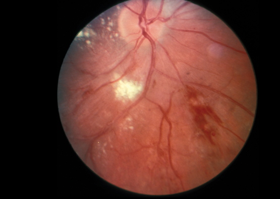
Box 1 –
Fundoscopic appearance of retinal vasculitis, showing retinal haemorrhages, cotton wool spots and perivascular hard exudates
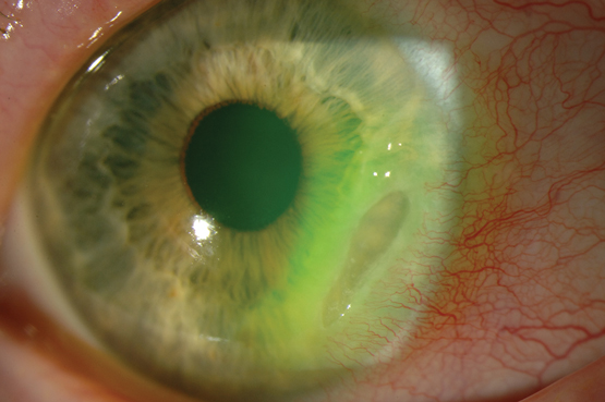
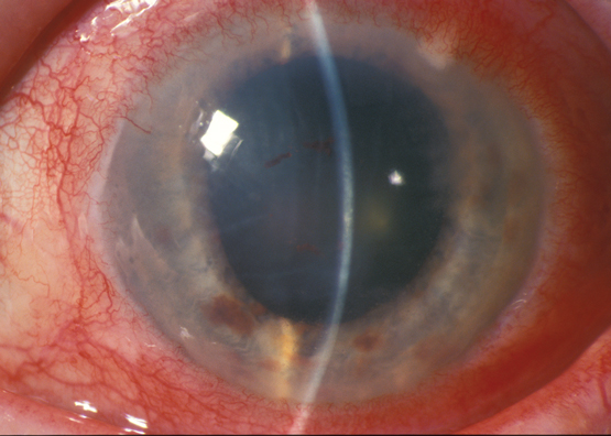
Box 2 –
Crescent-shaped corneal ulcer with neovascularisation and injection of ciliary blood vessels
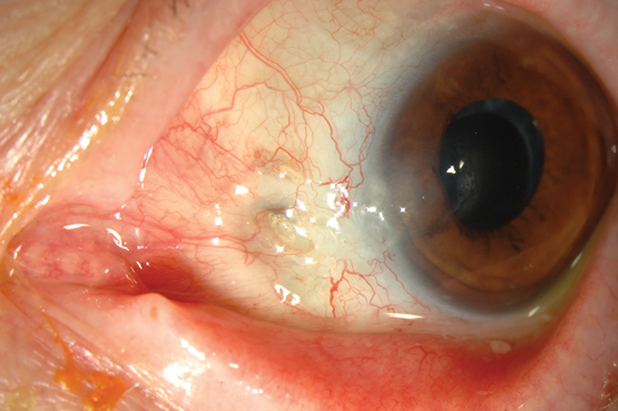
Box 3 –
Necrotising scleritis with exposure of the underlying uvea and associated area of scleral necrosis
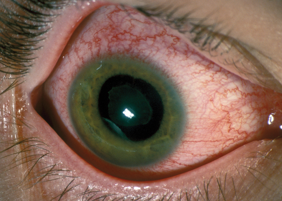
Box 4 –
Typical appearance of an eye in a patient with acute anterior uveitis, highlighting the injection of ciliary blood vessels

Box 5 –
Eye of a patient with iritis, showing a red eye with injection of ciliary blood vessels extending to the limbus

Advances in rheumatoid arthritis

This narrative review summarises the literature on contemporary understanding of adult rheumatoid arthritis (RA) focusing on current therapy, especially biological therapy. Articles were progressively identified by hand searching the list of contents in leading general and rheumatology journals, on a monthly basis over the past 6 years.

RA is a relatively common inflammatory arthritis (Box 1) and is self-reported by 2% of the Australian population (http://www.aihw.gov.au/rheumatoid-arthritis). It is diagnosed based on four criteria: number and pattern of joints involved, disease duration greater than 6 weeks, raised inflammatory markers (such as erythrocyte sedimentation rate [ESR] or C-reactive protein [CRP] level) and positive serology (the well established rheumatoid factor or the newer cyclic citrullinated peptide antibody [CCP])1 — CCP appears to be pathogenic and can be produced in gingival and lung tissues.2 RA cannot be diagnosed with only one involved joint. While there is considerable variation, either antibody is positive in about 50% of patients at presentation, with some overlap making about 25% seronegative. These antibodies are commonly misused in clinical practice, and their use should be restricted to patients with symptoms of inflammatory arthritis, especially involving peripheral joints, where they have both diagnostic and prognostic significance.

The cause of RA remains unknown, and many genes have been implicated.3 Each gene (with the exception of human leukocyte antigen) explains only a small amount of disease risk, but the involvement of a genetic pathway has proven useful for predicting response to therapy; for example, genes for tumour necrosis factor (TNF) and interleukin 6 (IL-6) receptor, but not interleukin 17 receptor, have been implicated to confer susceptibility to RA, and this closely mirrors positive clinical trial results for the first two, and negative trials for the latter. RA was not traditionally considered a lifestyle-related disease, but recent articles give clues about possible lifestyle modification. Smoking is a strong risk factor if a patient has the shared epitope of genetic predisposition, and smoking cessation appears to improve the disease outcomes, especially in patients who are CCP positive.4 The role of other lifestyle factors, such as sun exposure, which may be protective;5 salt intake, which may be deleterious;6 and alcohol, which may be protective,5 remain controversial. There have been great advances in our understanding of the pathophysiology of RA with the elucidation of critical cytokine pathways.7 Many of these have been targeted therapeutically with great success and considerable expense. Biological therapy for inflammatory arthritis is now costing the Australian taxpayer over $600 million each year. Using validated outcome measures of disease activity, patient-reported outcomes, and a treat-to-target approach of remission in early disease or low disease activity in established disease have led to a paradigm shift in RA management.

Traditional disease-modifying antirheumatic drugs (DMARDs) include methotrexate (MTX), which has become the cornerstone drug for the management of RA; sulfasalazine; corticosteroids; gold compounds, injectable and oral; and antimalarials. These agents are modestly effective but are proven to slow down radiographic progression, with the exception of antimalarials.8 They also decrease RA mortality rates, especially MTX,9 except prednisone, which increases death rates.10 However, historically, they were only used after damage was apparent, even though none have been shown to reverse progression. There is now evidence for a window of opportunity in the first 6 months of the disease, where therapies are more effective and have a long term effect on the disease, regardless of subsequent therapy.11 This means that RA should be diagnosed and treated with DMARD therapy as quickly as possible to maximise this benefit. Indeed, most rheumatologists in Australia have triage systems in place to see patients with RA quickly. The dosage has increased gradually over time, to a point where the mean dose is 20–25 mg per week, and many now use parenteral MTX, as oral absorption peaks at 15–20 mg weekly. MTX usage also maximises benefits and decreases side effects for many of the biological DMARDs (bDMARDs).12 A number of other agents such as d-penicillamine, cyclophosphamide and azathioprine are no longer used because of limited evidence of efficacy and poor safety.

Better disease control commenced with the use of higher doses of MTX, the development of leflunomide (the first of the targeted synthetic DMARDS), the use of combination therapy (most commonly with so-called triple therapy of MTX, sulfasalazine and hydroxychloroquine) and the use of fish oil as adjunctive therapy.13 Cyclosporine (a transplant medication), while of proven efficacy, is rarely used due to significant side effects.

The era of bDMARDs commenced in 1996, although the first of these agents did not become available in Australia until 2003. This was due to their approximate cost of around $20 000 per patient per annum. While they are very effective, with remission rates of up to 50%,14 they are only available with government subsidy on rheumatologist or immunologist prescription, with very strict entry and maintenance criteria. To start these agents, patients need to fail a 6-month intensive course of traditional DMARDs and have poorly controlled disease (ie, 20 or more affected joints, four or more large affected joints and raised ESR or CRP levels). Patients need a 50% improvement in joint count and a 20% improvement in ESR or CRP levels, documented every 6 months, to remain on these agents.

Box 2 summarises the mode of action of both biological and newer targeted synthetic DMARDs. In general, these agents have similar efficacy, making head to head studies desirable to choose between them.15 The only exception to this is the yet to be released baricitinib plus MTX — presented at the American College of Rheumatology Annual Meeting 2015 — which has recently been shown to be superior to adalimumab plus MTX.16 All bDMARDs improve signs and symptoms, blood markers, x-ray progression and important patient-reported outcomes, such as fatigue and quality of life. According to Pharmaceutical Benefits Scheme data, about 80% of patients meet the criteria for continuation, indicating that primary failure is rare. Usage in Australia reflects the length of time on the market, rheumatologists’ experience and mode of administration, with adalimumab and etanercept being the most commonly prescribed. Infliximab is rarely used due to the need for intravenous administration, infusion reactions and the well recognised tachyphylaxis over time — as this agent is a human–mouse chimeric antibody making it prone to provoking immunogenicity.

The initial therapy with biological agents is most often an antitumour necrosis factor (anti-TNF) agent plus MTX. If the latter is not tolerated or contra-indicated, then leflunomide, sulfasalazine or hydroxychloroquine may be used, with variable evidence of efficacy. Evidence favours IL-6 receptor blockade17 or tofacitinib18 if combination therapy is not appropriate. Despite this evidence, up to a third of Australian patients are on anti-TNF monotherapy, which works about as well as MTX, but costs much more. Switching for adverse effects or loss of efficacy over time between anti-TNFs is also common. However, recent evidence suggests that a switch to a different mode of action gives superior results in those with an insufficient response to the first anti-TNF.19 Anti-TNF agents work better in those who are seronegative, rather than in those who are seropositive, while rituximab, tocilizumab and abatacept work better in those who are seropositive.20 Anti-TNF agents should be avoided in those patients with tuberculosis, multiple sclerosis, cancer within the past 5 years, or heart failure.

Toxicity concerns

Managing the well documented toxicities of all available agents requires substantial rheumatological expertise, often in consultation with the general practitioner and other subspecialists. Common or serious side effects by class are listed in Box 3. Serious infection rates in this group of patients vary from 3% to 6% per annum.26 This appears to relate strongly to disease problems (as infection rates are higher in RA than other arthritides treated with bDMARDs), age, corticosteroid usage27 and medication. If a patient is at high risk of infection, then the first step is to minimise the dose of corticosteroid. In terms of bDMARDs, the agents with the lowest infection rates are abatacept, rituximab and etanercept,26 and they tend to be used more commonly in those aged over 65 years. Injection site reactions are also common, but rarely lead to discontinuation. Haematological monitoring is not required for anti-TNF agents, but all patients should be screened for tuberculosis, hepatitis B and C and human immunodeficiency virus before commencing therapy, and should be regularly rescreened if they live in endemic areas. IL-6 blockers require regular testing of full blood count, liver function tests and lipids. Abatacept and rituximab do not require screening tests or monitoring, although many rheumatologists check CD19 levels before re-treating with rituximab as these can take months or occasionally years to return to pre-dose levels; moreover, immunoglobulin levels should be monitored in those receiving long term rituximab as it may cause hypogammaglobulinaemia. Vaccination against influenza and pneumococcus is recommended for all patients with RA.28 In addition, patients should ideally receive herpes zoster vaccine before starting therapy,28 especially those on tofacitinib, even though this delays the initiation of medication.

Biosimilar or bio-originator?

The advent of biosimilars will result in significant decreases in the cost of bDMARDs. However, the process is not as straightforward as it is for generic small molecules, where bioequivalence is all that is required to be demonstrated for regulatory approval. bDMARDs are complex high molecular weight molecules that can vary in many ways, and the method of synthesis has changed over time. Thus, bioequivalence is not sufficient and, in general, regulators require large non-inferiority trials that compare the biosimilar with the bio-originator for at least one approved indication; for example, the biosimilar infliximab was studied in RA and ankylosing spondylitis,29,30 whereas the biosimilar etanercept was studied only in RA.31 If non-inferiority is demonstrated in these trials, then extrapolation to all previously approved indications is assumed to be valid. Development costs would be much higher if the original phase 3 program had to be fully replicated. The major concern relates to switching — which is freely allowed in Australia — as both of these agents are A flagged, meaning that they should not be substituted, but the reality is that they may be substituted by pharmacists depending on which agent is in stock. A single switch has been shown to be safe,32 but as multiple biosimilars of the same agent are approved, the safety of multiple switches is unknown. The concern is that the potential for formation of neutralising antidrug antibodies will be heightened by multiple switches, with resulting loss of efficacy and toxicity. At present, it is preferable that the patient remains on the same formulation (whether bio-originator or biosimilar) on a consistent basis.

Tapering

Studies are now underway to examine tapering and withdrawal of bDMARD therapy in patients in clinical remission, using power Doppler ultrasound scan to show absence of synovitis, and in those with normal CRP and ESR levels to reduce cost and risk of adverse effects. Withdrawal is especially suitable for the targeted synthetic DMARDs, as the development of antibodies against the compound is not a problem with small molecules.

Future directions

There is still an unmet need in RA, with most patients achieving at least a major clinical improvement, but with many still not achieving remission status. A number of novel agents are in development, including Australian discoveries such as mavrilimumab (a granulocyte-macrophage colony-stimulating factor antagonist)33 and novel ways of preventing RA using a vaccine-like approach, which induces dendritic cell tolerance in patients who are CCP positive.34 Thus, there is much to look forward to for patients with RA. The Australian Rheumatology Association has an excellent online resource for RA (http://rheumatology.org.au).

Box 1 –
Two examples of partially controlled rheumatoid arthritis showing synovitis in the metacarpophalangeal joints. Image A also shows some mild subluxation commonly seen with chronic disease, and image B shows proximal interphalangeal swelling and concomitant osteoarthritis in the distal interphalangeal joints

Box 2 –
Biological and targeted synthetic agents for rheumatoid arthritis and their mode of action

Agent

Mode of action

Mode of administration


Infliximab*

Anti-TNF

IV 8 weekly

Etanercept*

Anti-TNF

SC weekly

Adalimumab

Anti-TNF

SC fortnightly

Rituximab*

B cell blockade (CD20)

IV 6–12 monthly

Abatacept

T cell co-stimulation blocker

SC weekly or IV monthly

Golimumab

Anti-TNF

SC 4 weekly

Tocilizumab

IL-6 receptor blocker

SC weekly or IV 4 weekly

Certolizumab

Anti-TNF

SC 2 or 4 weekly

Tofacitinib

JAK inhibitor

PO BD

Coming soon

Baricitinib

JAK inhibitor

PO once daily

Sarilumab

IL-6 receptor blocker

SC fortnightly

Sirukumab

Direct IL-6 inhibition

SC


Anti-TNF = antitumour necrosis factor inhibition, BD = twice a day, IL-6 = interleukin 6, IV = intravenous, JAK = Janus kinase, PO = oral, SC = subcutaneous injection. * Now available as biosimilar.

Box 3 –
Common and serious side effects for biological and targeted synthetic DMARDs2125

Common side effects (> 1%)

Rare serious side effects


Anti-TNF

Injection site reactions
Serious infection

Drug-induced Lupus
Reactivation of TB
Sarcoidosis
Pustular psoriasis

IL-6 blockade

Neutropenia
Serious infection
Increased lipids
Abnormal liver function tests (with methotrexate)
Injection site reactions (SC version)

Anaphylaxis (IV version)

B cell blockade

Infusion reactions
Serious infections

Progressive multifocal leukoencephalopathy
Hypogammaglobulinaemia

T cell blockade

Serious infections

JAK inhibition

Zoster
Serious infections

Renal impairment
Anaemia


*Anti-TNF = antitumour necrosis factor inhibition, IL-6 = interleukin 6, IV = intravenous, JAK = Janus kinase, SC = subcutaneous, TB = tuberculosis.

Systemic lupus erythematosus: an update

Systemic lupus erythematosus (SLE) is a chronic multisystem autoimmune disease. Predominantly affecting young women, it continues to have an unacceptably high morbidity burden.1 Its clinical heterogeneity often makes diagnosis and management challenging even for experienced physicians. Additionally, the natural history of SLE is variable, with some patients experiencing fluctuating periods of relative inactivity contrasted by disease flare while others have persistently active disease.2 The classification criteria for SLE have recently been reappraised, and the new criteria have facilitated a more robust diagnostic process. The treatment approach of early and sustained control of disease activity is likely to lead to the prevention of irreversible end-organ damage,3 which correlates with early mortality.4 Advances have also been made in the therapeutic area, based on the exciting translational research identifying new targets that develop into therapies promising to ameliorate disease activity and lessen the reliance on treatments such as glucocorticoids. Patients with SLE experience poor health-related quality of life (HR-QoL),5 which can be worse than in groups with other chronic diseases such as congestive cardiac failure, myocardial infarction and diabetes.6 The determinants of HR-QoL in SLE are complex, but there may be disease- and treatment-related factors as well as factors associated with health care provision that may be amenable to change. Here, we present an overview of the key advances made in the diagnosis and management of this challenging disease, based on a review of recently published literature in peer-reviewed journals and international guidelines.

Diagnosis

By definition, SLE is a multisystem autoimmune disease, resulting in a wide spectrum of clinical manifestations, ranging from mucocutaneous, musculoskeletal and constitutional symptoms, to potentially life-threatening manifestations such as lupus nephritis or central nervous system (CNS) involvement. The use of classification criteria in rheumatology has allowed a more consistent description of the disease for the purpose of research and surveillance, and data from large observational cohorts using these classification criteria have become the basis on which clinicians derive important prognostic and treatment information on the disease.

The new Systemic Lupus International Collaboration Clinics (SLICC) classification criteria7 provide greater sensitivity with similar specificity to the previously used American College of Rheumatology (ACR) criteria.8 The more extended clinical and laboratory items that make up the SLICC classification criteria (see a summary in Box 1, and more details in the Appendix at mja.com.au) include conditions such as transverse myelitis, autoimmune haemolytic anaemia or immune-mediated cytopenias that carry separate weighting. Whereas previously, patient’s symptoms and test results may have been labelled as undifferentiated, the new criteria allow identification of otherwise missed SLE cases; in some cases this may result in earlier diagnosis.9

Autoantibody profiling can be useful in aiding the diagnosis of SLE. Antinuclear antibodies (ANA) are present in the vast majority of patients with SLE, but are not specific and can be seen at low to moderate titres in 5–13% of the normal population.10 In Australia, ANA can be detected using either indirect immunofluorescence or enzyme-linked immunosorbent assay (ELISA). It is important to recognise that the measurement of ANA is generally expressed as a titre, which reflects the number of dilutions at which ANA can still be detected. The laboratory reference range can vary, but generally a titre of 1:160 is considered as a weak positive ANA result. In its reporting, the staining pattern of the ANA is also included to provide clues to the specific underlying autoantibody, and clinical association with certain rheumatic diseases.

Detection of antibodies to double-stranded DNA (dsDNA) or Sm antigens are more specific for SLE, particularly for lupus nephritis, and they are present in about two-thirds of cases.11 The testing of anti-dsDNA in Australia has also undergone significant change, with many clinical immunology laboratories moving away from the conventional Farr assay. Although the Farr assay is the most robust test for anti-dsDNA, particularly in its association with disease flare, the efficiency of reporting is greater with the newer tests such as ELISA, fluorescent enzyme immunoassays (FEIA) and Luminex beads.12 Clinicians should be aware of the differences between these assays in their ability to detect different anti-dsDNA subsets and in their reference ranges. Other laboratory markers that may facilitate diagnosis include the presence of hypocomplementaemia, antiphospholipid antibodies, unexplained cytopenia and a positive direct anti-globulin test result. The latter illustrates the close relationship between immune-mediated haematological disorders and SLE.13

In practice, for general practitioners and other physicians, the diagnosis of SLE should be considered in any patient presenting with inflammatory joint pain with one or more extra-articular features, as arthralgia is one of the most common clinical manifestations (Australian Lupus Registry, Monash University, unpublished data). The diagnostic process relies on taking a good history in seeking confirmation of organ involvement for a variety of organs that are typically affected by this disease and, in some cases, laboratory tests that may reveal relatively “silent” features, such as haematological or renal manifestations. ANA testing should serve as a screening test and, in the right clinical setting, further serological testing including for autoantibodies to dsDNA or Sm, antiphospholipid antibodies, complement levels, and direct anti-globulin tests, may aid diagnosis (Box 2). In the Australian context, patients of Asian ancestry and Indigenous Australians are more likely to have clinically severe disease, with higher rates of lupus nephritis and autoantibody positivity, and higher levels of overall disease activity compared with their counterparts of European ancestry.14,15

While the use of classification criteria is widely accepted as a useful way to aid in the diagnosis of SLE, it is by no means a complete list of “diagnostic criteria”. When in doubt, categorising patients as having undifferentiated connective tissue disease is always the preferred option. Rheumatologists must weigh up the risk of delayed diagnosis (as many patients with SLE can present initially with undifferentiated symptoms and laboratory test results) against the risk of over-diagnosis purely based on criteria. Many studies have shown that most patients who do not meet classification criteria (ie, “possible SLE” cases) never develop SLE.16

Management

Pharmacotherapy

Glucocorticoids

Glucocorticoids (GCs) have rapid anti-inflammatory and broad immunosuppressive effects, and are often used as an adjunct in induction therapy of serious SLE manifestations such as lupus nephritis. In many instances, however, patients continue to require maintenance therapy with GCs as well as conventional immunosuppression, although recent literature and studies underway are challenging the need for long term use of GC in treating SLE.17 Although a “safe” maintenance dose of prednisolone is still being debated, studies assessing prednisolone use in rheumatic disease have shown that doses as low as 5 mg daily significantly increase the risk of osteoporosis,18 impaired glucose tolerance,19 and infections.20 The SLE Damage Index (SDI), which measures the accumulation of comorbid conditions commonly associated with SLE or its treatment, contains items that are classically related to GC exposure and other items that do not initially appear to be related. The progression of damage as measured by the SDI is commonly known as damage accrual in SLE, which correlates well with mortality. Recent research has shown that the use of GCs can independently predict damage accrual including the non-GC related domains within the SDI.21 A large cohort study has also shown that at GC doses of 6 mg or less, there appears to be insignificant impact on damage accrual in patients with SLE.22

Hydroxychloroquine

Hydroxychloroquine has been used to treat SLE since the 1960s, and may be considered an “anchor drug” in SLE therapy, just like methotrexate in rheumatoid arthritis. It is often adequate for controlling milder manifestations of SLE, has an independent protective effect on damage accrual, and confers a survival benefit in patients with SLE.23 There are some data to suggest that this drug can improve the efficacy of other immunosuppressive medications such as mycophenolate.24 It is generally well tolerated and is not associated with increased infection risk. Long term maintenance doses should be kept within the “safe level” of lower than 5 mg/kg wherever possible. The latest recommendation for annual screening after 5 years of therapy25 allows early detection of retinopathy and can prevent serious visual loss26 (Box 3). Screening with automated visual field testing offers a sensitive way of detecting hydroxychloroquine-related retinopathy, and the greater availability of new technologies, such as spectral domain optical coherence tomography, can assist with a more specific diagnosis.25

Immunosuppression

In the past decade, there has been a significant change in the choice of induction immunosuppression for lupus nephritis. Increasingly, mycophenolate is used, as it has been shown to be equivalent to pulse cyclophosphamide in inducing remission of renal disease and preventing relapse in a number of randomised control trials (RCTs).27 It has become the preferred first-line treatment for lupus nephritis in women of childbearing age because of a lack of gonadal toxicity (Box 3). A recent observational cohort study has shown that mycophenolate is also useful in a range of non-renal manifestations.28 Cyclophosphamide remains an important immunosuppressive therapy in managing refractory nephritis and other serious manifestations such as CNS disease. The Euro-Lupus Nephritis Protocol, which consists of a lower dose and shorter duration of cyclophosphamide therapy, has comparable renal remission rates, with a better safety profile.29

Biological therapy

In contrast to other rheumatic diseases, such as rheumatoid arthritis and ankylosing spondylitis, there is a lag in finding an effective targeted biological therapy for treating SLE. The reasons for this are multifactorial, including SLE having a more complex immunopathogenesis (Box 4), clinical disease heterogeneity, difficult trial designs with criticisms on the role of concomitant immunosuppression and GCs, and problematic outcome measures.30 While the understanding of lupus pathogenesis continues to advance (Box 4), many therapeutic targets that have shown great potential in preclinical studies and phase 2 trials have failed to show clinical efficacy in phase 3 RCT settings. The most notable examples are abatacept,31 which blocks costimulation of T cells, and rituximab, a B cell-depleting therapy.32 Pivotal phase 3 RCTs of rituximab in SLE have failed to reach their primary efficacy endpoints,33 despite widespread anecdotal experience suggesting positive results in at least some patients. For this reason, rituximab is still used off-label as rescue therapy based on observational data.

Belimumab, a monoclonal antibody inhibiting a B cell cytokine called BLyS (B lymphocyte stimulator), has been shown in phase 3 RCTs to be efficacious and is currently registered by the Therapeutic Goods Administration (TGA) as an add-on therapy for moderate to severe SLE. Multinational phase 3 trials showed clinical and serological improvement compared with placebo, particularly in patients with active musculoskeletal and mucocutaneous disease,34 and post hoc analysis showed it to be more effective in patients with serologically active disease (a low complement level and a high concentration of antibodies to dsDNA).35 Although statistically significant, the absolute effect size of belimumab over placebo appeared to be small, and the trials excluded patients with active lupus nephritis or CNS disease. Its role in lupus treatment is still to be defined, especially given that it has not been approved by the Pharmaceutical Benefits Scheme in Australia.

Because of the encouraging results of the belimumab trials, there has been a flurry of research focusing on alternative B cell targeted therapy, albeit without much success. Most recently, blockade of the type I interferon (IFN) pathway has gained significant interest, with anifrolumab, the first anti-IFNα receptor monoclonal antibody showing clinical efficacy in a phase 3 RCT.36 The interferon pathway is implicated in the process of loss of self-tolerance, and type I IFN is known to upregulate a panel of over a hundred genes that can be measured by blood microarray analysis. The so-called IFN signature is gathered by quantitative analysis of the downstream IFN-inducible genes, and can be used as biomarker for active disease in patients with SLE.37 A simplified interferon signature metric can be used to predict treatment response.38 Novel ways to target IFN are also in development, and it is to be hoped that, in time, one of these biological therapies may become a game changer in the management of SLE.

Treat-to-target in SLE

In addition to the advances in our understanding of the biological paradigm of SLE, there has been progress in the area of clinical science that may change the way physicians manage patients with SLE in the future. Analogous to the treat-to-target approach in other chronic diseases, an international taskforce was set up to define the principle of treat-to-target in SLE.39 Apart from having target organ response criteria (eg, in lupus nephritis), significant advances have been made in defining remission and overall low disease activity in SLE. The concept of lupus low disease activity state (LLDAS) has been shown to be an achievable target, and is associated with less damage accrual.40 A large multinational prospective cohort study is underway to validate the significance of LLDAS, and to determine its predictors.41 Future studies on treatment strategy may involve newer agents, but better still, may reveal ways that we can use existing therapies more optimally. One possibility is to consider selected use of therapeutic drug monitoring for the commonly used drugs in SLE to improve their efficacy and safety.

Quality of care in SLE

The challenge of translating clinical studies into everyday practice and health care decision making is another key area of interest in SLE management. Disease outcomes in SLE can be influenced by a complex interplay between genetic, biological, socio-economic and health system variables. The ability to implement the knowledge acquired by clinical studies and published in guidelines into clinical practice is the basis of implementation research. While SLE is not necessarily a common disease, patients often require frequent interaction with the health system, sometimes with different specialists.42 The quality of health care delivery can be benchmarked in accordance with published guidelines for monitoring, treatment and preventive care in SLE, and including the frequently associated comorbid conditions.43

Based on recommendations from published guidelines, a series of quality indicators has been defined which encompasses areas of diagnosis, disease assessment, cardiovascular risk assessment, osteoporosis management, drug monitoring, renal disease monitoring, immunisation, cancer screening, and pregnancy counselling (Box 3).44 It has recently been shown that improving the quality of care for patients with SLE can have an impact on disease outcomes, such as reductions in damage accrual45 and in-hospital mortality.46 Studies are currently underway to assess performance on SLE quality indicators within the Australian health system. While we await the results of these studies, simple preventive strategies should be considered and can be implemented by any doctor involved in the care of patients with SLE.

For example, the risk of cardiovascular death for patients with SLE is more than double that of age- and sex-matched counterparts.47 In addition to the traditional cardiovascular risk factors such as hypertension, hypercholesterolemia, diabetes and smoking, the risk of cardiovascular death is further compounded by prolonged active disease and use of GCs.48 Cardiovascular risk assessment can be easily done using the widely available absolute risk calculators. Recently, the University of Toronto Lupus Clinic researchers published a modified Framingham Risk Factor Score, proposing a doubling of the traditional score to better reflect the rate of cardiovascular disease captured in their cohort.49 Clinicians should also be suspicious when patients with SLE present with symptoms that could suggest premature coronary artery or peripheral artery disease, and proceed with appropriate investigations.

Patients with SLE are at increased risk of osteoporosis and fracture due to a combination of factors, such as a chronic inflammatory state, GC use, vitamin D deficiency and premature ovarian failure.50 Therefore, vitamin D status should be assessed in patients with SLE, especially as many may be avoiding sun exposure either because of their disease or medication increasing their sun sensitivity. Existing guidelines are available for any patient who is on chronic GC therapy with regard to bone density assessment and prevention of osteoporosis with anti-resorptive therapy, depending on the severity of osteopenia.

Conclusions

SLE is a chronic multisystem autoimmune disease that results in significant morbidity and loss of life expectancy. A lack of effective targeted therapies for SLE means that most patients are still treated with chronic GC therapy and broad spectrum immunosuppression. Our understanding of the relationship between disease activity and irreversible and progressive accrual of organ damage has advanced. Earlier clinical trials assessing novel biological agents failed because of a combination of factors, but there is ongoing interest in finding the optimal way to use B cell targeted therapy, and interferon blockade has shown some early success. Emerging definitions for remission and low disease activity are promising to not only change trial design, but to allow a “treat-to-target” approach in clinical practice. Meanwhile, research into quality of care for patients with SLE will shed light on areas of deficiency in clinical practice in a real world setting, and findings could potentially lead to evidence-based system redesign.

Box 1 –
Summary of classification criteria for systemic lupus erythematosus*

Systemic Lupus International Collaborating Clinics (SLICC) Criteria7

American College of Rheumatology (ACR) Criteria8


Clinical criteria

1. Acute cutaneous lupus

1. Malar rash

2. Chronic cutaneous lupus

2. Discoid rash

3. Oral ulcers

3. Oral ulcers

4. Non-scarring alopecia

4. Photosensitivity

5. Synovitis

5. Non-erosive arthritis

6. Serositis

6. Serositis

7. Renal

7. Renal disorder

8. Neurologic

8. Neurologic disorder

9. Haemolytic anaemia

9. Haematologic disorder

10. Leukopenia

11. Thrombocytopenia

Laboratory criteria

1. Antinuclear antibody

1. Antinuclear antibody

2. Anti-double-stranded DNA antibody

2. Immunological disorders

3. Anti-Sm antibody

4. Antiphospholipid antibody positivity

5. Low complement

6. Direct Coombs’ test

Requirement for diagnosis

  • Must meet 4 of 17 SLICC criteria (with at least one criterion being clinical and at least one criterion being immunological) OR,
  • Lupus nephritis proven by biopsy and at least one immunological criterion

  • Must meet 4 of 11 ACR criteria

* A more detailed classification is provided in the Appendix at mja.com.au.

Box 2 –
Recommendations for autoantibody profile testing in relation to systemic lupus erythematosus (SLE)

Recommendation 1

  • In making the diagnosis of SLE, ANA testing may be indicated if patients present with one of the following:
    • Inflammatory arthralgia/ arthritis;
    • Pleurisy or pericarditis;
    • Photosensitive rash;
    • Haemolytic anaemia, thrombocytopenia, leucopenia;
    • Raynaud’s phenomenon;
    • Acute brain syndrome in a young person.

Recommendation 2

  • The following supportive serological tests can be pursued in patients suspected of having SLE, after a positive finding of an ANA test:
    • anti-dsDNA;
    • ENA;
    • C3 and C4;
    • Anti-phospholipid antibodies (anticardiolipin antibodies, anti-beta-2 glycoprotein, and lupus anticoagulant);
    • Direct Coomb’s tests;
    • Consider ANCA, RF and anti-CCP as potential mimics.

Recommendation 3

  • ANA and ENA testing should normally not be repeated as they are not of monitoring value.
  • If an unexpected result is seen, or the clinical features of the patient change, then repeat testing can be considered.

ANA = antinuclear antibody; ANCA = anti-neutrophil cytoplasmic antibody; C3 = complement component 3; C4 = complement component 4; CCP = cyclic citrullinated peptide; dsDNA = double-stranded DNA; ENA = extractable nuclear antigen; RF = rheumatoid factor; SLE = systemic lupus erythematosus.

Box 3 –
Current recommendations for the management of systemic lupus erythematosus (SLE) from which quality indicator sets were derived

Diagnosis

  • For a suspected diagnosis of SLE, initial workup should include testing levels of ANA, FBE and creatinine, and urinalysis.
  • Once a diagnosis of SLE is made, evaluation of the baseline antibody profile should include ANA, dsDNA, Ro, La, RNP, Sm, antiphospholipid antibodies.

Disease and comorbidities monitoring

  • For patients diagnosed with proliferative renal disease (Class III or IV), treatment with prednisolone and other immunosuppressant should be initiated and documented within a month, unless contraindicated.
  • Disease activity should be recorded using a validated index at each visit.
  • For patients with renal disease and proteinuria, or eGFR < 60 and persistent hypertension, anti-hypertensive medication should be considered.
  • For patients with proteinuria > 300 mg/day, ACE inhibitor or ARB should be considered unless contraindicated.
  • Presence of comorbid conditions should be recorded at each visit.
  • For those requiring high-dose corticosteroids and/or immunosuppressive drugs, hepatitis B virus, hepatitis C virus and tuberculosis status should be evaluated and results recorded.
  • Risk factors for cardiovascular disease including smoking, blood pressure, body mass index, diabetes and serum lipid levels should be evaluated annually.
  • Damage accrual should be evaluated and documented using the Systemic Lupus International Collaborating Clinics Damage Index.
  • Quality of life should be evaluated at each visit.

General preventive strategy

  • Sun avoidance strategies should be discussed.

Medications-related monitoring

  • For newly prescribed medication, risks versus benefits should be discussed with the patient.
  • Drug toxicity should be evaluated at each visit, including review of laboratory results.
  • For patients receiving prednisolone ≥ 7.5 mg/day for 3 months.
    • bone mineral density testing should be done and results recorded unless the patient is already being treated with anti-resorptive therapy;
    • supplemental vitamin D and calcium should be recommended.
  • For patients receiving prednisolone ≥ 7.5 mg/day for 3 months, and who have a bone density test T score of ≤ −2.5 or already have a fragility fracture, an anti-resorptive or anabolic agent should be recommended.
  • For patients treated with hydroxychloroquine or long term corticosteroid, an ophthalmologic assessment according to current guidelines should be followed.
  • For patients taking prednisolone ≥ 10 mg/day for 3 months, an attempt should be made to taper the prednisolone dose, or to add or optimise doses of a steroid-sparing agent, unless contraindicated.

Immunisation

  • For patients treated with an immunosuppressive therapy:
    • annual inactivated influenza vaccination should be given unless contraindicated;
    • pneumococccal vaccination should be given unless contraindicated.

Pregnancy

  • Contraception and potential teratogenic risk should be discussed with women aged 18–45 years with childbearing potential who are starting therapy with methotrexate, leflunomide, mycophenolate, cyclosporine, cyclophosphamide or thalidomide.
  • Antiphospholipid, Ro and La antibody levels should be documented for pregnant women with SLE.
  • Women who have had an adverse pregnancy outcome due to antiphospholipid syndrome should be given aspirin and heparin/enoxaparin for any subsequent pregnancy.

ACE = angiotensin converting enzyme; ANA = antinuclear antibody; ARB = angiotensin II receptor blocker; BMI = body mass index; BP = blood pressure; dsDNA = double-stranded DNA; eGFR = estimated glomerular filtration rate; FBE = full blood examination; La = Sjögren’s-syndrome-related antigen B; RNP = ribonucleoprotein; Ro = Sjögren’s-syndrome-related antigen A; SLE = Systemic Lupus Erythematosus; SLICC = Systemic Lupus International Collaborating Clinics; Sm = Smith, as in anti-Smith antibodies. Key recommendations are based on available literature from which sets of quality indicators were derived.45

Box 4 –
Key cellular pathways in the immunopathogenesis of systemic lupus erythematosus (SLE)


BLyS = B lymphocyte stimulator; IFN = interferon; IL = interleukin; IRF = interferon regulatory transcription factor; NFkB = nuclear factor k-light-chain-enhancer of activated B cells; Th = T helper; TLR = toll-like receptor. (i) Excess apoptotic material may serve as a source of self-antigens, and are recognised by TLRs on plasmacytoid dendritic cells. Type 1 IFN is produced in response to this activation and drives subsequent activation of the immune system. (ii) Dysfunctional lymphocyte signalling pathways leading to the activation of autoreactive T and B cells, play important roles in the immune amplification pathways, and have been key targets of immunomodulation in SLE. (iii) Cytokines such as BLyS and IL17 play a pivotal role in promoting B cell survival, induction of other proinflammatory cytokines and chemokines, and mediating local tissue damage. (iv) Activated macrophages with upregulated monocyte chemoattractant protein 1 expression can further drive local injury. (v) Defective regulation of neutrophil extracellular traps has been shown to drive autoimmunity and tissue damage in SLE.

The Australian Lupus Registry and Biobank: a timely initiative

A collaborative effort to provide real world evidence for therapies for patients with lupus

Systemic lupus erythematosus (SLE) is a complex autoimmune disease with diverse clinical manifestations, which places an unacceptable level of burden on affected patients. Australian data on lupus are scarce, with figures suggesting a prevalence of SLE that ranges from 19 per 100 000 in people of European ancestry to 92 per 100 000 in Indigenous Australians,1 similar to other chronic diseases such as hepatitis C.2 Survival rates for SLE patients in the 1950s were as low as 50% at 5 years. With improvements in the treatment of renal disease and infection, survival rates in most studies improved to around 90% at 10 years by the 1990s. However, it is still a sobering thought that SLE, which typically presents in women in their second or third decade of life, confers a 1 in 10 chance of dying before the age of 40.3 Damage accumulation, long term medication side effects (particularly steroids side effects), fatigue and uncertainty profoundly affect quality of life.4 Fundamental data regarding age, geographic and ethnic distribution; natural history of the disease; currently used treatments; and unmet needs of patients in Australia have not been well defined.

Since it is a relatively rare and heterogeneous disease, longitudinal registry studies play an essential role in improving our understanding of SLE. Registry studies are ideally suited to capturing real world data on a large number of subjects, giving insights into disease course and treatment practices. Moreover, they may serve as a platform to inform planning of randomised controlled trials (RCTs) and subcohort studies. In contrast to RCTs, they typically have broader inclusion criteria and allow for long term follow-up. Registry findings can complement RCT results, as demonstrated by the increased risk of tuberculosis associated with the use of tumour necrosis factor inhibitors in patients with rheumatoid arthritis, which was not identified in clinical trials, but rather revealed and quantified through registry studies.5 Despite the inherent limitations, a great deal can be learnt from single-centre SLE registries, such as the seminal 1974 observation in the Toronto lupus cohort of a bimodal mortality pattern, with early deaths due to active disease and infection, and later deaths due to premature cardiovascular disease.6 However, much progress has been made in past 40 years, with large multicentre cohorts from predominantly Europe and North America contributing to our understanding of the disease, particularly with reference to real world epidemiology, clinical features, natural history and long term outcomes.7

A national registry based in Australia may be a late starter, but it has the potential to be a world leader, with carefully collected data that provide assessment of visit-to-visit disease activity and of medication exposure. It also enables the conduct of studies specific to the Australian health care settings and the demographics of a multicultural Australian population. The Australian Lupus Registry and Biobank (ALRB) was created in 2012, with seed funding from MOVE Muscle, Bone and Joint Health (formerly Arthritis and Osteoporosis Victoria) and contributions from the industry in the form of unrestricted grants.

The ALRB is an online platform that enables the longitudinal collection of systematic and comprehensive data. One of the first studies using the registry aimed at understanding the disease characteristics and treatment patterns in Australia. Using the framework provided by the registry, we have also undertaken a study to validate a consensus-based treatment target to determine whether this can be a data-driven treatment endpoint associated with better patient outcomes.8 The effects of long term use of immunosuppressive medication in SLE patients are not well understood, and data from the registry may give us a better understanding of the incidence of adverse effects and benefits, such as reduction in flares and accumulated damage over time. In addition, biomarker studies examining the interferon-α gene signature and disease manifestations, response to treatment, vitamin D status and disease manifestations, and patient-reported quality of life are in the planning stages.912

The registry also collects patient-reported outcomes, such as Short Form 36 and multidimensional health assessment questionnaires. Patients’ self-reported data complement physician-reported data in the ALRB to capture the breadth of experiences of patients with SLE in Australia and provide a meaningful assessment of the disease burden and treatment shortfalls.

There are now ten institutions recruiting patients with SLE to the ALRB across Victoria, New South Wales, South Australia and Western Australia, with the common goal of improving treatment and outcomes for people with SLE. The expansion of the ALRB to involve more practices across Australia may be easier if the platform imports from existing electronic medical records or health systems (administrative, laboratory or radiological) to avoid duplicated data entry — provided data fields are matched according to a stringent data dictionary. Periodic auditing, involving cross-checking data with source records, will be done by principal investigators at each site and will be reported to the ALRB Management Committee to ensure data completeness and accuracy. This committee may also request the de-identified source documentation for quality assurance purposes.

One of the key strengths of the ALRB rests in its ability to examine a variety of health care outputs over time. At present, in the complex Australian health care system, it is difficult to examine the different components of health care use; therefore, the true economic costs for a disease such as SLE are often grossly underestimated. The ALRB will allow the tracking of health care uses related to the care of the SLE in Australia and will provide data for benchmarking. With the rising costs of health care and a limited health budget, it is paramount that data are available to study the cost effectiveness of various management strategies. Health care use, based on annual patient self-reporting of hospitalisations, investigations and other health complications, may form the basis to derive cost. The ALRB information may help measure the health consequences of different health care interventions.

The overarching principle of the ALRB is to foster collaborative research and, with the same purpose, the simultaneous development of the Asia–Pacific Lupus Collaboration (www.asiapacificlupus.com) brings together researchers from Australia, China, Dubai, Hong Kong, Indonesia, Japan, Malaysia, Philippines, Singapore, Taiwan and Thailand. More than 2000 patients with lupus across the Asia–Pacific region have been recruited in the Lupus Low Disease Activity State study to validate a treatment target.8 This type of research is consistent with the Australian Research Council strategy to encourage international collaboration — especially where it has been led by the Australian site (www.arc.gov.au/international-collaboration).

Finally, the parallel development of a biobanking system to complement clinical data from the ALRB means that more questions into aetiology and novel biomarkers can be answered. The fostering of closer links between basic science researchers and clinicians is the foundation of good translational research. Linking clinical phenotype to genetic polymorphisms and novel laboratory parameters has been valuable in understanding pathogenesis and prognosis, and in predicting SLE manifestations and response to treatment in such a heterogeneous disease.13

The ALRB is still in its infancy and will require significant inputs from various funding sources to continue its growth. We expect that, as the registry grows, it will serve as a valuable resource for clinicians, scientists, epidemiologists, patient advocacy groups, industry and government to provide real world evidence of clinical effectiveness of existing or new therapies and management strategies in patients with SLE in Australia.

Examination of the shoulder joint

The shoulder joint could be described as a trade-off between range and stability. The striking range of movement of this joint is achieved by its shallow articulation and a dynamic cuff of the shoulder muscles and their associated tendons. It is therefore not surprising that ageing has more impact on the soft tissues of the shoulder than on other more stable and less dynamic joints.

An Australian population study1 showed the importance of the history and examination of the shoulder, rather than imaging. In this study, multiple asymptomatic changes in the magnetic resonance imaging (MRI) scan were observed in healthy individuals, and there was a poor correlation between shoulder pain and change in the MRI scan. In many cases, the clinical examination features are mainly diagnostic. Worldwide, there is a well defined, age-dependent increase in the frequency of rotator cuff tears in asymptomatic shoulders.2 In keeping with many joint examination techniques, a series of steps (including look, feel, move and special tests) remains valid with the shoulder.

Physical examination is not a blind or formulaic activity. We need to examine the patient in a sequence to answer specific questions based on diagnoses that may become more or less likely as the examination proceeds (Box 1). The first question should be: is the pain really emanating from the shoulder? If the shoulder is non-tender and moves easily, it is likely that the pain is coming from the neck or other structures, and proceeding with the shoulder examination is unlikely to yield a diagnosis.

If the shoulder joint is established as the source of pain, there are four main shoulder conditions to consider as the examination proceeds:

  • rotator cuff tendinitis and impingement (Box 2);

  • rotator cuff tear, in the case of older patients (Box 2);

  • frozen shoulder, that is, adhesive capsulitis (Box 3); and

  • arthritis of the glenohumeral joint.

In practice, we can consider shoulder problems to be divided into two main groups, based on whether there is restriction of passive shoulder movement.3 Restricted range of passive movement suggests either adhesive capsulitis (frozen shoulder) or arthritis of the glenohumeral joint. Absence of restriction of passive movement points to impingement syndrome (rotator cuff tendinitis) or a rotator cuff tear. After inspection and palpation, the key step is to examine the shoulder and take it passively through its range of movement.

Passive range of shoulder movement

A relatively unrestricted range of passive movement in all directions suggests that the problem is not emanating from the shoulder, and that it may be referred from the neck or other structures, or that it falls into the category of one of the rotator cuff problems, which include rotator cuff tendinitis, tendonopathy and subacromial bursitis.

If there is a global restriction of passive movement (ie, partial or full restriction in all directions), it may suggest arthritis of the shoulder or frozen shoulder. It is increasingly appreciated that the classic description of a frozen shoulder, in which there is a profound global restriction of movement, does not encompass all cases. For example, much milder global restriction of the shoulder — with a loss of 20–30° of movement in all directions — may be seen in some cases of frozen shoulder.

For these two conditions, which cause restriction of shoulder motion, an x-ray is required to discriminate between frozen shoulder and arthritis of the glenohumeral joint. This may resolve the problem with much greater reliability than any other examination.

Conditions not associated with significant restriction of passive movement include rotator cuff tendinitis or impingement and rotator cuff tear. There are two clues to differentiating between significant rotator cuff tear and tendinitis and impingement syndrome. The first is the age of the patient. Impingement syndrome and rotator cuff tendinitis are more commonly seen in people under the age of 40–50 years. In the population over 50 years, and with increasing frequency with each decade of age after 50 years, the underlying pathology usually involves degenerative change in the tendons of the rotator cuff, particularly supraspinatus, with frank tears.

Active range of shoulder movement and power testing

In a patient with preserved passive range of movement, pain on active and resisted abduction supports the idea of rotator cuff pathology. If there is weakness, particularly in resisted external rotation, this supports a diagnosis of rotator cuff tear.

An examination of power at the shoulder is, therefore, a major discriminator that indicates the presence of a rotator cuff tear of significance. If the rotator cuff is intact, even if inflamed, there will be good preservation of power.

It should be noted that pain on active horizontal adduction of the shoulder (across the body) can also be seen with osteoarthritis at the acromioclavicular joint.

Box 1 –
Shoulder joint examination sequence

Box 2 –
Rotator cuff tendinitis and impingement

Box 3 –
Frozen shoulder

Nocebo effects in practice: methotrexate myths and misconceptions

Providing patients with accurate information will help overcome obstacles to the use of an effective treatment for rheumatoid arthritis

Weekly low dose methotrexate is the first-line agent and arguably an essential disease-modifying antirheumatic drug (DMARD) in the treatment of rheumatoid arthritis (RA).1 Australian rheumatologists’ practice mirrors the recommendations of international rheumatology associations24 for early treatment with methotrexate, often combined with other DMARDs. A trial of methotrexate in combination with other DMARDs is an Australian prerequisite for therapy with biological disease-modifying drugs. About 20% of more than 3.4 million Pharmaceutical Benefits Scheme-subsidised DMARD prescriptions issued between 2003 and 2007 to 236 000 Australians for the treatment of RA were for methotrexate.5 As older agents such as injectable gold, azathioprine and cyclosporine fall out of favour, methotrexate usage will likely increase, not only for treatment of RA but also other rheumatological and inflammatory conditions such as psoriatic arthritis, juvenile idiopathic arthritis and systemic vasculitis.

Knowledge of the pharmacology of methotrexate is important to understand its potential toxicity. The inter-individual bioavailability of oral methotrexate is about 70%. Oral absorption of methotrexate can be variable at doses above 15–20 mg/week; hence parenteral administration may be appropriate in cases of apparent inefficacy or problematic tolerability. The serum half-life of methotrexate is between 6 and 8 hours following administration, 70% is eliminated unchanged by renal excretion, and it is undetectable after 24 hours.1 Methotrexate is not lipophilic6 and thus cannot be absorbed via the skin, whether in solid form (tablet) or liquid (solution for injection). Co-treatment with folate is recommended by a recent systematic review, in support of rheumatology association guidelines to reduce gastrointestinal side effects, mucositis, transaminitis and treatment discontinuations.7

Significant and avoidable toxicity, resulting in mortality, typically from inappropriate daily rather than weekly schedules and/or dosage errors, has been highlighted.8 While we agree that the potential for dosage and administration errors must be appreciated and eliminated, methotrexate is an indispensable medication for the treatment of RA as monotherapy or in combination.9

Nocebo effects are “biologically implausible negative effects of active therapies” that may arise from the anticipation of adverse effects from any intervention.10 There are several methotrexate “myths” that may be held by patients and health professionals, and propagated through word of mouth, information in print11 or on the internet that, if not simply inaccurate, may be contextually inappropriate and may generate prominent nocebo effects. Such negative anticipation responses — the expectation of routine adverse effects — may result in objections to treatment and misinterpretation and misattribution of symptoms developing during a treatment regimen. This can result in poor treatment adherence and potentially avoidable suboptimal patient outcomes. Routinely available consumer medical information leaflets12 list many adverse effects and may prove unnecessarily disturbing to patients for whom methotrexate is recommended. This is a particularly likely outcome if these written materials are used as a proxy or even a substitute for genuine discussion with the treating doctor.

In this article, we highlight commonly held myths and misconceptions regarding methotrexate treatment. We advocate that these be discussed with patients to assist in making appropriate decisions based on accurate and, most importantly, contextually relevant information.

Methotrexate myths

Methotrexate (for non-malignant diseases) is chemotherapy

Chemotherapy regimens for neoplasia may utilise cycling protocols where methotrexate may be used as monotherapy or in combination with other cytotoxics where single dosages might exceed 500 mg/m2. For rheumatic diseases, methotrexate is typically used in low dose weekly oral or parenteral regimens, in doses ranging from 10 to 30 mg weekly, usually less than 10 mg/m2 weekly.9

Methotrexate is unusual or strong treatment

Both the European League Against Rheumatism2 and the American College of Rheumatology3 recommend early treatment with methotrexate for RA. Methotrexate can be described as safe and efficacious, rather than unusual, treatment and has been in use since the mid-1980s.13,14 The concept of gaining control of inflammatory arthritis as early as possible to prevent joint damage and disability is compelling, and the words “strong” or, preferably, “effective” used in that context can be seen as much more positive and accurate.

Patients should not be co-prescribed non-steroidal anti-inflammatory drugs and methotrexate

Methotrexate is often prescribed along with non-selective non-steroidal anti-inflammatory drugs (NSAIDs) or cyclooxygenase-2 inhibitors to achieve analgesic and anti-inflammatory effects in addition to the disease-modifying effect of methotrexate; however, there is a common automatic alert generated in prescribing software and pharmacies, which can lead to confusion for patients. This myth may also be propagated through the internet via blogs and other grey sources.

However, there are no specific grounds to unreservedly prohibit the combination of NSAIDs and methotrexate.15

The context of compromised renal function deserves specific mention owing to the renal clearance of methotrexate. Renal function should be monitored and the dose of methotrexate appropriately adjusted. Patients are often unnecessarily alarmed by advice from health professionals to avoid this drug combination at all costs, and this may contribute to the nocebo effect.

Third parties must avoid (any) exposure to the body fluids of methotrexate-treated patients

Patients may be told by health professionals or lay persons to carefully dispose of their urine and faeces, avoid kissing, cuddling or nursing infants and children, and avoid both non-intimate and intimate kissing.

The warning to avoid any contact with pregnant women is so important that it is dealt with separately below.

Such statements conflate the concept of “methotrexate as chemotherapy” and the notion that “all cytotoxics are the same” in the absence of specific data pertaining to low dose weekly methotrexate for RA. Normal standards of personal hygiene are the principal safeguard for third parties.

Patients receiving methotrexate should avoid contact with pregnant women

Methotrexate is a pregnancy category X drug (exposure to which has a substantial risk of causing permanent damage to the fetus) and should not be administered to pregnant women or those with reproductive capacity unless reliable contraception measures are employed. There is a clear risk of congenital abnormalities in babies exposed to methotrexate in the first trimester.

However, the potential of a pregnant woman to have sufficient exposure to methotrexate from social contact with a methotrexate-treated individual with RA is negligible.16 Therefore, there is also a negligible risk in already pregnant women exposed to partners’ body fluids.

Misconceptions regarding parenteral methotrexate injections

Unlike in Australia, in the United States and the United Kingdom, methotrexate is commercially available as a pre-packaged preparation. Methotrexate has been administered via subcutaneous or intramuscular routes for RA since its introduction.1 Much of the problematic advice given to patients pertains to parenteral methotrexate. The most common are as follows.

Methotrexate injections must be prepared by an oncology pharmacist and administered by an oncology nurse

This misconception results from misreading and misapplying cytotoxic preparation and administration guidelines. Certainly methotrexate as chemotherapy must be so prepared under these guidelines.17 The guidelines do not refer to the specific context of low dose administration in non-neoplastic diseases. Methotrexate is neither a vesicant nor an irritant18,19 and is not an extravasation risk.20

Eye, mask and glove, and gown precautions are mandatory for staff administering injected methotrexate

Universal precautions and post-needlestick procedures are mandatory. If, despite universal precautions, a needle stick injury occurs, the amount of injected methotrexate is small and most likely equivalent to the dead space in a syringe (0.1 mL of the usual methotrexate concentration equates to a 2 mg dose).

Trace methotrexate exposures could be anticipated through contact with methotrexate-treated RA patients’ saliva, tears or urine21 or faecal material,22 but the practical likelihood of such exposure resulting in methotrexate toxicity is likely to be negligible with conventional personal hygiene.

If staff are unwilling to administer injectable methotrexate to RA patients, despite the above, self-administration should be recommended.

Self-administration is impractical and potentially unsafe

Self-injection can be safely recommended and instituted with appropriate education in RA patients treated with methotrexate,23 and this results in increased patient satisfaction relating to time-saving and convenience.24

Self-administration will obviate any potential concerns regarding exposure to third parties and should be encouraged where possible if parenteral administration is indicated.

Methotrexate should not be administered by pregnant health care workers

There is no specific evidence that health care workers with reproductive capacity are harmed by administering methotrexate for RA. Regarding male staff administering methotrexate, there is no record of increased fetal abnormalities in the offspring of men treated with methotrexate. Hence, there is no quantifiable reproductive risk to any worker or their partner from administering parenteral low dose methotrexate to RA patients.25,26

Summary

To improve trust, patient engagement, treatment adherence and therapeutic outcomes, nocebo-generating notions regarding methotrexate should be discussed openly with patients at the initiation of and during treatment.