×

AMA in the News

Your AMA has been active on policy and in the media on a range of issues crucial to making our health system better. Below is a snapshot of recent media coverage.

Print/Online

Lift freeze on Medicare rebate, AMA tells Libs, The Australian, 18 August 2016

Australian Medical Association President Dr Michael Gannon has called on Malcolm Turnbull and Health Minister Sussan Ley to “stare down” their cabinet colleagues and restore funding to the sector.

Health funds put profits first, Australian Financial Review, 18 August 2016

Dr Gannon wants the Federal Government and regulators to check private health insurers’ increasingly aggressive behaviour that he says puts profits above patients.

Show us the money, Adelaide Advertiser, 18 August 2016

Doctors will boycott the Federal Government’s Health Care Homes program unless it is better funded, the Australian Medical Association warns. Dr Gannon, in his first address to the National Press Club, listed primary prevention as one of the key priorities of the doctors’ group.

Health insurance fee crisis put down to prostheses costs, The Australian, 22 August 2016

Dr Gannon talked about claims by health insurers that the price of pacemakers and replacement hips and knees is the cause of Australia’s rising health insurance premiums. Dr Gannon said that he did not believe that Australia’s healthcare costs were out of control.

Australia produces more specialists, not enough GPs, The Age, 25 August 2016

The Australian Institute of Health and Welfare reported that while the number of registered medical practitioners overall has increased by 3.4 per cent a year, the ratio of general practitioners has remained steady. AMA Vice President Dr Tony Bartone, said the increase in specialists was needed, but the number of GPs remained too low, especially in rural and remote areas.

Doc drug spruiking revealed, Adelaide Advertiser, 1 September 2016

Drug companies have revealed they are paying Australian doctors up to $19,000 for overseas trips, and more than $18,000 in speaking and consultancy fees to spruik and critique their medicines. Dr Gannon said some of the payments helped doctors attend medical conferences to keep up to date with developments in their field.

Radio

Dr Michael Gannon, ABC 666 Canberra, 17 August 2016

Dr Gannon speaks about his upcoming National Press Club Address. He says health is not the problem in the Federal Budget and there will be inevitable increases in health spending due to the aging population.

Dr Michael Gannon, 2CC Breakfast, 17 August 2016

Dr Gannon talks about his upcoming address to the National press Club. He says the AMA is a voice independent from Government.

Dr Michael Gannon, ABC North West, 22 August 2016

Dr Gannon talks about a body representing private health insurance called “For Government Reforms” which they say will make private health insurance cheaper.

Dr Michael Gannon, 702 ABC Perth, 24 August 2016

Dr Gannon talks about a GP who has admitted assisting in hastening the death of a patient. Dr Gannon says that doctors have to act within the limits of the law and ethical code.

Dr Michael Gannon, 6PR Perth, 6 September 2016

Dr Gannon says the Federal Government is looking to drop the requirement for a doctor to issue medical certificates for sickness, dismissing Medicare costs and reducing the cost of the country’s medical services. Dr Gannon says doctors would miss out on health promotion opportunities.

Dr Michael Gannon, ABC 666 Canberra, 6 September 2016

Dr Gannon accused the Federal Government of unfairly blaming GPs for ballooning health costs after an interim report for the MBS Review was released.

Dr Michael Gannon, 2UE, 12 September 2016

Dr Gannon talks about a review into the Medicare Benefits Schedule. Dr Gannon said primary care lacks funding, which creates problems.

Dr Michael Gannon, Radio National, 13 September 2016

Dr Gannon talks about the warning signs of stillbirths, saying decreased foetal movement is not normal. Dr Gannon says decreased foetal movement is a sign that the baby is at risk due to placental deficiency and pregnant women who think that their babies are being quiet should take the time to rest and assess the foetal movement.

Dr Tony Bartone, Radio National, 13 September 2016
AMA Vice President Dr Tony Bartone commented about the latest report on Australian health which found alarming rates of chronic disease caused by lifestyle choices. Dr Tony Bartone said good preventive care is worth much more than the cost of consultation as many cases of chronic disease could have been avoided by preventive measures such as quitting smoking or reducing alcohol consumption.

Television

Dr Michael Gannon, ABC News 24, 17 August 2016
Address to the National Press Club by AMA President Dr Michael Gannon.

Dr Michael Gannon, Sky News, 2 September 2016

Dr Gannon discusses ethical implications from a court ruling that a child with brain cancer does not have to undergo treatment. Dr Gannon also discusses same sex marriage and foetal alcohol syndrome.

Dr Michael Gannon, Channel 7 Perth, 3 September 2016

Dr Gannon comments on swabs taken on hand rails, doors and ticket machines that revealed the presence of a range of germs responsible for many common respiratory and stomach infections.

Dr Michael Gannon, ABC News 24, 5 September 2016

Dr Gannon comments on the interim report of the Medical Benefits Schedule Review, which found patients visiting doctors for sick certificates, repeat scripts and routine test results cause costs to surge. He said patients who present for repeat prescriptions provided doctors with a health promotion opportunity. He says bashing GP as inefficient or expensive is not right.

Dr Michael Gannon, Channel 9 The Today Show, 10 September 2016

Dr Gannon talks about the MBS Review interim report, saying the current situation, with doctors prescribing medicines and pharmacists dispensing them, is working well and avoids ethical conundrums.


[Comment] Super-spreading events of MERS-CoV infection

Middle East respiratory syndrome coronavirus (MERS-CoV) was first identified in September, 2012, from a 68-year-old man who had died of severe pneumonia and multiorgan failure in Saudi Arabia in June, 2012.1 Since then, MERS-CoV infection has spread to 27 countries, including South Korea, where 186 cases had been confirmed within 2 months following the return of a Korean businessman (Patient 1) who had visited four countries in the Middle East between April 18 and May 3, 2015.2 This major outbreak in South Korea is characterised by five super-spreading events in hospital settings,3 of which two were related—one at Pyeongtaek St Mary’s Hospital (Pyeongtaek; by Patient 1)4 and one at Samsung Medical Center (Seoul; by Patient 14).

Costs force ill to skip care

Almost half of patients with depression, anxiety or other mental health conditions and a third of those suffering asthma, emphysema and other chronic respiratory illnesses are skipping treatment because of out-of-pocket costs.

As the Medicare rebate freeze forces a growing number of general practices to cut back on bulk billing and increase patient charges, researchers have found that out-of-pocket costs for medical services and medications are deterring many with chronic illnesses from seeing their doctor or filling their prescription, potentially making their health problems more difficult and costly to treat.

They found that those with mental illnesses were the most price sensitive – 44 per cent reported deferring an appointment or leaving a script unfilled because of cost, as did 32 per cent with asthma or chronic obstructive pulmonary disorder and 27 per cent of those with diabetes. Even a fifth of cancer patients reported skipping treatment because of the expense.

Out-of-pocket charges were cited as a barrier to care even for some of those without a long-term health problem – 9 per cent said deferred care because of cost.

Lead researcher Dr Emily Callander from James Cook University said that although, as a whole, Australia enjoyed good health outcomes, her study showed that out-of-pocket costs were a substantial barrier to care, particularly for vulnerable and at-risk patients like the chronically ill.

She said the problem was particularly acute for patients with mental health problems.

“Those with mental health conditions were shown to have particularly large out-of-pocket expenditure and be more likely to forgo care, which indicates that the costs of mental health services may be prohibitively high,” Dr Callander and her colleagues wrote.

Out-of-pocket costs high

The research, which is part of an international Commonwealth Fund health policy study and draws on data from a NSW Bureau of Health Information survey of 2200 respondents, found that out-of-pocket expenses for Australia patients were high by world standards.

It showed that Australians paid an average of $1185 in out-of-pocket costs, compared with $987 for Germans, $947 for Canadians, $639 for New Zealanders, $488 for British patients and $421 for the French.

Dr Callander’s said research showed expenses for patients with chronic illnesses can be particularly high.

Stroke survivors, for example, spent an average of $1110 a year on health costs, including up to $32,411 in the first 12 months following their stroke. Those with arthritis, meanwhile, paid out an average of $1513 a year on treatment, with some spending as much as $20,527.

Those with asthma, emphysema and COPD reported spending an average of $1642 a year on out-of-pocket expenses, while those with a mental illness spent $1350 a year and those with high cholesterol spent $1423 annually.

By comparison, those without a chronic illness spent on average $660 a year on out-of-pocket health costs.

Dr Callander said the point of her research was not that out-of-pocket costs were inherently wrong, but could have a much more significant effect on vulnerable patients, like the chronically ill, than the broader community.

“I don’t think that out-of-pocket costs per se are a bad thing,” the researcher said. “I am not saying that we should not have them.

“But while maybe a $30 GP out-of-pocket charge might not seem much to most of us, for someone with a chronic disease who is unemployed it might seem a lot. For them it might mean a choice between seeing the doctor or having some extra food.

“It is the disadvantaged – the people with low incomes and ones with chronic health problems – who are the worst affected.”

Chronic catch-22

The chronically ill are caught in a double-bind. They often face much higher health expenses than most, while the effect of their illness is often to make it difficult for them to work.

Dr Callander said it was well documented that people with chronic illnesses were on lower incomes, had less wealth, and were more likely to be in income poverty, “which is likely because of the effect that chronic health conditions have on their ability to participate in the labour force”.

And, of course, the more they defer or forgo treatment, the worse their health becomes and the more expensive the treatment required.

Dr Callander’s research found that the combined effect of high out-of-pocket expenses and low incomes was to force 25 per cent of chronically ill Australians to skip care, exceeded only by the United States, where 42 per cent of patients with a chronic illness said they deferred treatment because of cost. By comparison, just 8 per cent of chronically ill Swedish patients and 5 per cent of British patients admitted to the same thing.

While in the past research had concentrated on a lack of available services as a common barrier to care, Dr Callander said the effect of cost had often been overlooked.

“The [study shows] that the cost of health care does act as a barrier to receiving treatment, particularly for those with mental health conditions,” the researcher said. “These findings come at a vital time when there has been much discussion abour the possibility of raising the cost of healthcare to individuals.”

She said it was of “vital importance” that there be policies aimed at promoting affordable health care for at-risk and vulnerable patients, “to ensure that out-of-pocket cost is not a barrier to treatment and do not widen the gap in health status between those of high and low socioeconomic status”.

The study has been published in the Australian Journal of Primary Health.

Adrian Rollins


[Correspondence] The need for global R&D coordination for infectious diseases with epidemic potential

The relentless increase of public health crises caused by emergent, often life-threatening infectious diseases—eg, Nipah virus infection, severe acute respiratory syndrome, avian influenza, Middle East respiratory syndrome, Ebola virus disease, chikungunya, Zika virus infection, and now yellow fever—needs no introduction. In an increasingly globalised world, a coherent global response is needed, not only in the immediate care of patients and countermeasures to transmission but also in the prompt initiation of research efforts.

Contaminated mouth wash recalled

A common mouth wash and denture cleaner has been recalled after being blamed for a rash of infections among intensive care patients at a hospital.

Batches of Chlorofluor Gel, which is taken to help treat mouth infections and is often used as a post-operative treatment following teeth extraction and other oral surgery, have been found to be heavily contaminated with a bacteria that can cause serious infections in patients with chronic lung diseases such as cystic fibrosis.

The Therapeutic Goods Administration has called on all those with Chlorofluor Gel from with a batch number BK 119 to immediately stop using the preparation, and distributor Professional Dentist Supplies has undertaken a nationwide recall of the product.

The TGA said the contamination was discovered after a group of intensive care patients at an unnamed hospital were found to be colonised or infected with the bacterium Burkholderia cepacia.

Investigations found that Chlorofluor Gel used to treat the patients, as well as from unopened containers in the same batch, were contaminated with high levels of B. cepacia.  The contamination was found in all bottle sizes of the formula from the same batch.

The medicines watchdog said that although the bacterium posed little threat to healthy people, those with weakened immune systems, such as intensive care patients, might be more susceptible to infection and “at increased risk of associated health problems”.

“The effects of B. cepacia infection vary widely, ranging from no symptoms at all to serious respiratory infections, especially in patients with chronic lung diseases, such as cystic fibrosis,” the regulator said.

Chlorofluor Gel can be purchased over-the-counter, and those with products from the contaminated batch have been advised to return it to the place of purchase to get a refund, or to call Professional Dentist Supplies on 03 9761 6615 to arrange for the affected product to be collected and receive a refund.

Doctors treating patients who have used Chlorofluor Gel and who are showing signs of infection are being advised to include potential exposure to B. cepacia in clinical notes accompanying a pathology referral. The TGA said a test was unnecessary if patients were showing no signs of infection.

Adrian Rollins

Baggoley steps down

The former Deputy Chair of Health Workforce Australia has been appointed to replace Professor Chris Baggoley, who has retired as the nation’s Chief Medical Officer.

Professor Brendan Murphy, who served on the now-defunct HWA and has been Chief Executive Officer of Austin Health in Victoria since 2005, has been selected by Health Department Secretary Martin Bowles to succeed Professor Baggoley in the frontline role.

Mr Bowles announced Professor Baggoley’s departure last week, and praised the leadership he had shown in the CMO role in the past five years, particularly in advancing the nation’s response to mounting global antibiotic resistance, the threat of communicable diseases, and improved detection of non-communicable illnesses such as cancer.

The Health Department head singled out Professor Baggoley’s significant contribution to the international response to epidemics including Ebola, Middle East Respiratory Syndrome (MERS) and, most recently, the Zika virus.

In addition to his work on the World Health Organisation’s International Health Regulations Emergency Committee – which played a central advisory role during the Ebola, MERS and Zika outbreaks – Mr Bowles said Professor Baggoley had also been instrumental in work to improve the nation’s defences against, and response to, international health emergencies.

Professor Murphy will take up the CMO position on 4 October. In the interim, the position will be filled by Dr Tony Hobbs, who has been appointed Deputy CMO.

Adrian Rollins

[Case Report Comment] The inherent dangers of high-dose steroids for acute inflammatory conditions

The empirical administration of high-dose methylprednisolone was historically considered a “silver bullet” in the management of acute inflammatory conditions and exacerbations of chronic autoimmune disorders. Classic indications in the past included head trauma and spinal cord injuries, adult respiratory distress syndrome (ARDS), and fat embolism syndrome. However, more recent data from large-scale controlled randomised trials in the 21st century unveiled that high-dose steroids are indeed harmful—instead of beneficial—and associated with increased mortality in patients suffering from ARDS or traumatic brain injury.

Spirometry reference values in Indigenous Australians: a systematic review

Respiratory illnesses are a significant contributor to the morbidity and mortality of Aboriginal and Torres Strait Islander (Indigenous) Australians across all ages.1,2 Previous studies have highlighted the high incidence of respiratory disease in Indigenous Australians.3,4 In adults, asthma5 and chronic obstructive pulmonary disease (COPD)6 are the greatest contributors to the respiratory burden, with Indigenous Australians being 2.4 times more likely than non-Indigenous Australians to require hospitalisation.7

Spirometry is the most common test8 used to aid the diagnosis and management of respiratory diseases. As with all clinical tests, there are standards for performance and interpretation of results.8,9 The latter requires comparing the patient’s spirometry values (forced expiratory volume in 1 second [FEV1] and forced vital capacity [FVC]) against reference values obtained from a representative “healthy” population matched for age, sex, height and ethnicity.9,10 The American Thoracic Society (ATS)10 and European Respiratory Society (ERS)11 outline criteria for developing spirometry reference values that include recruiting individuals who (i) are lifelong non-smokers, (ii) are free from current respiratory symptoms or a diagnosed respiratory disease, and (iii) have spirometry performed in accordance with ATS–ERS test criteria.8 When validating an existing or new reference set using software developed by the Global Lung Function Initiative (GLI),12 it is a requirement to have a minimum of 300 participants who meet these criteria.

Anthropometric measures have a significant influence on spirometry values. While the contribution of (and hence adjustment for) age, height and sex is not controversial, the impact of ethnicity is less well defined. The 2005 ATS–ERS guidelines8 recommend using ethnic-specific reference values to interpret spirometry results, but these do not exist for many of the world’s ethnic groups. The GLI12 recently presented validated spirometry reference values for people of European ancestry and three other ethnic groups. African American and South-East Asian groups were found to have FEV1 and FVC values 10–16% lower than in people of European ancestry of matching sex, height and age, whereas values in North-East Asians were only 1–4% lower. Use of the correct reference values is important, as comparison against incorrect reference values could misclassify a person as having normal or abnormal lung function, and thus lead to possible misdiagnosis and either deprivation of necessary interventions or unwarranted overtreatment.

Given the high burden of respiratory disease in Indigenous Australians, the variations in ethnic adjustment factors and the implications for clinical care and outcomes, it is important to know how spirometry values in the Indigenous Australian population compare with those among people of European ancestry. This will inform what reference values or adjustment factors should be used. We undertook a systematic review of published and grey literature to explore community-based studies that reported on spirometry in healthy Indigenous Australians. Our objective was to determine whether (i) Aboriginal and Torres Strait Islander ethnicity influenced spirometry results; and (ii) any reliable healthy reference values or equations for spirometry (based on ATS–ERS guidelines) existed for Indigenous Australians.

Methods

Search strategy

We identified studies by searching PubMed and Cochrane Library databases with the search text: (spirometry or forced expiratory volume) and (indigenous or native or Aboriginal or Torres Strait Islander) and (lung function or respiratory or pulmonary). The last searches were conducted in April 2016. We set no limit on the earliest date of publications to include. Details of the search strategy are outlined in the Appendix. We also searched bibliographies of included studies and key Indigenous Australian health-related websites (grey literature) to identify possible additional studies.

Eligibility criteria

Studies were included in the review if they: (i) recruited Australian Aboriginal and/or Torres Strait Islander people; (ii) performed spirometry; (iii) compared spirometry results of Indigenous participants with those of other ethnic groups (people of European ancestry or other) or a standard reference range; and (iv) included healthy community members.

Studies were excluded if they: (i) did not include a healthy comparison group; or (ii) were either comment articles or study protocols.

Study selection and data extraction

Two of us (T B and M M) independently reviewed the search results. We identified potentially relevant studies from titles, abstracts or descriptions, then retrieved the full-text articles for review. We used specified criteria to independently select studies from among the full-text articles for inclusion. It was planned that a third person (A C) would adjudicate any disagreements.

Data extracted from each included study were: study location and population source, number of participants and their age range, participants’ medical history and smoking status, equipment used and parameters measured, exclusion criteria, comparison of spirometry results with those of a non-Indigenous population, statistical model used and factors examined. It was also noted whether the studies attempted to develop reference equations from their data. Any objective assessments (eg, chest x-rays and medical examination, skin prick tests, airway hyper-responsiveness [AHR] and white blood cell counts) were also recorded.

We used the most appropriate method to grade the included studies in terms of quality, which was to examine their adherence to current ATS–ERS guidelines for developing or validating reference equations.

Results

The database search identified 125 potential studies. Of these, 18 full-text publications were obtained for consideration for inclusion. Nine studies were excluded after reviewing the full text, leaving nine articles for inclusion. No further relevant studies were identified through searching bibliographies or the grey literature. Reasons for publication exclusions are shown in Box 1.

Study characteristics

The characteristics of the nine included studies are shown in Box 2. The studies took place in multiple locations around Australia: Queensland (3/9), Western Australia (3/9), Northern Territory (2/9), and both Queensland and the NT (1/9). All participants were recruited from rural and remote communities. Of the nine studies, four involved children and/or adolescents, one study focused solely on adults, and four studies included both children and adults. Overall, the studies recruited individuals aged about 5–85 years, although three of the studies did not record the oldest participant age. Seven of the studies tested participants of Aboriginal descent only, while two studies tested both non-Indigenous and Aboriginal Australians. No study stated inclusion of Torres Strait Islander people.

The type and depth of data obtained from the nine studies differed greatly. Atopy was objectively measured in two studies: using the skin prick test in one, and skin prick test and IgE serology in the other. Three studies assessed atopy status during physical examinations and/or by reviewing medical records, while the remaining four did not report any atopy information.

Respiratory history and current health were assessed using questionnaires and/or AHR tests in three studies, questionnaires and chest x-ray in one study and questionnaires alone in one study. Five studies reviewed medical charts to obtain previous hospitalisation data, and three studies recorded birth history. Physical examinations were undertaken in two studies. Smoking status was recorded in seven studies, but none used objective measures. Three studies used the British Medical Research Council questionnaire to assess previous and current smoking history. Three studies relied on self-reporting of current smoking status only, while one study asked only about mothers’ smoking history during pregnancy (maternal smoking).

Reported spirometry results

Comparisons of spirometry values of the Indigenous cohorts with other data are summarised in Box 3. Of the nine studies, only two16,19 directly compared results for Indigenous Australian participants with those of non-Indigenous Australians recruited to the study. The remaining seven studies compared results with previously published data; however, two studies13,14 did not specify the reference sets used. All the studies reported spirometry values in Indigenous people to be lower than those in people of European ancestry. However, there was a large variation in the differences observed. For example, Chandler and colleagues20 noted Indigenous FEV1 and FVC values to be ≤ 15% lower, whereas the studies by Verheijden and colleagues15 and Veale and colleagues17 reported results to be as much as 30% lower. Musk and colleagues13 and Bucens and colleagues14 noted that their spirometry results were similar to previous studies and lower than those in people of European ancestry, but did not specify how much lower.

Inconsistencies between the studies in how individuals were excluded from the sample population may have influenced the results. Three studies reported no exclusion criteria (Box 2). Six studies specified exclusion criteria, including not being able to perform the spirometry test. Two studies17,18 excluded participants with known or current respiratory symptoms or illnesses, but only Thompson and colleagues18 confirmed this by reviewing medical charts. No study excluded participants with current or previous smoking history.

All included studies involved prospective cohort groups and all followed appropriate ATS–ERS criteria when performing spirometry. The aims of the studies differed and included observing lung function,20 examining influencing factors1315 and developing reference equations for spirometry in Indigenous Australians (n = 5).1619,21 None of the included studies followed all the ATS–ERS and GLI criteria needed to develop reference equations or validate the spirometry results (Box 4). Only two studies14,17 recruited more than 300 participants, the minimum number required to validate new reference values for an ethnic population, according to GLI criteria.12

Discussion

To our knowledge, this is the first systematic review to critically examine studies that have measured spirometry values in Indigenous Australians, and to evaluate the validity of the reference values used or reference equations developed. All included studies reported lower spirometry values (15–30% lower) in Indigenous Australians compared with people of European ancestry. No study indicated having any Torres Strait Islander participants. The reported differences seen in these studies are even greater than those observed between many other ethnic populations and people of European ancestry.12,2630 However, as none of the studies adhered to the current ATS–ERS and GLI guidelines for spirometry reference data collection in healthy populations, it remains unknown if the reported lower levels reflect the true healthy reference range for Indigenous Australians. Application of the results from these studies to reference values in the clinical setting is therefore not appropriate, and further studies are required to develop suitable Indigenous-specific reference values for use in this population.

Validated ethnic-appropriate reference values are essential for correct interpretation of spirometry results, which subsequently influences the diagnosis of disease (eg, COPD) and the severity of disease (eg, asthma). We found five studies1619,21 that developed reference equations for Aboriginal Australians. However, these studies lacked the rigorous exclusion criteria and appropriate participant numbers essential for developing or validating reference values for spirometry. While most studies excluded participants with respiratory symptoms (ie, known respiratory conditions, presence of cough or wheeze), no study also excluded participants who had a history of smoking or evaluated their history adequately. Exclusion of participants who have previously been hospitalised for respiratory infections (especially in childhood) is important as it is well known that these are associated with lower lung function and chronic respiratory diseases such as COPD and bronchiectasis.3,31 Furthermore, the sample size of all studies was small, with only two studies14,17 recruiting more than 300 participants, the minimum number required by the 2012 GLI guidelines.12

Comparison of the reference equations developed in the studies highlights the potential for misinterpreting and misdiagnosing respiratory disease in the Indigenous population (Box 5). For example, in a hypothetical scenario, the reference equation for children generated by Watson and colleagues19 predicted FEV1 and FVC values up to 500 mL greater than those in the other three studies that developed reference equations for children.16,17,21 Results for predicted FEV1 and FVC values were also markedly different between the three studies that developed adult reference equations. Thompson and colleagues18 predicted FEV1 and FVC values up to 400 mL and 300 mL greater, respectively, than those predicted by Veale and colleagues17 and Bremner and colleagues.16 The end result of these differences is that some of the reference values developed in these studies would report lung function to be normal, while others would indicate the presence of disease in both children and adults.

The possible reasons given by the studies’ authors for the lower spirometry values observed in Indigenous Australians included external factors (ie, asthma, smoking, cough, repeated childhood infections) affecting lung growth during childhood13,14,16 and suboptimal environmental conditions causing a faster decline in lung function during adulthood.17,18 Differences between participant recruitment sites were considered the primary factor causing variation in spirometry results in Verheijden and colleagues’ study,15 as they noted lower spirometry values and more prevalent respiratory symptoms in the desert community compared with the tropical community. These factors, as well as access to quality health care, malnutrition, high levels of smoke exposure, premature birth and low birthweight, have been acknowledged in previous studies27,3234 as negatively influencing respiratory health in these populations. However, the differences observed between the studies in terms of the influence of population differences may have been due to study designs (eg, rural and remote recruitment, pathogen testing).

Examining the effect of ethnicity on lung function is complicated by the fact that “ethnicity” often includes other influences such as social and cultural factors,35 socio-economic status,36 nutritional status (diet trends),33 achieved educational level34 and genetic factors from diverse backgrounds.37 These factors are often difficult to quantify and measure. Unlike age or height, it is not yet known what influence ethnicity has on the development, decline and overall state of lung function in an individual. Also, self-identified Indigenous Australians are likely to have a range of Indigenous genetic ancestry, which may further affect interpretation of lung function measurements. It is therefore necessary to examine healthy lung function results from both Aboriginal and Torres Strait Islander populations. Comparison of results between the two populations will determine if a single Indigenous reference equation is clinically appropriate or if two ethnic-specific equations are needed.

All the studies included in this review have important shortcomings that limit the reported findings. In previous studies conducted by members of our group using unadjusted values for spirometry, Indigenous children with lung disease (specifically asthma38 and bronchiectasis39) often had results within the healthy range for people of European ancestry. For example, the mean FEV1 and FVC values in the asthma study were 95% predicted (SD, 18%) and 100% predicted (SD, 17%), respectively.38 It is possible that a study that includes only healthy Indigenous Australians (strictly defined and with careful examination of medical history) will find spirometry reference values for Indigenous Australians similar to those for people of European ancestry, or requiring only a small adjustment factor.

Our systematic review also has some limitations. It was restricted to only nine studies, and the differences between the studies in terms of methods and reporting of results affected our ability to make direct comparisons between them.

In conclusion, Indigenous Australians may have a healthy lung function range that differs from that in people of European ancestry, and ethnic-specific spirometry reference values should be investigated according to strict guidelines to ensure proper clinical applicability. While our systematic review found lower lung function values in Indigenous Australians compared with people of European ancestry, there were limitations in all the studies. None of the studies provided a reliable set of Aboriginal or Torres Strait Islander spirometry reference values that could be validated against ATS–ERS criteria. It therefore remains inconclusive as to what spirometry reference values should be used when testing Indigenous Australians. Given the importance of spirometry testing on respiratory diagnosis and treatment, there is an urgent need for accurate and validated reference values for Indigenous Australians. The inappropriate use of reference values developed for people of European ancestry could lead to a false-positive or false-negative diagnosis, subsequently affecting treatment and disease progression. Developing a set of Indigenous Australian-specific reference values for spirometry, according to current guidelines, will improve the accuracy of test interpretation, aid in diagnosis and clinical care and reduce morbidity from respiratory disease. Until these reference values are known, health care professionals should be aware of the limitations of using an ethnic correction or adjustment factor when managing Indigenous patients.

Box 1 –
PRISMA flow chart of included and excluded articles for systematic review of spirometry data in Indigenous Australians


AIHW = Australian Institute of Health and Welfare. AIATSIS = Australian Institute of Aboriginal and Torres Strait Islander Studies. NSW = New South Wales. NT = Northern Territory.

Box 2 –
Description of the nine included studies

Study, year

Location

Sample size

Age range (years)

Medical history and other data

Smoking status data

Equipment used

Exclusion criteria in publication

Final sample size


Musk et al,13 2008

WA, rural

251

> 5

Questionnaire (respiratory), IgG

Questionnaire

Vitalograph

Unable to perform test (n = 20)

230

Bucens et al,14 2006

NT, remote and rural

547

8–14

Physical examination, medical chart review

Self-reported (maternal only)

Vitalograph

Unable to perform test (n = 130)

417

Verheijden et al,15 2002

WA, remote

Not recorded

> 5

Questionnaire (respiratory), medical chart review, skin prick test, AHR, white blood cell count, IgE

Questionnaire

Vitalograph

Nil

292

Bremner et al,16 1998

WA, very remote

225

5–85

Questionnaire (respiratory), skin prick test, AHR

Questionnaire

Vitalograph

Unable to perform test (n = 18)

207 (82 < 18 years, 125 ≥ 18 years)

Veale et al,17 1997

Qld and NT, all rural

1161

7–80

Physical examination, AHR

Self-reported

Mijnhardt VRS 2000

Loose or chronic cough, wheeze, positive AHR, unable to perform test (n = 134), known respiratory disease

593 (261 < 20 years, 332 ≥ 20 years)

Thompson et al,18 1992

Qld, rural

288

> 20

Questionnaire (respiratory), medical chart review, chest x-ray

Self-reported

Vitalograph

Known respiratory disease, abnormal chest x-ray, loose cough, unable to perform test (n = 2), < 60% lung function

229

Watson et al,19 1986

NT, remote

154

11–15

Not recorded

Self-reported

Vitalograph

Nil

151

Chandler et al,20 1980

Qld, remote

92

5–13

Medical chart review, birthweight and weight at 1 year

Not recorded

Vitalograph

Unable to perform test (n = 36)

56

Chandler et al,21 1979

Qld, rural

Not recorded

5–13

Medical chart review, birthweight and weight at 1 year

Not recorded

Vitalograph

Nil

203


AHR = airway hyper-responsiveness. NT = Northern Territory. Qld = Queensland. WA = Western Australia.

Box 3 –
Comparison of spirometry results between Indigenous Australians and people of European ancestry

Study, year

Statistical model

Factors significant to spirometry results*

Comparison population data

Observed variation (Indigenous Australian v European ancestry)


Musk et al,13 2008

LR

Age, sex, height, weight, smoking

Unknown

Lower (non-specified)

Bucens et al,14 2006

LR

Age, sex, height, birthweight, gestational age, non-Aboriginal ancestry, residence, cough, hospitalisations for respiratory illness

Unknown

Lower (non-specified)

Verheijden et al,15 2002

χ2, Student t tests

Respiratory symptoms, white blood cell count

James et al22

≤ 30% lower

Bremner et al,16 1998

LR, non-LR

Age, sex, height, race, asthma, wheezing, cough, atopy, AHR

1020 recruited participants

≤ 25% lower

Veale et al,17 1997

MLR

Age, sex, height, smoking

Gore et al23

≤ 30% lower

Thompson et al,18 1992

MLR

Age, sex, height, smoking

Gibson et al24

≤ 25% lower

Watson et al,19 1986

MLR

Age, height, sex

246 recruited participants

≤ 20% lower

Chandler et al,20 1980

ANOVA

Age, height, weight

Polgar et al25

≤ 15% lower

Chandler et al,21 1979

MLR

Age, height, weight

Polgar et al25

≤ 25% lower


AHR = airway hyper-responsiveness. ANOVA = analysis of variance. LR = linear regressions. MLR = multiple linear regressions. * Statistically significant. † Published data on people of European ancestry. ‡ Non-Indigenous participants recruited as part of the study.

Box 4 –
Suitability of Indigenous spirometry data for developing healthy reference values, according to current ATS–ERS and GLI criteria

Study, year

Study aim

ATS–ERS and GLI criteria


Validated according to criteria?

No smoking history

No respiratory history or symptoms

≥ 300 participants


Musk et al,13 2008

Examine influence of previous infections on lung function

×

×

×

×

Bucens et al,14 2006

Examine influencing factors on lung function

×

×

×

Verheijden et al,15 2002

Examine differences in respiratory health between two communities

×

×

×

×

Bremner et al,16 1998

Examine prevalence of influencing factors on lung function and compare against data for people of European ancestry

×

×

×

×

Veale et al,17 1997

Develop reference equations

×

×

Thompson et al,18 1992

Develop reference equations

×

×

×

Watson et al,19 1986

Develop reference equations

×

×

×

×

Chandler et al,20 1980

Observe lung function in an Aboriginal community

×

×

×

×

Chandler et al,21 1979

Measure residual lung damage from childhood infections and develop reference equations

×

×

×

×


ATS = American Thoracic Society. ERS = European Respiratory Society. GLI = Global Lung Function Initiative.

Box 5 –
Interpretation of lung function results using five different reference equations

Study, year

Child scenario: 12-year-old boy, 145 cm tall, achieved FEV1 = 1.85 L


Adult scenario: 25-year-old man, 170 cm tall, achieved FEV1 = 2.70 L


Predicted FEV1

% predicted, interpretation*

Predicted FEV1

% predicted, interpretation*


Bremner et al,16 1998

2.21 L

84%borderline

3.11 L

86%normal

Veale et al,17 1997

2.02 L

92%normal

2.97 L

91%normal

Thompson et al,18 1992

na

na

3.40 L

79%low — disease

Watson et al,19 1986

2.57 L

72%

na

na

Chandler et al,21 1979

2.00 L

93%

na

na


FEV1 = forced expiratory volume in 1 second. na = not applicable. * Interpretation based on lower limit of normal (not shown). † Insufficient data for calculation of lower limit of normal.

Coal workers’ pneumoconiosis: an Australian perspective

“Pneumoconiosis” refers to a group of fibrotic lung diseases caused by the retention of dust in the lung. Coal workers’ pneumoconiosis (CWP), also known as “black lung”, is an irreversible interstitial lung disease resulting from chronic inhalation of coal dust.1 CWP has a long history, with the first case being reported in 1831.2 Workers exposed to coal dust are at risk of a range of chronic lung diseases including CWP,1 silicosis,1 mixed dust pneumoconiosis,3 chronic obstructive pulmonary disease4 and chronic bronchitis.4 In cases of heavy dust exposure, CWP may develop into progressive massive fibrosis (PMF),5 which can be fatal. In 2013, CWP resulted in 25 000 deaths globally.6 Most cases of CWP occur in the setting of poor occupational hygiene and dust control.7

Recent reports of CWP in Australia — six confirmed cases were reported by nominated medical advisers in the Queensland coal industry between May 2015 and February 20168 — are highly concerning and point to a potential decline in exposure control in Australian mines or a failure of the screening process, or both. If true, this is particularly disappointing given the historical success of Australian systems, such as the Joint Coal Board (formed in 19469), in reducing the burden of lung disease in the coal industry through comprehensive screening programs and oversight of dust exposure control. Given the potential resurgence of CWP, it is important that we understand the potential determinants of disease prevention. Here, we consider CWP in the Australian context but do not discuss the other lung diseases attributable to coal dust exposure.

Epidemiology of coal workers’ pneumoconiosis

There is a strong relationship between inhaled dust dose and the risk of developing CWP.10 Airborne respirable dust in coal mines consists of a number of dusts that are potentially dangerous to the lung. It originates from within the coal seam or from adjacent fractured rock and is caused by coal cutting and other operations such as roof bolting. The proportion of different dusts affects the type and severity of lung disease that may develop.

In 1990 and 2013, 29 000 and 25 000 deaths, respectively, were attributed to CWP globally, compared with 55 000 and 46 000 for silicosis.6 In the United States, there has been a resurgence of CWP. While there was a decrease in the prevalence of CWP in coal miners in the US from the 1970s to 1990s (from 6.5% to 2.1%), prevalence then increased from the 1990s to the 2000s (to 3.2%).11 This was accompanied by a substantial increase in the prevalence of coal mine workers with PMF (1990s, 0.14%; 2000s, 0.31%).11 While improvements in the quality of x-rays and reader technique may explain the some of the increase in CWP, the concomitant increase in PMF suggests that this is a true increase in disease prevalence. In the 2010 explosion that killed 29 miners in West Virginia, post mortem examination showed pathological changes consistent with CWP in 17 of 24 victims; 16 of these workers had started working after modern dust limits were applied.12

The recent increase in CWP is concerning and has been attributed to several potential causes: changes in the physicochemical characteristics of the dust, reduction in dust suppression activities, and increased workload (ie, increased exposure).11 CWP is still an important occupational lung disease, even in developed countries, and any reduction in vigilance regarding inhaled coal dust is likely to result in a significant increase in morbidity and mortality.

The prevalence of CWP varies considerably by country. In the United Kingdom, the incidence of CWP declined dramatically between 2004 and 2008 and has remained relatively stable since; it is unclear how these data reflect CWP prevalence in coal workers.13 In some countries, the prevalence of CWP in coal workers remains high, such as China (6.02%) and India (3.03%).14,15 In Australia, there are very few data regarding the true prevalence of CWP in coal workers, although long-term data from Coal Services (which replaced the Joint Coal Board) suggest that there have been no new cases of CWP in New South Wales since the 1980s.16 It is difficult to access information about cases due to a lack of mandatory reporting in Australia.

One retrospective study of pneumoconiosis mortality in Australia found that, of the more than 1000 deaths attributed to pneumoconiosis between 1979 and 2002, only 6% were classified as CWP, with the number of fatalities decreasing steadily over time.17 Data from the National Occupational Health and Safety Commission, based on potentially unreliable workers compensation statistics, suggest that between 2001 and 2003 there were 750 new cases of pneumoconiosis (including CWP, asbestosis and silicosis), with 92 deaths in 2003.18 Mortality from pneumoconiosis in this period of < 1 per 100 000 population is in stark contrast to the 1950s rate of 3.9 per 100 000.18 Again, it is difficult to determine the current prevalence of CWP among coal workers based on these data, and it is likely that CWP incidence is underestimated. One study comparing the prevalence of CWP in the US and NSW suggested that CWP was almost absent in Australian coal workers (prevalence < 0.5%).19 This is despite documented higher levels of dust in NSW19 than in the US, and clear evidence of increases in CWP-related morbidity from international studies, as discussed above. This also contradicts estimates of PMF in Australian coal workers (based on international data), which range from 1.3% to 2.9%.20 These discrepancies point to a need for standardising diagnosis and reporting of CWP nationally, and independent of industry, so that the true burden of disease in coal workers can be accurately monitored.

Symptoms and manifestations of coal workers’ pneumoconiosis

The symptoms and manifestations of CWP vary according to the stage of the disease and the physicochemical properties of the dust that has been inhaled. CWP has a long latency period (usually ≥ 10 years), and individuals with mild disease usually have no symptoms, making early diagnosis difficult at a time when prevention is most effective.1 Symptoms begin with mild cough, followed by increasing breathlessness, wheeze and cough productive of black sputum (melanoptysis) in later stages, accompanied by significant airflow obstruction,21 gas trapping and impaired diffusion capacity.22 Restrictive deficits may also occur as a result of fibrosis,3 and late complications include pulmonary hypertension,23 cor pulmonale24 and death. The symptoms of CWP are non-specific and identical to those of lung disease from other causes. Chronic bronchitis and emphysema are also well documented to occur with coal dust exposure,25 further complicating diagnosis.

Pure carbon, the main constituent of coal, is largely inert. However, the physicochemical properties of processed coal are complex and include organic and inorganic contaminants with known pro-inflammatory and carcinogenic properties, such as silica, iron, cadmium, lead, kaolin, pyrite and polycyclic aromatic hydrocarbons.1,26 The nature and extent of these, along with the physical properties of the carbon particles, have a significant impact on the risk of developing pneumoconiosis.27 Crystalline free silica, the commonest contaminant, independently causes silicosis28 and is often found in high quantities in dust associated with coal mining.29 Thus, CWP and silicosis have significant overlap.

CWP results from the aberrant repair processes that occur when prolonged exposure leads to the activation of pro-inflammatory and pro-fibrotic pathways in the lung.30 Coal dust stimulates pathways that lead to fibrosis due to the cytotoxic effects of the particles31 and the release of pro-inflammatory and pro-fibrotic mediators by cells responding to the particles.30 Central to this is the capacity of coal particles to produce abundant reactive oxygen species32 and induce oxidative stress.33

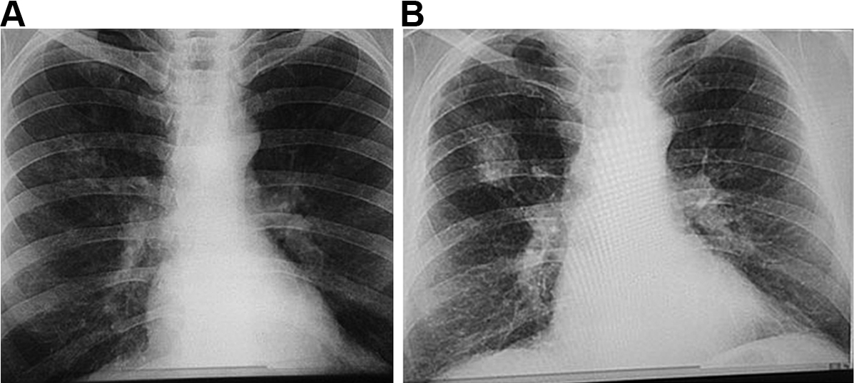
The manifestations of CWP can vary greatly between individuals, depending on the composition of the dust and duration of exposure, as well as host-related factors.34 Accumulation of dust occurs initially in the walls of the respiratory bronchioles, the adventitia of the blood vessels and the bronchoalveolar canals. Collections of dust-laden macrophages accumulate in the walls of the airways, particularly at their bifurcations, and in adjacent alveoli.1 Fibrous tissue is deposited, which later shrinks and leads to distortion of local lung structures.34 With increasing release of inflammatory mediators and deposition of fibrous tissue, these “macules” become larger and develop into more organised, dense, central, dust-pigmented lesions called micronodules,1 which can be palpated in the lung and seen on a chest x-ray (Box 1). Many larger rounded nodules then develop, particularly in the mid and upper zones of the lungs. Subsequently, centriacinar emphysema develops.35

Eventually, large masses of coal dust (Box 2), lymphocytes, dust-laden macrophages, reticulin and collagen may converge to form areas of PMF. These usually occur in the upper posterior parts of the lungs and appear as large rounded masses on the chest x-ray34 (Box 1, B). The presence of PMF represents “complicated” CWP and is associated with increasing symptoms and mortality.34

Radiological assessment relies on systematic objective assessment of good-quality chest x-rays. Scoring is important and should be performed to strict standards using the ILO (International Labour Office) International Classification of Radiographs of Pneumoconioses.36 The ILO classification provides a means of systematically recording the chest radiographic abnormalities that occur in any type of pneumoconiosis. The classification does not imply legal definitions of pneumoconioses for compensation purposes.36 While low-dose, high-resolution computed tomography (HRCT) scans are more sensitive for early disease,37 CT scanning is not currently used for screening purposes.

“Simple” pneumoconiosis is characterised by small, ill defined, rounded opacities in the outer thirds of the lung fields and the mid and upper zones. These can be categorised by size (categories q, r and s) and by density (categories 1–3). Identifiable radiological changes often occur well before changes in lung function or clinically significant disease. The whole of the lung fields may be involved in stage 3. PMF appears as rounded, sausage-shaped or ovoid opacities greater than 1 cm in diameter, which are well demarcated from the adjacent lung and may vanish if the contents are expectorated. Standard x-rays are available from the ILO for comparison purposes. Documentation of results includes an assessment of the technical quality of the chest x-ray, as well as relevant findings, and each detailed finding is assigned a standard code that facilitates documentation, diagnosis and monitoring.36

Prevention of coal workers’ pneumoconiosis

As the only cause of CWP is coal dust, prevention is straightforward — preventing exposure to coal dust prevents disease. This is important, as no effective treatments for CWP exist. Like all occupational diseases, prevention requires adherence to appropriate environmental standards and occupational health and safety guidelines by employers and workers.

Standards exist for limiting exposure to the respirable fraction of coal dust in most industrial settings. However, these standards vary considerably between states in Australia. For example, in Queensland the standard is 3.0 mg/m3, while in NSW it is 2.5 mg/m3.19 Both of these are significantly less stringent than the current US standard of 1.5 mg/m3.19 The Australian Institute of Occupational Hygienists has recommended that the limit be reduced to 1.0 mg/m3,38 and it could be argued that it should be even lower. The discrepancies in limits are compounded by variation in testing protocols between regions. For example, in Queensland, monitoring of dust exposure includes the travel time between the mine entrance and the coal face, whereas in NSW, exposure is only monitored during the individual miner’s period of underground work.19

Knowing the legislated standard for coal dust levels, it is up to the employer to implement measures to prevent exposure. However, the extent of implementation may depend on the costs associated with dust mitigation, which may explain why smaller mines tend to have a higher incidence of CWP.11 The management and oversight of dust sampling and monitoring is an important factor that can have a big impact on dust exposure; the intricacies of this are beyond the scope of this review. Given the variation in standards between states, in both dust monitoring and suppression, it is not possible to assess the overall extent of compliance with standards or the implementation of dust suppression strategies throughout Australia. However, a recent report by the Queensland Mines Inspectorate has raised concerns about the level of exposure in some situations,39 whereby 60% of mines exposed longwall operators to levels equal to or greater than the exposure limit in 2014, compared with 10% in 2012. Similarly, there have been increases in the percentage of mines that have exceeded regulatory limits (0 in 2012 v 25% in 2014).39

Taken together, these observations indicate that Australian standards are not based on the international understanding of the levels of coal dust that are likely to cause disease, and there are no consistent standards for monitoring dust levels. There is also some evidence to suggest that regulatory compliance may be a problem.

Screening for coal workers’ pneumoconiosis

Screening programs are crucial for monitoring the health of coal workers to identify individuals with early-stage disease and prevent progression from mild asymptomatic disease to PMF. Screening is mandatory in other countries and has been very effective at reducing the prevalence of pneumoconiosis in workers at risk of exposure to occupational dust.40 In situations where screening is voluntary, there is a low (about 40%) participation rate.19 The World Health Organization recommends that all workers exposed to coal and silica dust undergo lifelong health surveillance, including a baseline assessment (with chest x-ray) before commencing work, annual spirometry and symptom questionnaires, and follow-up chest x-rays every 2–5 years.41 Surveillance should continue after exposure and records should be kept for 30 years or longer after cessation of employment. Health surveillance is usually financed by the employer.

X-ray assessment using the ILO method is the current international standard for identifying disease.36 Questionnaires and spirometry are also effective in detecting other chronic lung diseases associated with dust exposure and can prompt referral to a respiratory specialist. Lung function assessment will identify respiratory disorders not visible on imaging and allow tracking of individual trajectories of lung decline. A computerised program (Spirometry Longitudinal Data Analysis [SPIROLA] software) is available free from the US National Institute for Occupational Safety and Health (NIOSH) to identify workers whose decline in lung function is greater than normal.

In the US, the NIOSH requires radiologists to be certified as “B readers” for classifying pneumoconiosis to ILO standards.42 This is not a requirement in Europe or Australia. The B Reader Program is a proficiency program to provide a pool of qualified readers capable of accurately using the ILO system,42 which has recently incorporated modern digital technologies.34 Use of digital techniques may improve the reproducibility of small-opacity profusion classification in some respects, but could also slightly reduce the frequency with which some readers identify large opacities.34 HRCT is acknowledged as being more sensitive than chest radiography for detecting parenchymal and pleural abnormalities and interstitial fibrosis,37 but it is limited by equipment availability, costs and radiation exposure, although modern CT scanners use a much lower radiation dose that is comparable to old chest x-rays.43 In Australia, the proficiency of radiologists for reporting surveillance films in accordance with ILO methods is determined by the Royal Australian and New Zealand College of Radiologists (RANZCR).

Data from a screening program must be carefully maintained to allow longitudinal assessment, which may identify important changes over time. A comprehensive surveillance program also requires a process for referral to specialist respiratory physicians and a means of explaining results to the worker, preferably with a written record of examination results.

Conclusions

Recent reports of CWP in Australia and international epidemiological data suggest an increase in CWP prevalence among coal workers and are a major concern. Given that CWP is a preventable but untreatable disease, no new cases should occur. Disease eradication must be the aim. We suggest the following actions, which are summarised in Box 3.

1. Dust exposure limits and monitoring

The current Australian standards for coal dust exposure limits, which vary between states, are less stringent than international recommendations, and exposure monitoring protocols vary considerably between sites. We strongly urge that the Australian guidelines be reviewed on the basis of current knowledge of CWP, in line with international standards, and that exposure limits and monitoring protocols are nationally standardised according to best practice guidelines.

2. Screening

We strongly advocate for a comprehensive screening program for workers at risk of exposure to coal or silica dust using a protocol based on international guidelines, which includes a questionnaire, medical imaging and lung function testing including measurement of diffusion capacity. This should be funded by the employer but preferably evaluated by physicians and radiologists not employed by the coal companies. We recommend implementing recent advances in lung imaging, including digital radiography and storing data in a single de-identified central system, which could be accessible to workers, unions, government agencies and employers. Workers should be given a copy of their results after each screening. The WHO recommends data storage for at least 30 years after the worker’s retirement, and we endorse this recommendation.

We acknowledge that chest radiography can be insensitive and non-specific for the diagnosis of CWP, but screening with low-dose CT is currently impractical for reasons of cost, availability and convenience. Digital radiography using ILO standards should be performed at the commencement of coal dust exposure and at least every 3 years, or more frequently, depending on the results of ongoing assessment. Sending radiological images overseas is unnecessary and the RANZCR register of radiologists who can assess to ILO standards should be used. We believe that further research is needed to examine the utility of more modern methods of early diagnosis; namely, the use of low-dose HRCT screening.

Workers who show an accelerated decline in lung function or a change in radiological or questionnaire scores should be recommended for further investigation with HRCT and full lung function testing, including measurement of gas transfer performed by accredited laboratory personnel according to international standards and interpreted by a specialist respiratory physician. Workers should be referred early to specialist respiratory physicians with an interest in occupational lung disease, and the costs of such investigations should be borne by employers. Where pneumoconiosis is diagnosed, we recommend a system of mandatory reporting to a centralised occupational lung disease register.

3. Medical practitioner training and referral

Pneumoconiosis is rarely diagnosed in the general practice setting. We suggest that training materials for medical practitioners be developed to assist with identifying current or retired workers at risk of pneumoconiosis. We also suggest that all dust-exposed workers with significant respiratory symptoms, whether in an existing surveillance program or after retirement, be referred to a specialist respiratory physician, preferably with expertise in occupational lung disease.

4. A centralised occupational lung disease register

Current data regarding occupational lung disorders in Australia are inadequate. The results of surveillance for CWP should be made publicly available and stored in a central repository. Thus, individual changes in respiratory health could be monitored, as well as changes in prevalence and incidence, and the system could be used to detect potential difficulties in surveillance and prevention processes. CWP and other occupational lung diseases should be made notifiable diseases, so that all diagnosed cases are recorded. Such information should be used to close the loop and feed back to employers to allow early implementation of change.

It is unacceptable that any new cases of CWP should be occurring in Australia in 2016, and our aim should be to eliminate CWP in Australia altogether.

Box 1 –
Chest x-rays of a coal worker (A) showing background nodulation and early progressive massive fibrosis (PMF) in the right upper zone, and (B) 12 years later, showing PMF
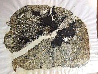
Box 2 –
Gough section of a coal worker’s lung showing coal workers’ pneumoconiosis with progressive massive fibrosis

Box 3 –
Recommendations for control of coal workers’ pneumoconiosis (CWP) endorsed by the Thoracic Society of Australia and New Zealand

Goal: Eliminate CWP in Australia

  1. 1. Exposure limits and monitoring protocols
    • Standardise across Australia and harmonise to international recommendations
  2. 2. Screening
    • Develop and implement a national screening program for at-risk workers
    • Questionnaire, imaging, lung function testing
  3. 3. Medical workforce training
  4. 4. A centralised occupational lung disease register

[Comment] REACTing: the French response to infectious disease crises

In the past decade, scientific and health systems have been challenged by an increase in the emergence of infectious diseases such as Middle East respiratory syndrome coronavirus, chikungunya, Ebola virus disease, and Zika virus.1–4 An effective and global public health response to these crises depends on our ability to anticipate these events and our level of preparedness. However, the development of research programmes in response to a rapidly emerging infectious disease in an emergency context is a challenge.