×

Kristine Whorlow AM retires as CEO of National Asthma Council

The National Asthma Council Australia’s inaugural chief executive officer Kristine Whorlow AM has retired.

NAC Chair Dr Jonathon Burdon said Ms Whorlow’s decision to retire caps a remarkable career of continuous service to the asthma community, both in Australia and internationally, including the Asia Pacific region.

“Kristine is a leader in her field and her expertise has established the NAC as the leading authoritative body for asthma in Australia with a considerable global reputation,” Dr Burdon said.

“We thank Kristine for her important contribution to improving asthma management in Australia. Her achievements are many, including facilitating asthma’s recognition as a national health priority and leading the ongoing development of asthma’s national treatment guidelines.

“Kristine has generated Australian Government program funding for asthma since 2001 and recently acquired Government funding for the fifth National Asthma Strategy now in the final stages of the AHMAC process.”

Dr Burdon also announced the appointment of the NAC’s new CEO, Siobhan Brophy, effective from August 1.

Ms Brophy was the NAC’s strategy and communications manager.

The NAC’s purpose is to reduce the health, social and economic impacts of asthma throughout Australia including free education workshops for GPs and allied health professionals funded by the Australian government through our Asthma & Respiratory Education Program.

Australia’s Institute for Health and Welfare’s data shows one in nine Australians have asthma– around 2.5 million people, based on self-reported data. The data also reports one in five people aged 15 and over with asthma have a written asthma action plan.

MEREDITH HORNE

[Comment] Offline: The G20 and health—platitudes and broken promises

Imagine “Anycountry”. A low-income nation-state with a weak health system. People have begun to report a mysterious respiratory infection that is rapidly fatal. The cause is an unknown virus. A localised outbreak quickly spreads from village to village. The virus is not only extremely pathogenic but also highly transmissible. The epidemic soon crosses the country’s border to neighbouring states. A global health emergency beckons. This scenario is fictional. But earlier this year G20 health ministers were locked in a room in Berlin to test their ability to deal with such a global health crisis.

The burden of chronic respiratory conditions in Australia

This report presents information on the disease burden of chronic respiratory conditions using data from the Australian Burden of Disease Study 2011. Respiratory conditions were the sixth leading contributor to total burden of disease in Australia, with chronic obstructive pulmonary disease (COPD), asthma and upper respiratory conditions being the greatest contributors to the respiratory burden. The burden due to respiratory conditions generally decreased over time, varied by condition severity and by population group, and some of the burden was attributed to modifiable risk factors such as tobacco use.

[Seminar] Infantile haemangioma

With a prevalence of 4·5%, infantile haemangiomas are the most common benign tumours of infancy, arising in the first few weeks of life and exhibiting a characteristic sequence of growth and spontaneous involution. Most infantile haemangiomas do not require therapy. However, to identify at-risk haemangiomas, close follow-up is crucial in the first weeks of life; 80% of all haemangiomas reach their final size by 3 months of age. The main indications for treatment are life-threatening infantile haemangioma (causing heart failure or respiratory distress), tumours posing functional risks (eg, visual obstruction, amblyopia, or feeding difficulties), ulceration, and severe anatomic distortion, especially on the face.

[Correspondence] Body-mass index and all-cause mortality – Authors’ reply

Associations of measured body-mass index (BMI) with mortality within just a few years of the BMI measurement can be strongly distorted by reverse causality (ie, by life-threatening neoplastic, respiratory, vascular, or other diseases having caused weight loss before the BMI was measured). By contrast, associations of measured BMI with mortality several years after the BMI measurement should be little affected by reverse causality, although they can still be strongly affected by confounding, especially in populations in which low BMI is correlated with smoking.

[Correspondence] Body-mass index and all-cause mortality

On the basis of finding that both overweight and obesity were associated with higher all-cause mortality in an Article in The Lancet1 the Global BMI Mortality Collaboration claimed that strategies are needed to “combat the entire spectrum of excess adiposity in many populations”. This assertion assumes that elevated body-mass index (BMI) is the primary cause of the higher mortality rates in overweight and obese adults reported in the Global BMI Mortality Collaboration study. However, this assumption fails to acknowledge the independent contributions of cardiorespiratory fitness and physical activity that substantially influence mortality risk.

[Comment] Respiratory medicine and critical care: a call for papers for ERS

The Lancet and The Lancet Respiratory Medicine would like to hear from authors of research papers in the fields of respiratory medicine and critical care, as the two journals are planning special issues to coincide with the 2017 European Respiratory Society (ERS) International Congress that will take place in Milan, Italy, on Sept 9–13, 2017.

[Seminar] Idiopathic pulmonary fibrosis

Idiopathic pulmonary fibrosis is a prototype of chronic, progressive, and fibrotic lung disease. Healthy tissue is replaced by altered extracellular matrix and alveolar architecture is destroyed, which leads to decreased lung compliance, disrupted gas exchange, and ultimately respiratory failure and death. In less than a decade, understanding of the pathogenesis and management of this disease has been transformed, and two disease-modifying therapies have been approved, worldwide. In this Seminar, we summarise the presentation, pathophysiology, diagnosis, and treatment options available for patients with idiopathic pulmonary fibrosis.

[Comment] Offline: The message of mental health

One entirely avoidable and indefensible tragedy of the global response to non-communicable diseases has been its extraordinary failure to embrace mental health. Instead, advocates and international institutions, such as WHO, have preferred to emphasise the simple (and simplistic) message of “four-by-four”—four key diseases (heart disease, cancer, diabetes, and chronic respiratory diseases) and four key risk factors (tobacco use, alcohol, unhealthy diets, and physical inactivity). It is not that those who preside over global health today have been ignorant of the mental health crisis befalling our world.

Complicated silicosis resulting from occupational exposure to engineered stone products

Clinical record

A 54-year-old man, formerly a smoker, presented with a 6-year history of chronic cough and exertional breathlessness without previous respiratory illnesses. Born in Vietnam, he came to Australia as a refugee at the age of 20 years. A screening chest x-ray was performed on his arrival in Australia; as the patient was not informed about any abnormality, this was assumed to be normal. He commenced work as a labourer; he denied exposure to silica-containing materials and did not participate in activities typically associated with silica exposure (such as jack-hammering) during this period. About 15 years later, the patient started a job manufacturing stone benchtops. He cut, ground, finished and installed the benchtops, using a popular brand of engineered stone comprising > 85% crystalline silica. Occasionally, he made benchtops from granite and marble. During the first 7 years of this work, the patient did not use any respiratory protective equipment, but later used a simple paper mask. Despite some dust extraction facilities in the factory, he reported that the environment was visibly dusty and that dust suppression with water was hardly ever used.

At presentation, chest examination showed scattered fine crackles and bronchial breath sounds bilaterally in the upper zones. Spirometry showed restriction (forced expiratory volume in 1 second [FEV1]/forced vital capacity [FVC], 1.9/2.6 L [73% and 82% predicted, respectively]) and no bronchodilator reversibility; gas transfer was reduced (diffusing capacity of the lungs for carbon monoxide [Dlco], 60% predicted, carbon monoxide transfer coefficient [Kco], 92%).

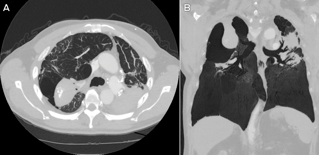
Multiple sputum specimens tested negative for acid-fast bacilli. Levels of inflammatory markers were within reference levels, and the result of a screening test for autoimmunity and vasculitis was negative. A computed tomography (CT) scan of the chest (Box 1) showed confluent bilateral calcified fibrotic masses in the upper zones, with marked volume loss, distortion and perilesional bullae. Occasional small peripheral lung nodules were present, predominantly distributed in the upper zones. There was calcified mediastinal lymphadenopathy.

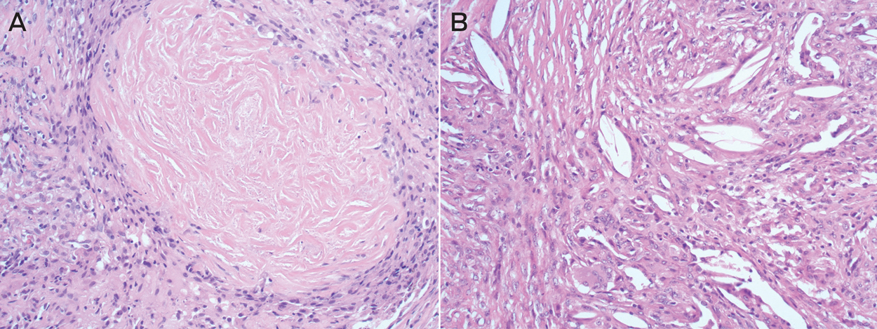
Bronchoscopic washings tested negative for acid-fast bacilli and malignant cells. Positron emission tomography (PET) scanning showed intense uptake in the confluent densities (maximum standardised uptake value, 10.6) and mediastinal lymphadenopathy. CT-guided fine-needle aspiration biopsy was negative for malignant cells. Transbronchial fine-needle aspiration biopsy of lymph nodes using endobronchial ultrasound was non-diagnostic. An open biopsy of the right upper lobe lesions and paratracheal lymph nodes was performed. Histological analysis of the lung sample showed numerous large sclerotic silicotic nodules surrounded by collections of histiocytes (Box 2). Analysis of lymph node sections confirmed prominent nodular silicosis.

Overall, the findings, including PET, were compatible with a diagnosis of complicated silicosis with progressive massive fibrosis. The patient continued to experience worsening breathlessness, despite treatment with bronchodilators and inhaled corticosteroids. He subsequently developed bilateral pneumothoraces, requiring temporary intercostal drain insertion, and is now listed for lung transplantation.

Silicosis refers to a spectrum of progressive and debilitating occupational lung diseases caused by the inhalation of free crystalline silica. Once established, there is no effective treatment. Prevention is therefore paramount. Exposure to silica has been unequivocally associated with an increased risk of lung cancer, as well as an increased frequency of tuberculosis and possibly also autoimmune disease. Implementation of appropriate workplace standards to minimise exposure is crucial to reduce incident and severe cases of silicosis.

Silicon dioxide (silica) is the most abundant mineral on Earth and is present in almost all types of rock, sand, clay and gravel. The most common crystalline forms of silica are quartz, cristobalite and tridymite. Silica exposure may occur in many work settings. Despite a global downward trend, new outbreaks of silicosis have recently been reported, with life-threatening silicosis occurring after exposure to a relatively new type of engineered stone product used for kitchen and bathroom benchtops.1,2 These products (known as “artificial quartz conglomerate” and “artificial stone”), which contain a high content of free crystalline silica (70–90%), are increasingly used in preference to their marble and granite counterparts because of their low cost, improved durability and hardness.

Risk of exposure to high levels of crystalline silica from engineered stone is present at all levels of this industry, from manufacturing (stone cutting, shaping and finishing) to assembly and installation.3 In a recent study, 25 ornamental stone workers who had been employed in dry-cutting a synthetic stone product developed advanced silicosis.1 In another report, silicosis was diagnosed in 46 men working in the manufacture of artificial stone for kitchen benchtops.2

Following these reports, an alert issued in the United States highlighted potentially dangerous levels of silica exposure associated with suboptimal practices in the artificial stone industry.4 Further calls to action have come from Europe, with a report from Tuscany citing seven cases of silicosis in workers exposed to crystalline silica during benchtop manufacturing.5 In almost all reported cases, there was little adherence to basic protection measures, such as provision of appropriate ventilation systems and use of personal protective equipment.

The Safe Work Australia workplace exposure standard for respirable crystalline silica (time-weighted average) is 0.1 mg/m3, which is designed to prevent the occurrence of silicosis.6 Our case reaffirms the need for vigorous enforcement of dust reduction regulations, particularly in the growing industry of engineered stone products. Benchtop stonemasonry is a potentially dangerous occupation, and medical practitioners should have a heightened awareness of this newly described occupational hazard.

Lessons from practice

  • Silicosis is a disabling but entirely preventable occupational lung disease caused by exposure to inhaled free crystalline silica.

  • Recent outbreaks of silicosis have been associated with the manufacture of relatively new engineered stone products that are used for kitchen and bathroom benchtops.

  • Medical practitioners should be aware that cutting and installing these engineered artificial stone products can be a hazardous occupational exposure.

  • Appropriate dust suppression practices and the use of respiratory protective equipment should be reinforced to prevent silica-related diseases.

Box 1 –
Axial (A) and coronal (B) computed tomography scans of the patient’s lungs


The scans show confluent mass-like fibrosis in the posterior upper lung zones, with adjacent bullae and compensatory expansion of the lower lobes. Calcification is seen within the fibrotic masses as well as within the hilar and mediastinal lymph nodes. There are occasional small peripheral nodules, most obvious in the right lung.

Box 2 –
Typical features of silicosis on lung histopathology slides


The biopsy specimen (A) shows the whorled appearance of a silicotic lung nodule, consisting of concentric laminated collagen fibres (haematoxylin–eosin stain, magnification 10×). A higher magnification image (B) highlights clefts containing faintly visible silicate particles surrounded by macrophages and giant cells (magnification 20×).