×

Renal replacement therapy associated with lithium nephrotoxicity in Australia

In 1949, John Cade reported the use of lithium in the treatment of manic–depressive illness, thus ushering in the era of psychopharmacology.1 Clinicians soon observed that patients receiving long-term lithium therapy developed polyuria, suggesting that it might cause renal tubular damage.2 Many years later, evidence arose that it might also reduce renal function, although the initial report included some patients with lithium toxicity.3 A meta-analysis of 1172 patients across 14 studies found that 15% of patients had a reduced glomerular filtration rate (GFR).4 This led to a discussion of the lithium level sufficient to control symptoms but not to create permanent renal damage.5

Controversy regarding the level of risk persisted. One authority rejected the possibility of renal damage;6 however, a recent meta-analysis of 365 studies disagreed with this but concluded that the risk of renal replacement therapy (RRT) was low (18/3369 patients [0.5%]).7

The need for an epidemiological survey was advocated;8 however, until now, this has only been done within restricted geographical foci, with uncertain denominators associated with incidence rates. A study involving two Paris hospitals and a questionnaire sent to all nephrologists in France found that 0.22% of all RRT patients had received lithium treatment, without any other symptoms of lithium intoxication.9 A Swedish study of two regions found that 0.81% of RRT patients had kidney disease attributed to lithium-induced nephropathy (LiN), and 1.2% of lithium-treated patients had raised serum creatinine levels. These patients had consumed lithium for at least 12 years and their incidence of end-stage renal disease (ESRD) was sixfold greater than that of the general population.10 Another study found that patients receiving lithium for 15.6 years had a lower GFR compared with that of controls.11 The evidence suggests that ESRD occurs after long exposure to lithium, up to 23 years.1012

Our study addresses the need for a nationwide survey of the epidemiology of ESRD associated with LiN. We investigated details of all patients commencing RRT within Australia with clinically diagnosed LiN. Analysis was performed on a de-identified data extract from the Australia and New Zealand Dialysis and Transplant Registry (ANZDATA). Release of the data for this purpose was approved by the ANZDATA Executive.

Methods

We collected data from ANZDATA for all patients who commenced RRT with renal failure attributed to lithium toxicity between 1 January 1991 and 31 December 2011. We compared these patients with those who commenced RRT with renal failure due to other causes over the same period. We noted whether patients at the time of commencing RRT were smokers, whether diagnosis was via biopsy (available after 1 April 1997), and serum creatinine levels at time of entry (available after 1 April 1998). Estimated GFR was calculated using the four-variable Modification of Diet in Renal Disease formula.13 ANZDATA contains information about patients who receive chronic RRT (dialysis or transplantation) in Australia,14 and is noted for its completeness. We used Australian Bureau of Statistics information concerning number, age and sex distribution of the Australian population.15

We used Pearson χ2 and Mann–Whitney U tests to compare patient characteristics between groups. We compared biopsy rates and trends over time between LiN and other patients using logistic regression, with time by LiN interaction included. We directly standardised incidence rates for age and sex to the 1 June 2006 Australian population, with age in 10-year cohorts for patients aged 0–79 years, then a single cohort of patients aged ≥ 80 years. We used linear regression to investigate changes in age of commencing RRT, and Poisson regression to investigate the mean number of comorbidities.

Results

No patients commenced RRT associated with LiN before 1991. However, between 1991 and 2011, 187 patients did so, compared with 38 316 patients who commenced RRT with renal disease attributed to other causes.

LiN patients were more likely than other patients to be women, to be white, to smoke and to have a higher body mass index, but were of similar age and less likely to have undergone renal biopsy (Box 1). Among 172 LiN patients with available data, 42 (22.4%) underwent a diagnostic kidney biopsy. Biopsy rates of LiN patients decreased over time (odds ratio per year, 0.74; 95% CI, 0.64–0.84; P < 0.001), which was more rapid than biopsy rates in non-LiN patients (P < 0.001 for the time by LiN interaction).

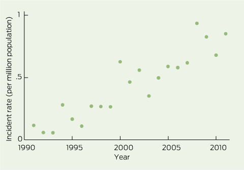
The number of patients commencing RRT associated with LiN increased over time, both in raw numbers, per population and as a proportion of all RRT patients. There were 0.14 cases per million population per year (95% CI, 0.06–0.22) in 1992–1996 and 0.78 (0.67–0.90) in 2007–2011. As a proportion of all incident RRT patients, LiN increased from 0.19% in 1992–1996 to 0.70% in 2007–2011 (Box 2). Age- and sex-standardised incidence rates were slightly lower than crude incidence rates for recent years (Box 2 and Box 3).

Discussion

We found a marked increase in the incidence of clinically diagnosed LiN leading to RRT, rising from 0.14 cases per million population per year in 1992–1996 to 0.78 in 2007–2011. LiN accounted for 0.19% of all new RRT cases in 1992–1996, and 0.70% in 2007–2011. The former figure is close to the incidence reported in France in the late 1990s,9 while the latter is close to the incidence found in a recent Swedish study (0.81%).10 The similarity between standardised and crude incidence rates suggests that demographic changes do not explain the increase in the incidence of LiN.

Registry data have some limitations. Some cases of LiN may have been missed, especially early in the time series, when awareness of LiN may have been low. LiN is largely a clinical diagnosis, and most patients do not undergo biopsy. LiN patients commencing RRT were less likely to undergo biopsy, and rates are decreasing compared with those for other RRT patients. Diagnostic bias is possible with any registry data. Nephrologist awareness of irreversible LiN may have increased over time, with the publication of recent epidemiological studies10 and a systematic review.7 As such, the propensity to diagnose LiN as a cause of ESRD may have increased over time. Unknown confounders may also potentially increase rates of both bipolar disorder and kidney disease. Existing data do not allow us to determine numbers of patients with ESRD associated with LiN who are not recorded in ANZDATA, because they do not receive RRT. However, most people with ESRD in Australia do commence RRT.16 Data on the number of patients receiving long-term lithium treatment in Australia are not readily available.

We conclude that LiN is an uncommon cause of ESRD but is becoming more common. Many cases of LiN could be avoided through careful diagnosis of bipolar illness, restricted prescription of lithium, and careful follow-up of lithium serum levels and renal function. Improved record keeping, such as a lithium-monitoring book17 or electronic records, is important. We suggest that renal function (based on estimated GFR derived from serum creatinine levels, and proteinuria tests) and serum lithium levels should be monitored more frequently than 6 months, and certainly more than every 12 months, which some authors have suggested.18 This is most important for patients who have received lithium for many years. Clinicians should consider stopping lithium and using other suitable mood stabilisers (eg, sodium valproate19) if two consecutive readings suggest decreasing renal function, or if the estimated GFR is < 45 mL/min/1.73 m2.20

1 Characteristics of patients commencing renal replacement therapy (RRT) with lithium-induced nephropathy and all other patients in Australia, 1991–2011

Characteristic

No. (%)* of patients with
lithium-induced nephropathy

No. (%)* patients with other
types of kidney disease

P


Number of patients

187

38 316

Median age, years (IQR)

60 (53–66)

61 (47–71)

0.99

Men

76 (40.6%)

22 714 (59.3%)

< 0.001

Diabetes

28 (15.0%)

14 133 (36.9%)

< 0.001

Median body mass index, kg/m2 (IQR)

28 (23–33)

26 (22–30)

< 0.001

Smoker

38 (20.3%)

4749 (12.4%)

0.001

Median eGFR, mL/min/1.73 m2 (IQR)

7.2 (5.4–9.9)

7.2 (5.3–9.7)

0.78

European ancestry

183 (97.9%)

30 733 (80.2%)

< 0.001

Biopsy

42 (22.4%)

10 079 (26.3%)


eGFR = estimated glomerular filtration rate, calculated using four-variable Modification of Diet in Renal Disease formula;13 only available for patients who commenced RRT after 1 April 1998. IQR = interquartile range. * Unless otherwise indicated. Not available before 1997.

2 Number of patients commencing renal replacement therapy (RRT), 1987–2011

Years

No. of patients receiving
lithium

Crude incidence rate/year/million population (95% CI)

Standardised incidence
rate/year/million population (95% CI)*

Proportion of all incident RRT patients (95% CI)


1987–1991

2

0.02 (0–0.07)

0.02 (0.00–0.07)

0.20% (0–0.49%)

1992–1996

12

0.14 (0.06–0.22)

0.13 (0.05–0.21)

0.19% (0.08%–0.30%)

1997–2001

36

0.36 (0.20–0.52)

0.32 (0.18–0.47)

0.40% (0.27%–0.53%)

2002–2006

52

0.51 (0.42–0.59)

0.43 (0.36–0.51)

0.48% (0.35%–0.62%)

2007–2011

85

0.78 (0.67–0.90)

0.65 (0.54–0.76)

0.70% (0.55%–0.85%)


* Standardised incidence rates were directly age- and sex-standardised to the 1991 populations. Both patients in 1991.

3 Crude incidence rates of renal replacement therapy associated with clinically diagnosed lithium-induced nephropathy in Australia, 1991–2011

Renal sympathetic nerve denervation for the treatment of resistant hypertension

Long-term safety and durability data for this novel approach to treating resistant hypertension are eagerly awaited

Hypertension is responsible for more deaths and disease than any other cardiovascular risk factor worldwide.1,2 For each 20 mmHg increment in systolic blood pressure (BP) or 10 mmHg increment in diastolic BP, the risk of cardiovascular disease doubles.2,3 Despite lifestyle modification and pharmacotherapy, achieving BP targets poses a significant challenge. Resistant hypertension is BP that remains above target levels in spite of adherence to three antihypertensive agents, one of which should ideally be a diuretic.1 The United States prevalence of resistant hypertension of 8.9%, reported in the National Health and Nutrition Examination Survey (2003–2008), is likely to be an underestimate.4,5 A promising alternative treatment modality for resistant hypertension — renal sympathetic denervation (renal denervation; RDN) — has recently been developed.

The procedure

Numerous RDN devices are currently under development, and most use an endovascular catheter-based approach to ablate renal sympathetic nerves by delivering radiofrequency (RF) or ultrasound energy via an electrode catheter or balloon catheter. The only RDN device currently approved by the Therapeutic Goods Administration is the Symplicity Catheter (Medtronic). The renal artery is accessed via a femoral arterial approach, the denervation catheter is connected to an RF generator, and then the denervation catheter is advanced via a guide catheter into the main renal artery. Multiple RF treatments are applied (120 seconds in duration each) from distal to proximal in a circumferential pattern to maximise renal sympathetic nerve disruption in the artery. The individual treatments are separated from one another by about 5 mm and, depending on the length of the renal artery, four to eight RF treatments are delivered. During each RF treatment, the patient can experience transient visceral pain; adequate analgesia is of utmost importance.6

The proof of concept

The clinical effect of RDN is presumed to result from its effect on sympathetic overactivity, which has been proven to have a role in the maintenance of high BP.3,6 Historically, the role of the sympathetic nervous system in BP control was demonstrated in patients who had surgical splanchnicectomy in the early 1900s.7 Profound improvements in BP were achieved, but this was at the cost of significant morbidity, so the procedure was abandoned. Modern antihypertensive pharmacotherapy was introduced in the early 1970s.

After promising data on RDN were obtained in swine models of hypertension, Symplicity HTN-1 was conducted as the proof-of-concept study for RDN.6 It was a multicentre feasibility, safety and efficacy trial of 45 patients with resistant hypertension. At baseline, the mean office BP was 177/101 mmHg. At 1 month, there was a significant 14/10 mmHg reduction in BP. The role of the sympathetic nervous system was supported in a subgroup of patients who underwent renal vein sampling for noradrenaline spillover, a marker of sympathetic activity; spillover was reduced by 47%.

Sustained reduction in blood pressure

Longer-term durability and safety of RDN was demonstrated in a cohort study of 153 patients with resistant hypertension, which included Symplicity HTN-1 patients.8 When compared with baseline (mean office BP, 176/98 mmHg), BP was reduced by 32/14 mmHg at 24 months. Unpublished 36-month data showed an ongoing durable response (American College of Cardiology Annual Scientific Session, Chicago, 24–27 March 2012, personal communication). Ninety-two per cent of patients had a greater than or equal to 10 mmHg reduction in systolic BP at 24 months.8 The average number of antihypertensive medications taken by patients did not change within 24 months. There were four procedural complications (three femoral pseudo-aneurysms and one renal artery dissection caused by the guide catheter), which were managed without further sequelae. At 6 months, one patient had renal artery stenosis (not related to the site of RF ablation) and was treated with percutaneous intervention. During the first year of follow-up, renal function (measured by estimated glomerular filtration rate [eGFR]) was stable for all 153 patients.

The randomised controlled trial

Symplicity HTN-2 was a multicentre, randomised controlled trial of 106 patients who were assigned to RDN immediately (RDN group) or after a 6-month control period (control group).9 When compared with baseline (mean office BP, 178/96 mmHg), BP in the RDN group was reduced by 32/12 mmHg at 6 months (SD, 23/11 mmHg; P < 0.0001), and there was essentially no change (1/0 mmHg) in the control group. At 6 months, systolic BP was less than 140 mmHg in 39% of patients in the RDN group, compared with 6% of those in the control group. Twenty per cent of patients in the RDN group had their number of antihypertensive medications reduced during this follow-up period, compared with 6% of those in the control group (P = 0.04). There were no serious procedure-related complications. Renal artery imaging at 6 months did not show renal artery stenosis. Renal function was unchanged from baseline in both groups and was not adversely affected, even in patients with mild to moderate renal impairment.9

Limitations of current evidence

There were two major criticisms of the first two Symplicity trials that will be addressed in Symplicity HTN-3. First, potential bias will be reduced with the introduction of a sham procedure in the control group. Second, ambulatory BP monitoring, the gold standard for predicting cardiovascular morbidity and mortality,10 will be conducted for all patients. Only 42% of Symplicity HTN-2 patients underwent ambulatory BP monitoring, and they were not randomly assigned to this investigation. The mean reduction in ambulatory BP in these patients after 6 months was significant (11/7 mmHg; P = 0.006 for systolic BP change, P = 0.014 for diastolic BP change), but not as large as the reduction in office-based BP measurements.

Although the numbers of antihypertensive medications taken by patients were recorded in the first two Symplicity trials, observed benefits should be viewed with the knowledge that compliance was not strictly evaluated and changes to antihypertensive regimens were permitted during the trial.

The long-term durability of RDN has been questioned based on the observation that sympathetic re-innervation has been described in cardiac transplantation. Although it is possible that re-innervation could similarly occur in the kidney, there is currently no evidence for this.

Long-term safety data are currently lacking, but are eagerly anticipated given that medial and adventitial fibrosis in the renal artery has been demonstrated in a swine model11 and a human case report has described renal artery stenosis at the site of RF ablation.12 Whether the RDN procedure caused the stenosis is unknown, but vigilance should be encouraged.

Cost-effectiveness studies will be required if renal denervation is to remain a viable intervention. Based on Symplicity HTN-2, a cost–benefit model suggested that RDN is a cost-effective strategy that may improve cardiovascular outcomes.13 Nevertheless, this was based on several assumptions that the Symplicity HTN-3 study should clarify. In Victoria, procedural and postoperative care costs are co-funded by the state government and public hospitals.

Indications and contraindications for RDN

RDN is currently indicated in patients who have an office-based systolic BP greater than or equal to 160 mmHg and in patients with type 2 diabetes who have a systolic BP greater than or equal to 150 mmHg. All patients should be compliant with three or more antihypertensive agents (one of which should ideally be a diuretic).6,8,9 The current major contraindications are previous renal artery intervention (balloon angioplasty or stenting), renal artery stenosis greater than 50 per cent, presence of multiple main renal arteries or main renal arteries less than 4 mm in diameter or less than 20 mm in length, and/or eGFR less than or equal to 45 mL/min/1.73 m2.

Future developments and applications

Further research is required to understand the variable effects of RDN. Responders are more likely to be those with excess renal sympathetic activity, which may explain why older patients are poorer responders (ie, because of a greater burden of arteriosclerosis relative to increased sympathetic drive). Future challenges include developing biochemical and procedural markers of efficacy, assessing long-term health outcomes, and exploring novel applications of RDN, such as atrial fibrillation,14 obstructive sleep apnoea,15 diabetes mellitus,16 chronic kidney disease,17 heart failure18 and polycystic ovary syndrome.3,19 Finally, more efficacious, safer, multi-electrode catheters and non-invasive ablation techniques are currently being tested in clinical trials;20 preliminary data on these were presented at international cardiology meetings held in 2012 (Transcatheter Cardiovascular Therapeutics, American College of Cardiology Annual Scientific Session, and EuroPCR).

Summary

Improvements in BP control with pharmacotherapy undoubtedly translate into significant individual and population health benefits. RDN results in impressive reductions in BP, which are likely to be associated with further improvements in clinical outcomes. With more clinical trials and concurrent development of new devices, we hope that RDN holds up to its current accolades.

Mercury poisoning from home gold amalgam extraction

This is the first Australian report of confirmed minimal change disease with nephrotic syndrome, which occurred in a 62-year-old man who inhaled mercury vapour in his home. This case highlights the immediate and delayed effects of such poisoning on multiple organs. Prompt and sometimes prolonged treatment may prevent long-term damage.

Clinical record

A 62-year-old man first presented to his general practitioner complaining of a cough, dyspnoea and lethargy for which he was prescribed oseltamivir phosphate, as his symptoms were presumed to indicate influenza. He re-presented 2 days later with worsening dyspnoea, at which point he revealed a history of mercury exposure. He had attempted to extract gold from an amalgam containing mercury by heating the amalgam in an aluminium pan inside his home. He was exposed for 3 hours — initially, to fumes, and subsequently, through direct skin contact as he attempted to clean up some spilt liquid amalgam. The windows were open and he used a tea towel covering his nose as protection from the fumes. A chest x-ray showed extensive alveolar shadowing consistent with pneumonitis. He was admitted to a regional hospital, and was given supplemental oxygen and antibiotics. His blood mercury level was 5933 nmol/L (level of concern, > 70 nmol/L). On the advice of a toxicologist, he was transferred to a tertiary hospital for chelation therapy.

On arrival at the tertiary hospital, his oxygen saturation on room air was 92% and signs on examination and repeat chest x-ray results were consistent with severe pneumonitis. His other vital signs were stable. Findings of other clinical examinations, including neurological examination, were unremarkable. Laboratory investigations showed he had an increased white cell count of 16.3 × 109/L (reference interval [RI], 4.0–11.0 × 109/L), a platelet count of 617 × 109/L (RI, 150–400 × 109/L), a serum albumin level of 21 g/L (RI, 34–48 g/L), and mild abnormalities in liver function test results (total protein, 60 g/L [RI, 65–85 g/L]; globulin, 39 g/L [RI, 21–41 g/L]; total bilirubin, 15 µmol/L [RI, 2–24 µmol/L); γ-glutamyl transpeptidase, 274 U/L [RI, < 60 U/L]; alkaline phosphatase, 228 U/L [RI, 30–110 U/L]; alanine aminotransaminase, 92 U/L [RI, < 55 U/L]; aspartate aminotransferase, 102 U/L [RI, < 45 U/L]; and lactate dehydrogenase, 294 U/L [RI, 110–230 U/L]). In a spot urine sample, the mercury concentration was 7556 nmol/L and the mercury : creatinine ratio was 2519 nmol/mmol (level of concern, > 5.8 nmol/mmol). Pulmonary function testing was suboptimal owing to the patient’s inability to suppress coughing on inspiration, but the results suggested a restrictive deficit. Analysis of a spot urine sample showed a protein concentration of 60 mg/L (RI, < 150 mg/L) and a protein : creatinine ratio of 18 mg/mmol (RI, < 12 mg/mmol). The patient’s serum creatinine level was 82 µmol/L (RI, 50–120 µmol/L) and his estimated glomerular filtration rate was > 60 mL/min/1.73 m2.

The patient was treated with dimercaptosuccinic acid (DMSA) chelation therapy, 800 mg three times a day for 7 days, after which the dose was reduced to 800 mg twice daily for a further 14 days. He was also treated with prednisolone 50 mg daily, gradually tapering the dose to zero over 3 weeks. Box 1 shows the 24-hour urinary mercury excretion in relation to exposure to mercury vapour, serum albumin concentrations and treatment with chelation therapy.

During this initial hospital stay, the patient’s dyspnoea decreased markedly, and subsequent chest x-rays showed resolution of interstitial shadowing. His liver function test results also normalised, and his serum albumin level rose to 33 g/L. The mild vertigo he had reported, with no other neurological symptoms or deficits, resolved spontaneously.

He was discharged on DMSA 800 mg twice daily and prednisolone 15 mg daily (tapering dose), and it was planned to repeat the assessment of his mercury levels after completion of the initial 3-week course of DMSA. This reassessment showed ongoing elevated levels (blood mercury, 418 nmol/L; urinary mercury, 1019 nmol/24 h), so the patient was rechallenged with DMSA at 800 mg three times a day for 34 days, commencing on Day 66 after exposure. Improved clearance resulted, as evidenced by a subsequent increase in urinary mercury excretion (from 1762 nmol/24 h on Day 62 to a maximum of 5790 nmol/24 h on Day 67). Consequently, treatment with DMSA at 800 mg twice daily was resumed, with the intention of giving a prolonged course, and his blood mercury levels and renal mercury clearance were assessed periodically. His serum albumin level had normalised by this point, and his prednisolone course had been completed.

Approaching 1 month into this prolonged course of DMSA, he re-presented with a history of fatigue, increasing peripheral oedema, nausea and vomiting. Physical examination revealed significant peripheral oedema with intravascular volume depletion. Biochemical analysis showed a serum albumin level of 6 mg/L (nadir, 3 g/L), urinary protein level of 13.4 g/24 h (RI, < 150 mg/24 h), serum creatinine level of 133 µmol/L (peak, 220 µmol/L) and serum cholesterol level of 13 mmol/L, indicating severe nephrotic syndrome. A renal ultrasound scan was unremarkable. In a renal biopsy specimen, light microscopy showed glomeruli of normal appearance and electron microscopy showed podocyte effacement consistent with minimal change disease (Box 2). He was treated with prednisolone, regular infusions of concentrated albumin, and diuretics, and was restricted to 1.2 L of fluids daily. Treatment with an HMG-CoA (3-hydroxy-3-methylglutaryl-coenzyme A) reductase inhibitor and prophylactic warfarin therapy were also commenced. His nephrotic syndrome abated and renal function gradually normalised over the course of 4 months.

The patient experienced significant nausea, which necessitated cessation of chelation therapy after a total of 7 weeks. At this point, his blood mercury level was 112 nmo/L; within 1 month it fell below the level of concern. Three months after cessation of treatment with DMSA, his urinary mercury level was also normal. When last reviewed, his serum albumin level was 35 g/L and urinary protein excretion was 0.25 g/24 h. Resolution of his respiratory injury was almost complete, without evidence of developing neurotoxicity.

Discussion

We present a case of prolonged exposure to mercury vapour with characteristic “fume fever” illness followed by pneumonitis and nephrotic syndrome, and an associated body mercury burden requiring prolonged chelation therapy.

Heating of mercury forms mercury vapour, which is actively absorbed in the lungs. About 80% of mercury vapour formed from amalgams is absorbed through inhalation.1 Once absorbed, metallic mercury is rapidly oxidised to mercurous and mercuric ions,2 and distributes in a variety of tissues including the brain, kidneys, liver, testes, thyroid gland and oral mucosa. A small amount of elemental mercury remains in the blood and can easily pass through the blood–brain barrier and the placental barrier.

The symptoms and signs of mercury inhalation vary according to the concentration of mercury to which the patient is exposed and the duration of exposure. Acute exposure to high levels primarily causes respiratory symptoms such as dyspnoea, chest pain, tightness, and dry cough, secondary to chemical pneumonitis. Absorption at the alveolar and bronchiolar levels causes capillary damage, pulmonary oedema, and desquamation and proliferation of airway lining cells, leading to the obliteration of air spaces.3 Airway obstruction and capillary leakage may cause alveolar dilatation, pneumothorax and, in severe cases, acute respiratory distress syndrome.4 Systemically, mercurous and mercuric ions can bind with sulfhydryl groups, leading to inactivation of sulfhydryl-containing enzyme systems and structural proteins and alteration of cell-membrane permeability.5

The evolution of clinical symptoms after mercury vapour inhalation may be described in three phases.6 The initial phase is typically an influenza-like illness occurring 1 to 3 days after exposure. The intermediate phase is dominated by severe pulmonary toxicity and may involve renal, hepatic, haematological, and dermatological dysfunction. Our patient had hypoalbuminaemia and a mild and transitory abnormality in liver enzyme levels, followed by a delayed onset of minimal change disease with severe nephrotic syndrome. It is possible that DMSA chelation may have contributed to the nephrotic syndrome by subjecting nephrons to a high load of chelated mercury. Both his initial hypoalbuminaemia and subsequent nephrotic syndrome appeared to respond to steroid therapy. The most commonly reported histological abnormality in the kidney associated with mercury exposure is membranous glomerulopathy;7 however, minimal change disease, with negative findings on light microscopy and confirmed by characteristic findings from immunofluorescence and electron microscopy, has been previously described.8 The late phase is characterised by gingivostomatitis, tremor and erethism, which we have not seen in our patient.

Aggressive supportive care including continuous cardiac monitoring and pulse oximetry, supplemental oxygen therapy and mechanical ventilation4 remains the cornerstone of therapy after acute inhalational mercury poisoning. Several chelating agents bind mercury, increasing its water solubility and augmenting its renal elimination. Historically, dimercaprol, D-penicillamine and N-acetyl-penicillamine have been used.5 Recently, DMSA has proven to be more effective and less toxic. It is unclear to what extent chelation therapy reduces mercury tissue burden or prevents long-term neurological injury.9 Awareness among clinicians of the immediate and the delayed consequences of mercury poisoning, with prompt treatment, may help to avoid long-term organ damage.

1 Twenty-four-hour urinary mercury excretion in relation to exposure to mercury vapour, serum albumin concentrations and treatment with chelation therapy


DMSA = dimercaptosuccinic acid; shading indicates periods of therapy.

2 Micrographs of the patient’s renal biopsy specimen, showing features consistent with minimal change disease


A: Light micrograph showing glomeruli of normal appearance (haematoxylin and eosin stain; original magnification, × 200). B: Electron micrograph showing flattening of podocyte foot processes (arrows).

International treatment guidelines for anaemia in chronic kidney disease — what has changed?

Balancing the risks and benefits of erythropoietin-stimulating agents and iron therapy

One in nine Australians has chronic kidney disease (CKD),1 although the condition may often not be recognised in primary care.2 There are five stages of CKD, ranging from Stage 1, in which patients have normal renal function but urinary abnormalities, structural abnormalities or genetic traits pointing to kidney disease, through to Stage 5, in which patients have end-stage disease.1

Although anaemia in patients with CKD is multifactorial in origin, it is primarily associated with relative erythropoietin production deficiency3 as the glomerular filtration rate (GFR) falls. Once the estimated GFR trends below 60 mL/min/1.73 m2 (Stage 3a CKD), erythropoietin production by the kidneys falls, and anaemia may develop.

The history of anaemia management in CKD and associated clinical practice guidelines has been one of contradiction and perceived industry influence.4 The publication in August 2012 by the Kidney Disease: Improving Global Outcomes (KDIGO) group (an international collaboration of nephrologists) of a guideline that updates and informs clinical practice in this area5 was therefore welcome.

The KDIGO guideline contains 47 recommendations with varying grades of evidence (Box).5 It is noteworthy that the conclusions, recommendations and ungraded suggestions for clinical practice in the KDIGO guideline are largely consistent with those currently provided in the Kidney Health Australia–Caring for Australasians with Renal Impairment (KHA-CARI) guidelines.6,7 Of particular note, the KDIGO guideline takes into account the importance of balancing the risks and benefits of erythropoietin-stimulating agents (ESAs) and iron therapy.

A key aspect of the KDIGO guideline is that it recommends more caution in the use of erythropoietin. The cloning of human erythropoietin 30 years ago was an important breakthrough that led to the clinical development of recombinant human erythropoietin and thus to the use of ESAs for treating anaemia in patients with CKD. Numerous observational studies suggested that patients with lower targeted haemoglobin levels had worse outcomes than those with higher targets.4 This conclusion was supported by the clinical consequences of anaemia (including fatigue, exercise intolerance, cognitive impairment and exacerbation of cardiovascular disease)3 and led clinicians to virtually demand ESAs for the treatment of CKD-associated anaemia. It was therefore ironic that the first randomised controlled trials to evaluate the outcomes of higher versus lower haemoglobin targets, using recombinant human erythropoietin, found the unexpected reverse outcome of negative effects (increased cardiovascular events and mortality) in patients randomly assigned to the higher targets.8,9

The evidence now seems to suggest that haemoglobin target levels between 100 and 115 g/L should be the aim for patients with CKD, and certainly not levels > 130 g/L,10 which have the potential to cause harm. In fact, the KDIGO guideline reaffirms the very strong (Grade 1A) KHA-CARI recommendation of not targeting haemoglobin concentrations > 130 g/L.7,10 There are also newer cautionary recommendations, not covered by the KHA-CARI guidelines, regarding ESA use in patients with active malignancy (1B), a history of stroke (1B) or a history of malignancy (2C), where there is greater potential for harm.

The recommendations in the KDIGO guideline relating to the use of iron supplementation are similar to those in the KHA-CARI guidelines.6 They include the recommendation that oral iron supplementation is inadequate compared with injectable iron. This is problematic for most general practitioners, as intravenous iron cannot be administered in general practice, due to the need for monitoring. However, intravenous iron therapy may sometimes be arranged for the patient at a local hospital. Further, the KDIGO guideline recommends the use of iron indices to help guide therapy, with considerations of infection risk from excess iron and suboptimal ESA responsiveness.

The KDIGO guideline also covers the use of red cell transfusion in patients with CKD,5 again emphasising the importance of balancing risks and benefits. For example, for patients who may undergo future organ transplantation, red cell transfusion carries a risk of sensitisation to various tissue-type antigens; and, for all patients, excessive blood transfusions can lead to iron overload diseases. Symptom change (eg, decrease in fatigue) in an individual patient, rather than a haemoglobin concentration threshold, is a better measure of successful improvement.

Importantly, the first section of the KDIGO guideline considers the diagnosis and evaluation of anaemia in later stages of CKD (Stage 3a and beyond), where anaemia is most common.3 The recommendations, while not graded, provide protocol-type approaches to the frequency of testing and a rational approach to diagnosis that is relevant and appropriate in the Australian context. As GPs frequently manage patients with Stage 3a and 3b CKD, when CKD-associated anaemia is most likely to develop, it is expected that the KDIGO guideline will assist them in investigating and excluding other reversible causes of anaemia in the earlier stages of CKD.

In summary, for GPs and other non-nephrologist clinicians, the key KDIGO guideline recommendations are to: (i) consider CKD in any diagnostic workup of anaemia; (ii) recognise that injectable (rather than oral) iron is the first-line treatment for CKD-associated anaemia; (iii) consider ESA use only when the haemoglobin level is about 100 g/L — refer the patient to a nephrologist at that time; and, lastly, (iv) be appropriately cautious with the use of transfusions (blood and platelets) if transplantation potential exists.

Number of KDIGO guideline recommendations,5 by grade
of evidence*

KDIGO = Kidney Disease: Improving Global Outcomes. * Using the Grading of Recommendations Assessment, Development, and Evaluation (GRADE) system.

The urgency of monitoring salt consumption and its effects in Aboriginal and Torres Strait Islander Australians

To the Editor: There is convincing evidence that high salt diets are linked to elevated blood pressure — a major risk factor for chronic diseases.1 Premature mortality due to chronic diseases is a major contributor to
the life expectancy gap between Indigenous and non-Indigenous Australians.2 Although much has been written about the health benefits of a traditional hunter-gatherer diet and the detrimental effects of colonisation on diet and exercise patterns,3 we know little about how colonisation has affected salt intake among Indigenous Australians.

Currently in Australia, most efforts to prevent disease caused by high blood pressure focus on the clinical management of hypertension. Such programs can only affect individuals identified and treated for high blood pressure. In population terms, a
small proportion of the total blood pressure-related disease burden
is adequately addressed by this approach.4 Salt reduction programs, on the other hand, have the potential to address much more of the disease burden. A modest reduction in salt intake has been found to have a significant blood pressure-lowering effect.5 Furthermore, salt reduction programs that are implemented by modifications to the food supply have the potential to prevent disease at a very low cost.6

Salt consumption data in Australia are limited. A recent study in regional New South Wales, Drop the Salt Lithgow, confirmed that Australians are eating too much salt. The average salt intake in this Australian population was 9 g/day (Mary-Anne Land, Research Assistant, The George Institute for Global Health, unpublished data), which is 50% higher than the upper limit of 6 g/day recommended by the National Health and Medical Research Council.7 Unfortunately, this study only captured a small number of Indigenous Australians.

We suggest expanding the scope
of this research to Indigenous communities in New South Wales (by adapting the methods used in Drop the Salt Lithgow) to determine the types of food bought, prepared and eaten that have high levels of salt. This would involve: collecting baseline data on salt intake (by measuring 24-hour urinary sodium excretion) and dietary sources
of salt (via 24-hour dietary recall questionnaires); implementing a salt-reduction program; and collecting follow-up data on dietary sources
of salt (by repeat questionnaires). Surveying 500 people at baseline
and follow-up would provide power to detect a difference in salt consumption that would translate to real differences in individual and population health.

We believe that research aimed at recruiting a community-based sample of Indigenous adults to precisely and reliably measure salt intake and to better understand behaviours that underpin dietary patterns is a public health priority.