×

Toxic epidermal necrolysis — an investigation to dye for?

We report the first case in Australia, as confirmed by the Therapeutic Goods Administration, of toxic epidermal necrolysis associated with the iodinated contrast medium iopamidol. It serves as a warning about the use of contrast in imaging and cardiac catheterisation and a reminder of the need for increased awareness of the issue.

Clinical record

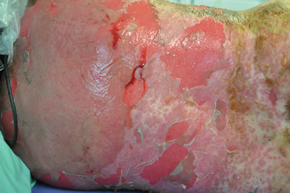
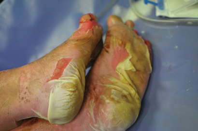
A 44-year-old woman presented to the emergency department with a 3-day history of a progressive rash, fever, malaise and mucosal ulceration. She met the diagnosis of toxic epidermal necrolysis (TEN) based on the following criteria: bullae and desquamation affecting about 84% of the body surface (Box 1 and Box 2), buccal and vaginal ulceration, a positive Nikolsky sign (this is a useful sign in bullous skin diseases and can be demonstrated by rubbing the skin surface, which will blister within a few minutes if the sign is positive), fever, tachycardia and mild hypotension. She also had abnormal results of liver function tests: bilirubin level, 69 µmol/L (reference interval [RI], < 20 µmol/L); alkaline phosphatase level, 180 U/L (RI, 25–100 U/L); γ-glutamyl transferase level, 499 U/L (RI, < 30 U/L); alanine aminotransferase level, 1730 U/L (RI, < 30 U/L); and aspartate aminotransferase level, 638 U/L (RI, < 30 U/L). She had a white cell count of 4.3 × 109/L (RI, 4.0–10.0 × 109/L) and a raised C-reactive protein level of 53.1 mg/L (RI, < 5 mg/L).

The patient was immediately transferred to the burns unit and managed with nanocrystalline silver dressings, intravenous immunoglobulin, aggressive fluid and electrolyte balance therapy, analgesia and intravenous antibiotics. She was discharged home on Day 15.

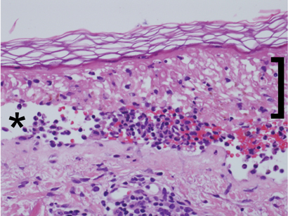
Histopathological examination (Box 3) showed extensive epidermal necrosis and subepidermal clefting with a sparse superficial perivascular infiltrate of lymphocytes, occasional neutrophils and eosinophils, and exocytosis of cells into the epidermis. Results of staining for immunofluorescence were negative. This was consistent with the clinical diagnosis of TEN, and the possibility of pemphigus vulgaris was excluded.

The patient’s only recent exposure to medications included 150–200 µg of thyroxine sodium daily for 19 years and 2.5 mg of indapamide daily for 6 months. The patient underwent a computed tomography (CT) neck scan with the contrast medium iopamidol about 4 weeks before the development of symptoms. The patient had recently (3 weeks before onset of rash) stopped taking a herbal “liver cleanser”. She had been taking this intermittently for 2 months. She had no recent travel history or vaccinations.

The patient recalled having a previous CT scan of the neck before her surgery 17 years ago. Unfortunately, however, any records of this had been destroyed.

The patient’s past medical history was notable for mild stable hypertension, hypothyroidism and a benign mixed salivary gland tumour electively excised 17 years earlier.

Discussion

Toxic epidermal necrolysis (TEN), or Lyell syndrome, is a rare and life-threatening severe systemic condition associated with dramatic cutaneous sloughing of up to 100% of the body surface area. The incidence of TEN is two cases per million person-years.1 It is characterised by necrosis and subsequent detachment of the epidermis from the dermis in more than 30% the body surface. If not treated and managed promptly, the consequences can be fatal; patients are vulnerable to infections and sepsis leading to death. The mortality associated with TEN is high, at 30%–40%.2

At the other end of the spectrum, and more common, are mild-to-moderate skin reactions to contrast media (CM). These include, in increasing severity, lichenoid reaction, erythema multiforme and Stevens–Johnson syndrome.3 Patients at risk of late skin reactions are those with a previous history of CM reactions.3,4

TEN is attributed to medications in 80% of cases.1 The most commonly associated medications include sulfonamides, penicillin and other antibiotics, anticonvulsants, oxicam nonsteroidal anti-inflammatory drugs, allopurinol and corticosteroids.5 TEN commonly occurs 1–3 weeks after the start of therapy. Other triggers include infections, malignancy and vaccination.1

The dermatological reactions caused by CM can be classified as early or late reactions. Early reactions occur soon after injection of the contrast medium, and late reactions occur within a week. The incidence of late adverse reactions is 2%.3,6 They commonly present as maculopapular erythema, angioedema and urticaria. Evidence to date suggests that late reactions are more common with non-ionic CM, in particular dimers,3 despite non-ionic CM being touted as having fewer adverse reactions. Late reaction incidence with non-ionic CM varies between 8% and 71%.3

Iodinated CM can be divided into ionic and non-ionic contrast medium. The ionicity pertains to the osmolality the CM create in blood; ionic CM create higher osmolality leading to CM reaction. The move from the use of ionic CM to non-ionic CM was based on the need for an agent with fewer adverse effects and equal or slightly improved diagnostic efficacy.7 The morbidity and mortality associated with non-ionic CM were less than for ionic CM.8 Although non-ionic CM have these advantages, ionic CM are still in use today. A recent study highlighted that although non-ionic CM are the best tolerated in the early phase, they are associated with a higher level of adverse effects such as late skin reactions.8 Iopamidol is a non-ionic contrast medium.

There have been several cases of TEN caused by CM reported in the literature. Commonly, the cases have involved repeated exposure or sensitisation to the CM in the cardiac catheter laboratory over a period of days to even years.911 TEN occurs with subsequent exposures to the CM administered. In our case report, there was a history of prior CT scan of the neck; however, records of the scan are no longer held by the radiologist to verify the date and contrast medium used.

TEN has also been shown to be caused by gastrointestinal oral CM.12

Only two published cases of TEN have been attributed to the administration of iopamidol. The first case is of a young boy with subsequent exposure to iopamidol.10 The case was not biopsy-proven TEN, but was based on clinical diagnosis. The second case is that of a patient who underwent intravenous urography for investigation of systemic lupus erythematosus with renal involvement.13 The patient died despite intensive care and support. Our case report would be the third reported case of TEN caused by iopamidol.

Although it is difficult to be sure that iopamidol was responsible for the development of TEN in our patient, it is highly likely to be the cause. She had been on indapamide for a period and had been taking the herbal “liver cleanser” intermittently. One recent case of herbal medicines and TEN has been reported.14 However, as noted by the author, it was difficult to determine the causative agent.

It is important to be aware of the risk of CM and to think twice about the necessity of CM in imaging. Although rare, life-threatening adverse effects such as TEN should lead to reconsideration of contrast dyes, as patients may suffer unnecessarily or lose their lives.

1 Bullae and desquamation of back

2 Desquamation of both feet

3 Skin punch biopsy sample showing extensive epidermal necrosis (bracket) and subepidermal clefting (asterisk) with mixed inflammatory infiltrate and negative results of staining for immunofluorescence

Dysphagia, regurgitation and weight loss in an elderly man

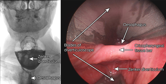
An 84-year-old man reported unintentional weight loss of 15 kg over 18 months and dysphagia and regurgitation of undigested food over 3 months. Barium swallow showed a large pharyngo-oesophageal (Zenker) diverticulum. These are caused by elevated intraluminal pressure at the Killian dehiscence, an area of hypopharyngeal weakness between the cricopharyngeus and inferior pharyngeal constrictors.

Zenker diverticuli are rare, but can cause significant morbidity and mortality. A barium swallow should precede gastroscopy, as there is a risk of perforation and mediastinitis. We performed endoscopic stapling, as it is less invasive than open repair.

First, do no harm

To the Editor: Maintenance and improvement of physical and mental health should be based on the principle of primum non nocere, meaning “first, do no harm”.

I would like to discuss this issue using the example of a common clinical presentation to general practitioners — spinal pain with features suggesting idiopathic or biomechanical spinal pain syndrome. A diagnosis can be difficult to make because the anatomy of the spine and adjacent soft tissues is complex. Diagnosis is based on the history, clinical examination and, when necessary, laboratory procedures and imaging.

It should be noted that fewer than 1% of presentations of spinal pain in primary care are associated with a serious disorder (such as fracture, malignancy, infection, cauda equina syndrome).1

When deciding which imaging is most likely to aid in diagnosis, the risk versus the potential benefit of the procedure should be considered, especially with regard to the patient’s exposure to ionising radiation. This is important as radiation exposure from medical imaging substantially increases the risk of cancer.1 When considering computed tomography (CT) or magnetic resonance imaging (MRI) scans, it is important to note that there is substantial evidence on CT scans2,3 to suggest they should be avoided, whenever possible, in children and young adults, to minimise potential risks of ionising radiation;3 the risks are negligible in older patients. A study of more than 680 000 Australians exposed to CT scans in childhood or adolescence showed that, overall, they had a 24% greater incidence of cancer than those not exposed.2

Australia’s Medicare allows only medical specialists to request a rebatable MRI spinal investigation, unless patients are aged under 16 years and meet specialised criteria.
If Medicare is not changed to allow GPs to refer young adults for a rebatable spinal MRI, patients aged 16 years and over will continue to undergo unnecessary doses of CT ionising radiation, with potential risks to their long-term health. As cancer risk is summative, spinal CT imaging needs to be evaluated with regard to a patient’s lifetime total radiation risk.4

I believe this potential health
risk cannot be ignored. Whenever imaging is clinically indicated,
non-carcinogenic spinal MRI should be used instead of potentially carcinogenic CT imaging. The consequent reduction in exposure
of the at-risk community to ionising radiation would lead to an important contribution to public health.

Taking the inferior out of inferior vena cava filter follow-up

To the Editor: Follow-up of inferior vena cava (IVC) filters after insertion is a task that is variably successful. This was highlighted
by a recent article describing poor removal rates of IVC filters at our institution between 2007 and 2009.1 Since that time, the interventional radiology (IR) department has established a filter database and clinic with the aim of improving IVC filter monitoring and removal. This radiology-driven initiative has been integrated into the standard interventional procedures and has proven extremely effective. Based
on the success of this program, we advocate strongly that IVC filter follow-up should be the responsibility of those who provide the insertion service.

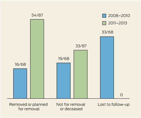
Between 2011 and 2013, all 87 IVC filters inserted by IR at St Vincent’s Hospital Melbourne have been accounted for; 44 have been removed, 33 were planned not for removal (eg, patients with a poor prognosis or contraindications), and 10 are awaiting removal or review in our IR clinic. The average indwelling time for IVC filters inserted between 2011 and 2013 was 23 weeks (range, 1–130 weeks), with 71/87 filters remaining in situ for less than 6 months. The average time in situ fell from 35 weeks in 2011 to 14 weeks
in 2013. These results contrast with 2008–2010 data from our institution, where only 16/68 IVC filters were removed or planned for removal,
19/68 were planned not for removal or the patient was deceased, and
33/68 were lost to follow-up (Box).

Concerns about inadequate IVC filter removal and follow-up have been reported in the literature, particularly as the use of filters has increased.2,3 Potential complications associated with “forgotten” IVC filters include thrombogenesis, caval perforation, and filter fracture and migration. However, the important role of IR in tracking and removing lost filters is becoming widely recognised, with a number of studies outlining improvements in IVC filter follow-up that have resulted from radiology-led initiatives.4,5 Our data support the conclusion that IR departments, through easily implemented strategies, can and should take responsibility for tracking and removing all IVC filters they insert.

Inferior vena cava filter outcomes at St Vincent’s Hospital Melbourne

Lack of appropriate imaging before breast augmentation can have serious patient consequences

Advice on preoperative imaging for different groups of women is now available

Breast augmentation has become increasingly popular in recent years, with 316 848 procedures reported in the United States in 2011.1 While there is a lack of accessible data about breast augmentation procedures in Australia, the breast surgical oncology community has ongoing concerns about the potential for breast implants to deter women from participating in appropriate symptom assessment or routine screening and to impair early detection of breast cancer.2

The situation is complicated by a lack of high-level evidence about health outcomes for women with implants who subsequently develop breast cancer. In an observational study of 129 women who had undergone breast augmentation and 3953 women who had not, those who had undergone augmentation presented more frequently with palpable lesions, invasive tumours and axillary nodal metastases, and were more likely to have a false-negative mammogram result. However, overall there was no significant difference in stage of disease, tumour size, recurrence rates or survival between the two groups.3 It was postulated that breast implants may facilitate palpation of tumours and make the breast easier to examine, explaining why lesions of the same size were more frequently palpable in patients with implants.3 This explanation fits with our clinical experience, but there are other possible reasons for the higher rates of invasive tumours and axillary nodal metastases in this group. These explanations relate to the higher likelihood of a false-negative mammogram result in women with implants who do have regular screening, and to the lower rate of screening in women with implants because of reluctance to have mammograms for fear of damaging the implants.

The discrepancy between the higher rates of invasive tumours and axillary node metastases among women who have had breast augmentation on the one hand, and a lack of significant differences in cancer staging between women with and without breast implants on the other, may be explained, in part, by methodological limitations. These include use of the χ2 test, which may not have enough power to detect trends, and lack of consideration of confounding factors, such as age. According to a systematic review of observational studies, women with breast implants who develop breast cancer have later-stage tumours at diagnosis, while meta-analysis of these studies suggested that these women may also have an increased risk of non-localised breast tumours and lower rates of breast cancer-specific survival.4 The authors noted the need for further research to clarify the impact of breast augmentation on cancer detection and prognosis.

What is clear is that patients with breast implants present specific challenges for radiologists involved in breast imaging. Breast implants are radio-opaque and restrict the visualisation of breast tissue with mammography.4 Implants can also compress breast tissue, which can limit detection of subtle findings such as architectural distortion and microcalcifications. Twenty-six experts from five countries reviewed the best available evidence and concluded in a consensus statement that preoperative breast imaging, with mammography and/or ultrasound, should be considered essential, except in rare circumstances, before any breast augmentation procedure.5 Appropriate imaging may vary depending on the woman’s age, risk factors for breast cancer, breast density and clinical examination findings, but would usually involve mammography, possibly ultrasound and sometimes magnetic resonance imaging.

Failure to provide appropriate imaging not only has serious implications for the woman herself, but also for the health system, which is left paying for expensive surgery and adjuvant therapies that may have been avoided had the cancer been detected earlier, as a recent case from our practice demonstrates (Box 1). This case highlights the serious consequences of a probable missed opportunity to diagnose breast cancer 6 months earlier, when the disease most likely would have been entirely in situ. There are two implications for clinical practice. First, thorough clinical examination and risk assessment, as well as appropriate imaging, should become a routine component of the preoperative work-up for women who undergo breast augmentation. Second, if a general practitioner is aware of a patient’s intent to undergo breast augmentation, in Australia or overseas (given the increase in medical tourism), he or she should inform the patient of the importance of preoperative screening and arrange the appropriate imaging.

As a result of such cases, Breast Surgeons of Australia and New Zealand has developed a position statement on preoperative assessment of women who undergo breast surgery, which outlines the consensus view of what imaging is appropriate for different categories of women.2 The recommendations are summarised in
Box 2.

1 Case of breast cancer in a patient with breast implants

A 46-year-old woman presented with odd sensations in her right breast 6 months after undergoing bilateral breast augmentation surgery without preoperative breast imaging.
A mammogram and ultrasound revealed a 6.5 cm area of malignant calcifications. Analysis of a core biopsy specimen showed ductal carcinoma in situ (DCIS) and the patient underwent right mastectomy and sentinel node biopsy. The final pathology report described 9 cm of high-grade DCIS with multiple small foci of oestrogen receptor-negative and human epidermal growth factor receptor 2-positive invasive breast cancer. The sentinel nodes were free of disease. After multidisciplinary consultation, we recommended chemotherapy and trastuzumab treatment, which the patient agreed to.

2 Appropriate preoperative investigations for women undergoing breast augmentation or reduction, according to age and risk status*

Clinical
examination

Mammography

Ultrasound

Magnetic resonance imaging


Age < 40 years

Average risk

Yes

Consider

Consider

No

Increased risk§

Yes

Yes

Yes

Consider

Difficult to clinically assess

Yes

Consider

Yes

No

Age ≥ 40 years

Average risk

Yes

Yes

Consider

No

Increased risk§

Yes

Yes

Yes

Consider

Difficult to clinically assess

Yes

Yes

Yes

Consider


* Reproduced with permission from Breast Surgeons of Australia and New Zealand.2 Significant clinical findings, including abnormal results of clinical examination or abnormal symptoms, require full assessment by a breast physician or breast surgeon and may require a biopsy depending on the nature of the problem. Magnetic resonance imaging is specifically indicated only if recommended by a breast specialist. It can only be claimed on Medicare for patients in Cancer Australia’s risk category 3. § Defined as significant family history (Cancer Australia risk category 2 or 3) or previous clinical history that indicates high risk (eg, prior biopsy specimens that show proliferative breast disease such as lobular carcinoma in situ, atypical ductal hyperplasia, atypical lobular hyperplasia or multiple papillomatosis). Defined as suspicious or inconclusive findings, or situations where assessment is difficult due to anatomical features such as dense breasts.

Over 150 potentially low-value health care practices: an Australian study

To the Editor: We read the recent study by Elshaug and colleagues1 with interest, but were concerned by the inclusion of radiotherapy for spinal cord disease as a potentially low-value intervention. The lack of clinical review in the identification process appears to be the key driver in coming to this erroneous conclusion.

Elshaug and colleagues recognise the need for clinical expertise in assessing trials demonstrating no difference between a health care service and an active comparator
by excluding these from analysis. However, we suggest that the application of this expertise should extend to all publications included in the analysis. Of the three publications identifying radiotherapy for spinal cord compression, none disputed its role in managing this condition.24 One randomised trial, assessing the addition of decompressive surgery to radiotherapy, supported the role of radiotherapy for this disease.2 The intervention being assessed was, in fact, surgery, but this was not recognised.

The study by Elshaug et al also presented a list of 13 services which were identified by more than one search strategy and flagged as being potential priority candidates for review. Radiotherapy for spinal cord compression was again identified by this “triangulation” strategy, but two review articles3,4 were flagged by referencing the same randomised trial,2 essentially “double dipping”.

We commend the authors on attempting to develop a strategy to identify potential low-value interventions, but this process cannot be divorced from clinical judgement. Indeed, this is the entire raison d’être for expert working groups and consensus guidelines, which evaluate the quality and validity of trials in a clinical context.

Over 150 potentially low-value health care practices: an Australian study

To the Editor: Elshaug and colleagues recently attempted to identify low-value health care practices.1 They further identified 13 services found by more than one of their search methods, implying that these services may be of particularly low value.

One such practice identified is “radiotherapy for patients with metastatic spinal cord disease”.1 However, this terminology is loose at best, and the following quotes from Elshaug et al’s own sources do not offer ready support for the authors’ conclusion that this treatment may be of low value:

  • “Radiation therapy is a mainstay of treatment for spinal metastases, and continues to play an important role in pain relief, prevention of pathological fractures, and stabilization of neurological function”;2 and

  • “Offer fractionated radiotherapy as the definitive treatment of choice to patients with epidural tumour without neurological impairment, mechanical pain or spinal instability ”.3

Two of Elshaug et al’s references refer specifically to the treatment of metastatic spinal cord compression (MSCC),3,4 and it may be that this was what the authors really meant to review. Here, there is a valid question as to which patients may be better served by surgery — as suggested by the randomised study of surgery in highly selected patients.4 However, patient selection is key — “Selecting patients who will benefit from resection remains a challenging undertaking”2 — and surgery is not suitable for all — “Offer urgent radiotherapy . . . to all patients with MSCC who are not suitable for spinal surgery . . .”.3

In summary, radiotherapy for patients with metastatic spinal cord disease does not appear to be a
low-value practice and is actually recommended by Elshaug et al’s
own sources.

Over 150 potentially low-value health care practices: an Australian study

In reply: I thank Bece and colleagues and Pitson for engaging constructively with this quality improvement agenda. Bece et al challenge the inclusion of radiotherapy for spinal cord disease in our list of 150 candidate treatments potentially warranting further review under a quality improvement program.1 Pitson calls attention to “loose terminology” in the same example. This latter point, I concede. Precision of language in relation to treatment type and patient indications is essential in this debate.

However, I respectfully disagree with the assertion from Bece et al that listing radiotherapy for spinal cord disease represents an “erroneous conclusion”. In our article we state: “The list includes examples where practice optimisation (ie, assessing relative value of a service against comparators) might be required.”1 Later, I further qualify this point2 and extend the debate.3 The study we cited investigated the relative value of surgery with adjuvant radiotherapy compared with radiotherapy alone.4 The authors of that study concluded: “Direct decompressive surgery plus postoperative radiotherapy is superior to treatment with radiotherapy alone for patients with spinal cord compression caused by metastatic cancer.”4 Hence, rather than listing radiotherapy for spinal cord disease being an “erroneous conclusion”, it seems the relative value of radiotherapy alone is open for legitimate debate, and, therefore, for inclusion in our list based on a-priori inclusion criteria.

As Pitson goes on to highlight, appropriate selection of patients for what might become high-value as opposed to low-value applications of these procedures (or combinations thereof) is key. Surely these complexities and treatment choices support calls for further practice (as well as funding) optimisation in this area. I am hopeful that with a tightening of the language, any disagreement might be diminished.

Where is the next generation of medical educators?

In reply: We thank Hart and Pearce for supporting the views raised in our editorial, noting the unmet demand for medical education expertise.

We also thank Kandiah for his response, and agree that medical graduates should be “clinically competent, reliable, keen to learn and show compassion to patients and colleagues”. We believe this outcome is best achieved by strong collaborations among “skilled clinicians and excellent mentors” and medical educators, many of whom are also practising clinicians. Clinicians provide critical input to ensure the validity and authenticity of what is taught and assessed, and are an essential element of the “triad” of patient, student and clinician in clinical learning.1 Collaboration between clinicians and medical educators is not difficult because they are often embodied within the same people.

The question of proof in medical education is the subject of much activity and, as Kandiah notes, there is an increasing output of scholarship in medical education. Moreover, the quality and rigour of this output is increasing, with a growing evidence base for medical educational practice.2 Generating new knowledge and applying it to medical student education is a key goal of an increasingly professionalised medical education community.

Medical education research is confounded by multiple factors, not the least being the powerful and uncontrolled effects of the diverse clinical environments in which students learn and practise as graduates.3 These make causal pathways difficult to unpick. While researching the effect of medical educators may be desirable, we believe that researching the effects of medical education interventions is more fruitful. For example, if one were to substitute medical educators with radiologists, how could one “prove” that radiologists have improved the health of the Australian population? Yet we are convinced that radiology does play an important role, based on multiple individual studies showing contributory evidence for this claim.

We welcome opportunities to work with health services and the community to examine the long-term performance of our students and their impact on the health system. Collaboratively defining and answering specific questions is likely to be much more productive than making artificial distinctions between clinicians and educators.

Injuries to the head and face sustained while surfboard riding

To the Editor: Surfboard riding is an iconic pastime in Australia. Injuries to the head and face constitute a considerable proportion of surfing injuries;15 26% of acute surfing injuries are to the head and face, and these make up 42% of emergency department presentations by surfers.3

We conducted a retrospective review at our tertiary referral hospital of patients who underwent medical imaging for injuries sustained to the head and face while surfboard riding from January 2008 to January 2012. We searched the hospital radiology databases for patient records containing the terms “surfboard”, “surfer” or “surfing”. Patients were included if they were injured while surfboard riding and were excluded if they were injured during other water-based activities (eg, bodyboarding, kitesurfing, bodysurfing, paddleboarding). Twenty-nine patients were identified: 23 males and six females (mean age, 34 years; range, 10–73 years). Of the 26 who had acute injuries, 17 had imaging of the head only, seven had imaging of the head and cervical spine and two underwent a trauma protocol (computed tomography scans of the head, spine, chest and abdomen). Fifteen patients had been struck in the head by their own board, nine had other mechanisms of injury (primarily involving contact with the sea floor and associated neck pain), one collapsed while surfing and one was retrieved from the surf unconscious (mechanism of injury was unknown). The most common significant injuries were facial fractures (five of 26 patients, all of whom had been struck by their own board). One patient ruptured their left globe after being struck by their own board. No intracranial trauma (eg, intracranial haemorrhage, contusion) was identified.

Surfboard design and surfing accessories have evolved significantly over the past 20 years. Lighter, shorter boards are now commonly used and provide greater manoeuvrability in the water. Leg ropes are universally used to ensure that surfer and board do not become separated (Box). However, lighter boards and leg ropes might increase the risk of being struck and injured by one’s own board during a “wipe-out”. The pointed nose and fins on the undersurface of the board are also potentially injurious (Box).

Understanding injury mechanisms can drive surfboard and surfing accessory design to reduce the risk of injury and death. Protective devices — such as helmets, protective eyewear and nose guards that cover the tip of the surfboard — have been marketed, but there is no evidence of their effectiveness in injury reduction.

Innovations in surfboard design that are potentially injurious to the surfer

A: Leg rope. B: Pointed nose at the front of the board. C: Fins on the undersurface of the board (a three-fin design is the most common configuration on a modern shortboard).