×

News briefs

Severe head trauma mortality drops at Royal Darwin

Mortality rates for severe head trauma at the Royal Darwin Hospital are down 40% from the 79% rate reported in a study 10 years ago, according to the ANZ Journal of Surgery. The study reviewed clinical service between 2008 and 2013, highlighting the continuing challenge of remoteness to the delivery of emergency medicine and surgery in the Top End. Alcohol remains a major player in hospitalisation, with 57% of patients having evidence of alcohol involvement and 39% of patients with traumatic brain injury having alcohol as a factor in their presentations. Indigenous persons were also overrepresented, accounting for 39% of all procedures as well as being considerably younger by a median of 15 years than their non-Indigenous counterparts. Resident generalist surgeons are reliant upon interstate neurosurgeons, who provide ongoing education, training and support, both by way of outreach visits and by 24-hour telephone and teleradiology consultation over 2600 km away.

Maternal, neonatal tetanus eliminated in India

Maternal and neonatal tetanus has been reduced to less than one case per 1000 live births in India, according to a WHO report. Until a few decades ago, India reported 150 000 to 200 000 neonatal tetanus cases annually. According to Dr Poonam Khetrapal Singh, WHO Regional Director for South-East Asia, the Indian government used a mix of existing and new programs to make elimination possible. “India’s re-energized national immunization program and the special immunization weeks and the most recent ‘Mission Indradhanush’, helped ensure that children and pregnant women are reached with vaccines”, he said. “The ‘National Rural Health Mission’ promoted institutional deliveries with a focus on the poor. The ‘Janani Suraksha Yojana’ encouraged women to give birth in a health facility.” Maternal and neonatal tetanus in South-East Asia now exists in just a few districts of Indonesia.

Hazard alert for hip replacement component

The Therapeutic Goods Administration has issued a hazard alert for one model of the Profemur cobalt-chrome femoral neck (part number PHAC1254 – “long 8-degree varus”) due to the potential for the component to fracture. The manufacturer, Surgical Specialities, is also undertaking a recall of unimplanted stock. Component fractures are extremely rare; however, the manufacturer reported that there had been 27 reports of fracture of the PHAC1254 component in the approximately 9800 units sold worldwide over the previous 5 years. Only 32 units have been sold in Australia. “If you are treating patients who have had a hip replacement and are concerned about the above issue, advise them to be alert to the potential symptoms of a femoral neck component fracture (the sudden onset of symptoms such as pain, instability and difficulty walking or performing common tasks).”

Elevated lead levels in 30 NT children

The Northern Territory Health Department has confirmed that 30 children have been found with elevated blood lead levels in three separate locations across remote areas of the territory, the ABC reports. Children in Palumpa and Peppimenarti, in the West Daly region, and the Emu Point outstation, had higher than expected lead levels, probably due to contact with lead shot, used for shooting magpie geese, according to NT Health Minister John Elferink. NT Chief Health Officer Professor Dinesh Arya said that the children and their families were being interviewed to determine the cause, and all the children were receiving treatment from “specialist paediatricians”.

Ebola vaccination trial extended to Sierra Leone

The WHO reports that a new case of Ebola virus in Sierra Leone, after the country had marked almost 3 weeks of zero cases, has set in motion the first “ring vaccination” use of the experimental Ebola vaccine in the country. A swab taken from a woman who died aged around 60, in late August in the Kambia district, tested positive for Ebola virus. “The Guinea ring vaccination trial is a Phase III efficacy trial of the VSV-EBOV vaccine. Interim results published last July show that this vaccine is highly effective against Ebola. The ‘ring vaccination’ strategy involves vaccinating all contacts — the people known to have come into contact with a person confirmed to have been infected with Ebola (a ‘case’) — and contacts of contacts.”

Cancer nanomedicine: challenges and opportunities

Nanotechnology holds enormous promise for personalised cancer medicine — translation is the key

Medicine is on the cusp of a revolution. Personalised, precision medicine — designed and tailored at a molecular level for an individual’s own physiological make-up — will become an inevitable reality in the 21st century. As with all paradigm shifts in medicine, this will be driven by new science and technology, and the technology of the 21st century is nanotechnology.

Nanomedicine is a rapidly evolving paradigm where nanoscience and nanotechnology are applied to medicine. The science underpinning nanotechnology is that some materials, when reduced from everyday, bulk scales down to nanoscales (billionths of a metre; smaller than the size of a typical virus), exhibit dramatically different physical properties. Harnessing and customising these unique nanoscale properties offer unique advantages to health and medicine for two reasons. First, many key molecules involved in biochemical processes responsible for regulating biological function have nanometre (nm) sizes (eg, a glucose molecule is about 1 nm), so nanoscale probes offer a means for molecular-based interrogation and intervention strategies. Second, because of their size, nanoprobes offer a relatively discreet, non-invasive strategy for disease detection and targeted therapy (although the immune system inevitably catches up).1

An important example of how nanoscale properties can be harnessed for medical applications is magnetic resonance imaging. Here, image contrast is enhanced using magnetic nanoparticles, usually based on gadolinium or iron oxide, which exhibit strong magnetism only when reduced to scales of 20 nm or less.2

Nanoparticles: size matters when it comes to targeting tumours

In cancer nanomedicine, a wide range of nanoparticles continue to be developed for better tumour-targeted delivery of therapeutics (chemotherapy and radiotherapy). These include liposome-, polymer- and micelle-based nanoparticles for encapsulated delivery, and metallic nanoparticles (eg, gold), which have been investigated for targeted radiotherapy.13 There are two types of tumour-targeting approaches with nanoparticles: passive and active. Passive targeting relies on tumour vasculature, which has larger endothelial gap junctions compared with healthy tissue. Nanoparticles greater than 8 nm can pass through these gaps to reach tumour cells. An enhanced permeability and retention effect results from the combination of larger gap junctions and defective lymphatic drainage, particularly around fast-growing tumours, facilitating preferential accumulation and prolonged retention in the tumour tissue.1 In active targeting, however, nanoparticles are conjugated with targeting agents, such as antibodies, that are specific to proteins highly expressed by certain tumours (eg, human epidermal growth factor receptor 1 in non-small cell lung cancer).3

Challenges: clinical translation

Despite ongoing progress in basic and preclinical cancer nanomedicine research, arguably the single most important challenge is clinical translation.4 However, most of the many different nanoplatforms developed for cancer therapy have not progressed past Phase II clinical trials.1 Very few have achieved United States Food and Drug Administration approval (eg, liposome-encapsulated doxorubicin and daunorubicin for breast and ovarian cancers, and Kaposi sarcoma). New efforts are focusing on the potential to extend the capabilities of other therapeutic and imaging nanoplatforms developed and approved for non-cancer indications. For example, ferumoxytol is an iron oxide nanoparticle used for treating anaemia and it is also a magnetic resonance imaging contrast-enhancing agent.5 Nanotheranostics — the use of nanoplatforms combining targeted therapy and diagnostic imaging functionality — is a rapidly growing trend.

Why is bench-to-bedside so challenging for cancer nanomedicine? The problems are many. Key difficulties include controlling nanoparticle size and preventing nanoparticle aggregation in vivo, which are critical for clearance by the kidney or liver. Biocompatibility, blood circulation time and the ability to elude the immune system long enough to release a therapeutic cargo, are similarly difficult to clinically validate. Additional practical challenges that need to be overcome for clinical translation include tumour cell specificity, cellular uptake and localisation, and controlled release and functionality of the cancer therapeutic.3

Opportunities: clinical translation

The challenges presented by clinical translation could equally be viewed as opportunities. This is the approach taken by the European Foundation for Clinical Nanomedicine (https://www.clinam.org). Similarly, the US National Cancer Institute (NCI) integrates translational and basic science research in its Alliance for Nanotechnology in Cancer (http://nano.cancer.gov). Launched in 2004, the Alliance held a strategic workshop in 2013, the outcomes of which highlighted several recommendations for future opportunities in cancer nanotechnology.6 These include supporting the development of new techniques and clinical translation in parallel; supporting a stronger focus on developing active targeting strategies; and giving a high priority to imaging probes and lower priority to developing in-vitro nano-enabled techniques. The NCI report also highlighted the importance of interdisciplinary collaboration — bringing together clinical and basic science researchers from diverse backgrounds is the key to creating unique opportunities for genuine breakthrough discoveries in cancer nanomedicine.

Splenic abscess complicating gastroenteritis due to Salmonella Virchow in an immunocompetent host

Clinical record

A 20-year-old man was admitted to a regional hospital with fevers, rigors, anorexia and left upper quadrant pain. It was his fourth presentation to the emergency department in the preceding 10 days. On the first two presentations, he had been sent home with a provisional diagnosis of renal colic. After review by his general practitioner, he had undergone outpatient imaging that identified filling defects in the pulmonary arteries of his left lower lobe, which were reported as being consistent with pulmonary emboli. In addition, two hypodense splenic lesions were identified, as well as collapse and possible consolidation of the left lower lobe. His GP had referred him to the emergency department for further review (his third presentation), after which he had commenced therapeutic anticoagulation for a presumed diagnosis of pulmonary emboli.

The patient’s history was notable for a self-limiting episode of gastroenteritis 6 weeks before his initial presentation, with sick family contacts. On his fourth presentation, he described progressive left upper quadrant and flank pain over the preceding 10 days, with intermittent fevers and rigors. He had no other focal infective symptoms on review.

On examination, he was found to have a fever (temperature, 39.3°C), sinus tachycardia (heart rate, 154 beats/min), tachypnoea (respiratory rate, 28 breaths/min), hypotension (blood pressure, 97/66 mmHg), decreased breath sounds at the left base of his lung fields and mild left upper quadrant tenderness. Investigations showed a white cell count of 16 × 109/L (reference interval [RI], 4.0–11.0 × 10⁹/L), with a predominant neutrophilia (neutrophils, 14 × 109/L [RI, 2.0–7.0 × 10⁹/L]). Results of his liver function tests and electrolyte, urea and creatinine levels were all within reference intervals.

A computed tomography scan of the chest and upper abdomen again showed two low-density lesions of unclear aetiology in the spleen, as well as a left-sided pleural effusion and collapse of the left lower lobe. Given the possibility that the hypodense splenic lesions represented septic emboli from a cardiac source, the patient was treated empirically with benzylpenicillin, flucloxacillin and gentamicin for a provisional diagnosis of endocarditis. However, a transthoracic echocardiogram performed the next day did not support this diagnosis, with no abnormalities detected. Beyond the radiological findings described, there were no other clinical grounds to support a diagnosis of endocarditis.

Blood cultures taken on Day 1 of admission were positive for gram-negative bacilli, with confirmation of a non-typhoidal Salmonella species (later confirmed as Salmonella Virchow) the following day. This allowed targeted antibiotic therapy, once susceptibilities were known, with ampicillin (2 g every 6 hours). Cultures of stool samples taken at admission were positive for the same isolate, consistent with the patient’s self-limiting episode of gastroenteritis 6 weeks before his first presentation.

Magnetic resonance imaging of the abdomen suggested that the two splenic lesions were likely to represent abscesses in this clinical context (Figure). Given our patient’s ongoing sepsis, a decision was made to perform a laparoscopic splenectomy for source control on Day 5 of admission. Surgical specimens tested positive for Salmonella Virchow. Histopathological testing identified cystic lymphangiomas of the spleen. Despite problems with postoperative pain and a prolonged ileus, the patient made a full recovery. He received appropriate post-splenectomy vaccinations, along with a total of 2 weeks’ intravenous ampicillin, followed by a 2-week course of oral amoxicillin.

Non-typhoidal salmonellae are common foodborne pathogens. In Australia, they are the second most frequent bacterial isolates identified in cases of acute gastroenteritis, after Campylobacter jejuni. In 2010, OzFoodNet sites reported 11 992 cases of Salmonella infection, a rate of 53.7 cases per 100 000.1 Salmonella Virchow was the third most common isolate, after Salmonella Typhimurium and Salmonella Enteritidis. Non-typhoidal Salmonella infection outbreaks are most commonly associated with consumption of poultry and eggs, but have also been linked to fresh produce and, increasingly, contact with pet reptiles.2

Up to 8% of patients with gastroenteritis secondary to non-typhoidal Salmonella infection develop bacteraemia.3 Risk factors for invasive infection include extremes of age, immunosuppressed states, malignancy, HIV infection and use of tumour necrosis factor-blocking medication.4 Our case is unusual in that bacteraemia occurred in an otherwise immunocompetent host.

Extraintestinal focal infections have been reported to occur in 5% to 10% of patients with non-typhoidal Salmonella bacteraemia.3 The best recognised complications are endovascular infections, most commonly involving the aorta, that result from seeding of atherosclerotic plaques and aneurysms.5 However, focal infections of almost all organ systems have been reported.

Splenic abscesses are most commonly seen as a complication of infective endocarditis, occurring in about 5% of patients.6,7 They are also found as a rare complication of non-typhoidal Salmonella infections. In one case series of 49 patients from southern Taiwan, Salmonella species were the third most common pathogens isolated from splenic abscesses, accounting for 11% of cases.8 The most common presentations among the 49 patients with splenic abscesses were fever (47 patients), abdominal pain confined to the left upper quadrant (33 patients), left pleural effusion and splenomegaly (both 27 patients), all of which were present in our patient.

About 50% of patients presenting with splenic abscesses have pre-existing anatomical abnormalities.9 The cystic lymphangiomas identified in our patient almost certainly predisposed him to developing splenic abscesses.

According to the literature, the mainstay of treatment for splenic abscesses is splenectomy. Data from 287 cases published between 1987 and 1995 suggested that non-operative management, which included invasive treatment with percutaneous aspiration and catheter drainage, had a success rate of less than 65%.10 The same series suggested that antimicrobial therapy alone had a success rate of less than 50%. Salvage splenectomy, however, was not shown to result in increased mortality. Another retrospective study of 51 patients in a tertiary hospital between 1998 and 2003 reported survival rates of 48% with antimicrobial therapy alone, 45% with pigtail catheter insertion and drainage in addition to antimicrobial therapy, and 100% with splenectomy and antimicrobial therapy.11 These results may be influenced by selection bias but do suggest improved outcomes with splenectomy over less invasive strategies.

Lessons from practice

  • Splenic abscesses are a rare but potentially life-threatening complication of non-typhoidal Salmonella bacteraemia.
  • Splenic abscesses should be considered as a possible source of infection in patients presenting with unexplained fevers and left upper quadrant or left flank pain.
  • Splenectomy plus appropriate antimicrobial therapy remains the mainstay of treatment for splenic abscesses.
  • Interventional radiological techniques should be considered as a spleen-preserving strategy on a case-by-case basis and where experienced radiologists are available.

Splenic abscesses are a rare but serious complication of non-typhoidal Salmonella bacteraemia that may occur in otherwise immunocompetent individuals. Splenic abscesses should be suspected in patients with unexplained fevers and left upper quadrant pain. The mainstay of treatment is splenectomy with appropriate antimicrobial therapy.


A: Axial T2-weighted magnetic resonance image (MRI) of the abdomen, without contrast, showing an abscess in the inferior pole of the spleen (circle).


B: Saggital T2-weighted MRI of the abdomen, without contrast, showing two splenic abscesses (circles).


The spectacular recent trials of urgent neurointervention for acute stroke: fuel for a revolution

How should we redesign our stroke services in light of neurointerventional advances?

In 2013, neutral results from three trials of neurointervention for treating ischaemic stroke were simultaneously published — a triad of gloom.13 In just over 2 years since, five positive trials have been reported.48 What explains this extraordinary turnaround, and what are the implications for stroke services in Australia and around the world? The answers to these questions are surprising and reflect a mixture of science, technology and policy.

The roles of science, technology and policy

The science involved is the culmination of a decade of work on proving that brain imaging can identify the ischaemic penumbra — the area of the brain that has shut down and is on the path to infarction but, with successful reperfusion, is potentially salvageable. By recruiting patients with a favourable profile for reperfusion therapy (so-called target mismatch, where the ratio of perfusion lesion to established infarct is > 1.8, the perfusion lesion volume is > 15 mL, and the established infarct volume is < 70 mL),9 we are now able to identify those who are likely to respond well. In addition, computed tomography (CT) angiography is now widely available and can demonstrate major cerebral vessel occlusion — a clear target for therapy.9

In contrast to the neutral trials, the recent trials all used either advanced imaging to identify patients with the “reperfusion responder” profile or angiography to prove major vessel occlusion, or both, then randomly assigned this population of likely responders to receive endovascular reperfusion (usually in addition to alteplase thrombolysis) or standard acute stroke care. The combination therapy resulted in potent reperfusion and a dramatic treatment effect (Appendix), such that three of the five neurointervention trials were stopped early.

The technology is all about the device. In today’s fast-moving world, it is almost impossible to design, fund and complete a trial of a device without it becoming obsolete by the time the trial has finished — the fate of the previous studies.13 Unlike in coronary intervention, the thrombus or embolus in ischaemic stroke must be physically extracted, and the new generation of retrievable stents are a major advance in this regard. One of the recent trials, MR CLEAN, demonstrated that carotid stenting (for extracranial occlusion) was also required for 13% of the patients receiving intra-arterial treatment.4

Finally, the unanticipated influence of policy can have profound effects. One of the neutral trials, IMS III, was conducted in the United States at a time when neurointervention was generously compensated.1 This, together with the attractiveness of the technology (despite its lack of evidence), meant that most people were treated outside the trial. The difficulties in recruitment (only one or two patients per centre per year) and a possible selection bias of recruiting only “difficult” patients might have had an effect on the results of IMS III.

In contrast, the Dutch MR CLEAN trial provides an important lesson.4 All the neurointervention centres in the Netherlands participated in this trial, and from 2013 there was no reimbursement for people treated outside the trial. This allowed some 500 patients to be recruited from 16 sites in just over 3 years, compared with IMS III, which needed 7 years to recruit 656 patients from 58 sites. If trial-only reimbursement for unproven devices were enforced, it is likely that reliable data on efficacy would have been available much earlier, potentially saving hundreds, if not thousands, of lives.

Implications for practice

Implementing intravenous thrombolysis has been a difficult and protracted affair, and we are still researching the best ways to achieve it.10 However, there has been progress in Australia, thanks to the hard work of clinicians in the state stroke networks. For example, in New South Wales, the Agency for Clinical Innovation led the establishment of a state-wide stroke reperfusion strategy that involved training paramedics to screen for potential thrombolysis candidates with the FAST (Face, Arms, Speech, Time) test and fast-tracking potentially eligible patients to 24-hour thrombolysis centres.

Our challenge is how to redesign our stroke services and how best to build capacity in the neurointerventional workforce. What is the required infrastructure, support and training in advanced imaging selection that would work in our hospitals? The London model of hyperacute stroke centres might work in our capital cities, but the “drip and ship” model of starting thrombolysis followed by urgent transfer to a comprehensive stroke centre may be a solution for outer metropolitan and regional thrombolysis centres. It is abundantly clear that we will never be able to provide on-site neurointervention at all stroke thrombolysis-capable hospitals, nor achieve complete equity of access to endovascular therapy for stroke patients from rural and remote communities.

A potential solution to the shortage of neurointerventionalists is the emerging model to train neurologists in interventional neuroradiological skills. Given there is broad acceptance that a stroke physician (with appropriate training) does not necessarily have to be a neurologist, there is growing support in the US and Australia for the position that a neurointerventionalist does not have to be a radiologist, provided he or she has had appropriate training.11

The exact shape of future neurointerventional stroke centres is still uncertain, but the endovascular revolution has arrived, and the stroke community needs to work quickly to redesign stroke care services and build the workforce of specialists trained in endovascular therapies. Drivers for change will include the new national stroke care standard (launched on 10 June 2015)12 and the next revision of the national Clinical guidelines for stroke management.

Stroke medicine has come a long way from the nihilism of two decades ago, with numerous interventions now supported by high-level evidence. Immediate brain imaging will identify strokes that are due to haemorrhage, and rapid blood pressure lowering and stroke unit (or intensive) care are the mainstay of treatment for these patients, with surgery needed only for a select few. For patients with ischaemic stroke, revascularisation with appropriate intravenous thrombolysis should be sought, followed by advanced brain imaging to identify patients suitable for additional endovascular therapy.

What is the future? Colleagues in the US and Germany are exploring the utility of an ambulance with an onboard CT scanner (Box), with anecdotal reports of excellent responses to alteplase when given within minutes of major stroke onset. The search is also on for more effective intravenous thrombolytic drugs, with Australia leading an international trial of tenecteplase versus the current standard, alteplase.13 However, none of this will be effective without further public health interventions to improve awareness of stroke and the importance of immediately calling 000 for any suspected stroke patient. When it comes to stroke, time is brain.

Ambulance with a computed tomography scanner

General practitioner-referred magnetic resonance imaging for musculoskeletal conditions: not a substitute for plain x-ray

To the Editor: In November 2012, the Australian Government extended requesting rights for Medicare-eligible magnetic resonance imaging (MRI) scans to general practitioners for “a small set of clinically appropriate indications”1 in patients under the age of 16 years. The purpose was

avoiding exposure of children to unnecessary radiation associated with other types of diagnostic imaging like computed tomography (CT) scans.1

With musculoskeletal MRI, a Medicare-eligible scan is performed “following radiographic examination”.1

Plain x-ray, rather than MRI, remains the gold standard for diagnosing most musculoskeletal conditions in children, and we report two cases where the initial use of MRI rather than x-ray led to a delay in diagnosis.

First, an 11-year-old girl was investigated for a 3-month history of left knee pain and locking. An ultrasound and MRI scan (Box 1, A) were performed, although an x-ray was not done. As no cause for the symptoms was identified on the ultrasound or MRI, the patient was referred to an orthopaedic surgeon, who requested an x-ray (Box 1, B). This showed a large proximal tibial spur tethering the semitendinosis tendon. This was poorly visualised on MRI and had not been reported. The patient’s symptoms resolved after excision of the spur.2

Second, a 13-year-old boy with a 3-week history of left hip pain and limp underwent MRI (Box 2, A) without a prior x-ray for suspected Perthes disease. The report stated “left hip joint effusion, without other identifiable abnormality”. When symptoms persisted, the patient was referred to a paediatric emergency department. The magnetic resonance images were reviewed and a slipped capital femoral epiphysis (SCFE) was evident (but had not been reported). Standard hip x-rays showed an obvious SCFE (Box 2, B and C). X-ray is always the initial investigation of choice for Perthes disease and SCFE (MRI should be reserved for equivocal cases), and SCFE is a far more common diagnosis than Perthes disease during adolescence.3

Over 20 000 GP-referred Medicare-rebated MRI scans were performed in patients under the age of 16 in the 2013–14 financial year (Box 3). The number of referrals per GP in Australia was 0.44 in 2012–13 and doubled to 0.90 in 2013–14.4,5

In contrast, specialist referrals for paediatric MRI were 0.31 scans per specialist in 2007–08 and 0.37 in 2013–14;4,5 the number of MRI scans referred per specialist has not altered appreciably.

While it is important to decrease radiation from CT scans in children under 16 years, it is also important to remember that MRI is mostly unhelpful in diagnosing paediatric musculoskeletal complaints — most of which can be confirmed on plain x-ray after taking an accurate history and examining the patient. Due to the significant cost to the health care system of MRI, we suggest that evidence of prior x-ray imaging should be a requirement before Medicare funds an MRI scan.

1 Magnetic resonance image (A) and x-ray image (B) of the left knee of the 11-year-old girl

2 Magnetic resonance image (A), pelvic x-ray, anteroposterior view (B) and pelvic x-ray, frog-leg view (C) of the pelvis of the 13-year-old boy

3 Number of Medicare rebates for magnetic resonance imaging scans in patients < 16 years of age4,5

Eagle Syndrome as a potential cause of Tapia Syndrome

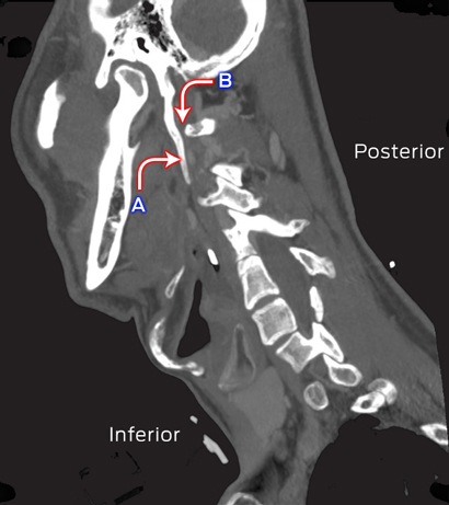
A 38-year-old man presented with influenza A (H1N1) pneumonia complicated by acute respiratory distress syndrome, resulting in an extended intensive care unit stay (37 days). After he was extubated, he was found to have left-side IX, X and XII cranial nerve palsies. Computed tomography of his neck showed bilateral elongated styloid processes (5.0 cm long; Figure), consistent with Eagle syndrome.1 The left styloid process was closely opposed to the transverse process of the first cervical vertebra, causing effacement of the internal jugular vein and compression of the IX, X and XII cranial nerves, which converge in this area. The eponym used to describe concurrent paralyses of the X and XII cranial nerves is Tapia syndrome,2 with prolonged neck flexion potentially contributing to the condition in our case.

Sagittal computed tomography image of the neck of the patient. A: Elongated styloid process (left); B: Area of compression of IX, X and XII cranial nerves.

Can magnetic resonance imaging solve the prostate cancer conundrum?

The debate about prostate-specific antigen (PSA) testing for the diagnosis of prostate cancer continues to rage. The United States Preventive Services Task Force (USPSTF) and the equivalent Canadian body have both advised against PSA screening.1,2 On the other hand, many authoritative bodies, including the American Urological Association,3 the National Comprehensive Cancer Network (US), the American Cancer Society, the European Association of Urology,4 and our own Urological Society of Australia and New Zealand recommend that, instead of population-based screening, decisions about PSA testing of men aged 55–69 years should be shared by doctors and individual patients.

While the USPSTF acknowledged the potential benefit of PSA screening for reducing cancer mortality and morbidity, it decided that the significant potential harms outweighed this benefit. The ongoing impact on the patient’s quality of life of the side effects of the various treatments, such as erectile dysfunction and urinary incontinence, are well documented,5 but the diagnostic biopsy itself is also associated with potential morbidity and, if rarely, mortality.6,7

These harms can be summarised as the overdiagnosis and overtreatment of indolent prostate cancer. The root cause of this problem is the troubling inaccuracy of the current diagnostic process that is still considered the standard of care — an elevated PSA level leading to a transrectal ultrasound-guided (TRUS) biopsy.

The poor specificity of PSA for significant prostate cancer is well known.8 What may not be such common knowledge is that about half of all TRUS biopsies are negative for cancer.9 This might seem to imply that a large proportion of men undergoing this invasive procedure do not have prostate cancer. We know, however, that some do, in fact, have significant prostate cancer, but the TRUS biopsy, being a blind random sampling of the gland, has missed it.10

In addition, a large proportion of the prostate cancer currently diagnosed by the combination of an elevated PSA level and a TRUS biopsy is actually low-risk disease.11 The vast majority of such cases require nothing more than active surveillance, but many of the men involved, unfortunately, are subjected to unnecessary treatments.12

Multiparametric magnetic resonance imaging

A diagnostic test, that can detect significant prostate cancer, but can exclude indolent disease, has therefore been desperately needed. Multiparametric magnetic resonance imaging (mpMRI) may turn out to be that test.

Prostate assessment by MRI has been available for many years, but only recently has it shown real promise. This has been achieved by combining multiple parameters, including T2-weighted imaging, diffusion-weighted imaging and dynamic contrast enhancement, and by reporting results in a standardised fashion, such as the framework provided by the Prostate Imaging–Reporting and Data System.13

Recently published mpMRI research by two Australian groups may provide the breakthrough for this approach.

In Brisbane, Pokorny and colleagues undertook a prospective study that compared 12-core TRUS biopsy with mpMRI. Targeted biopsies of suspicious lesions were performed to confirm the mpMRI findings. It was found that mpMRI was considerably more sensitive than TRUS biopsy in detecting significant cancer, with negative predictive values (NPVs) of 97% and 72%, respectively. That is, only 3% of significant cancers were missed by mpMRI, compared with 28% not detected by TRUS biopsy. At the same time, performing only mpMRI-targeted biopsies reduced the diagnosis of indolent disease by as much as 89%, and the need for biopsy by 51%.14

In Sydney, Thompson and colleagues compared mpMRI with 30-core transperineal biopsy, presumed to be a more accurate assessment of the presence of cancer. They also found excellent NPVs of 92%–96% for mpMRI, depending on the definition of “significant cancer”. The overdiagnosis of low-risk cancer by 34% would have been reduced by mpMRI-targeted biopsies, and the need for biopsy by 50%.15

The future role of mpMRI in prostate cancer diagnosis

These landmark reports argue in favour of a significant future role for mpMRI in the diagnosis of prostate cancer. It has already entered prostate cancer guidelines in the United Kingdom, where the National Institute for Health and Care Excellence recently recommended that mpMRI be considered for men with a negative TRUS biopsy, to determine whether a further biopsy is required.16

Although mpMRI might initially seem an expensive addition to the diagnostic algorithm, recent evidence suggests that it may ultimately be cost-effective.17 In Australia, however, the costs of an mpMRI prostate assessment ($400–$1000) are not currently reimbursed by Medicare.

It is important to note that the excellent results obtained in the Australian studies relied heavily on the immense mpMRI experience of the radiologists and urologists involved. Such expertise remains unusual. Further, the results need to be validated by larger multicentre studies before mpMRI be considered for routine use.

In the meantime, a prostate mpMRI should be ordered by urologists only with caution and after discussing it with radiologists with the necessary expertise. At our institution, we conduct a regular multidisciplinary prostate mpMRI meeting, a practice we recommend both as a means of quality control and for accelerating the development of expertise.

It is also imperative that long-term patient outcomes are investigated in settings where MRI has been included in the diagnostic algorithm, so that the value of this exciting advance can be accurately assessed. For this purpose, we have maintained an institutional ethics committee-approved prospective database of all such patients, and look forward to sharing our findings in the future.

The role of mpMRI in prostate cancer detection is rapidly evolving. By enabling targeted biopsies that exclusively detect significant cancer, mpMRI may provide the diagnostic accuracy that has been so sorely lacking. As such, it has the potential to revolutionise both the diagnosis and treatment of prostate cancer.

Tech advances intensify demands on time-poor specialists

Radiologist workloads are increasing “exponentially” as the complexity of tests and demands on time multiply, intensifying workplace pressure and compromising training, the AMA’s Radiology Specialty Group has warned.

The group told the AMA Federal Council that technological advances meant diagnostic imaging tests were producing much more data than ever before, making the task of interpretation and diagnosis far more complex and demanding.

For instance, the group said, the data produced by a computed tomography scan of an oncology patient now took between 20 and 30 minutes to interrogate, much longer than a decade ago when data sets were much smaller.

But it said hospital administrators had not kept up with such changes, and time allowances had become increasingly inadequate.

Not only were radiologists interpreting bigger and more complex data sets, but were also being required to devote an increasing amount of time to multidisciplinary meetings.

The group said it took 60 minutes to prepare for a typical one-hour meeting, but because there was no billing involved, such demands were not taken into account when assessing radiologist productivity.

“[Multidisciplinary meetings], review of previous studies, ad hoc consultations and problem solving patient-related issues in the public sector take up about 20 per cent of a radiologist’s time in non-reporting patient management which is not recognised by hospital admin,” the report said, adding that this did not include time spent teaching a training registrars and trainees.

It warned that time constraints and other pressures were compromising training and imposing a significant burden on radiologists, particularly junior specialists.

“There is a shortage of staff, and the service requirements are impacting on training,” the group said. “The increases in workload are placing undue pressure on radiology trainees, in terms of on-call, supervision and checking of their reports, as well as apprentice model teaching.”

Adrian Rollins


An incidentaloma not to be missed

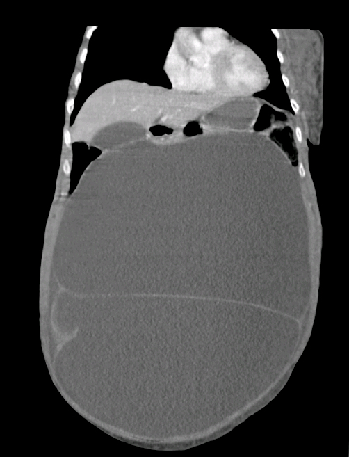
A frail 92-year-old woman presented with pelvic and femoral fragility fractures after a fall. She had synchronous gross abdominal distension which was diagnosed as ascites. Computed tomography was requested to exclude malignancy before performing paracentesis.

Formal imaging showed a large intraperitoneal structure. The 4-Hounsfield unit attenuation was consistent with simple fluid. However, identification of septations, together with rim enhancement, led to a revised diagnosis of a cystic mass. The lesion measured 24 cm × 28 cm × 33 cm and, at an estimated volume of 16 L, displaced most of the abdominal and pelvic viscera. Fortunately, this was recognised before paracentesis.

Wasteful overinvestigation — ultrasound in groin hernias and groin pain

To the Editor: Surgeons detest the waste involved in routine ultrasounds for all patients with groin hernias and those with groin pain only.

An inguinal hernia is a groin swelling with a cough impulse, clinically obvious in nearly all patients. No additional value is obtained from an ultrasound, which does not change the management.

In addition, nearly all patients with groin pain without any noticeable swelling are now unfortunately appearing with an ultrasound “diagnosing” a hernia. Pain is often exercise-induced, felt in the inner thigh, and often present at night or when rolling over in bed. The ultrasound report will invariably declare “there is an indirect inguinal hernia containing fat” (sometimes the fat is labelled omentum). Commonly this is embellished with “a neck of 4 [or 6 or 8] mm”.

Unfortunate consequences ensue from mislabelling a mobile blob of fat protruding through the deep inguinal ring as an indirect hernia. The ultrasound, the second general practitioner visit and the surgeon’s visit add to delay and cost, and the true musculoskeletal, or occasionally neurogenic, cause of the groin pain is not assessed and treated.

The ultrasound report not infrequently leads to totally unnecessary surgical exploration of the groin to cure the “inguinal hernia”. The original musculoskeletal cause of groin pain persists, leading to prolonged incapacity and sometimes a chronic pain syndrome. Complications of hernia surgery or mesh are blamed for the continuing pain; this often results in a degree of permanent impairment, and litigation.

There are a few useful indications for groin ultrasounds. They can help to exclude recurrent herniation. They may also detect a narrow-necked hernia in the rare but classic patient with an intermittently presenting, reducible groin swelling. An ultrasound can help to exclude a direct hernia defect plugged with fat in a patient with groin pain on sitting, no musculoskeletal pain source and no detectable clinical hernia. Ultrasound can also be used to determine whether a fixed solid groin lump is an incarcerated hernia or a lymph node.

Otherwise, time and money should not be wasted on unnecessary groin ultrasounds.