×

Pathologists on the warpath

Pathology and diagnostic imaging providers have vowed to flex their political muscle as part of an election-year campaign to force the Federal Government to dump controversial cuts to bulk billing incentives.

In a stark warning to Government MPs, pathology and diagnostic imaging groups have vowed to mount a vigorous campaign over last December’s decision to save $650 million by axing the bulk billing incentive for pathology services and reducing it for diagnostic scans.

Emphasising their political impact, industry leaders said there were about 5000 pathology collection centres around the country that were used regularly by millions of Australians.

Sonic Healthcare Chief Executive Colin Goldschmidt told Fairfax Media that his company alone had around 2000 collection centres.

“We reach something like 1 million to 2 million patients per month through those collection centres,” Mr Goldschmidt said. “We have access to a lot of people.”

The Government’s cuts have particularly angered pathology providers, who have not had an increase in the Medicare rebate for their services in 17 years.

Primary Health Care Chief Executive Peter Gregg said the decision was “ludicrous” because it would force providers to begin charging a co-payment, which would in turn deter some patients – including those with chronic conditions such as diabetes – from being tested as regularly, resulting in more serious and expensive health problems later on.

Related: Pathology services ‘cost Aust too much’

Mr Gregg told The Australian pathology services could not absorb any more Government cuts without changing their business model, and said Primary, which operates 71 medical centres, more than 2000 collection centres and 168 radiology clinics, had begun trials of co-payments for some pathology and diagnostic imaging tests to gauge their effect on demand.

Mr Goldschmidt said Sonic currently bulk billed 98 per cent of its services and, although it had not yet moved to introduce more co-payments, “we are tending in that direction”.

But both executives insisted their preferred option was to block the bulk billing incentive cuts altogether.

The change was announced by the Government in its Mid Year Economic and Fiscal Outlook. Health Minister Sussan Ley argued the incentive, worth between $1.40 and $3.40, had done little to boost bulk billing rates, and had instead served to plump up the bottom line of providers like Primary and Sonic.

She said the companies could comfortably absorb the cut.

Related: Graham Jones: Pathology power

But AMA President Professor Brian Owler said the Government’s real intent was to introduce a co-payment “by stealth” by forcing pathology and diagnostic imaging providers charge out-of-pocket expenses for their services.

“It’s very clear that to be viable, that if these bulk billing incentives are taken away, then of course they’re going to have to pass those fees onto patients,” Professor Owler said. “That’s what this strategy is all about. It’s about the Government saying ‘no, we’re not paying any more; we’re going to make the provider charge you a fee’.”

The AMA President said the likely fee providers would have to charge would be considerably more than the incentive, because providers would have to introduce and operate billing systems, chase up bad debts, make provisions for losses and other additional tasks.

“They’ve got to actually introduce a whole new system to enable this to work, so of course they’re going to start to charge more. They’re not going to charge one of three dollars; it’s going to be much more than that,” he said.

Adding to the pressure on Government MPs, the ACTU has revealed it will mount a campaign involving doorknocking and targeted advertising in Coalition marginal seats.

The campaign has been triggered by the cuts to bulk billing incentives and the Government’s plan to outsource the Medicare payments system to the private sector.

Adrian Rollins

Latest news:


Massive subdural haematoma and dementia: a “yin and yang” paradigm

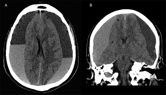
An 85-year-old man with a 2-year history of cognitive decline and falls, and signs of brain atrophy on previous computed tomography imaging, presented with severe drowsiness. Axial and coronal non-contrasted cerebral computed tomography images (Figure, A and B respectively) showed massive bi-hemispheric acute (hyperdense) and chronic (hypodense) subdural haematoma, with extensive parenchymal compression. Dementia is strongly associated with brain atrophy and frequent falling, both of which are risk factors for subdural bleeding1 caused by stretching and tearing of subdural veins2. The formation of a subdural haematoma can have further adverse effects in terms of cognitive decline. Surgical decompression of the subdural haematoma led to progressive improvement of the patient’s neurological status in the following weeks. The axial image resembles the black and white “yin and yang” symbol, which is conceptually relevant to the patient’s clinical history of subdural haematoma and dementia — two interdependent entities giving rise to each other.

Figure

A rare cause of intrathoracic mass

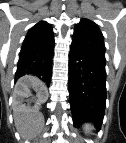
A 47-year-old woman presented complaining of cough and dyspnoea. Corrective surgery for scimitar syndrome had been performed 4 years earlier, with relocation of the draining scimitar vein to the left atrium via right thoracotomy. A computed tomography scan of the chest revealed right middle lobe pneumonia and a cephalad malposition of the right kidney in a diaphragmatic hernia, giving an intrathoracic appearance. The patient had normal renal function according to biochemistry results. Malpositioning of the kidneys and renal ectopia are rare causes of intrathoracic mass and should be considered in the differential diagnosis.

Pitfalls in photographing radiological images from computer screens

Using mobile phones to acquire images in clinical practice enables rapid, collaborative decision making1 and is increasingly common. However, the practice is not completely foolproof, as a recent “near miss” at our institution demonstrates.

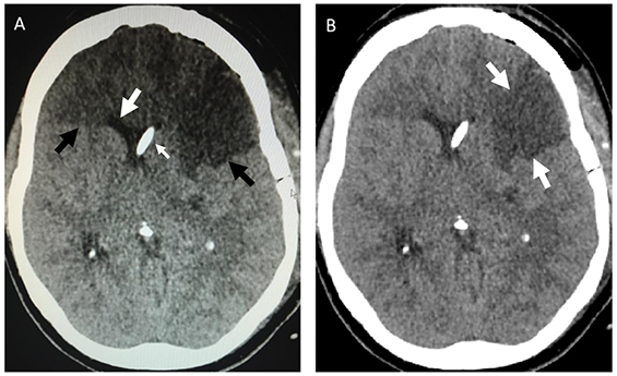
A 45-year-old woman presented with spontaneous subarachnoid haemorrhage secondary to a ruptured anterior communicating artery aneurysm. The anterior communicating artery aneurysm and an unruptured left middle cerebral artery aneurysm were clipped via craniotomy and a ventricular drain was inserted. Serial post-operative computed tomography (CT) brain scans showed an evolving infarction in the left middle cerebral artery territory, presumed to be secondary to temporary clipping at surgery, which became fully established after 28 hours. All cerebral vessels were patent, visualised on a post-operative CT angiogram. Elevated intracranial pressure (> 40 mmHg) and neurological fluctuation prompted a repeat CT scan, a photograph of which was taken from a computer screen using a mobile phone (Box, A). This image was sent by the intensive care unit consultant to the mobile phone of the on-call neurosurgeon, who noted apparent extensive bifrontal infarction. The patient was urgently transported to the operating room for decompressive craniectomy; however, on reviewing the scans at a radiology workstation before surgery (Box, B), the neurosurgeon noted the discrepancy and the procedure was cancelled. The patient recovered well and was neurologically intact and independent 6 months after discharge.

Although others report success using mobile phones to photograph CT brain scans displayed on computer monitors,2 our case highlights the need for doctors to appreciate the limitations of display technology. For example, many computer monitors exhibit viewing angle-dependent reductions in luminance and contrast ratio,3 which render images susceptible to artefact, particularly when viewed at close range.4 Moreover, mobile phone screens do not meet the technical requirements of a medical imaging display device.5

We confirmed that viewing angle-dependent reductions in luminance were responsible for the spurious frontal lobe darkening evident in the mobile phone image. Clearly, spatial variations in image brightness can dramatically affect image interpretation, with potentially disastrous results.

Guidelines on mobile device photography in the health care setting address privacy concerns but not technical aspects.1 Therefore, we offer some suggestions on preventing similar cases from occurring:

  • Use original images wherever possible.

  • Compare the photo with the original before sending.

  • When photographing computer screens, position the camera perpendicularly to, and at arm’s length from the screen, enlarging the image with digital zoom as required.

  • Before making clinical decisions, review the original imaging, including confirming the correct patient details with an observer or peer.

  • After photographing, ensure that images are deleted from the phone and any online data storage accounts, and record in writing the image use in the case notes.

  • Teach undergraduates as well as practising clinicians the technical aspects of the use of mobile phone images.

We hope this case serves to remind doctors of the need for caution when reviewing photographs of digital images, and that our suggestions will be helpful in preventing similar situations from occurring.

Box –
Mobile phone photograph of the cranial computed tomography (CT) scan compared with the original image


A: Photograph of axial CT image sent via a mobile phone, showing apparent hypodense frontal lobes suggestive of infarction (black arrows). Note however the anterior horns of the lateral ventricles are not distorted or effaced, as would be expected with such an extensive infarct (large white arrow). A ventricular catheter is located in the right lateral ventricle (small white arrow). B: The original axial CT image showing only a wedge-shaped, left middle cerebral artery infarct (white arrows).

Vertebroplasty is not a do-not-do treatment

Vertebroplasty has been controversial but remains clinically useful and new evidence awaits publication

Duckett and colleagues have classified vertebroplasty as a do-not-do treatment.1 They referenced two randomised controlled trials (RCTs)2,3 as definitive proof of this. However, the authors failed to heed our clinical opinion published in the MJA that these two trials were “not relevant to the patient group that we treat with vertebroplasty”.4 We have the largest clinical vertebroplasty experience in Australia, yet our published advice was apparently ignored. In the article by Duckett and colleagues, Box 1 illustrated the selection process that the authors used to determine do-not-do procedures. The process supposedly excluded evidence which was “contested” or “which was not supported by consulted clinical experts”. Accordingly, vertebroplasty should have been deleted from the list.

The authors used the United Kingdom National Institute for Health and Care Excellence (NICE) for clinical guidance. Current NICE guidance5 states that “vertebroplasty and kyphoplasty can be considered appropriate interventions for people with recent, unhealed osteoporotic vertebral compression fractures in whom the pain is severe and ongoing despite optimal pain management”.

From 1208 potential treatments, the authors excluded 1200, leaving five apparently incontrovertible do-not-do treatments. The fact that at least one of the five is wrongly included (by the authors’ own criteria) demonstrates the failure of the proposed model and the danger of adopting this kind of formula to influence clinical practice in hospitals.

The evidence for and against vertebroplasty is inconclusive. There is disparity in measured outcomes between blinded RCTs2,3 of vertebroplasty for fractures up to 12 months old and a larger, open-label RCT6 of fractures less than 6 weeks in duration. The blinded trials found no significant benefit of vertebroplasty over placebo, whereas the open-label RCT found significant benefit of vertebroplasty over conservative care. This disparity is well described in the NICE guidance.5

For the past 10 years, my vertebroplasty practice has been confined to treating fractures less than 6 weeks old.7 It is clear to me that the published blinded trials tested a different approach and are not relevant to the patient group that my practice treats with vertebroplasty for two principal reasons: the fractures were mostly non-acute; and the volume of cement used in these trials (2.6 cm3 on average in both trials) would have been insufficient to stabilise an acutely collapsing vertebral fracture.

Attempting to answer the acute fracture conundrum, the authors of the blinded RCTs published a meta-analysis of 52 patients from both trials with fractures less than 6 weeks duration.8 Only outcomes at 2 weeks and 1 month were presented and the evidence is hardly definitive.

The onus was placed on vertebroplasty practitioners to provide high-quality blinded data in this group of patients. For this purpose, my co-investigators and I embarked on the Vertebroplasty for Acute Painful Osteoporotic fractURes (VAPOUR) trial.9 Five years ago, we reconfigured the protocol from the INvestigational Vertebroplasty Efficacy and Safety Trial (INVEST),2,10 the larger of the two blinded vertebroplasty trials. We excluded crossover, which was permitted at 1 month in INVEST. We changed the selection criteria to include only fractures less than 6 weeks duration (average fracture age in the VAPOUR trial was 2.6 weeks compared with 18 weeks in INVEST) with pain scores greater than 7/10 and with either magnetic resonance imaging or single-photon emission computed tomography evidence of acute fracture. In-patients, already hospitalised with acute fractures, comprised 59% of the VAPOUR trial enrolment but were excluded in INVEST. The procedural technique was different in the VAPOUR trial, where we attempted maximum fill of the vertebral body to stabilise the fracture and prevent ongoing collapse. The average cement volume of 7.5 cm3 in the VAPOUR trial was three times that in INVEST. The method of blinding and data collection was similar for the two trials.

Our trial team included four Sydney centres with established vertebroplasty programs. The VAPOUR trial completed enrolment of 120 patients in December 2014 and is the largest RCT and the only acute fracture RCT of vertebroplasty in Australia. Statistical assessments of outcomes are nearing completion and the results of the trial will soon be published.

Change treatment for small tumours: expert

A leading UK cancer expert says Australian patients would benefit from a change in the way small tumours are treated.

Professor Andreas Adam from King’s College London is in Australia for the Clinical Oncology Society of Australia’s Annual Scientific Meeting in Hobart this week.

He says treatment of small tumours using minimally invasive techniques with medical imaging helps avoid invasive surgery and longer hospital stays.

Interventional oncology is particularly important in the treatment of kidney cancers.

“Typically, smaller tumours in the kidney have been monitored by urologists rather than operated on. The risks of surgery for these patients have been considered not to be justified, because they rarely give rise to secondary tumours,” he says.

“However, there is increasing evidence from large studies that small kidney tumours give rise to secondaries more often than was previously thought. This means we need to be more proactive in assessing and treating this type of cancer.

Related: Skin rash, a kidney mass and a family mystery dating back to World War II

“Interventional radiologists can destroy these tumours using techniques that have lower risks than surgery, thus justifying treatment at an earlier stage rather than waiting for them to grow.”

These learnings could be applied in Australia, Clinical Oncology Society of Australia President, Professor Mei Krishnasamy says.

“Interventional radiation oncology is an emerging field in Australia and the shortage of practitioners could be having a negative impact on our cancer patients,” Professor Krishnasamy says.

“Sharing knowledge about the benefits of this innovative form of treatment is crucial to ensuring that interventional oncology and radiology gets the recognition it deserves.”

The theme of this year’s COSA ASM is Rare cancers – Common goals’.

Latest news:

The dangers of diagnosing cystic neck masses as benign in the era of HPV-associated oropharyngeal cancer

Clinical record

A 59-year-old woman presented to her general practitioner with a lump in the left neck. She was a non-smoker, non-daily drinker and had no significant past medical history. Fine needle aspiration biopsy (FNAB) was performed and led to the diagnosis of a branchial cleft cyst. The mass collapsed after aspiration and the patient was managed conservatively by observation. In the year after diagnosis, re-emergence of the mass was noted by the GP at follow-up. Further investigations were declined on the patient’s presumption that the lesion was a benign branchial cleft cyst.

Two years after her initial diagnosis, the patient re-presented with a 1- to 2-month history of a sore throat and further increase in the size of the neck mass. On examination, a 7 cm mass was palpable, there was no overlying skin invasion and the mass was mobile to deep structures. Repeat FNAB of the neck mass was performed and revealed squamous cell carcinoma (SCC) with p16 positivity.

A staging computed tomography (CT) scan detected a lobulated tumour centred on the left tonsil invading the vallecula, base of tongue and parapharyngeal space. Further, a 27 mm cystic mass consistent with her initial lump was identified in the left level II group of lymph nodes, with additional necrotic nodes present at levels III and IV. A subsequent positron emission tomography scan confirmed increased uptake in these regions.

The patient was diagnosed with a T3 N2b M0 oropharyngeal SCC and was referred for radical chemoradiation. Expression of the surrogate marker p16 on cytological testing was highly suggestive that this was a human papilloma virus (HPV)-positive oropharyngeal SCC.

This case illustrates the diagnostic challenges faced in differentiating cystic nodal metastases from branchial cleft cysts, in the context of the increasing prevalence of HPV-related oropharyngeal SCC in Australia over the past two decades.1

HPV-related oropharyngeal SCC differs from traditional head and neck cancer in both its aetiology and clinical features; and because it is a relatively new clinical condition, GPs, radiologists and pathologists often do not recognise it as a potential diagnosis. Unlike traditional head and neck cancer, it occurs in a younger population who are frequently non-smokers and not heavy drinkers. Clinically, it most frequently presents with a neck mass, and often primary lesions are not easily discernible, as they are commonly small and in clinically difficult areas to examine, such as the base of the tongue or tonsil.

The natural disease pattern of oropharyngeal SCC is to metastasise to the cervical lymph nodes. In the setting of HPV-related cancer, these nodal metastases are frequently cystic in morphology and, as previously indicated, are frequently the first mode of presentation.2 Given the differing clinical features of HPV-related oropharyngeal cancer compared with non-HPV-associated oropharyngeal cancer, it is not uncommon for these nodal metastases to be misdiagnosed as branchial cleft cysts, as described in this case report. The proportion of metastatic SCCs in cysts initially presumed to be of branchial cleft origin has been reported to range from 11% to 21%.3,4

Misdiagnosis can negatively affect patient prognosis as it delays treatment, allows for further disease progression and increases the potential for metastatic spread. Alternatively, proceeding to excisional biopsy of a cystic mass suspected to be a branchial cleft cyst without adequate investigation for an occult primary can lead to tumour spillage into the surrounding tissues.

Differences in the demographics between branchial cleft cysts and cystic nodal metastases may aid clinicians in accurate diagnosis. While branchial cleft cysts may occur at any age, they most commonly present in early adulthood in the second and third decade of life.5 The reported mean age for cervical cystic masses histologically confirmed as branchial cleft cysts ranges from 32 to 37 years.6,7 In contrast, cystic nodal metastases present later, with a reported mean age ranging from 53 to 57.8 years.6,7 Particular attention must be paid to cervical cysts in patients over 40 years of age, as 44% of cystic masses in this patient population are reported to be malignant in origin.8 The most commonly represented lesions associated with cystic metastases are HPV-related head and neck cancer and thyroid papillary carcinoma.9

CT is a mainstay in the diagnosis and staging of head and neck cancer and plays an important role in differentiating benign lesions of the neck from malignant cystic lymphadenopathy. On contrast-enhanced CT, there is homogeneous attenuation throughout the substance of branchial cleft cysts.6 Features suggestive of malignancy include the presence of septations, heterogeneous attenuation and extracapsular spread.6,7 Significant overlap between the radiological features of benign and malignant cysts is, however, present. Repeated local infection involving a branchial cleft cyst may confer a radiological appearance similar to that of nodal metastasis of an SCC.7 In one study, 31% of cystic nodal metastases were reported as benign in appearance, while 38% of branchial cleft cysts had aggressive features mimicking nodal metastases.6 Evidently, isolated use of CT in evaluating a cystic neck mass confers a high degree of misdiagnosis.

FNAB cytology is the current standard of care in diagnostic workup of solid masses of the neck and is reported to have an overall sensitivity of 92% and a positive predictive value of 100% for head and neck carcinomas.10 Unfortunately, this has not been the common experience with cystic lesions, for which its role remains controversial. Reported sensitivities range from 33% to 55%,11 with a false-negative rate of up to 50%.2 The aspirate of cystic metastatic nodes may be difficult to interpret as a result of hypocellularity from the dilutional effect of the cyst fluid.2 There is commonly an associated inflammatory reaction within the cystic nodes, resulting in large quantities of degenerating epithelial cells, inflammatory cells and cellular debris within the aspirate. These cytological features overlap considerably with those of branchial cleft cysts12 and can lead to misdiagnosis.

Despite these limitations, FNAB still retains some relevance in the diagnosis of cystic lesions. Further, with the emergence of molecular analysis techniques, the detection of HPV DNA and thyroglobulin within fine needle aspirates may facilitate the pathological diagnosis of malignant cystic lymphadenopathy and detection of occult primary tumours. The presence of HPV DNA or thyroglobulin in aspirates is strongly correlated with HPV-related oropharyngeal SCC and thyroid cancer, respectively.13,14 Although these tests are primarily used for research purposes at present, their utility may expand to the clinical setting in future to help to differentiate benign and malignant cystic neck lesions.

Differentiating cystic nodal metastases from branchial cleft cysts is an important, albeit sometimes difficult, diagnostic challenge. With the growing prevalence of HPV-related oropharyngeal SCC in Australia, we conclude that metastatic lymphadenopathy should be considered as the primary provisional diagnosis in the adult population with cystic neck masses until proven otherwise. We encourage caution in the interpretation of neck masses as benign by isolated use of either CT or FNAB. Clinicians should use these modalities in conjunction with each other and, if necessary, include referral for an ear, nose and throat specialist opinion to increase diagnostic accuracy.

Lessons from practice

  • HPV-related oropharyngeal cancer is an increasingly common and relatively new entity that differs from traditional head and neck cancer in its aetiology, epidemiology, clinical features and prognosis.

  • Lateral cystic neck masses in adults are often misdiagnosed as branchial cleft cysts and should be considered as metastatic lymphadenopathy until proven otherwise.

  • Isolated use of computed tomography or fine needle aspiration biopsy in evaluating a neck mass can lead to misdiagnosis. Instead, these modalities should be used in conjunction with each other and, if necessary, include referral for an ear, nose and throat specialist opinion to increase diagnostic accuracy.

  • The emergence of molecular analysis techniques for detecting HPV DNA and thyroglobulin in fine needle aspirates may assist clinicians in the diagnosis of occult primary tumours with cystic nodal metastasis.

Abortion laws need updating in line with fetal medicine advancements

Women whose babies are diagnosed with a fetal abnormality are being disadvantaged by Australia’s patchwork abortion laws, experts say.

Professor Caroline de Costa, from James Cook University, and Professor Heather Douglas, from the University of Queensland write about the barriers these women face in today’s Medical Journal of Australia.

“Medicare-funded diagnosis of fetal abnormality is now routinely offered to all pregnant Australian women — with the implication that a woman may choose to terminate the pregnancy if a serious abnormality is detected”, they write.

However they say that abortion laws haven’t kept pace with these developments.

Abortion is decriminalised in ACT, Victoria and Tasmania with various timeframe restrictions.

“Fetal abnormality is specifically discussed in the legislation in Western Australia, South Australia, Tasmania and the Northern Territory, and covered by the decriminalisation of abortion in Victoria and the ACT; in practice, however, late abortion is restricted by health regulations in WA, SA and the NT”, de Costa and Douglas write.

Related: MJA – Termination of pregnancy: a long way to go in the Northern Territory

“In Queensland and New South Wales, the law does not refer to fetal abnormality at all.”

They are concerned these difference in laws could lead to extensive abortion ‘tourism’ to states that are decriminalised.

“In 2015, there is an urgent need for legislative uniformity across Australia so that the law is in step with modern medical practice, and so that women, regardless of where they live, have equal access to abortion services”.

To read the full article, visit the Medical Journal of Australia.

Latest news:

Roller coasters and cervical artery dissection

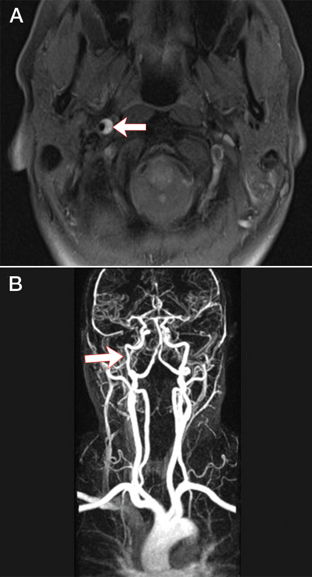
A 42-year-old woman with no vascular risk factors was admitted with neck pain and right Horner syndrome after riding a roller coaster. Magnetic resonance imaging (MRI) of her neck showed a characteristic crescent-shaped intramural haematoma in the right internal carotid artery1 (Figure, A), confirmed by magnetic resonance angiogram (MRA) (Figure, B). T1-fat-suppressed MRI and arterial wall imaging are currently the most sensitive techniques for diagnosis of dissection. Riding roller coasters has been associated with shearing neck injury leading to cervical artery dissection.2 Our patient was treated with warfarin for 6 months, leading to the intramural haematoma resolving and the Horner syndrome greatly improving.

Figure


A. T1-fat-suppressed MRI of neck. Arrow: intramural haematoma in right internal carotid artery. B. MRA of neck. Arrow: narrowing of C1 segment of right internal carotid artery.

Forcing GPs to adopt half-baked e-health record a dud idea: AMA

The Federal Government has hit a major snag in its overhaul of the troubled e-health record system after the AMA rejected plans to link GP incentive payments to the adoption of the scheme.

The Government has proposed that Practice Incentive Program e-health payments be tied to doctor use of the MyHealth Record (MyHR) system being developed to replace the $1 billion Personally Controlled Electronic Health Record scheme. The PCEHR has been dumped amid dismal take-up rates among patients, doctors and medical practices.

But AMA President Professor Brian Owler said the MyHR system was far from fully developed, so using PIP incentives to get doctors to sign up was ill-considered and premature.

“The MyHealth Record is not at a stage where it can be adopted by practices, so it should not be linked to the PIP scheme,” Professor Owler said. “There are fundamental issues with the design of the MyHR that are yet to be fully addressed.”

The AMA has detailed a long list of problems with the current version of the system in a submission to the Health Department, including:

  • the ability of patients to remove information from view, making the record potentially incomplete and of no clinical value;
  • no flags to indicate if information has been removed from view;
  • radiology or pathology results are not yet included;
  • the shared health summaries are not automatically updated, rendering them quickly out-of-date; and
  • inaccuracies occur in the upload of data.

In addition MyHR, in its current iteration, remains an ‘opt-in’ system.

The reliance on patients to sign up for an e-health record was seen as a fatal weakness of the PCEHR, and a three-person review of the system recommended that MyHR be an opt-out scheme.

But Health Minister Sussan Ley has indicated that the opt-out approach will first be trialled next year before being adopted.

“It’s important that all Australians are signed up to ensure we have a functioning system, and trialling an opt-out model means we can do it carefully, methodically and ensure the appropriate protections are in place to give patients peace of mind,” Ms Ley said.

“If automatic registration for a digital health record in the opt-out trials leads to higher participation in the My Health Record system, the Government will consider adopting opt-out on a national scale.”

Professor Owler said this lengthy catalogue of unresolved problems with MyHR meant it was unfair to expect GPs to adopt it.

“Until the problems with the MyHR have been rectified, so that it is easy to use and offers real clinical benefits for patients, it is unreasonable to expect GPs to actively use it,” the AMA President said.“The AMA has been a strong advocate for a well-designed and governed e-health record which can deliver real benefits for patients, but the current MyHR model has well-known flaws that must be fixed.”

 The AMA has recommended the Government focus on rectifying problems with MyHR rather than trying to force GPs to use a system that is cumbersome and incomplete.

Even when the system is complete and fit for use, the AMA has argued that, instead of using the existing e-PIP incentive, the Government instead create a Medicare Benefits Schedule item and a Service Incentive Payment scheme to promote its use.

To help establish MyHR, Ms Ley has announced the appointment of former National Mental Health Commission Chief Executive Robyn Kruk to head an 11-member eHealth Implementation Taskforce Steering Committee.

The Committee, which includes Dr Hambleton, will design, implement and oversee the establishment of the Australian Commission for eHealth.

For its part, the Commission will oversee the operation and development of e-health systems, including operating the My Health Record System.

Revised eligibility requirements for the e-Health Incentive are due to be announced in November 2015, and to commence from 1 February 2016.

The AMA submission can be viewed at: at submission/ama-submission-proposed-changes-pip-ehealth-incentive

Adrian Rollins