×

Prevalence of microcephaly in an Australian population-based birth defects register, 1980–2015

The known Microcephaly has recently received increased attention because it is associated with congenital Zika virus infection. The World Health Organization recommends that countries at risk of Zika virus transmission collate baseline data on the historical prevalence of microcephaly. 

The new We report the prevalence and characteristics of microcephaly in a geographically defined Australian population over a 35-year period. Significant rate differences between Aboriginal and non-Aboriginal births were identified. 

The implications Our study provides a useful baseline that will allow changes in microcephaly prevalence to be measured, and raises important matters for consideration by other jurisdictions reviewing their birth defects surveillance systems. 

Microcephaly is a birth defect in which a child’s head is smaller than expected for their age, sex and ethnicity; there are, however, varying operational definitions of how much smaller (eg, head circumference below the first or third percentile, or more than two or three standard deviations below the mean value). The lack of a universally accepted definition complicates the comparison of prevalence estimates, which range from 2 to 12 per 10 000 live births in the United States,1 0.5 to 10 per 10 000 total births in EUROCAT registries (source: http://www.eurocat-network.eu/accessprevalencedata/prevalencetables), and 2 to 6 per 10 000 total births in Australia.24

Known causes of microcephaly include genetic conditions, metabolic diseases, teratogens (eg, alcohol), severe malnutrition (eg, extreme placental insufficiency), and transplacental infections.5 For many children the cause is unknown.

Microcephaly has recently gained increased attention because of its association with congenital Zika virus infection.6 It is predicted that more than 2.2 billion people live in areas where there is a risk of Zika virus infection.7 Local transmission has recently been reported in Singapore8 and in other common travel destinations for Australians.9 The most common vector, the mosquito Aedes aegypti, is found in northern and central Queensland; although local transmission of Zika virus has not been reported, returning travellers with Zika viraemia could infect local mosquitoes and thereby cause a local outbreak. A. aegypti was once present in other Australian states and territories, and there is concern that the mosquito could become re-established outside Queensland.10

Because of the many travellers returning from Zika-affected countries and the potential for Zika virus transmission to become established in Australia, we report baseline data on the prevalence and characteristics of microcephaly in a geographically defined Australian population over a 35-year period (1980–2015).

Methods

Cases of microcephaly were identified in the Western Australian Register of Developmental Anomalies (WARDA), a population-based statutory register with high ascertainment of developmental anomalies (cerebral palsy and birth defects).3,11 Birth defects are defined as structural or functional abnormalities present from conception or before the end of pregnancy and ascertained after a live birth (diagnosed by 6 years of age), stillbirth (minimum 20 weeks’ gestation), or a pregnancy terminated because of detected fetal anomalies (regardless of gestational age). Each defect (up to ten per case) is coded according to the British Paediatric Association extension to the International Classification of Diseases, ninth revision (BPA–ICD9).12

WARDA defines microcephaly as an occipito-frontal head circumference below the third percentile or more than two standard deviations (SDs) below the mean sex- and age-appropriate distribution curve. Any known primary cause (eg, infection, clinical syndrome) is recorded, but the head circumference measurements are not recorded. All identified cases fulfilled the third percentile criterion; either the head circumference measurement was validated by WARDA staff, or the notifier (specialist, genetics services, neonatal units) confirmed applying this criterion. The definition and registration criteria have not changed since 1980.

We identified all cases of microcephaly (BPA–ICD9, 742.10) recorded during January 1980 – December 2015. Each case was reviewed and classified according to aetiology:

  • cause unknown;

  • microcephaly associated with a chromosomal defect;

  • monogenic disorder known to affect head size;

  • known environmental cause (eg, fetal alcohol spectrum disorder [FASD]);

  • congenital infection; or

  • other cause.

We also grouped cases according to whether microcephaly was the only defect (“isolated”), or further major birth defects were reported (“associated”). We examined the age at diagnosis and notification sources.

Microcephaly prevalence was defined as the total number of cases (in live births, stillbirths and terminations of pregnancy for fetal anomaly) divided by the number all births in WA, expressed as number per 10 000 births. Tabulated denominator data for all WA live and stillbirths of at least 20 weeks’ gestation during the study period were obtained from the statutory WA Midwives Notification System. The average annual change in prevalence was calculated by Poisson regression with an offset term. Birth prevalence rates were analysed according to sex, mother’s Aboriginal status, region of residence, known v unknown cause, and isolated v associated microcephaly. We examined associated defects by organ system in cases with known and unknown causes. The prevalence of microcephaly of known and unknown cause in Aboriginal births (5.7% of births during the study period) was compared with that in non-Aboriginal births as prevalence ratios (PRs) with 95% confidence intervals (CIs). Analyses were performed in SPSS 23 (IBM) and EpiBasic (Aarhus University, Denmark).

Ethics approval

Ethics approval was obtained from the Human Research Ethics Committee of the WA Health Department (reference, 2016/19).

Results

Among 963 126 births during 1980–2015, 478 cases of microcephaly were ascertained, a prevalence of 5.0 per 10 000 births (95% CI, 4.53–5.43). For births during 1980–2009 (ie, with at least 6 years’ follow-up and therefore with complete case ascertainment), 416 cases were identified, a prevalence of 5.5 per 10 000 births (95% CI, 4.95–6.02), or 1 in 1830 births.

Of the cases from 1980–2009, 389 (93.5%) involved live births; there were seven stillbirths (1.7%) and 20 terminations of pregnancy for fetal anomaly (4.8%). A cause of microcephaly was identified in 186 cases (45%), and more frequently for Aboriginal (64 cases, 70%) than non-Aboriginal births (122, 38%). The most frequent known cause in Aboriginal births was FASD (11 per 10 000 births; 95% CI, 8.2–14.6); in non-Aboriginal births the most frequent causes were monogenic disorders (0.68 per 10 000 births; 95% CI, 0.51–0.90) and chromosomal defects (0.59 per 10 000 births; 95% CI, 0.42–0.79) (Box 1).

The prevalence of microcephaly of unknown cause was higher among Aboriginal than non-Aboriginal births (6.1 v 2.8 per 10 000 births; PR, 2.2; 95% CI, 1.38–3.22); this was also true for each type of known cause, but was statistically significant only for known environmental (PR, 200; 95% CI, 73–756) and infectious causes (PR, 4.2; 95% CI, 1.40–10.6) (Box 1).

There was no significant temporal trend in microcephaly prevalence during 1980–2009 (Wald χ2 test, P = 0.23; Box 2). The prevalence of microcephaly of unknown cause declined from 3.4 per 10 000 births (95% CI, 2.7–4.2) during 1980–1989 to 2.6 per 10 000 births (95% CI, 2.0–3.2) during 2000–2009, a mean decline of 1.3% per year (95% CI, –2.4% to –0.2%; P = 0.02); that of microcephaly with a known cause increased by a mean 0.4% per year (95% CI, –1.3% to 2.0%; P = 0.67). The average annual increases in the prevalence of microcephaly associated with chromosomal defects (0.6%; 95% CI, –2.1% to 3.4%; P = 0.64), monogenic disorders (0.6%; 95% CI, –2.2% to 3.4%; P = 0.67), and environmental causes (2.6%; 95% CI, –0.3% to 5.4%; P = 0.08) were statistically non-significant. We found a significant 5.6% average annual decline in the prevalence of microcephaly associated with congenital infection (95% CI, –10.0% to –1.1%; P = 0.01); the change was evident for both Aboriginal (4.4% decline; 95% CI, –12.9% to 4.2%; P = 0.32) and non-Aboriginal births (6.0% decline; 95% CI, –10.6% to –1.5%; P = 0.008), but was statistically significant only for non-Aboriginal births.

The prevalence of microcephaly was similar for male and female children, but higher for Aboriginal than non-Aboriginal Australians (PR, 4.5; 95% CI, 3.55–5.73; Box 1). Prevalence was much higher in remote regions, particularly that of microcephaly of known cause (Box 3); this was influenced by the higher proportion of Aboriginal births (and FASD) in these areas (data not shown).

The 81 cases of isolated microcephaly (20% of all cases) were all of unknown cause. All 186 cases with a known cause were associated with other major birth defects, compared with 65% of the 230 cases without a known cause (Box 4).

The overall prevalence of isolated microcephaly declined by a mean 3.4% per year (95% CI, –5.3% to –1.4%; P = 0.001); there was no significant change in the prevalence of microcephaly with associated defects (P = 0.86) (Box 2). Temporal changes in the availability of folate supplements and the introduction of folic acid fortification of some foods were not associated with changes in the prevalence of microcephaly (data not shown).

Microcephaly was diagnosed after the age of 12 months in 27% of cases, increasing from 19% during the 1980s to 29% in the 1990s and 31% during 2000–2009 (Box 5). Isolated cases were more likely to be diagnosed later (35% after 12 months of age), as were cases with a known cause (30% after 12 months of age). For most cases with a known cause diagnosed after 12 months of age, the cause was FASD (52%).

The major notification sources were geneticists, paediatricians, and obstetric and paediatric hospitals. While paediatric and obstetric hospitals maintained steady notification rates, genetics services played an increasingly important role; they contributed more than one-quarter of reported cases during 2000–2009 (data not shown). Most cases were notified by more than one source (mean, 2.8 notifications per case [SD, 1.76]; median, 2 notifications per case [interquartile range, 1–4]).

Discussion

Our study, the first descriptive epidemiological investigation of microcephaly in Australia, provides baseline data on the prevalence of microcephaly in Western Australia over a 35-year period. Most cases of microcephaly were in live-born infants (93.5%), and the overall prevalence was 5.5 per 10 000 births, with annual rates ranging between 2.9 and 7.7 per 10 000 births.

There are few published epidemiological studies of microcephaly, and comparisons are complicated by differences in case definition, period of ascertainment, pregnancy outcomes included, underlying population differences, and environmental variables. These difficulties were illustrated by a recent report of important variations in the prevalence of microcephaly recorded by 16 European birth defects registries.13 In Australia, national estimates are difficult because of major differences between states in the scope of birth defects data collections.14 The most recent national data (for births during 2002–2003),2 which did not include data from the Northern Territory, found a lower prevalence of microcephaly (1.9 per 10 000 births) than we did. Our results, however, are similar to those reported by other registers using the same case definition and equivalent follow-up periods, including the Atlanta Congenital Defects Program1 and the South Australian Register4 (5.0–5.2 per 10 000 births).

Our prevalence estimates, and those reported by birth defects registries in other countries, are well below the rate of 230 cases per 10 000 births expected when a case definition of an occipito-frontal head circumference more than two SDs below the mean for age and sex is applied. This implies that specialists are moderating their notifications according to criteria other than head circumference; these may include notifying cases when they have clinical concern about the child, or when there is an association with a particular cause (eg, trisomy 18). This moderation probably operates in most surveillance systems, including our own. Although head circumference or percentile measures were checked before registering a case of microcephaly in the database, the measurements themselves are not recorded by WARDA. Adding these data to the collection is planned, facilitating assessment of case severity and comparisons with registries using different registration criteria.

We identified a cause for microcephaly in 45% of cases, and more frequently for births to Aboriginal (70%) than to non-Aboriginal mothers (38%). The figure for non-Aboriginal births was similar to those reported from Atlanta (40%)15 and by a German clinic-based study in which the most common cause was genetic in nature (29% of 680 cases;16 similar to the 28% of non-Aboriginal births in our study). We found an average annual decline in microcephaly cases of unknown cause of 1.3%; this may be attributable to improved genetic diagnostic technologies and increasing diagnosis of FASD, factors that may also account for the declining number of cases of isolated microcephaly.

Our results highlight the contribution of FASD to microcephaly in Aboriginal children, with a prevalence of FASD-associated microcephaly (11 per 10 000 births) 260 times that for non-Aboriginal births (0.04 per 10 000 births). FASD also contributed to higher prevalence rates in remote areas of WA. A recent study found that the prevalence of fetal alcohol syndrome in remote WA Aboriginal communities among 7–8-year-old children was 12%, and that 69% of these children had microcephaly;17 another study reported a twofold increase in FASD notifications to WARDA for both Aboriginal and non-Aboriginal births during 1980–2010, coinciding with increased state and national awareness of FASD.18 Nevertheless, FASD is still underdiagnosed, and this may partly explain the higher prevalence of microcephaly of unknown cause in Aboriginal children (6.1 v 2.8 per 10 000 non-Aboriginal births), and may also account for some cases of unknown cause in non-Aboriginal infants. The higher prevalence of microcephaly of unknown cause among Aboriginal Australians may also be related to the relative lack of access to or engagement with health care services and the paucity of genomic reference data for Aboriginal Australians.19 The release of the Australian guide for diagnosing FASD20 and the Partnership Grants (“Models and quality of genetic health services for Aboriginal and Torres Strait Islander people”) co-funded by the National Health and Medical Research Council and the Lowitja Institute may advance aetiological investigations. In addition, a major research program for investigating the diagnosis, prevention and management of FASD is underway in WA.21

In 27% of cases, microcephaly was diagnosed in children more than 12 months old. This large proportion would be missed were ascertainment periods shorter. This factor may be particularly important when differentiating cases of known and unknown cause, especially when microcephaly is associated with conditions (such as FASD and specific genetic anomalies) that are often diagnosed at a later age.18

Further major birth defects were associated with 80% of cases of microcephaly. The most frequent in cases of unknown cause were those affecting the nervous system (61% of associated defects), including neural tube defects (13%). The US National Birth Defects Prevention Network suggests excluding microcephaly associated with neural tube defects, holoprosencephaly, craniosynostosis, and conjoined twins.5 We favour broader inclusion criteria, especially in light of the evolving understanding of the full spectrum of defects that may be associated with congenital Zika infection. One in five definite or probable cases of congenital Zika syndrome in a large case series from Brazil were associated with brain abnormalities in infants without microcephaly, suggesting that surveillance should not focus on microcephaly alone.22 Other defects associated with Zika infection include intracranial calcifications, craniofacial malformations, severe arthrogryposis, and eye defects.23 WARDA includes each of these in its data collection.

Zika virus is the first mosquito-borne virus associated with human birth defects, and is also sexually transmissible. A recent editorial in the MJA24 highlighted the robust Australian systems that enable rapid responses to newly identified communicable diseases, but our capacity for monitoring birth defects was not discussed. Medical practitioners have been advised to ask pregnant women about their recent travel history and to offer testing to those who have visited Zika-affected areas. Birth defect registries that record the causes of birth defects are therefore likely to capture cases of microcephaly or other defects associated with Zika virus exposure in returned travellers. Our capacity to detect changes in the prevalence of microcephaly caused by local transmission of the virus is less clear, as infection is frequently asymptomatic, and the collection of data on birth defects in this country is not standardised, with wide variability between states in case ascertainment.14

As remote regions in Northern Australia either already harbour or are at risk of harbouring A. aegypti mosquitoes, enhanced monitoring of birth defects in these areas (and also of FASD) is important. The Australian Paediatric Surveillance Unit project for the surveillance of microcephaly in infants under 12 months of age is important for responding to the challenges of geographically equitable case ascertainment.25

Attempts to standardise state and territory birth defects data collections have been unsuccessful.26 In the absence of national data, our study provides a useful baseline for measuring changes in microcephaly prevalence, and highlights important matters for consideration by other jurisdictions reviewing their birth defects surveillance systems.

Box 1 –
Prevalence of microcephaly (per 10 000 births) by cause and Aboriginal status of the mother, Western Australia, 1980–2009

Cause

All cases


Aboriginal


Non-Aboriginal


Prevalence ratio (95% CI)

Number

Prevalence (95% CI)

Number

Prevalence (95% CI)

Number

Prevalence (95% CI)


Total number of cases

416

5.5(4.95–6.02)

91

21(16.5–25.2)

325

4.5(4.05–5.05)

4.5(3.55–5.73)

Unknown cause

230 (55%)

3.0(2.64–3.44)

27

6.1(4.01–8.86)

203

2.8(2.46–3.25)

2.2(1.38–3.22)

Known cause

186 (45%)

2.4(2.10–2.82)

64

14(11.11–18.43)

122

1.7(1.41–2.03)

8.5(6.17–11.6)

Chromosomal

47 (11%)

0.62(0.45–0.82)

5

1.1(0.37–2.63)

42

0.59(0.42–0.79)

1.9(0.59–4.86)

Monogenic

54 (13%)

0.71(0.53–0.93)

5

1.1(0.37–2.63)

49

0.68(0.51–0.90)

1.6(0.51–4.12)

Environmental

53 (13%)

0.70(0.52–0.91)

49*

11(8.2–14.6)

< 5

0.06(0.01–0.14)

200(73–756)

Fetal alcohol spectrum disorder*

52

0.68(0.51–0.90)

49*

11(8.2–14.6)

< 5

0.04(0.01–0.12)

260(85–1320)

Infectious

29 (7%)

0.38(0.26–0.55)

6*

1.4(0.50–2.94)

23

0.32(0.20–0.48)

4.2(1.40–10.6)

Cytomegalovirus*

24

0.32(0.20–0.47)

5*

1.1(0.37–2.63)

19

0.26(0.16–0.41)

4.2(1.24–11.8)

Other infections

5

0.07(0.02–0.15)

< 5

0.22(0.01–1.26)

< 5

0.06(0.01–0.14)

4.0(0.08–40.8)

Other cause

< 5

0.05(0.01–0.14)

0

< 5

0.06(0.01–0.14)


* One child was diagnosed with both cytomegalovirus infection and fetal alcohol spectrum disorder.

Box 2 –
Prevalence of microcephaly in Western Australia, 1980–2015: overall prevalence, and prevalence of cases with and without associated anomalies*


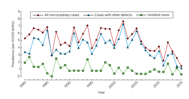
* From 2009, there is less than 6 years’ follow-up of births, so that ascertainment of cases will be incomplete.

Box 3 –
Prevalence (per 10 000 births) of microcephaly in Western Australia, 1980–2009

All cases


Microcephaly of known cause


Microcephaly of unknown Cause


Number

Prevalence (95% CI)

Number

Prevalence (95% CI)

Number

Prevalence (95% CI)


All

416

5.5 (4.95–6.02)

186

2.4 (2.10–2.82)

230

3.0 (2.64–3.44)

Sex

Male

199

5.1 (4.41–5.85)

85

2.2 (1.74–2.69)

114

2.9 (2.41–3.51)

Female

216

5.8 (5.08–6.66)

101

2.7 (2.22–3.31)

115

3.1 (2.56–3.72)

Missing data

1

1

Aboriginal status of mother

Aboriginal

91

21 (16.52–25.2)

64

14 (11.11–18.4)

27

6.1 (4.01–8.86)

Non-Aboriginal

325

4.5 (4.05–5.05)

122

1.7 (1.41–2.03)

203

2.8 (2.46–3.25)

Residential location*

Metropolitan

261

4.8 (4.24–5.42)

103

1.9 (1.55–2.30)

158

2.9 (2.47–3.40)

Rural

74

5.2 (4.12–6.50)

31

2.2 (1.49–3.12)

43

3.0 (2.21–4.10)

Remote

81

11 (8.56–13.4)

52

6.9 (5.17–9.07)

29

3.9 (2.59–5.54)


* According to the postcode of the mother as recorded by WARDA at the time of her child’s birth. The eight WA Department of Health residential location categories were grouped into three classes: metropolitan; rural (Great Southern, South-West, Wheatbelt, Midwest Murchison); and remote (Kimberley, Pilbara–Gascoyne, Goldfields–Southeast).

Box 4 –
Association of microcephaly (of known and unknown cause) with other congenital anomalies, Western Australia, 1980–2009

All cases

Microcephaly of known cause

Microcephaly of unknown Cause


Total number of cases

416

186

230

Isolated microcephaly

81 (20%)

0

81 (35%)

Microcephaly associated with other defects

335 (80%)

186 (100%)

149 (65%)

Associated defects by diagnostic category

335

186

149

Nervous system, apart from microcephaly

141 (34%)

50 (27%)

91 (61%)

Neural tube defects

25 (6%)

6 (3%)

19 (13%)

Chromosomal

47 (11%)

47 (25%)

0

Cardiovascular

49 (12%)

29 (16%)

20 (13%)

Respiratory

15 (4%)

10 (5%)

5 (3%)

Gastrointestinal

48 (12%)

20 (11%)

28 (19%)

Musculoskeletal

72 (17%)

35 (19%)

37 (25%)

Urogenital

68 (16%)

43 (23%)

25 (17%)

Eye

36 (9%)

24 (13%)

12 (8%)

Ear, face, neck

8 (2%)

3 (2%)

5 (3%)

Integument

4 (1%)

2 (1%)

2 (1%)

Other major diagnoses

158 (38%)

142 (76%)

16 (11%)

Monogenic condition

58 (14%)

54 (29%)

4 (3%)

Fetal alcohol spectrum disorder

52 (12%)

52 (28%)

0


Box 5 –
Number of cases of microcephaly in Western Australia, 1980–2015, by age at diagnosis

Age at diagnosis

Follow-up period


At least 6 years (416 cases)


Less than 6 years (62 cases)

1980–1989

1990–1999

2000–2009

2010–2015


Prenatal

16 (12%)

25 (17%)

18 (13%)

18 (29%)

Within first month of life

47 (36%)

49 (33%)

40 (29%)

16 (26%)

> 1 month to 1 year

33 (26%)

26 (18%)

36 (26%)

16 (26%)

> 1 year to 3 years

16 (12%)

24 (16%)

27 (19%)

7 (11%)

> 3 years to 6 years

9 (7%)

19 (13%)

17 (12%)

3 (5%)

Post mortem

8 (6%)

4 (3%)

2 (1%)

2 (3%)


The Australasian Society for Infectious Diseases and Refugee Health Network of Australia recommendations for health assessment for people from refugee-like backgrounds: an abridged outline

There are currently more than 65 million people who have been forcibly displaced worldwide, including 21.3 million people with formal refugee status, over half of whom are aged under 18 years.1 More than 15 000 refugees have resettled in Australia in the 2015–16 financial year, which includes a proportion of the 12 000 refugees from Syria and Iraq recently added to Australia’s humanitarian intake.2 In addition, around 30 000 asylum seekers who arrived by plane or boat are currently in Australia awaiting visa outcomes.3

People from refugee-like backgrounds are likely to have experienced disruption of basic services, poverty, food insecurity, poor living conditions and prolonged uncertainty; they may have experienced significant human rights violations, trauma or torture. These circumstances place them at increased risk of complex physical and mental health conditions. They face numerous barriers to accessing health care after arrival in Australia, such as language, financial stress, competing priorities in the settlement period, and difficulties understanding and navigating the health care system.46 Most people require the assistance of an interpreter for clinical consultations.7 Offering a full health assessment to newly arrived refugees and asylum seekers is a positive step towards healthy settlement, and helps manage health inequity through the provision of catch-up immunisation and the identification and management of infectious and other health conditions.

These guidelines update the Australasian Society of Infectious Diseases (ASID) guidelines for the diagnosis, management and prevention of infectious diseases in recently arrived refugees8 published in 2009 and previously summarised in the MJA.9 When these recommendations were first published, more than 60% of humanitarian entrants arriving in Australia were from sub-Saharan Africa10 and had a high prevalence of malaria, schistosomiasis and hepatitis B virus (HBV) infection.1115 The initial guidelines were primarily intended to help specialists and general practitioners to diagnose, manage and prevent infectious diseases. Since then, there have been changes in refugee-source countries — with more arrivals from the Middle East and Asia and fewer from sub-Saharan Africa16,17 — and an increased number of asylum seekers arriving by boat,18 alongside complex and changing asylum seeker policies and changes in health service provision for these populations. In this context, we reviewed the 2009 recommendations to ensure relevance for a broad range of health professionals and to include advice on equitable access to health care, regardless of Medicare or visa status. The revised guidelines are intended for health care providers caring for people from refugee-like backgrounds, including GPs, refugee health nurses, refugee health specialists, infectious diseases physicians and other medical specialists.

This article summarises the full guidelines, which contain detailed literature reviews, recommendations on diagnosis and management along with explanations, supporting evidence and links to other resources. The full version is available at http://www.asid.net.au/documents/item/1225.

Methods

The guideline development process is summarised in Box 1. The two key organisations developing these guidelines are ASID and the Refugee Health Network of Australia. ASID is Australia’s peak body representing infectious diseases physicians, medical microbiologists and other experts in the fields of the prevention, diagnosis and treatment of human and animal infections. The Refugee Health Network is a multidisciplinary network of health professionals across Australia with expertise in refugee health.20

We defined clinical questions using the PIPOH framework (population, intervention, professionals, outcomes and health care setting).21 The chapter authors and the Expert Advisory Group developed recommendations based on reviews of available evidence, using systematic reviews where possible. Australian prevalence data also informed screening recommendations; for example, the low reported prevalence of chlamydia (0.8–2.0%) infections and absence of gonorrhoea infections in refugee cohorts in Australia13,2224 (and in other developed countries2527) informed the new recommendation for risk-based sexually transmitted infection (STI) screening.

Despite the intention to assign levels of evidence to each recommendation, there was limited published high level evidence in most areas, and virtually all recommendations are based on expert consensus. Consensus was not reached regarding the recommendations relating to human immunodeficiency virus (HIV) and STIs.

The term “refugee-like” is used to describe people who are refugees under the United Nations Refugee Convention,28 those who hold a humanitarian visa, people from refugee-like backgrounds who have entered under other migration streams, and people seeking asylum in Australia. “Refugee-like” acknowledges that people may have had refugee experience in their countries of origin or transit, but do not have formal refugee status.

Current pre-departure screening

All permanent migrants to Australia have a pre-migration immigration medical examination 3–12 months before departure,29 which includes a full medical history and examination. Investigations depend on age, risk factors and visa type,30 and include:

  • a chest x-ray for current or previous tuberculosis ([TB]; age ≥ 11 years);

  • screening for latent TB infection with an interferon-γ release assay or tuberculin skin test (for children aged 2–10 years, if they hold humanitarian visas, come from high prevalence countries or have had prior household contact);

  • HIV serology (age ≥ 15 years, unaccompanied minors);

  • hepatitis B surface antigen (HBsAg) testing (pregnant women, unaccompanied minors, onshore protection visas, health care workers);

  • hepatitis C virus (HCV) antibody testing (onshore protection visas, health care workers); and

  • syphilis serology (age ≥ 15 years, humanitarian visas, onshore protection visas).

Humanitarian entrants are also offered a voluntary pre-departure health check depending on departure location and visa subtype.31 The pre-departure health check includes a rapid diagnostic test and treatment for malaria in endemic areas; empirical treatment for helminth infections with a single dose of albendazole; measles, mumps and rubella vaccination; and yellow fever and polio vaccination where relevant. The current cohort of refugees arriving from Syria will have extended screening incorporating the immigration medical examination and pre-departure health check, with additional mental health review and immunisations.

People seeking asylum who arrived by boat have generally had a health assessment on arrival in immigration detention — although clinical experience suggests that investigations and detention health care varies, especially for children. However, asylum seekers who arrived by plane will not have had a pre-departure immigration medical examination.

General recommendations

Our overarching recommendation is to offer all people from refugee-like backgrounds, including children, a comprehensive health assessment and management plan, ideally within 1 month of arrival in Australia. This assessment can be offered at any time after arrival if the initial contact with a GP or clinic is delayed, and should also be offered to asylum seekers after release from detention. Humanitarian entrants who have been in Australia for less than 12 months are eligible for a GP Medicare-rebatable health assessment. Such assessments may take place in a primary care setting or in a multidisciplinary refugee health clinic. Documented overseas screening and immunisations, and clinical assessment should also guide diagnostic testing.

Health care providers should adhere to the principles of person-centred care when completing post-arrival assessments.32,33 These include: respect for the patient’s values, preferences and needs; coordination and integration of care with the patient’s family and other health care providers; optimising communication and education, provision of interpreters where required (the Doctors Priority Line for the federal government-funded Translating and Interpreting Service is 1300 131 450) and use of visual and written aids and teach-back techniques to support health literacy.34 It is important to explain that a health assessment is voluntary and results will not affect visa status or asylum claims.

Specific recommendations

Recommendations are divided into two sections: infectious and non-infectious conditions. Box 2 provides a checklist of all recommended tests, and Box 3 sets out details of country-specific recommendations. A brief overview is provided below. For more detailed recommendations regarding management, follow-up and considerations for children and in pregnancy, see the full guidelines.

Infectious conditions

TB:

  • Offer latent TB infection testing with the intention to offer preventive treatment and follow-up.

  • Offer screening for latent TB infection to all people aged ≤ 35 years.

  • Children aged 2–10 years may have been screened for latent TB infection as part of their pre-departure screening.

  • Screening and preventive treatment for latent TB infection in people > 35 years will depend on individual risk factors and jurisdictional requirements in the particular state or territory.

  • Use either a tuberculin skin test or interferon-γ release assay (blood) to screen for latent TB infection.

  • A tuberculin skin test is preferred over interferon-γ release assay for children < 5 years of age.

  • Refer patients with positive tuberculin skin test or interferon-γ release assay results to specialist tuberculosis services for assessment and exclusion of active TB and consideration of treatment for latent TB infection.

  • Refer any individuals with suspected active TB to specialist services, regardless of screening test results.

Malaria:

  • Investigations for malaria should be performed for anyone who has travelled from or through an endemic malaria area (Box 3), within 3 months of arrival if asymptomatic, or any time in the first 12 months if there is fever (regardless of pre-departure malaria testing or treatment).

  • Test with both thick and thin blood films and an antigen-based rapid diagnostic test.

  • All people with malaria should be treated by, or in consultation with, a specialist infectious diseases service.

HIV:

  • Offer HIV testing to all people aged ≥ 15 years and all unaccompanied or separated minors, as prior negative tests do not exclude the possibility of subsequent acquisition of HIV (note that consensus was not reached regarding this recommendation).

HBV:

  • Offer testing for HBV infection to all, unless it has been completed as part of the immigration medical examination.

  • A complete HBV assessment includes HBsAg, HB surface antibody and HB core antibody testing.

  • If the HBsAg test result is positive, further assessment and follow-up with clinical assessment, abdominal ultrasound and blood tests are required.

HCV:

  • Offer testing for HCV to people if they have:

    • risk factors for HCV;

    • lived in a country with a high prevalence (> 3%) of HCV (Box 3); or

    • an uncertain history of travel or risk factors.

  • Initial testing is with an HCV antibody test. If the result is positive, request an HCV RNA test.

  • If the HCV RNA test result is positive, refer to a doctor accredited to treat HCV for further assessment.

Schistosomiasis:

  • Offer blood testing for Schistosoma serology if people have lived in or travelled through endemic countries (Box 3).

  • If serology is negative, no follow-up is required.

  • If serology is positive or equivocal:

    • treat with praziquantel in two doses of 20 mg/kg, 4 hours apart, orally; and

    • perform stool microscopy for ova, urine dipstick for haematuria, and end-urine microscopy for ova if there is haematuria.

  • If ova are seen in urine or stool, evaluate further for end-organ disease.

Strongyloidiasis:

  • Offer blood testing for Strongyloides stercoralis serology to all.

  • If serology is positive or equivocal:

    • check for eosinophilia and perform stool microscopy for ova, cysts and parasites; and

    • treat with ivermectin 200 μg/kg (weight ≥ 15 kg), on days 1 and 14 and repeat eosinophil count and stool sample if abnormal.

  • Refer pregnant women or children < 15 kg for specialist management.

Intestinal parasites:

  • Check full blood examination for eosinophilia.

  • If pre-departure albendazole therapy is documented:

    • if there are no eosinophilia and no symptoms, no investigation or treatment is required; and

    • if there is eosinophilia, perform stool microscopy for ova, cysts and parasites, followed by directed treatment.

  • If no documented pre-departure albendazole therapy, depending on local resources and practices, there are two acceptable options:

    • empirical single dose albendazole therapy (age > 6 months, weight < 10 kg, dose 200 mg; weight ≥ 10 kg, dose 400 mg; avoid in pregnancy, class D drug); or

    • perform stool microscopy for ova, cysts and parasites, followed by directed treatment.

Helicobacter pylori:

  • Routine screening for H. pylori infection is not recommended.

  • Screen with either stool antigen or breath test in adults from high risk groups (family history of gastric cancer, symptoms and signs of peptic ulcer disease, or dyspepsia).

  • Children with chronic abdominal pain or anorexia should have other common causes of their symptoms considered in addition to H. pylori infection.

  • Treat all those with a positive test (see the full guidelines for details, tables 1.5 and 9.1).

STIs:

  • Offer an STI screen to people with a risk factor for acquiring an STI or on request. Universal post-arrival screening for STIs for people from refugee-like backgrounds is not supported by current evidence.

  • A complete STI screen includes a self-collected vaginal swab or first pass urine nucleic acid amplification test and consideration of throat and rectal swabs for chlamydia and gonorrhoea, and serology for syphilis, HIV and HBV.

  • Syphilis serology should be offered to unaccompanied and separated children < 15 years.

Skin conditions:

  • The skin should be examined as part of the initial physical examination.

  • Differential diagnoses will depend on the area of origin (see table 11.1 in full guidelines for details).

Immunisation:

  • Provide catch-up immunisation so that people of refugee background are immunised equivalent to an Australian-born person of the same age.

  • In the absence of written immunisation documentation, full catch-up immunisation is recommended.

  • Varicella serology is recommended for people aged ≥ 14 years if there is no history of natural infection.

  • Rubella serology should be completed in women of childbearing age.

Non-infectious conditions

Anaemia and other nutritional problems:

  • Offer full blood examination screening for anaemia and other blood conditions to all.

  • Offer screening for iron deficiency with serum ferritin to children, women of childbearing age, and men who have risk factors.

  • Check vitamin D status as part of initial health screening in people with one or more risk factors for low vitamin D.

  • People with low vitamin D should be treated to restore their levels to the normal range with either daily dosing or high dose therapy, paired with advice about sun exposure.

  • Consider screening for vitamin B12 deficiency in people with history of restricted food access, especially those from Bhutan, Afghanistan, Iran and the Horn of Africa.

Chronic non-communicable diseases in adults:

  • Offer screening for non-communicable diseases in line with the Royal Australian College of General Practitioners Red Book35 recommendations, including assessment for:

    • smoking, nutrition, alcohol and physical activity;

    • obesity, diabetes, hypertension, cardiovascular disease, chronic obstructive pulmonary disease and lipid disorders; and

    • breast, bowel and cervical cancer.

  • Assess diabetes and cardiovascular disease risk earlier for those from regions with a higher prevalence of non-communicable diseases, or those with an increased body mass index or waist circumference.

Mental health:

  • A trauma informed assessment of emotional wellbeing and mental health is part of post-arrival screening. Being aware of the potential for past trauma and impact on wellbeing is essential, although it is generally not advisable to ask specifically about details in the first visits.

  • Consider functional impairment, behavioural difficulties and developmental progress as well as mental health symptoms when assessing children.

Hearing, vision and oral health:

  • A clinical assessment of hearing, visual acuity and dental health should be part of primary care health screening.

Women’s health:

  • Offer women standard preventive screening, taking into account individual risk factors for chronic diseases and bowel, breast and cervical cancer.

  • Consider pregnancy and breastfeeding and offer appropriate life stage advice and education, such as contraceptive advice where needed, to all women, including adolescents.

  • Practitioners should be aware of clinical problems, terminology and legislation related to female genital mutilation or cutting and forced marriage.

Box 1 –
Guideline development process


  • An EAG, consisting of refugee health professionals, was formed and it included two ID physicians, an ID and general physician, two GPs, a public health physician, a general paediatrician and a refugee health nurse. An editorial subgroup was also formed.
  • The EAG determined the list of priority conditions in consultation with refugee health specialists and RACGP Refugee Health Special Interest Group clinicians, incorporating information from consultations with refugee background communities19 and previous ASID refugee health guidelines.
  • Each condition was assigned to a primary specialist author with paediatrician and primary care or specialist co-authors. Twenty-eight authors from six states and territories were involved in writing the first draft.
  • The EAG reviewed the first draft to ensure consistency with the framework and the rest of the guidelines. They were then revised by the primary authors.
  • External expert review authors reviewed the second draft and they were then revised by the primary authors.
  • The EAG and the refugee health nurse subcommittee reviewed the third draft.
  • The stakeholders reviewed the fourth draft: ASID, NTAC, RHeaNA, RACGP Refugee Health Special Interest Group, RACP, RACP AChSHM, the Victorian Foundation for the Survivors of Torture, the Multicultural Centre for Women’s Health, the Asylum Seeker Resource Centre, the Ethnic Communities Council of Victoria and community members.
  • The comments from the stakeholders were returned to the authors for review and the EAG compiled the final version.
  • ASID, RACP, NTAC and AChSHM endorsed the final version.

AChSHM = Australasian Chapter of Sexual Health Medicine. ASID = Australasian Society for Infectious Diseases. EAG = Expert Advisory Group. GP = general practitioner. ID = infectious diseases. NTAC = National Tuberculosis Advisory Council. RACGP = Royal Australian College of General Practitioners. RACP = Royal Australasian College of Physicians. RHeaNA = Refugee Health Network of Australia. Adapted from the ASID and RHeaNA Recommendations for comprehensive post-arrival health assessment for people from refugee-like backgrounds (2016; https://www.asid.net.au/documents/item/1225) with permission from ASID.

Box 2 –
Short checklist of recommendations for post-arrival health assessment of people from refugee-like backgrounds

Offer test to

Test

Comments and target condition


All

Full blood examination

Anaemia, iron deficiency, eosinophilia

Hepatitis B serology (HBsAg, HBsAb, HBcAb)

HBsAg testing introduced overseas in 2016 for Syrian and Iraqi refugee cohort and may have been completed in other groups

Strongyloides stercoralis serology

Strongyloidiasis

HIV serology*

≥ 15 years or unaccompanied or separated minor
Also part of IME for age ≥ 15 years

TST or IGRA

Offer test if intention to treat. All ≤ 35years; if≥ 35 years, depends on risk factors and local jurisdiction. TST preferred for children < 5 yearsTST or IGRA testing introduced in 2016 as part of IME for children 2–10 years (humanitarian entrants, high prevalence countries, prior household contact)
LTBI

Varicella serology

≥ 14 years if no known history of disease
Determine immunisation status

Visual acuity

Vision status, other eye disease

Glaucoma assessment

Africans > 40 years and others > 50 years

Dental review

Caries, periodontal disease, other oral health issues

Hearing review

Hearing impairment

Social and emotional wellbeing and mental health

Mental illness, trauma exposure, protective factors

Developmental delay or learning concerns

Children and adolescents
Developmental issues, disability, trauma exposure

Preventive health as per RACGP35

Non-communicable diseases, consider screening earlier than usual age

Catch-up vaccinations

Vaccine preventable diseases, including hepatitis B

Risk-based

Rubella IgG

Women of childbearing age
Determines immunisation status

Ferritin

Men who have risk factors, women and childrenIron deficiency anaemia

Vitamin D, also check calcium, phosphate, and alkaline phosphatase in children

Risk factors if dark skin or lack of sun exposure
Low vitamin D, rickets

Vitamin B12

Arrival < 6 months, food insecurity, vegan diet or from Bhutan, Afghanistan, Iran or Horn of Africa
Nutritional deficiency, risk for developmental disability in infants

First pass urine or self-obtained vaginal swabs for gonorrhoea and chlamydia PCR

Risk factors for STI or on request*

Syphilis serology

Risk factors for STIs, unaccompanied or separated minors. Part of IME in humanitarian entrants aged ≥ 15 years

Helicobacter pylori stool antigen or breath test

Gastritis, peptic ulcer disease, family history of gastric cancer, dyspepsia

Stool microscopy (ova, cysts and parasites)

If no documented pre-departure albendazole or persisting eosinophilia despite albendazoleIntestinal parasites

Country-based (Box 3)

Schistosoma serology

Schistosomiasis

Malaria thick and thin films and rapid diagnostic test

Malaria

HCV Ab, and HCV RNA if HCV Ab positive

HCV, also test if risk factors, regardless of country of origin


HBcAb = hepatitis B core antibody. HBsAb = hepatitis B surface antibody. HBsAg = hepatitis B surface antigen. HCV = hepatitis C virus. HCV Ab = hepatitis C antibody. HIV = human immunodeficiency virus. IGRA = interferon-γ release assay. IME = immigration medical examination. LBTI = latent tuberculosis infection. PCR = polymerase chain reaction. TST = tuberculin skin test. * The panel did not reach consensus on these recommendations. See full guideline at http://www.asid.net.au/documents/item/1225 for details.

Box 3 –
Top 20 countries of origin for refugees and asylum seekers2,3,16 and country-specific recommendations for malaria, schistosomiasis and hepatitis C screening*

Country of birth

Malaria36

Schistosomiasis37

Hepatitis C38


Afghanistan

No

No

No

Bangladesh

Yes

No

No

Bhutan

Yes

No

No

Burma

Yes

Yes

No

China

No

No

No

Congo

Yes

Yes

Yes

Egypt

No

Yes

Yes

Eritrea

Yes

Yes

No

India

Yes

Yes

No

Iran

No

No

No

Iraq

No

Yes

Yes

Lebanon

No

No

No

Pakistan

Yes

No

Yes

Somalia

Yes

Yes

No

Sri Lanka

Yes

No

No

Stateless

Yes

Yes

No

Sudan

Yes

Yes

No

Syria

No

Yes

Consider

Vietnam

No

No

No


* There are regional variations in the prevalence of these conditions within some countries. We have taken the conservative approach of recommending screening for all people from an endemic country rather than basing the recommendation on exact place of residence. Note that some refugees and asylum seekers may have been exposed during transit through countries not listed here. See full guideline for further details. † People with risk factors for hepatitis C should be tested regardless of country of origin. ‡ “Stateless” in this table refers to people of Rohingyan origin. Adapted from the ASID and RHeaNA Recommendations for comprehensive post-arrival health assessment for people from refugee-like backgrounds (2016; https://www.asid.net.au/documents/item/1225) with permission from ASID.

Treatment of latent tuberculosis infections in the Darwin region

As it is estimated that one-third of the world population have a latent tuberculosis infection (LTBI), treatment to prevent active tuberculosis is an essential component of the World Health Organization “End TB Strategy”.1

To inform and evaluate practice, we undertook a cohort study of people in the Darwin region (estimated population, 140 000; 25% Indigenous Australians, 25% overseas-born residents) diagnosed with LTBI according to Northern Territory guidelines2 during June 2013 – July 2014. Diagnosis was based on a positive Mantoux test result2 and the absence of radiological and clinical evidence for active tuberculosis. Demographic and treatment acceptance and compliance data were collected from the sole treatment centre serving the region. The recommended therapy was 9 months’ treatment with isoniazid, or 4 months’ treatment with rifampicin if isoniazid was contraindicated.2 Treatment adherence was assessed monthly on the basis of clinic attendance, self-reported adherence, and collection of the medication.

During the study period, 573 people were diagnosed with LTBI, of whom 422 (74%) were overseas-born, 81 (14%) were Indigenous Australians, and 70 (12%) were non-Indigenous Australians. The age range was 0–77 years (median, 32 years); 61% were male. The proportions of people diagnosed with LTBI who were offered, accepted and completed treatment are shown in the Box. Uncertainty on the part of the physician about an individual’s ability to complete treatment was the most common reason for not offering treatment, including to members of transient populations, such as those with short prison sentences and immigration detainees. Most patients who did not complete therapy had been lost to follow-up, either moving interstate (31%) or defaulting without a reason being recorded (25%). Outcome data for people moving interstate were not collected, as there are no mechanisms for routinely sharing such data between Australian states, and forwarding addresses were unavailable. The 55% completion rate therefore probably underestimates the proportion of those who completed treatment.

Parents and guardians of all 28 children under 6 years of age accepted treatment for their children, 12 of whom (43%) completed treatment, including six of 11 who were contacts of people with active tuberculosis, four of 16 immigration detainees, and three of seven refugees. Five children did not, however, commence treatment (three immigration detainees, two refugees), and 11 moved interstate without completing treatment. Nine of those moving interstate were immigration detainees, highlighting the transiency of this population and the uncertainty of outcomes arising from a lack of feedback between states about LTBI treatment compliance.

Indigenous Australians were significantly more likely to accept treatment than overseas-born people (odds ratio [OR], 4.46; 95% CI, 1.55–12.8) or non-Indigenous Australians (OR, 7.69; 95% CI, 2.33–25.4). Overseas-born patients were less likely to complete treatment than Indigenous (OR, 1.34; 95% CI, 0.66–2.72) or non-Indigenous Australians (OR, 1.38; 95% CI, 0.56–3.41). This finding, however, was not statistically significant, and potentially confounded by the fact that all 36 patients who moved interstate were overseas-born, so that completion for this population was possibly higher.

Similar to the findings of other Australian studies, 45% of patients who accepted treatment did not complete it, representing missed opportunities for preventing disease.3,4 Uncertainty about treatment adherence by overseas-born people moving interstate indicates that national data sharing and collaboration between tuberculosis services should be improved. LTBI treatment could then be evaluated according to WHO recommendations, and targeted measures to improve treatment outcomes for this high-risk population implemented.1,3 Further, the reasons for not completing treatment were often unknown; communicating with non-adherent patients would identify problems and enable targeted interventions for improving compliance.

Encouragingly, we found high uptake of treatment by Indigenous Australians, which may help reduce the disproportionately high incidence of active tuberculosis in this population, compared with non-Indigenous Australians.

Box –
The proportions of people diagnosed with latent tuberculosis infection who were offered, accepted and completed treatment, Darwin, June 2013 – July 2014

Total

Age group (years)


0–5

6–15

16–35

> 35


People diagnosed with a latent tuberculosis infection (LTBI)

573

32

56

244

241

Reasons for LTBI screening: asylum seeker/refugee (24%); health care worker (19%); tuberculosis contact (17%); (pre-)immunosuppression (7%); school student (overseas-born) (6%); medical referral (6%); incarcerated (6%); immigration health undertaking (3%); defence force personnel (3%); other (9%)

Offered treatment

374 of 573 (65%)

28 (88%)

48 (86%)

153 (63%)

145 (60%)

Reasons for not offering treatment: short term detention/prison sentence (physician uncertain about future adherence) (37%); low risk (35%); excessive alcohol use or liver disease (6%); prior LTBI treatment (4%); pregnant/lactation (4%); depression (2%); other (14%)

Accepted treatment

265 of 374 (71%)

28 (100%)

38 (79%)

103 (67%)

96 (66%)

Completed treatment

147 of 265 (55%)

12 (43%)

26 (68%)

56 (54%)

53 (55%)

Reasons for incomplete treatment: moved away from treatment centre (31%); no reason given/defaulted (25%); did not commence treatment (20%); elevated liver enzyme levels (5%); peripheral neuropathy (3%); patient died of disease other than tuberculosis (3%); rash (2%); other (12%)


Death from an untreatable infection may signal the start of the post-antibiotic era

The ASID perspective on the most important infectious diseases problem of 2017 and beyond

On 12 January 2017, the United States Centers for Disease Control and Prevention reported that a woman in Nevada had died from an untreatable Gram-negative infection resistant to all available classes of antibiotics.1 The woman had sustained a fractured femur, complicated by osteomyelitis, while travelling in India, necessitating hospitalisation and intravenous antibiotic treatment. After returning to the US in mid-2016, she was admitted to hospital with systemic inflammatory response syndrome, probably secondary to a hip seroma that developed after the earlier surgery, and a pan-resistant Klebsiella pneumoniae was isolated from a tissue specimen; the woman died of untreatable septic shock.

Although infections by antimicrobial-resistant organisms are now common, we and other infectious diseases physicians, microbiologists, and public health experts in Australia and around the world are deeply alarmed by this report, as it may herald a post-antibiotic era in which high level antimicrobial resistance (AMR) is widespread, meaning that common pathogens will be untreatable. Should this be the case, it would profoundly affect all areas of health care, and society. Simple childhood infections would once again be life-threatening events, major surgery would be associated with high mortality, chemotherapy for cancer and organ transplantation would no longer be possible.

There is increasing international recognition that AMR is one of the major public health problems of our time. An independent review of AMR prepared for the United Kingdom government recommended a global public awareness campaign, reducing unnecessary antibiotic use in agriculture, and providing incentives for both AMR diagnostics and new drug development.2 The authors of the report emphasised that these goals could not be achieved without the concerted participation of the United Nations and G20 group. In September 2016, the G20 declared AMR a serious threat to public health, economic growth, and global economic stability, and called for prudent antibiotic use and action to tackle AMR.3 The UN General Assembly held a special summit later the same month at which several countries affirmed national action plans for dealing with AMR.4

The Australian government has been proactive in its response to AMR, promptly forming the Australian Antimicrobial Resistance Prevention and Containment Steering Group, led by the secretaries of the federal Departments of Health and Agriculture. Australia’s first National Antimicrobial Resistance strategy was released in June 2015, supporting a “One Health” approach to mitigating AMR (that is, recognising that human, animal and environmental health are interrelated),5 and was soon followed by an implementation plan.6 Our challenge is to translate this plan “into swift, effective, life-saving actions across the human, animal and environmental health sectors”, as the Director-General of the World Health Organization, Margaret Chan, has urged.4

The per capita consumption of antibiotics by people in Australia is among the highest in the world.7 Australian prescribers and consumers need to reduce antibiotic use in both humans and animals. The National Health and Medical Research Council National Centre for Antimicrobial Stewardship is leading national initiatives to adapt human antimicrobial stewardship to busy clinical practices in both the hospital and community settings, with the aim of improving prescribing behaviour.8 The Royal Australasian College of Physicians and the Australasian Society for Infectious Diseases (ASID) have recently developed a list of the top five low value interventions in infectious diseases,9 as discussed by Spelman and colleagues in this issue.10 Four of the five recommendations are related to reducing antibiotic use in settings where they are of limited value: asymptomatic bacteriuria, leg ulcers without clinical infection, upper respiratory tract infections, and treating faecal pathogens in the absence of diarrhoea.9 The Australian Veterinary Association has released guidelines for the prescribing of veterinary antibiotics, but antimicrobial stewardship in animals and agriculture is yet to be established.11

To have an impact on AMR, we will need to address all its drivers in Australia, including unrestrained use of antibiotics and poor infection control in both humans and animals, the decline of antibiotic research and development, and the introduction of AMR by ingesting imported food products (eg, seafood and meat) that contain AMR organisms, particularly if antibiotics were employed during their production, and through international travel. Coordination of these actions will be critical, but also complex in Australia, as health departments, antimicrobial prescribing, and communicable diseases surveillance are regulated by state-based authorities, while the federal government regulates quarantine, biosecurity, and the licensing and subsidising of medicines. The Australian Medical Association has recently called for immediate establishment of an Australian National Centre for Disease Control, “with a national focus on current and emerging communicable disease threats, engaging in global health surveillance, health security, epidemiology and research”.12 Such a body could operate in a similar manner to the European Centre for Disease Prevention and Control (ECDC), complementing and coordinating existing state- and territory-based activities.

The recent death from an untreatable infection in Nevada provides a preview of a future without effective antibiotics. A list of tangible actions against each of the drivers of AMR, coordinated across human and animal health and agriculture, must be an urgent priority. ASID, the Australian Society for Antimicrobials, and animal health societies will host government representatives and stakeholders in June 2017 at the second Australian AMR Summit in Melbourne, with the aim of drafting this action list.

Is Australia prepared for the next pandemic?

Pieces of the plan are in place, but we must continue to strengthen preparedness research capacity

Infectious diseases continue to threaten global health security,1 despite decades of advances in hygiene, vaccination and antimicrobial therapies. Population growth, widespread international travel and trade, political instability and climate change have caused rapid changes in human populations, wildlife and agriculture, in turn increasing the risk of infection transmission within and between countries and from animal species.2 New human pathogens have emerged, and previously “controlled” diseases have re-emerged or expanded their range.2 In the past decade alone, the global community has experienced infection outbreaks of pandemic influenza, Ebola and Zika viruses and Middle East respiratory syndrome (MERS).

Planning for an effective response to the next pandemic is complex and requires extensive engagement between public health experts, clinicians, diagnostic laboratory staff, general and at-risk communities and jurisdictional and federal agencies. An effective response also requires access to real-time data, management of uncertainty, clear and rapid communication, coordination and, importantly, strong leadership. Are all these pieces of the plan currently in place in Australia?

The 2009 influenza pandemic tested Australia’s capacity to respond to a highly transmissible emerging infectious disease.3 Public health units and frontline practitioners around the country were affected in different ways. The pandemic reached our states and territories at different times, leading to staggered and varied responses and pointing to clear gaps and challenges in logistics and governance. Although higher than usual rates of hospitalisation and intensive care admission, particularly among Aboriginal and Torres Strait Islander people and pregnant women, were observed early in the pandemic, most cases were mild.3 As the spectrum of disease became apparent, the existing plan, which had been based on a Spanish influenza-like worst-case scenario, was modified. The focus shifted from containing or limiting the spread of disease at a whole-of-population level to mitigation strategies targeted at key risk groups.3

Alongside their roles as primary effectors of response, frontline clinical, public health and laboratory staff were required to gather key information to inform best practice. At the same time, surge capacity was limited. Given the requirement for comprehensive laboratory testing to support diagnosis and case management in the initial phases, laboratory resources in the states affected early in the pandemic were stretched beyond their limits.4 Antivirals were available but sub-optimally deployed in some areas.4 It was therefore not possible to determine the impact of antivirals on rates of hospitalisation, the need for critical care, or death in Australia, as was reported elsewhere.5 As in other settings, the pandemic vaccine only became available after the “first wave”. However, a 20% increase in the proportion of the population with antibodies to the pandemic strain, which was associated with vaccine uptake, likely contributed to low levels of disease activity in the 2010 influenza season.6

In keeping with similar exercises globally,7 the Review of Australia’s health sector response to pandemic (H1N1) 2009 identified a need for greater flexibility in implementation plans to achieve an optimal response.4 Improved data sharing and synthesis within and between jurisdictions and internationally was defined as a key priority, to enable better understanding of the situation and evolving needs, to advise evidence-based practice and to inform clear, consistent messaging. The review also recommended development of a set of overarching ethical principles, to guide resource allocation in alignment with community expectations and values and to identify feasible interventions that are not disproportionately disruptive to society (disruptive interventions include social distancing measures such as school and workplace closures or travel restrictions). Failure to engage key populations at risk, including Aboriginal and Torres Strait Islander peoples, in preparedness activities before the 2009 pandemic was recognised as a critical deficiency.4

Much has happened since 2009. The Australian health management plan for pandemic influenza, redrafted in 2014, is a nationally agreed plan for flexible and scalable responses in the health sector. It was developed in consultation with key stakeholders, including state and territory health departments and practitioner groups involved in implementing responses.8 The plan emphasises engaging with existing committees and practitioners to provide input to decision making under the leadership of the Australian Health Protection Principal Committee (AHPPC), the key decision maker in a national health emergency. Pandemic response phases and key responsibilities in Australia, as outlined in the plan, are summarised in Box 1 and described in detail elsewhere.9 The AHPPC is advised by two expert standing committees — the Communicable Diseases Network Australia and the Public Health Laboratory Network, on which states and territories are represented by their chief health officers — and by practitioner groups including the General Practice Roundtable and National Aboriginal Community Controlled Health Organisation. The plan’s recommendations on the use of infection control measures and pharmaceuticals, including antivirals and vaccines, are based on a wealth of national and international evidence emerging from the 2009 experience.8 Corresponding efforts have gone into strengthening the National Medical Stockpile and ensuring onshore vaccine manufacturing capacity to safeguard against the emergence of novel influenza strains.

However, influenza is not the only threat to Australia’s health security. Recent outbreaks of MERS and Ebola and Zika virus infections have provided opportunities for the AHPPC and key stakeholders to practise and refine coordination and communication strategies to prevent, prepare for and respond to threats posed to Australians. These new threats highlighted the need to develop response plans that are agile, can be adapted to known and unknown pathogens and syndromes and are well coordinated with international responses. The CDPLAN: Emergency response plan for communicable disease incidents of national significance, released in September 2016, provides a generic national framework for a primary response to outbreaks for which there is no pre-existing disease-specific plan.10 This plan is supported by the National framework for communicable disease control,11 a roadmap to improve national information sharing and facilitate a coordinated response to events of public health importance.

Research readiness to identify and generate key information needed in health emergencies is also crucial. The World Health Organization’s research and development blueprint, released in May 2016, draws on lessons learned from past responses to improve preparedness and reduce the time needed to make diagnostics, therapeutics and vaccines available.12 Three main approaches are required: improved coordination and an enabling environment; acceleration of research and development processes; and new norms and standards tailored to the epidemic context.12 Both pre-emptive and responsive efforts are needed. Establishment of the Global Research Collaboration for Infectious Disease Preparedness has enabled global financial support for both. This collaboration is a network of organisations that fund research,13 of which Australia’s National Health and Medical Research Council (NHMRC) is a member.

The NHMRC has recently funded four centres of research excellence (CREs) that are focused on effective information acquisition and use, laboratory diagnostics, clinical trials, modelling and community engagement. All will contribute to Australia’s emergency response to infectious diseases (Box 2). One of these CREs, the Australian Partnership for Preparedness Research on Infectious Disease Emergencies (APPRISE), was funded under a novel paradigm in July 201614 — the requirement to complete an initial, broad stakeholder consultation to achieve an agreed research plan. Central to this consultation is the development of ethical frameworks to support implementation of an emergency response, with clear emphasis on planning with (not for) key populations, including Aboriginal and Torres Strait Islander peoples. Pre-emptive activities undertaken by the four complementary CREs will promote collaboration and information sharing between researchers, frontline responders and the community, including developing pre-approved protocols for emergency response research and implementation. This process will accelerate development and testing of novel diagnostics and therapeutic interventions; facilitate rapid acquisition, collation and interpretation of clinical and epidemiological data to support decision making; and, ultimately, enhance emergency responses.

The networks established by these CREs recognise the need for an interdisciplinary and cross-sectoral approach to preparedness research and are developing local skills and capacity to support emergency responses. Critically, they represent a focal point of engagement with similar international efforts, including PREPARE (Platform for European Preparedness against [Re-]emerging Epidemics; https://www.prepare-europe.eu) in Europe, and the REACTing (Research and action targeting emerging infectious diseases) consortium in France.15 Such links enable rapid information sharing and synthesis to inform local responses. They also facilitate participation in multinational clinical trials of sufficient power to rapidly determine effectiveness of novel infection control, therapeutic and preventive interventions, including vaccines. Equally important is the trilateral engagement between researchers, public health practitioners and policy makers in defining the research agenda. Priority needs are linked to training programs, ensuring that research activities and skills feed into ongoing policy and practice.

We are unable to predict when the next pandemic will occur or which new pathogen may appear, emphasising that every country must be well prepared. Australia has many pieces of the plan in place, but we must continue to fill gaps, test and refine existing systems and continually review what works to make sure we are as ready as possible for the next emerging infectious disease challenge. Louis Pasteur once said, “Gentlemen, it is the microbes who will have the last word”. We need to ensure that he was wrong!

Box 1 –
Pandemic response phases and key responsibilities defined by the Australian health management plan for pandemic influenza,8 superimposed on a representative epidemic curve


The AHPPC coordinates the national response under the leadership of the CMO, and the PHLN and CDNA provide critical capability to support and inform the response.

AHPPC = Australian Health Protection Principal Committee. CDNA = Communicable Diseases Network Australia. CMO = Chief Medical Officer. ED = emergency department. PHLN = Public Health Laboratory Network. WHO = World Health Organization.

Box 2 –
Current NHMRC centres of research excellence (CREs) engaged in emergency infectious diseases preparedness research*

Centre of research excellence

Stated goals


Centre of Research Excellence in Emerging Infectious Diseases (CREID)http://www.creid.org.au

  • Develop and integrate new technologies, including profiling the entire gene complement of microorganisms and creating new electronic communication platforms to improve the precision and speed of public health responses
  • Develop ethics research-based policy frameworks to enable implementation of these technologies into public health practice and policy

Centre of Research Excellence, Integrated Systems for Epidemic Response (ISER)https://sphcm.med.unsw.edu.au/centres-units/centre-research-excellence-epidemic-response

  • Conduct cross-sectoral collaborative research and engagement
  • Convene and lead multidisciplinary systems research in epidemic response across health, government, international law and security, at both national and international levels

Centre of Research Excellence in Policy Relevant Infectious Diseases Simulation and Mathematical Modelling (PRISM)http://prism.edu.au

  • Develop new methods for the study of disease distribution and transmission using expertise in infectious disease epidemiology, public health and mathematical and computational modelling

Australian Partnership for Preparedness Research on Infectious Disease Emergencies (APPRISE)https://www.nhmrc.gov.au/media/releases/2016/infectious-disease-emergency-response-research-funding

  • Support a single, multidisciplinary, nationally focused team who will establish a collaborative network to undertake infectious disease emergency response research in the Australian health system
  • Lead a cohesive approach to priority setting for infectious disease emergency response research
  • Conduct research in accordance with these priorities
  • Facilitate rapid Australian research responses to urgent infectious disease threats

NHMRC = National Health and Medical Research Council. * Information sourced from the NHMRC website (https://www.nhmrc.gov.au/grants-funding/research-funding-statistics-and-data), CRE websites and funding applications, where available.

News briefs

Hidden risk population for thunderstorm asthma

Research presented at the Thoracic Society for Australia and New Zealand (TSANZ) Annual Scientific Meeting in Canberra last month identified “a potentially hidden and significant population susceptible to thunderstorm asthma”. “This is a wake-up call for all of Australia, but particularly Victoria as it prepares for its next pollen season,” said Professor Peter Gibson, president of TSANZ. “Many more people than previously thought are at risk of sudden, unforeseen asthma attack. It is essential that we invest more research into this phenomenon and educate our health services and public to take preventative and preparedness measures.” Nine people died in Victoria late last year and over 8500 required emergency hospital care when a freak weather event combining high pollen count with hot winds and sudden downpour led to the release of thousands of tiny allergen particles triggering sudden and severe asthma attacks. Those most seriously affected were people who were unaware they were at risk of asthma and therefore had no medication to hand. In the study of over 500 health care workers, led by the Department of Respiratory and Sleep Medicine, Eastern Health, Victoria, almost half the respondents with asthma experienced symptoms during the thunderstorm event. Most took their own treatment, a few sought medical attention and one was hospitalised. More alarming was the 37% of respondents with no prior history of asthma who reported symptoms such as hayfever, shortness of breath, cough, chest tightness and wheeze during the storms. The study also found that people with a history of sensitivity to environmental aeroallergens (eg, ryegrass or mould) were far more likely to report symptoms than those with a history of either no allergy or allergy to dust mite/cats. Physical location, described as predominantly indoors versus outdoors, was not a risk factor. “This study gives us an indication of the proportion of our population that might be at risk of thunderstorm asthma, but are unaware of it as they have no history of asthma. It also suggests that a history of hayfever is one of the greatest risk factors,” said lead researcher Dr Daniel Clayton-Chubb. “The key message from our work is that anyone with hayfever should ensure that they have ready access to quick-acting asthma treatments such as bronchodilators at all times, but particularly in pollen season or if thunderstorms are predicted. Severe thunderstorm asthma symptoms can strike rapidly and without warning.”

New genetic causes of ovarian cancer identified

A major international collaboration has identified new genetic drivers of ovarian cancer, findings which have been published in Nature Genetics. The study involved 418 researchers from both the Ovarian Cancer Association Consortium, led by Dr Andrew Berchuck from the United States, and the Consortium of Investigators of Modifiers of BRCA1/2, led by Professor Georgia Chenevix-Trench from QIMR Berghofer Medical Research Institute. Professor Chenevix-Trench said it was known that a woman’s genetic make-up accounts for about one-third of her overall risk of developing ovarian cancer. “This is the inherited component of the disease risk,” Professor Chenevix-Trench said. “Inherited faults in genes such as BRCA1 and BRCA2 account for about 40% of that genetic risk. Other variants that are more common in the population (carried by more than one in 100 people) are believed to account for most of the rest of the inherited component of risk. We’re less certain of environmental factors that increase the risk, but we do know that several factors reduce the risk of ovarian cancer, including taking the oral contraceptive pill, having your tubes tied and having children. In this study, we trawled through the DNA of nearly 100 000 people, including patients with the most common types of ovarian cancer and healthy controls. We have identified 12 new genetic variants that increase a woman’s risk of developing the cancer. We have also confirmed that 18 variants that had been previously identified do increase the risk. As a result of this study, we now know about a total of 30 genetic variants in addition to BRCA1 and BRCA2 that increase a woman’s risk of developing ovarian cancer. Together, these 30 variants account for another 6.5% of the genetic component of ovarian cancer risk.”

[Comment] Offline: Difficult truths about a post-truth world

In 2012, I took a train to Brussels to attend a meeting on harm reduction. The invitation came from Anne Glover, Chief Scientific Adviser to JosĂ© Manuel Barroso (then President of the European Commission). The organiser of the meeting was a communications agency called SciCom. The gathering included people I knew and respected (such as Michel Kazatchkine), as well as some of the scientific glitterati of European policy making—Helmut Greim (Chair of the European Commission’s Scientific Committee on Health and Environmental Risks) and Jim Bridges (Chair of the European Commission’s Scientific Committee on Emerging and Newly Identified Health Risks).

HPV vaccine coverage is increasing in Australia

In 2016, the tenth year of quadrivalent human papillomavirus (HPV) vaccine delivery in Australia by the national immunisation program, it is encouraging to report recent increases in HPV vaccine coverage recorded by the National HPV Vaccination Program Register (NHVPR). The program commenced in April 2007, with a catch-up program for all females aged 12–26 years until the end of 2009, and routine school vaccination at age 12–13 thereafter. In 2013, routine immunisation of boys (12–13 years) against HPV commenced, with a 2-year catch-up program for boys up to 15 years old. Quadrivalent HPV vaccine is routinely given at school to both girls and boys aged 12–13 years, with a three-dose schedule. The NHVPR maintains records of HPV vaccinations, and there is almost complete notification of school doses. Coverage is routinely reported at age 15, as recommended by the World Health Organization and to facilitate consistency in national reporting (the age of vaccination and course completion varies slightly between states and territories). The NHVPR uses Australian Bureau of Statistics estimated resident population data as the denominator for calculating coverage. Notification of doses delivered in general practice is not compulsory, resulting in some undernotification.1

As shown in the Box, HPV vaccine coverage in girls by age 15 had in 2015 reached 86%, 83% and 78% for doses 1, 2 and 3 respectively. Coverage in 14-year-old girls in 2015 was 87%, 85% and 79%, indicating that coverage at age 15 will increase further. This improvement has occurred in the context of systematic assessment and action to identify barriers to completing the HPV vaccine course in school-based vaccination programs, in the light of relatively stable coverage since the program commenced; research has found that logistical barriers in program delivery are the major problem.24 It is notable that coverage for the third HPV vaccine dose increased by 10 percentage points in New South Wales by moving the catch-up of missed doses into the school program of the next school year, rather than relying on delivery by a general practitioner.4 Coverage of boys by age 15 in 2015 for the three doses was 78%, 75% and 67% respectively; at 14 years it was 82%, 79% and 74%.

It is likely that coverage will continue to improve, especially if a two-dose HPV vaccine schedule, now recommended by the WHO as clinically equivalent for those under 15 at the first dose, is implemented in Australia.5 Further, the expansion of Australia’s immunisation registers into a whole-of-life system, with complete electronic capture of all vaccine doses, promises to streamline reporting of GP-delivered doses of HPV vaccine, reducing the current problem of under-reporting. As first-dose coverage has been relatively stable over time, barriers to consent need to be further investigated and overcome. The availability of up-to-date information explaining the rationale for HPV vaccination and providing data that support its safety and effectiveness are also important. Our sex-neutral HPV vaccination program will hopefully become a routine rite of passage for all pre-adolescents as a safe and effective cancer prevention strategy.

Box –
National human papillomavirus (HPV) vaccination coverage for girls at age 15, by dose number and year, Australia, 2007–2015*


* Data as held on the National HPV Vaccination Program Register on 19 January 2017 (available in online Appendix). Age is at date of Australian Bureau of Statistics (ABS) estimated resident population (30 June) for the specified year. Coverage estimates have been revised from earlier reports because of revised vaccination data and finalised ABS population estimates (except 2015), resulting in an increase in coverage estimates for most years.

German Chancellor presented Australian statement on global health

German Chancellor Dr Angela Merkel has received a position statement on global health from Australian scientists.

Australian Academy of Science President, Professor Andrew Holmes, and his colleagues from the S20 Science Forum presented the position statement late in March ahead of the G20 Summit in July.

“The Ebola and Zika epidemics have shown how disease in one country can have global impact. Infectious diseases are causing at least 15 per cent of cancer cases. And 15 per cent of tuberculosis cases may be linked to type II diabetes,” Professor Holmes said.

This issues illustrate why health will be an important focus at the G20 Summit, along with economic growth and financial market regulation.

The Science Academies of the G20 states have drawn up recommendations on improving global health and are playing an active role in the G20.

In their joint statement, the Academies offer strategies and tools to tackle communicable and non-communicable diseases and to strengthen public health systems. The joint document provides a basis for the G20 Summit consultations.

Professor Holmes was in Germany for the Science 20 Dialogue Forum where the statement was presented.

“Global health – specifically the management of both infectious and non-infectious diseases – still causes issues world-wide for individuals, health systems and economies alike,” he said.

“We are calling for strong short and long-term evidence-based strategies to address these issues.”

In the statement the G20 Academies of Sciences call for:

  • the strengthening of healthcare and public health systems;
  • applying existing and emerging knowledge;
  • addressing the broader social and environmental determinants of health; 
  • reducing serious risk factors for disease through education and promotion of healthy life styles;
  • ensuring access to health resources globally; and
  • enhancing and extending robust strategies for surveillance and information-sharing. 

Furthering research is a prerequisite for providing knowledge and new tools to meet these challenges.

You can read the full statement at: www.science.org.au/media

Chris Johnson