×

Private insurers being brought to account

The AMA’s activities over several years to shed light on the egregious behaviour of certain private health insurers is now bearing fruit.

The Australian Competition and Consumer Commission (ACCC), the Commonwealth Ombudsman and the Federal Government are now taking action to curb unacceptable practices and shift the focus onto consumer needs, informed by AMA advice and submissions.

As part of its work in this area, the AMA recently made a submission to the Government’s review of private health insurance policy. Our submission called for the Government to abolish ‘junk’ policies; prevent insurers from arbitrarily introducing exclusions in policies and benefit payment schedules without prior advice; and prohibit insurers from encouraging consumers to purchase a product, or downgrade their cover to a level that is inappropriate to their health care needs.

In addition, the AMA’s inaugural AMA Private Health Insurance Report Card issued in February this year sent a clear message that consumers could not take at face value information provided by their health insurer. We warned consumers to avoid ‘junk policies’ – those that provide cover only for treatment in public hospitals – and to ensure they clearly understood the level of benefits paid by their insurer and likely out-of-pocket costs.

In response, the Government has now announced that it will eliminate junk policies as a part of its program of private health insurance reforms.

The Government also intends to create a three-tiered system of policies that will allow consumers to more easily choose a product that is right for them. It will mandate minimum levels of cover for policies, and develop standardised terminology for medical procedures.

These proposals will require detailed consideration to ensure an appropriate balance between private and public health care is maintained. This work will keep the Medical Practice Committee busy this year.

The Government has also responded to our complaints that the operations of third party comparator sites for private health insurance are not transparent; ‘comparisons of best value’ exclude some policies and commissions are kept secret. The Government will require third party comparator sites to publish commissions they receive, similar to the requirements for other financial services.

The Commonwealth Ombudsman is also investigating those insurers who are insisting on seeking ‘pre-approvals’ for plastic and reconstructive procedures. Many of our surgeon members have been affected by this practice in which insurers require private hospitals to get surgeons to fill in and ‘certify’ a form providing clinical details of the procedure and the reasons why it is necessary.

While insurers continue to claim that this process is not compulsory and does not constitute a ‘preapproval’, we understand that patients, hospitals and medical practitioners are being told that if forms are not submitted, benefits will not be paid.

In direct response to AMA concerns, the Department of Health wrote to all insurers in 2015 reminding them that, under law, they must pay benefits for a hospital treatment when an insured member undergoes a procedure for which a Medicare benefit is payable, and which is covered by their health insurance product.

Clearly this advice has been ignored, but the Ombudsman’s investigation will hopefully put a stop to this practice.

Finally, the ACCC is taking legal action against Medibank Private for allegedly misleading consumers – specifically, failing to give notice to members on its decision to limit benefits paid for in-hospital pathology and radiology services.

As mentioned earlier, we raised the issue of arbitrary changes to policies and benefits in our submission to the Government’s private health insurance review last year, but we also brought this to the attention of the ACCC in our 2016 submission concerning insurer activities designed to erode the value of private health insurance cover and maximise insurer profits.

Commenting on its legal action, the ACCC said: “Consumers are entitled to expect that they will be informed in advance of important changes to their private health insurance cover, as these changes can have significant financial consequences”.

The AMA wholeheartedly agrees.


[Clinical Picture] Embolic central retinal artery occlusion after subcutaneous auricular steroid injection

A healthy 12-year-old boy had a subcutaneous triamcinolone acetonide steroid injection at the site of a keloid on his left earlobe in November, 2008, at a plastic surgery clinic. Within 5 min, he developed left-sided facial numbness, diaphoresis, dizziness, hypaesthesia of the left side of the mouth and tongue, left upper lid ptosis, and nearly complete vision loss in his left eye. Several hours later his systemic symptoms had resolved, he had regained partial vision in his left eye, and his right eye was asymptomatic.

Call yourself a cosmetic surgeon? New guidelines fix only half the problem

The dangers associated with cosmetic surgery, detailed in a report by the Health Care Complaints Commission in 1999, are well known and still present. The new cosmetic guidelines for doctors, published this week by the Medical Board of Australia, are welcome, but tackle only half the problem.

The guidelines for registered medical practitioners who perform cosmetic medical and surgical procedures cover all phases from patient assessment through to aftercare. They will be a valuable tool for cosmetic surgeons. But two areas of public concern – the use of the title cosmetic surgeon and the facility in which the surgery is performed – remain untouched.

Who is a cosmetic surgeon?

The public can be forgiven for thinking a person who calls themselves a cosmetic surgeon has surgical qualifications. But this is not the case. Any registered medical practitioner can use the title, even those without any specialist training.

The 1999 cosmetic surgery report recommended any medical practitioner performing invasive cosmetic surgical procedures should have adequate surgical training equivalent to that required for fellows of the Royal Australasian College of Surgeons.

It is unthinkable today that any registered doctor could start performing non-cosmetic surgical operations without qualifications from an accredited training body. But this remains permissible for surgery that is “cosmetic”, although cosmetic surgery is as complicated and risky as other forms of surgery.

The guidelines tackle this problem by recommending procedures should be provided only if the medical practitioner has the appropriate training, expertise and experience to perform the procedure and to deal with all routine aspects of care and any likely complications.

This guideline is steering doctors towards having appropriate qualifications and training. However, it stops short of mandating a minimum qualification. The decision as to the level of expertise or experience is left to the doctor.

The effect of not requiring a minimum surgical qualification or accreditation is that when doctors provide surgery without the required knowledge, expertise and experience, it will primarily leave patients responsible for bringing them to the medical board’s attention using the complaint system.

We know qualified surgeons also harm patients and generate complaints. But the fact that the public, in the case of qualified surgeons, can trust the level of training and supervision involved is some reassurance a minimum level of skill has been obtained.

The guidelines would have had more force, and offer more protection to the public, if the requirement was for any person who performs major cosmetic medical or surgical procedures to have a minimum qualification of general surgery awarded by an accredited training body.

Where is cosmetic surgery performed?

Doctors performing cosmetic surgery such as breast augmentations in their rooms, outside of a licensed hospital or day surgery centre, are prohibited from administering a general anaesthetic. They may only use conscious sedation – where medications to help patients relax and to block pain during a medical procedure are given. The patient stays awake but can’t usually speak.

This is because the real risks associated with general anaesthesia and unconscious patients require a properly equipped theatre and trained staff.

Cosmetic surgeons have been able to bypass this requirement and perform surgical procedures in unlicensed facilities because they use local anaesthetic agents and sedatives. Risks such as seizures, cardiac arrest and rapid heartbeat are associated with overdosing of local anaesthetic agents and sedatives.

Patients need to be still during any procedure. So, if a patient is moving about or agitated, additional local anaesthetic may be administered. But this could lead to an overdose, which is potentially fatal. Local anaesthetic overdose is a preventable adverse event, which can be avoided if the surgery is in a licensed facility with the equipment to manage an emergency.

Since March 2015, the Health Care Complaints Commission has identified 33 patients who had breast augmentation surgery where the level of sedation was of concern. Six had an adverse event associated with sedative drug combinations consistent with deep sedation – some even at the level of general anaesthesia.

The new guidelines require doctors to perform surgery in a facility that is appropriate for the level of risk involved in the procedure. Facilities should be appropriately staffed and equipped to manage possible complications and emergencies. The judgement as to risk and equipment is left to the doctor to decide.

Cosmetic surgeons have a significant conflict of interest – putting patient interest ahead of profits. The guidelines make clear cosmetic surgeons must place the safety of their patients first. But the Medical Board of Australia has no jurisdiction over facilities and cannot mandate the level of facility for major cosmetic medical and surgical procedures. Only the state and territory governments can do this.

The new guidelines present for the first time an opportunity to bring the cosmetic “industry” back into medicine – where ethical obligations to patients override profits. This is an important first step, but it cannot be the last.

The ConversationMerrilyn Walton, Professor of Medical Education (Patient Safety), University of Sydney

This article was originally published on The Conversation. Read the original article.

Other doctorportal blogs


New cosmetic surgery guidelines encourage cooling off periods

The Medical Board of Australia has introduced a range of new guidelines in a bid to crackdown on the cosmetic surgery industry.

The guidelines aim to inform medical professionals and the community about the expectations the Board has for doctors who perform cosmetic surgery procedures.

According to Board Chair, Dr Joanna Flynn, “The guidelines will help keep patients safe, without imposing an unreasonable regulatory burden on practitioners.”

Related: Delay implants, women advised

The 6 page guidelines were developed after draft guidelines were circulated in March 2015.

“The Board listened to stakeholder feedback, and responded with a new set of guidelines that will best keep patients safe,” Dr Flynn said.

“The changes prioritise patient safety and reduce some of the regulatory requirements proposed in the previous draft guidelines, when either there was no evidence of improved safety or the costs significantly outweighed the benefits of a proposal,” she said.

Related: Cosmetic clinic under fire over surgeries

9 key points from the guidelines include:

  • There should be a 7 day cooling off period for all adults before any major procedure (anything that involves cutting beneath the skin);
  • Adult patients should be referred to a psychologist, psychiatrist or general practitioner if there are any indications of underlying psychological problems;
  • There should be a 3 month cooling off period for all under 18s before major procedures and a mandatory evaluation from a registered psychiatrist, psychologist or general practitioner;
  • There should be a 7 day cooling off period for all under 18s before minor procedures (cosmetic medical procedures that do not involve cutting beneath the skin but may involve piercing the skin);
  • The treating medical practitioner must take responsibility for any post-operative care;
  • The treating medical practitioner must make sure there are emergency facilities when using sedation, anaesthesia or analgesia;
  • There needs to be a mandatory consultation (either by person or by video conference) before medical practitioner prescribes schedule 4 cosmetic injectables;
  • Medical practioners need to provide detailed, written information to the patient to ensure they are making informed consent. Information should include the range of possible outcomes, complications and recovery times associated with the procedure and the qualifications and experience of the medical practioner;
  • Medical practitioners need to provide patients with detailed written information about costs including any costs for follow-up care or any potential revision surgery or treatment.

The new guidelines will take effect on 1 October 2016. Read the Cosmetic Surgery Guidelines.

Latest news:

[Seminar] Testicular germ cell tumours

Testicular germ cell tumours are at the crossroads of developmental and neoplastic processes. Their cause has not been fully elucidated but differences in incidences suggest that a combination of genetic and environment factors are involved, with environmental factors predominating early in life. Substantial progress has been made in understanding genetic susceptibility in the past 5 years on the basis of the results of large genome-wide association studies. Testicular germ cell tumours are highly sensitive to radiotherapy and chemotherapy and hence have among the best outcomes of all tumours.

Quality of life increasing focus after breast cancer surgery

With the 5-year survival rate of breast cancer now at 90%, quality of life outcomes have never been more important.

According to a clinical focus in the Medical Journal of Australia, cancer specialists have changed their approach when determining the best outcomes for their patients.

“As survival has improved, the focus of surgical management has rightly undergone a major evolution to recognise the importance of aesthetic and other quality-of-life outcomes, including less extensive axillary surgery and sentinel node biopsy for most patients,” Professor Andrew Spillane from the University of Sydney and President of Breast Surgeons of Australia and New Zealand explained.

Treatment for breast cancer is now thought of as a package of care which allows for flexibility in treatment options and a range of benefits for the patient.

Related: Missing link in breast cancer gene mutations

Cancer Australia recommends all breast cancer patients are managed by a multidisciplinary team comprising of relevant surgeons, nursing, medical and supportive care specialists who meet at diagnosis and any major treatment hurdle.

“The multidisciplinary input into individual cases helps to alleviate the biases that individual specialists may have,” Professor Spillane explains.

Neoadjuvant chemotherapy (chemotherapy before surgery) is increasingly recommended due to its increase in aesthetic and quality-of life outcomes for the patients. Although it has no proven survival advantage, it does help the surgeon as tumours that respond well to neoadjuvant chemotherapy tend to receive the best prognosis.

Related: Breast cancer more deadly for the young

Other surgeries such as oncoplastic breast surgery and immediate breast reconstruction both improve the quality of life and reduce the trauma of the surgery by improving the aesthetic appearance of the breast.

There is also increased information available to patients and their doctors about breast cancer risks from lifestyle choices, family history and previous hormonal exposures which gives women an opportunity to reduce risk factors.

Latest news:

What is new in the surgical management and prevention of breast cancer?

Breast cancer is the most common malignancy in Australian women. Cancer Australia predicted there would be 15 740 new cases and 3065 deaths from breast cancer in 2015.1 Ideally, treatment is multidisciplinary, with cooperation between a range of medical, nursing and supportive care specialties combining to give each woman access to the best available individualised treatment. With refinements and developments in therapy and the reasonably widespread use of screening, breast cancer survival rates continue to steadily improve, with the overall 5-year survival now about 90%.1 Recent incremental improvements in survival and reduced morbidity have resulted from refinement of therapies related to the understanding of breast cancer subtypes (luminal A, luminal B, human epidermal growth factor receptor 2 [HER2]-enriched, and triple negative)2 and the more personalised application of adjuvant therapies to those most likely to benefit. Commercially available gene expression profiling tools add to that refinement and will hopefully soon become publicly funded in Australia.

As survival has improved, the focus of surgical management has rightly undergone a major evolution to recognise the importance of aesthetic and other quality-of-life outcomes, including less extensive axillary surgery and sentinel node biopsy for most patients. If the best treatment for breast cancer is thought of as a package of care, rather than a series of independent therapies, then understanding the multifaceted implications of each component allows for increased flexibility in delivery of care and a range of benefits for the patient. To facilitate flexibility in decision making, the maximum amount of information has to be available at the time of diagnosis. This means high-quality breast imaging, which may include tomosynthesis and breast magnetic resonance imaging, as well as core biopsy of the tumour, with tumour phenotype identification including an estimate of grade and oestrogen receptor (ER), progesterone receptor (PR) and HER2 immunohistochemistry. This information facilitates multidisciplinary discussion of all possible options before commencing any therapy.

In this article, I highlight areas that demonstrate the major changes occurring in breast surgery to meet the challenges presented in this environment of steady improvements in outcome.

The role of the surgeon in multidisciplinary management

Cancer Australia recommends all breast cancer patients be managed by multidisciplinary teams, which includes presentation at a multidisciplinary meeting (MDM) at diagnosis and again at any major intersection of treatment.3 The multidisciplinary input into individual cases helps to alleviate the biases that individual specialists may have. The members of the multidisciplinary team comprise representatives from the relevant medical, nursing and supportive care specialties, including radiological and histopathology specialists. Surgeons are the most common source of new patients presented at MDMs, as most patients newly diagnosed with breast cancer require surgery and those presenting with distant metastatic disease may come to a surgeon first. It is the surgeon’s responsibility to present an accurate history and explain the planned or completed operative intervention, including margins and lymph node assessment. It is often the surgeon who is responsible for relaying the recommendations from the MDM back to the patient. Patients should be notified if there are any outlying opinions expressed in the MDM, even if there was general consensus.

Neoadjuvant chemotherapy

Based on the various patient and tumour factors, it can be confidently predicted at presentation that some patients will need chemotherapy as part of their multidisciplinary treatment. Multiple randomised controlled trials assessed in several meta-analyses and a Cochrane review46 have demonstrated equivalence in overall survival and loco-regional control for neoadjuvant chemotherapy (NACT; chemotherapy before surgical treatment) compared with adjuvant chemotherapy, providing all planned therapies are used. The extent of tumour response to NACT is a very powerful indicator of prognosis, with patients who achieve a pathological complete response having the best prognosis.

Persistent anxiety that giving NACT prevents the oncologist from knowing the exact size, grade and lymph node status of the tumour has largely been overcome by improvements in breast imaging and information available from core biopsy at diagnosis to define phenotype based on grade, ER, PR and HER2 status. Despite having no proven survival advantage, NACT has benefits that lead to better aesthetic and quality-of-life outcomes for the patient. However, in Australia, the benefits of NACT are not being fully realised and it is underused.7 Its potential benefits include:

  • a higher rate of breast conservation surgery (BCS) and improved aesthetic outcomes for patients who are already suitable to receive BCS;

  • lower rates of axillary lymph node (ALN) involvement and most likely lower rates of ALN dissection;

  • time for genetic testing before deciding on BCS or possibly bilateral mastectomy if a BRCA gene mutation is identified;

  • time for the woman to consider her surgical preferences, including contralateral surgery, and to seek multiple opinions;

  • demonstration of the tumour’s chemosensitivity to the drugs being given; and

  • enrolment in clinical trials evaluating new drugs or new drug combinations, including trials specifically investigating the role of additional chemotherapy for patients with significant residual disease after receiving NACT.

Oncoplastic breast surgery

Oncoplastic breast surgery (OBS) combines the principles of breast oncological surgery, to achieve adequate staging and local control of breast cancer, with aesthetic techniques, including some borrowed from plastic surgery. These involve a range of simple through to complex rearrangements of breast volume (volume displacement) or breast volume replacement, where adjacent or remote tissues are used for various types of flap reconstructions, to maintain breast shape. OBS techniques can increase the proportion of patients who can achieve BCS and improve its aesthetic results, lowering the need for patients to have a mastectomy. Some of the techniques, such as therapeutic mammoplasty, may lead to better aesthetic outcomes if a contralateral symmetry procedure is also performed, opening up the possibility of improving the patient’s pre-operative aesthetic appearance without compromising cancer treatment. A range of OBS techniques that may be used are listed in the Box. Many breast surgeons who have embraced OBS concepts are also performing breast reconstruction (BR) as a natural extension of the broader range of aesthetic surgical skills.

Immediate breast reconstruction

BR is recognised to improve both quality of life and recovery from the psychological trauma of a mastectomy. In New York, BR is a legislated right;8 in the United Kingdom, the National Institute for Health and Care Excellence recommends it be made available when medically appropriate,9 as does Cancer Australia.10 Despite this, reported rates of BR in Australia (8%–12%) have remained low compared with similar countries, such as the UK (21%) and United States (up to 25%).1113 In the UK, structural reform regarding what constitutes a breast unit and advances in the training of new breast surgeons, in particular oncoplastic breast surgeons, has led to significant progressive improvements in the BR rate, as documented in the National Mastectomy and Breast Reconstruction Audit.11 Plastic and reconstructive surgeons continue to be the main providers of BR services in Australia, particularly for highly specialised free flap reconstructions. However, with an expanding number of breast surgeons with training in OBS techniques, breast oncology surgeons are more often performing immediate BR (IBR), mostly using implant-based techniques. Increasingly, patients are having skin-sparing, skin-reducing or nipple-sparing (total skin-sparing) mastectomy and IBR with implants, performed as either a one-stage or two-stage procedure by the breast surgeon. The one-stage, direct-to-implant reconstruction techniques (rather than two-stage, with an initial expander implant, followed by a permanent implant at a second operation) are facilitated by the expanding range of acellular dermal matrices derived from porcine, bovine or cadaveric human sources or synthetic mesh materials. As a result of these changes, some Australian centres are now reporting rates of IBR over 40%.14 As more breast surgeons become trained in these techniques, the expectation is that the national rate of BR will increase.

Risk reduction and preventive treatments

Much information is available to inform patients (and their physicians) about their risk of breast cancer. Influences on risk include family history of breast or ovarian cancer, length of endogenous and exogenous hormonal exposures during life, prior breast biopsy samples showing atypia, breast density, alcohol use and body mass index (BMI). Women can be assigned a risk category using various risk calculation tools that are generally based on a combination of these factors. For example, Cancer Australia offers an online tool, Familial Risk Assessment — Breast and Ovarian Cancer (FRA-BOC), that categorises risk level as 1 (normal), 2 (moderately elevated) or 3 (significantly elevated) based on a woman’s family history.15

The breast surgeon can instigate strategies to reduce a woman’s risk of developing breast cancer. The full range of options, as outlined below, should be discussed with the patient.

Lifestyle changes

Lifestyle changes to reduce risk of breast cancer are to reduce BMI, increase exercise, eat less fatty food and red meat, and consume less alcohol. Women, especially those at elevated risk, should limit prolonged exposure to exogenous hormones, especially combined hormone replacement therapy. It would be reasonable for every woman to get a breast cancer risk assessment, including mammographic breast density, around the age of 40 years and adjust lifestyle factors and frequency of screening accordingly.

Systemic therapies

Tamoxifen and other selective ER modulator drugs (SERMs), such as raloxifene, can moderate breast cancer risk by about 50% if used for 5 years, and are often offered to women in FRA-BOC risk categories 2 and 3. The possible negative impacts of these drugs include exacerbation of menopausal side effects and a slightly higher risk of thromboembolic events and uterine cancer (especially with tamoxifen).16 Aromatase inhibitor drugs are likely more effective than SERMs for prevention, but they only work in post-menopausal women and can have more pronounced menopausal symptoms, particularly arthralgia, and can worsen osteopenia.17

Bilateral prophylactic mastectomy

Risk management in the setting of a proven BRCA1 or BRCA2 gene mutation is a special situation requiring a multidisciplinary approach that includes genetic and gynaecological oncology expertise. For these women, risk-reducing bilateral salpingo-oophorectomy halves breast cancer risk, as well as minimising ovarian or fallopian tube cancer risk, and is recommended at an appropriate age (based on each woman’s individual circumstances). These women also receive greater benefits from risk-reducing medication and bilateral prophylactic mastectomy (BPM) than do non-mutation carriers.18 Situations exist where BPM is performed on non-BRCA mutation carriers, usually when a high level of risk has been otherwise established and after full discussion of the real, rather than perceived, risk of developing breast cancer and the complications of the procedure. Many more women initially present for a discussion of BPM but never have it after consideration of these factors.

Contralateral prophylactic mastectomy

Several studies have reported increased rates of contralateral prophylactic mastectomy (CPM) at the time of diagnosis with a unilateral breast malignancy.19 This is largely driven by patient preference, as many patients request CPM. When it is explained that this additional procedure does not affect overall survival, that the rates of complications are just as high for the procedure in the normal breast as the diseased breast, and that any complications may lead to delay in receiving important cancer therapy, most patients choose not to have, or at least to defer, the CPM. However, even when it is understood that CPM does not improve survival, it is still often requested. The reasons for this seem to be largely related to anxiety about the potential for having to go through treatment, especially chemotherapy, again, and the impact this will have on the patient’s family and quality of life. There is also a realisation that many women will be offered a contralateral symmetry procedure (most often reduction mammoplasty) if IBR is performed, so the logical argument presented, usually by the patient, is that she may as well remove and reconstruct both breasts. This is especially the case where deep inferior epigastric artery perforator and transverse rectus abdominis myocutaneous flap reconstructions are the patient’s preferred method of BR, as these flap reconstructions can only be performed once. Improvements in IBR, including those described above, have facilitated this increasing trend in CPM. The breast surgeon must fully inform patients of the potential negative impacts of CPM to try to moderate this demand and ensure that well informed decisions are made.

The importance of auditing and measuring high-quality breast surgical cancer care

As has been highlighted, breast surgery is increasingly complicated and there is pressure on established breast surgeons to expand their skill base and provide many of the options discussed here. The only Australian organisation representing breast surgeons as a craft group is Breast Surgeons of Australia and New Zealand (BreastSurgANZ; http://www.breastsurganz.org). Since its formation in 2010, it has been compulsory for full members of BreastSurgANZ to audit all their cases of breast cancer by contributing de-identified data to the BreastSurgANZ Quality Audit. This invaluable quality assurance tool allows members to assess their rates for five key performance indicators (KPIs) on the audit portal (https://www.bqa.org.au). By meeting threshold recommendations for these KPIs, the surgeon provides evidence that he or she is offering the best available cancer care. BreastSurgANZ is now auditing compliance of its membership, checking that individual surgeons are auditing all their cases and assessing their rates for the KPIs. It is not unreasonable for referring doctors or patients to ask their breast surgeon if he or she is meeting these KPIs or participating in an equivalent audit if the surgeon is not a BreastSurgANZ member.

Training breast surgeons in Australia and New Zealand

New breast surgeons are now mostly trained within a structure overseen by BreastSurgANZ. This requires 2 years of subspecialty training after attaining a Royal Australasian College of Surgeons Fellowship in General Surgery. Training increasingly aims to expose post-Fellowship trainees (PFTs) to at least two different breast units to enable them to see a broader range of techniques. In addition, PFTs are required to complete a log book, attend PFT training days, complete clinical ultrasound and communications courses, and participate in OBS Level 1 and Level 2 courses conducted by BreastSurgANZ or equivalent organisations overseas. Commencing this year, BreastSurgANZ and the University of Sydney have collaborated to develop a Graduate Certificate in Surgery (Breast Surgery).20 This provides a knowledge curriculum to assist breast surgeons with meeting the challenges of contemporary breast surgical practice.

Box –
Examples of oncoplastic breast surgery procedures

Description

Notes


Volume displacement techniques

Dual plane mobilisation

Simple, done in most low-volume resection cases by mobilising the breast parenchyma off the subcutaneous and deep tissues, closing the defect and redraping the skin over it

Round block techniques

Circumareolar incision, wedge resection, then volume redistribution

Grisotti flap reconstruction

For central tumours where the NAC has to be removed

Batwing mastopexy

Simple technique for central upper-pole tumours; results in mammoplasty

Tennis racquet method

Simple sectorial resection, with repositioning of the NAC

Therapeutic mammoplasty

Range of techniques and pedicles ± secondary pedicles to resect tumour and perform reduction mammoplasty and reshaping; often requires contralateral procedure

Volume replacement techniques

Latissimus dorsi miniflap; rarely free flaps (eg, TRAM, DIEP or SGAP flap)

Pedicled or free flap — importing muscle and/or overlying fatty tissues into the breast defect to reshape breast after significant volume resection (> 15–25% of breast volume)

TDAP, LICAP, AICAP, SAAP or SEAP flap

All variations on the theme of importing local fatty tissues into the breast defect on random or pedicled flap


AICAP = anterior intercostal artery perforator. DIEP = deep inferior epigastric artery perforator. LICAP = lateral intercostal artery perforator. NAC = nipple-areolar complex. SAAP = serratus anterior artery perforator. SEAP = superior epigastric artery perforator. SGAP = superior gluteal artery perforator. TDAP = thoracodorsal artery perforator. TRAM = transverse rectus abdominis myocutaneous.

The Burns Registry of Australia and New Zealand: progressing the evidence base for burn care

Burn injuries are associated with high personal and financial costs,1 and acute care costs for severely injured burn patients may represent as little as 20% of the long term financial burden of burn injury.2 Despite the devastating consequences of burn injuries and the complexity and resource-intensive nature of burn care, the evidence base for burn management practice is severely inadequate. A study in 2009,3 reporting on the number and quality of trials in burn care, found only 257 randomised control trials in the burns literature over a 58-year period. Further, the quality of reporting in this small number of trials was poor, and it was noted that the heterogeneity of patients, injuries, interventions and outcome measures were significant impediments to conducting trials in burns patients.

The profound evidence gap resulting from the difficulty of designing and conducting randomised control trials in burn care, and the rapid emergence of new technologies for wound management and critical care have each contributed to a strong worldwide interest in developing burn injury databases to inform prevention strategies and to benchmark the quality of care. The National Burn Repository of the American Burn Association4 and the United Kingdom National Burn Injury Database5 are two examples. The Burns Registry of Australia and New Zealand (BRANZ) was launched in 2009 with the primary aim of improving the quality of burn care in the two countries. The BRANZ is a collaboration between the peak body for Australian and New Zealand burn clinicians, the Australian and New Zealand Burn Association (ANZBA), and the Department of Epidemiology and Preventive Medicine at Monash University.

The aim of our study was to provide a summary and analysis of the first 4 years of data collected by the BRANZ, and to highlight specific areas of practice where there is variation between units that may affect the efficiency and efficacy of treatment. We examined four basic features of burn care in Australia and New Zealand after examination of the raw data had suggested specialist units differed on these features: two management items, rates of admission to intensive care units (ICUs) and rates of skin grafting; and two outcome measures, length of hospital stay and mortality.

Methods

Setting

ANZBA is the multidisciplinary peak body for health care professionals delivering care to burn patients in Australia and New Zealand, where burn care is highly centralised, with a coordinated approach to care. Australian states have designated adult and paediatric burn centres, and the New Zealand National Severe Burn Injury Service also designates hospitals for treating burn-injured patients. In total, 17 hospitals in Australia and New Zealand have dedicated units for treating significant burn injuries. This centralised organisational structure for delivering acute burn care provides an opportunity to collect standardised burn-specific data from the relatively low number of sites that treat almost all patients with major burn injury (percentage of total body surface area burned [%TBSA] > 20%) in Australia and New Zealand, as well as for many with less extensive burns.6

Intervention

In 2008, ANZBA submitted a successful tender to the Australian Commission on Safety and Quality in Health Care (ACSQHC) to test and validate the draft operating principles and technical standards for Australian clinical quality. This provided project funding for establishing the Bi-National Burn Registry of Australia and New Zealand as a clinical quality registry.7 The registry was subsequently renamed the BRANZ. Patients admitted to Australian and New Zealand burn units within 28 days of an acute burn are eligible for inclusion in the registry. Nineteen quality indicators (structural, process and outcome measures) were developed by a multidisciplinary working party of paediatric and adult burn clinicians in accordance with ACSQHC guidelines and are embedded in the registry. As detailed in previous reports, these measures were chosen according to best available evidence and were based on an extensive review of the literature.8

Data were retrieved from clinical notes and hospital information systems and entered into a web-based database. International Classification of Diseases, 10th revision, Australian modification (ICD-10-AM) codes for diagnoses and procedures were electronically extracted and uploaded. Data were submitted quarterly. Formalised governance structures for managing and overseeing the registry were established in accordance with operating principles published by the ACSQHC and endorsed by the Australian Health Ministers’ Conference.9

Participants

Data for all adult patients (16 years and over) registered by BRANZ with a date of admission between July 2010 and June 2014 were extracted for analysis. Ten contributing sites managed adult cases and contributed data: the Alfred, Royal Perth Hospital, Royal North Shore Hospital, Concord Hospital, Royal Darwin Hospital, Royal Adelaide Hospital, Royal Hobart Hospital, Middlemore Hospital, Christchurch Hospital, and Hutt Hospital. One burn unit providing statewide service for adults, at the Royal Brisbane Hospital in Queensland, did not contribute data to the BRANZ, but has now commenced the process necessary for participation. As Waikato Hospital in New Zealand only commenced contributing data in 2012–13, its data were excluded from our analysis.

Data management and analysis

Demographic, burn cause, burn severity, management and in-hospital outcome data were extracted for eligible cases. The %TBSA was the primary measure of severity. Summary statistics were used to describe the included cases: frequencies and percentages for categorical variables, and means and standard deviations (SDs) or medians and interquartile ranges (IQRs) for continuous variables. Data were presented by %TBSA group and by burn unit. Differences between burn units in casemix and outcomes were assessed using χ2 tests for categorical variables, Kruskal–Wallis tests for continuous variables not conforming to a normal distribution, and analysis of variance (ANOVA) for continuous variables conforming to a normal distribution.

Variation in practice between the burn units was assessed with multivariable, mixed effects linear and logistic regression modelling. The burn unit was treated as a random effect to account for correlation between cases within each unit, and the fixed effects were the covariates describing the difference in casemix between the burn units known to affect the outcomes of interest: %TBSA, age, sex, presence of an inhalation injury, and the cause of the burn injury. Mixed effects logistic regression was used for the outcomes of in-hospital death, ICU admission, and skin grafting. From these models, the estimated probability and corresponding 95% confidence intervals (CIs) for each outcome were calculated for each burn unit. Hospital length of stay (LOS) was analysed in a mixed effects linear regression model, with LOS log-transformed for the analysis and the estimated mean LOS (with 95% CI) for each burn unit calculated after back-transformation. P < 0.05 was defined as statistically significant; all analyses were performed in Stata 13 (StataCorp).

Ethics approval

Ethics approval was obtained for all participating sites: Monash University (reference CF08/2431–2008001248); the Royal Adelaide (no reference number); the Alfred (243/09); the Royal Perth (EC 2009/065); the Severe Burn Injury Service (HREC/08/CIPHS/53); the Royal North Shore (SSA/09/HARBR/8); Concord (HREC/08/CIPHS/53; site-specific assessment: 009/CRGH/1); the Royal Hobart (H0010538); the Royal Darwin (HoMER01/09); and the participating New Zealand hospitals (CEN/10/EXP/21). Monash University and the registry employ an opt-out process for consent at all sites except one, where written informed consent is obtained. Opt-out consent is associated with higher rates of participation than an opt-in approach,10 and is the recommended type of consent for participating units.

Results

From July 2010 to June 2014, there were 7184 adult admissions to adult burn units in Australia and New Zealand, for 6955 of which (96.8%) a valid %TBSA was recorded; 70% were men (Box 1). Most injuries (79%) involved less than 10% of TBSA. Inhalation injury was described in 7% of patients. Transfer via a non-BRANZ hospital occurred in 50.6% of cases, and 22.7% were transferred to a BRANZ hospital directly from the scene of the incident. During admission, 14.5% were admitted to an ICU. Blood cultures were taken from 1723 patients, of which 132 (1.9%) were positive; 35 of these patients had sepsis caused by multiply resistant organisms. The median LOS was 5.6 days, and 106 patients (1.5%) died. The in-hospital mortality rate was 1.3% (65 of 5074) for men and 2.0% (41 of 2101) for women.

Unit profile

The profile of admissions by unit is shown in Box 2. There were differences between the ten contributing units in the numbers of patients admitted and the severity of burn injuries treated; five units admitted more than 1000 patients during the reporting period, while five admitted fewer than 480.
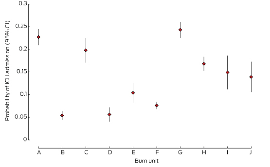
ICU admission

There were differences in the proportions of patients admitted to the ICU by different units (Box 2, Box 3). One low volume (D) and two high volume units (B, F) had the lowest rates of ICU admission.
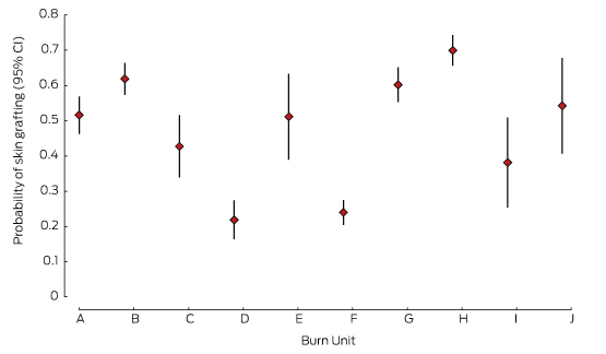
Skin grafting

Nearly three-quarters of patients (74%) underwent at least one surgical procedure. There were marked differences in skin grafting rates between units (Box 2, Box 4); one high volume (F) and one low volume unit (D) reported the lowest rates of skin grafting.
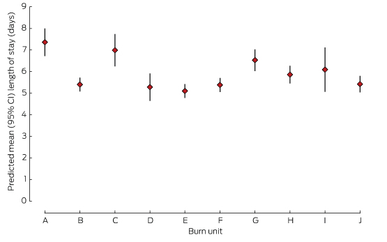
Length of stay

For survivors, LOS increased exponentially with %TBSA. The mean adjusted hospital LOS differed by more than 2 days between the hospitals with the shortest and longest LOS (Box 2, Box 5).
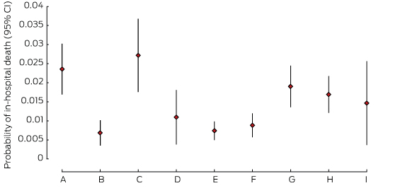
Mortality

There were differences between units in the adjusted odds of mortality, with three units reporting significantly lower estimated probabilities of death (B, E, F) than the two units with the highest estimates (A, C), despite large CIs (Box 2, Box 6). There were no deaths in unit J, which was therefore excluded from this analysis.

Discussion

This report highlights the significant differences in practice and outcomes that exist between specialist burn units that treat a well defined group of patients in Australia and New Zealand. The LOS and mortality rates in burn units in Australia and New Zealand are comparable with those reported internationally.11,12 However, our analysis of 4 years of BRANZ data identified considerable variation in practice in areas fundamental to the treatment of burn injury, and also provides evidence of significant variations in outcomes that are not explained by simple differences in casemix alone.

Scarring and its associated symptoms, functional deficits and deformity, are inevitable outcomes of burn injury, and they determine the capacity for successful rehabilitation and social re-integration. Surgical treatment is one of the few techniques for influencing scarring and other outcomes,13 and our data indicate that the rates of grafting differ between units. The relationship of these differences in surgical management with long term outcomes remains to be determined. The introduction of grafting protocols based on objective determination of the depth of burn injury is required to address variation in this fundamental aspect of burn care, and to provide evidence that supports such protocols. There were also considerable differences in the rates of admission to ICUs in the higher volume statewide services, and in the rates of diagnosis of inhalation injury. The rates of ICU admission differed between hospitals despite our adjusting for inhalation injury. While it might be hypothesised that diagnosis of inhalation injury that increases the risk of airway obstruction drives pre-hospital intubation and subsequent ICU admission, this does not appear to be the case in our study. We suggest that specific transfer protocols in certain jurisdictions can affect rates of intubation as patients are stabilised for early transfer. Demonstrated differences in practice are not insignificant in terms of resource requirements and interventions that are not without risk. Further investigation of pre-burn hospital management practices and diagnostic methods for inhalation injury will inform improvements in protocols for care delivered to patients suspected of having sustained an inhalation injury.14

The hospital stay caused by burn injury is longer than for other kinds of trauma, and is the most significant contributor to the cost of acute burn treatment.1 Attention to variables that influence LOS is particularly important for ensuring that care is delivered efficiently. BRANZ data indicate significant variation in LOS between units. Time to surgery has been reported to influence several aspects of burn care outcomes, including LOS;13 however, other factors could also affect this outcome, such as mobilisation protocols and social and geographical constraints on discharge from hospital. The variation we identified constitutes a flag that signals the need to examine possible contributors at the hospital level that may be amenable to changes that will improve care.

The differences in mortality between units found in this study have also been reported for burn patients in Australian and New Zealand ICUs,15 so that further investigation of practice is necessary. On the basis of our analysis, one burn unit is assessing policies on withholding active treatment for patients with acute burn injury. More detailed analysis of other BRANZ data that may relate to mortality will be undertaken, but is beyond the scope of this article.

The management of burn injury is a relatively small area of clinical practice, conducted in only a few centres. Benchmarking of practice across states and countries is paramount for ensuring best practice and for developing evidence-based practice for burn care. Accuracy and completeness of data, together with robust risk adjustment and analysis methods, are fundamental prerequisites for clinicians’ confidence in the improvement process. While variation is not necessarily in itself a sign of inferior treatments, it does signal the need for further investigation, especially where outcome indicators are also found to vary. For example, it may be that variations in LOS reflect differing rehabilitation protocols rather than delay in treatment. Examination at the hospital level will be required to determine cause and effects. The introduction of protocols based on the best available evidence will assist this endeavour. Protocols should not be interpreted as binding directives, but the identification of key factors that determine long term outcomes, in particular, is assisted by documentation that records instances of deviations from the protocol and the reasons for these deviations.

In response to the findings reported here, ANZBA launched the Burns Quality Improvement Program (BQIP) in 2013. This program will use data generated by BRANZ to develop evidence-based standards of care (few of which are currently available for burn care) and to provide a framework that drives change. Ongoing data collection and analysis will provide information that will link quality indicators with outcomes and thus validate them as meaningful indicators, as few currently used indicators are based on high quality evidence. It will also support establishing standards for compliance. Analysis of the quality indicators embedded in the dataset is currently underway in order to establish their validity for benchmarking purposes. Participation in BQIP will become a fundamental component of the burn unit accreditation process administered by ANZBA. BRANZ has contributed to increased cooperation and collegiality among burn care clinicians, as evidenced by the broadly representative membership of the BRANZ steering and reference committees, and the creation of a BRANZ/BQIP position on the ANZBA board. There is significant international interest in the processes and indicators developed by BRANZ, and in sharing this information to support the introduction of globally consistent indicators that will facilitate international benchmarking.

Conclusions

A clinical quality registry for burn care has been established in Australia and New Zealand. Our results demonstrate the feasibility of collecting data, as well as the need for such data, given the evidence of variations in practice and outcomes that we identified. The information provided by BRANZ provides a unique opportunity for significantly improving the quality of care for burns patients in Australia and New Zealand.

Box 1 –
Profile of cases by total body surface area burned

Population descriptor

Total body surface area burned (%TBSA)


< 10%

10–19%

20–29%

30–39%

40–49%

≥ 50%


Number of admissions (% of all cases)

5472 (78.7%)

925 (13.3%)

274 (3.9%)

125 (1.8%)

54 (0.8%)

105 (1.5%)

Mean age (SD), years

41.2 (18.2)

40.1 (18.6)

40.9 (18.7)

39.5 (17.4)

40.7 (16.7)

39.9 (16.2)

Age group, number (%)

16–19 years

465 (8.5%)

91 (9.8%)

29 (10.6%)

12 (9.6%)

5 (9.3%)

8 (7.6%)

20–29 years

1387 (25.3%)

250 (27.0%)

69 (25.2%)

30 (24.0%)

15 (27.8%)

26 (24.8%)

30–39 years

984 (18.0%)

174 (18.8%)

51 (18.6%)

30 (24.0%)

9 (16.7%)

26 (24.8%)

40–49 years

945 (17.3%)

160 (17.3%)

41 (15.0%)

20 (16.0%)

7 (13.0%)

13 (12.4%)

50–59 years

750 (13.7%)

91 (9.8%)

31 (11.3%)

14 (11.2%)

11 (20.4%)

15 (14.3%)

60–69 years

457 (8.3%)

78 (8.4%)

31 (11.3%)

12 (9.6%)

3 (5.6%)

12 (11.4%)

≥ 70 years

484 (8.8%)

81 (7.9%)

22 (8.0%)

7 (5.6%)

4 (7.4%)

4 (4.8%)

Sex, number (%)

Male

3794 (69.5%)

702 (75.9%)

216 (78.8%)

96 (76.8%)

39 (72.2%)

76 (72.4%)

Female

1669 (30.5%)

223 (24.1%)

58 (21.2%)

29 (23.2%)

15 (27.8%)

29 (27.6%)

Cause, number (%)

Flame

2006 (36.7%)

677 (73.2%)

224 (81.7%)

102 (81.6%)

45 (83.3%)

95 (90.5%)

Scald

1715 (31.3%)

184 (19.9%)

38 (13.9%)

13 (10.4%)

2 (3.7%)

3 (2.9%)

Contact

898 (16.4%)

24 (1.5%)

2 (0.7%)

1 (0.8%)

0

0

Other

853 (15.6%)

50 (5.4%)

10 (3.7%)

9 (7.2%)

7 (13.0%)

7 (6.7%)

Median time from injury to admission (IQR), hours

26 (5–123)

6 (2–15)

5 (2–9)

6 (2–10)

5 (1–13)

5 (2–10)

In-hospital mortality rate, deaths (%)

18 (0.3%)

13 (1.4%)

8 (2.9%)

7 (5.6%)

7 (13.0%)

50 (47.6%)

Treatment withdrawn, number

10

10

5

3

6

32

ICU admission rate, number (%)

378 (6.9%)

255 (27.6%)

149 (54.8%)

91 (72.8%)

45 (83.3%)

94 (90.4%)

Median ICU length of stay (IQR), hours

43 (12–82)

55 (29–110)

89 (43–233)

142 (48–307)

194 (101–348)

223 (19–480)

Median time ventilated in ICU (IQR), hours

14 (0–37)

26 (8–72)

40 (10–130)

76 (22–212)

137 (33–251)

118 (14–300)

Median length of stay (IQR), hours

All cases

4.5 (1.9–8.8)

11.7 (6.9–18.6)

20.6 (13.8–30.5)

29.0 (18.5–43.6)

30.6 (17.4–54.1)

13.6 (0.7–62.2)

Survivors

4.4 (1.8–8.8)

11.7 (6.9–18.7)

20.7 (14.5–30.7)

29.2 (19.0–43.6)

37.9 (22.3–55.7)

60.3 (31.8–84.0)

Surgical management

4022 (73.7%)

754 (81.6%)

243 (88.7%)

119 (95.2%)

47 (87.0%)

72 (68.6%)

Debridement and grafting

2632 (65.5%)

489 (64.9%)

194 (79.8%)

104 (87.4%)

36 (76.6%)

52 (72.2%)

Debridement only

838 (20.8%)

160 (21.2%)

70 (28.8%)

34 (28.6%)

14 (29.8%)

32 (44.4%)

Debridement and skin closure

805 (20.0%)

280 (37.1%)

91 (37.5%)

57 (47.9%)

27 (57.5%)

43 (59.7%)

Debridement and skin cell product

422 (10.5%)

77 (10.2%)

28 (11.5%)

18 (15.1%)

7 (14.9%)

8 (11.1%)

Median time from injury to grafting (IQR), days

6.7 (3.7–10.6)

4.6 (2.5–7.8)

4.9 (2.5–7.5)

4.3 (2.6–6.5)

4.4 (2.4–9.5)

5.3 (2.9–12.4)


ICU = intensive care unit; IQR = interquartile range; SD = standard deviation.

Box 2 –
Profile of adult cases managed at each burn unit

Population descriptor

Burn unit


A

B

C

D

E

F

G

H

I

J


Number of admissions

1108

1056

475

429

243

1378

1052

1070

194

179

Mean age (SD), years

42.2 (18.8)

38.8 (16.8)

40.1 (17.7)

37.7 (16.3)

44.5 (19.2)

42.4 (18.5)

42.3 (18.4)

40.2 (18.6)

40.9 (18.5)

41.3 (18.4)

Sex, males (%)

807 (72.8%)

749 (70.9%)

329 (69.6%)

295 (69.7%)

156 (64.2%)

949 (68.9%)

753 (71.7%)

770 (72.0%)

137 (70.6%)

129 (72.1%)

Cause, number (%)

Flame

633 (57.2%)

509 (48.2%)

222 (46.7%)

117 (27.3%)

82 (33.7%)

544 (39.5%)

474 (45.1%)

472 (44.1%)

84 (43.3%)

76 (42.4%)

Scald

253 (22.8%)

272 (25.8%)

134 (28.2%)

85 (19.8%)

63 (25.9%)

498 (36.1%)

307 (29.2%)

295 (27.6%)

45 (23.2%)

61 (34.1%)

Contact

81 (7.3%)

166 (15.7%)

81 (17.1%)

55 (12.8%)

56 (23.1%)

162 (11.8%)

131 (12.4%)

164 (15.3%)

37 (19.1%)

27 (15.1%)

Other

141 (12.7%)

109 (10.3%)

38 (8.0%)

172 (40.1%)

42 (17.3%)

174 (12.6%)

1400 (13.3%)

139 (13.0%)

28 (14.4%)

15 (18.4%)

Mean %TBSA (SD)

9.4 (13.1)

6.1 (9.3)

10.2 (15.1)

5.7 (9.1)

4.3 (6.1)

5.0 (8.2)

8.0 (11.9)

6.7 (10.0)

7.6 (10.7)

7.1 (6.3)

%TBSA < 10%

749 (68.8%)

865 (82.2%)

292 (70.2%)

352 (88.0%)

194 (87.4%)

1178 (88.4%)

773 (74.0%)

828 (77.5%)

139 (72.8%)

27 (16.6%)

%TBSA, 10–19%

217 (19.9%)

112 (10.6%)

56 (13.5%)

30 (7.5%)

18 (8.1%)

94 (7.0%)

164 (15.7%)

170 (15.9%)

37 (19.4%)

27 (16.6%)

%TBSA ≥ 20%

123 (11.3%)

76 (7.2%)

68 (16.3%)

18 (4.5%)

10 (4.5%)

61 (4.6%)

107 (10.3%)

71 (6.6%)

15 (7.8%)

9 (6.5%)

Inhalation injury, number (%)

157 (14.2%)

31 (2.9%)

24 (5.1%)

13 (3.0%)

8 (3.3%)

41 (3.0%)

106 (10.1%)

96 (9.0%)

18 (9.3%)

8 (4.5%)

In-hospital deaths, number (%)

27 (2.4%)

5 (0.5%)

15 (3.2%)

2 (0.5%)

2 (0.8%)

11 (0.8%)

20 (1.9%)

20 (1.9%)

4 (2.1%)

0

ICU admission, number (%)

261 (23.6%)

53 (5.0%)

85 (17.9%)

21 (4.9%)

27 (11.1%)

104 (7.6%)

259 (24.6%)

180 (16.8%)

27 (14.1%)

21 (11.7%)

Median length of stay (IQR), days

8.1 (3.1–15.0)

6.8 (3.9–11.4)

5.1 (2.3–11.1)

7.8 (3.2–14.1)

5.2 (2.1–12.1)

4.7 (2.8–7.9)

5.1 (1.0–14.9)

3.5 (0.4–10.2)

4.1 (2.0–10.1)

4.0 (2.2–8.9)

Grafting procedure performed, number (%)

573 (51.7%)

665 (63.0%)

194 (40.8%)

93 (21.7%)

121 (49.8%)

336 (24.4%)

644 (61.2%)

462 (71.2%)

75 (38.7%)

96 (53.6%)

Median time to grafting from injury (IQR), days

4.6 (2.6–8.2)

5.7 (3.8–8.9)

8.2 (4.0–14.2)

8.7 (5.0–12.6)

11.0 (8.3–15.2)

3.6 (1.1–8.1)

6.5 (3.7–10.4)

6.3 (3.7–10.4)

6.8 (3.5–10.5)

7.7 (3.7–12.5)


ICU = intensive care unit; IQR = interquartile range; SD = standard deviation; %TBSA = total body surface area burned.

Box 3 –
Probability of admission of patients to the intensive care unit (ICU) of the ten hospital burn units

Box 4 –
Probability of skin grafting for patients at the ten hospital burn units

Box 5 –
Predicted hospital length of stay for patients at the ten hospital burn units

Box 6 –
Probability of in-hospital death for patients at the ten hospital burn units

Emergency department presentations with mammalian bite injuries: risk factors for admission and surgery

Animal bites, particularly by mammals, are common in Australia,1,2 and their treatment is a substantial public health burden.3 Clinical assessment and the subsequent decision to transfer patients to surgical centres may be challenging, especially for primary health care providers, paramedics and rural emergency departments. There have been few investigations into predictors of hospital admission and surgery for bite injury patients.2,4 We retrospectively analysed the characteristics of all mammalian bite injuries with which patients presented to seven major hospital emergency departments in Victoria during a 2-year period.

Methods

Study design

A retrospective review of all patients presenting with mammalian bite injuries to seven Victorian emergency departments (at the Alfred Hospital, Austin Hospital, Royal Melbourne Hospital, Frankston Hospital, Monash Medical Centre, St Vincent’s Hospital and Western Hospital) during the 2-year period 1 January 2012 – 31 December 2013 was undertaken. Patients were identified using International Statistical Classification of Diseases and Related Health Problems, tenth revision, Australian modification (ICD-10-AM) codes for animal-related injury, and by searching patient record systems for the terms “bite” and “animal-related injuries”. Injuries not involving mammalian bites were excluded.

Descriptive and univariate analysis

All statistical analysis was performed with SPSS Statistics 22 (IBM). Graphs were created in Excel 2013 (Microsoft) and Prism 5 (GraphPad). P < 0.05 (two-tailed) was defined as statistically significant.

The associations between each predictor and outcome of interest were analysed with univariate methods, χ2 tests, analyses of variance (ANOVAs) and Kruskal–Wallis tests as appropriate. Post hoc Bonferroni corrections were performed when appropriate. The choice of potential predictors was based on reports in the literature; age, sex, smoking status, diabetes mellitus, immunosuppression, time to presentation, type of animal and site of injury were assessed. The measured outcomes were hospital admission, surgery, readmission, reoperation, and positive microbiological culture.

Multiple regression analysis

We conducted multiple logistic regression analyses, with stepwise backward elimination by likelihood ratio tests, to further clarify the associations between predictors and outcomes. The probability for stepwise elimination was set at 0.10. This method allowed us to examine the effects of multiple predictors on an outcome. For each predictor, the category with the lowest rate of the outcome of interest was designated as the baseline or reference category. Each regression model was assessed with the Hosmer-Lemeshow test, the Nagelkerke R2, percentage of correct predictions, and area under the receiving operating characteristic curve.

Ethics approval

The investigation was approved by all hospitals involved in this research (Alfred Health Human Research Ethics Committee, reference QA535/13; St Vincent’s Hospital Human Research Ethics Committee, reference QA004/15; Western Health Human Research Ethics Committee, reference QA2014.02; Melbourne Health Office for Research, reference QA2013161; Monash Health Human Research Ethics Committee, reference 14386Q; Frankston Hospital Human Research Ethics Committee, reference QA13PH36; Austin Health Human Research Ethics Committee, reference LNR/15/Austin/525).

Results

Epidemiology of mammalian bites

We identified a total of 717 patients who presented with mammalian bite injuries to the seven Melbourne emergency departments during the study period. Their mean age was 36.5 years, with an equal number of males and females (sex unspecified in one case). Almost all cases (96.1%) involved bites to only one anatomical region; 60.9% involved the upper limbs, 18.7% the head and neck, 14.5% the lower limbs, and 2.0% another part of the body (trunk, back or perineum). Most patients had presented to an emergency department (84.5%) within 24 hours of the injury. The overall rate of hospital admission was 50.8%, and the mean length of stay was 2.7 days. Intravenous antibiotics were administered in 46% of cases; surgery was undertaken in 43.1% of cases. The reoperation rate was 4.5%, the readmission rate was 3%.

A comparison of the demographic and other data for patients presenting with bites by different mammals is shown in Box 1. Almost all bites sustained by patients aged under 15 years were dog bites (92%). Further, 63.1% of patients aged 0–15 years with dog bites were bitten on the head and neck (compared with 13.3% of older patients with dog bites). Dog and human bites were significantly more likely to be seen in male than in female patients (54% and 75%, respectively were sustained by males; P < 0.05); the reverse was true for cat bites (72% were sustained by females).

Patients presenting with cat bites were on average older (mean age, 46.8 ± 19.3 years) than those presenting with bites by other mammals (mean age, 35.7 ± 20.5 years; P < 0.0001). Cat bites comprised 24.0% of all bites in patients aged 60 years or over, compared with 15.9% in other age groups. Cat bites were seen significantly more frequently in female than in male patients (72.1% v 27.9%; P < 0.05). There was no seasonal trend in the frequency of presentation of mammalian bites according to type of bites (data not shown).

Box 2 summarises the rates of hospital admission and the management outcomes for patients presenting with the different bite types. Patients with dog bites usually presented to a hospital on the day of the injury, while presentation with bites by cats and other mammals was often delayed for up to 2 days. Hospital admission rates were significantly higher for cat bites (64% v 48% for all other bites; P < 0.05), and surgery rates were significantly higher for patients with dog bites (48% v 30% for all other bites; P < 0.05). Patients with cat and dog bites were more likely to receive intravenous antibiotics than were those with bites from other mammals (P < 0.05).

Predictive factors for admission to hospital

Patient age, type of animal, the site of injury, and time to presentation of 2 days or more were all significantly associated with admission to hospital (P < 0.01 for all tests). Children under 15 years of age and adults over 60 years of age were more likely to be admitted; the probability also increased with age from the age of 30 years. Admission was more frequent for patients with cat and dog bites than for bites by other mammals. Patients with isolated bites to the head and neck or an upper limb were more likely to be admitted than those with bites to the trunk, back or perineum. However, bites to multiple sites were associated with the greatest risk for admission (Box 3). Finally, smoking was identified by multiple regression analysis as a significant risk factor for admission (adjusted odds ratio [aOR], 1.99 v non-smokers; 95% CI, 1.21–3.28). Sex, immunosuppression and diabetes were not significant risk factors for admission.

Predictive factors for surgery, re-admission and re-operation

Surgery was significantly more frequent for patients with bites by dogs (P < 0.05) and bites to the head and neck, upper limb or multiple sites, and was more frequent in patients who smoked. Patients aged 15–29 years were more likely to undergo surgery as a result of their bite, but this difference was not statistically significant (P < 0.10). Sex, diabetes and immunosuppression were not statistically significantly associated with surgery (Box 4).

After pooling data for all mammalian bites, time to presentation of greater than 2 days was associated with an increased risk of re-operation (OR, 4.41; 95% CI, 1.39–13.95; P = 0.019).

Discussion

Our findings show that presentations by patients to emergency departments with animal bites are frequent, and that a substantial proportion of these patients are hospitalised or undergo surgery. Our data identified certain trends that are consistent with other findings in the literature. Males were more likely to sustain bite injuries, especially by dogs,59 and cat bites were more common in females, as in previous reports.1 In children under 15 years of age, dog bites were more common than other bites (92% of all mammalian bites in this age group were dog bites); further, 20.2% of all dog bites were presented by children under 15 years of age, similar to other reported findings.1,57,10

We also confirmed that the average age of patients presenting with cat bites was higher than for patients presenting with other animal bites.10 The most common site of injury for animal bites of any type was an upper limb, consistent with previous studies,1 although some authors found that the lower limbs were the predominant site of injury for dog bites.8,9 In our study, dog bites more frequently caused head and neck injuries in younger patients than in adults. It has been proposed that children are at particular risk because of their shorter stature, lower capacity for self-defence, and poorer risk awareness with regard to potentially provocative behaviour.7,9

Patients with dog bite injuries usually presented to hospital on the day of the injury, while presentations with bites by cats and other mammals were often delayed. This could be explained by the smaller wound sizes of cat bites, so that patients do not seek medical attention until after infections have developed.11,12 That patients with dog bites had the highest rate of surgery is reasonable, given the depth and complexity of dog bite wounds.5,13 Higher hospital admissions of patients with cat bites may be related to the need for prophylactic intravenous antibiotics, as is currently recommended.2,14

Our study found a relatively high admission rate of 50.8% for mammalian bites. This is at the high end of a broad range of admission rates reported in the literature (4.7–51%).69,15 The variability of these estimates may be explained by differences in the sources of the collected data; some studies analysed surveillance data based on presentations to general practice clinics,1,15 while the emergency department presentations in our study may include a larger proportion of more serious injuries. We also found that delayed presentation for treatment increased the risk of hospitalisation, surgery and reoperation. This is consistent with most studies,4 with the exception of one which found that smoking, an immunocompromised state, and location of the bite over a joint or tendon sheath were associated with hospitalisation.14 Our study confirmed the previously reported association between higher age and the risk of hospitalisation for bite injuries.4 Some traditional risk factors, such as diabetes and immunosuppression, were not significantly associated with hospitalisation, surgery or complications in our study, perhaps because only 3% of our sample were affected by these factors.

A limitation of this study was the retrospective nature of the data collection. Further, the outcomes we analysed were limited to reported hospitalisation and surgery; there was no long term patient follow-up, so that there were no recorded data about any subsequent disabilities.

Our study identified risk factors associated with hospitalisation and surgery. Further analysis of surgical findings in patients who have sustained bite injuries is needed, as this could allow the derivation of risk-stratifying scoring systems from regression models that predict whether a patient will require hospitalisation or surgery. A scoring system could prove beneficial for guiding primary health care providers and emergency physicians in identifying low-risk and high-risk patients who can be managed conservatively or in the outpatient setting, as well as who require timely interhospital transfer or assessment by a surgical unit.

Box 1 –
Demographic data for patients presenting with bites from different mammals

Variable

Source of bite


P

Dog

Cat

Human

Other*


Number of patients

509

122

36

50

Mean age (SD), years

34.8 (21.0)

43.9 (19.3)

37.4 (16.5)

34.9 (17.1)

0.0002

Age group

< 0.0001

< 15 years

103 (20.2%)

4 (3.3%)

1 (3%)

4 (8%)

15–29 years

127 (25.0%)

27 (22.1%)

12 (33%)

19 (38%)

30–44 years

114 (22.4%)

38 (31.1%)

12 (33%)

12 (24%)

45–59 years

97 (19.1%)

29 (23.8%)

8 (22%)

10 (20%)

≥ 60 years

68 (13.4%)

24 (19.7%)

3 (8%)

5 (10%)

Sex

< 0.0001

Female

232 (45.6%)

88 (72.1%)

9 (25%)

29 (58%)

Male

276 (54.2%)

34 (27.9%)

27 (75%)

21 (42%)

Unknown

1 (0.2%)

0 (0%)

0 (0%)

0 (0%)

Site of bite

49

< 0.0001

Head and neck

119 (23.4%)

4 (3.3%)

10 (28%)

1 (2%)

Upper limb

272 (53.4%)

105 (86.1%)

20 (56%)

39 (80%)

Lower limb

84 (16.5%)

12 (9.8%)

1 (3%)

7 (14%)

Trunk, back, perineum

11 (2.2%)

0 (0%)

2 (6%)

1 (2%)

Multiple sites

23 (4.5%)

1 (0.8%)

3 (8%)

1 (2%)

Diabetes mellitus

14 (3.5%)

9 (8.7%)

1 (3.8%)

1 (2%)

0.12

Current smoker

71 (18.0%)

19 (18.6%)

10 (38.5%)

14 (35%)

0.007

Immunosuppression

12 (3.0%)

3 (2.9%)

2 (7.4%)

1 (2%)

0.62


All percentages are column percentages. * Monkey, rat, possum and bat bites. † Data on diabetes, smoking and immunosuppression status were not recorded for all patients.

Box 2 –
Outcomes for patients presenting with mammalian bites

Clinical outcome

Source of bite


P

Dog

Cat

Human

Other*


Number of patients

509

122

36

50

Median time to presentation (IQR), days

0 (0–0)

0.5 (0–1.75)

0 (0–1)

0 (0–4)

< 0.0001

Time to presentation of 2 days or more

54 (11.4%)

29 (24.2%)

5 (14.7%)

17 (34.0%)

< 0.0001

Admission

262 (51.5%)

78 (63.9%)

12 (33.3%)

12 (24.0%)

< 0.0001

Surgery

246 (48.3%)

41 (33.6%)

11 (30.6%)

11 (22.0%)

< 0.0001

Median length of stay (IQR), days

1 (0–2)

2 (1–3.75)

2 (0–3)

2 (1–2.75)

< 0.0001

Positive wound or blood culture

32 (6.3%)

28 (23.0%)

4 (11.1%)

3 (6.0%)

< 0.0001

Administration of intravenous antibiotics

227 (44.6%)

79 (64.8%)

13 (36.1%)

11 (22.0%)

< 0.0001

Re-admissions (percentage of prior admissions)

7 (2.7%)

1 (1.3%)

1 (8.3%)

2 (16.7%)

0.021

Re-operation (percentage of prior operations)

8 (3.3%)

3 (7.3%)

1 (9.1%)

2 (18.2%)

0.074


IQR = interquartile range. All percentages are column percentages. * Monkey, rat, possum and bat bites.

Box 3 –
Univariate and multivariable predictors and prediction score for hospital admission following a mammalian bite injury

Risk factors for admission

Admission rate

Univariate tests (n = 717)


Multiple logistic regression model (n = 545)


Unadjusted OR (95% CI)

Adjusted OR (95% CI)

P


Animal type

< 0.0001

Other

24%

1

Human

33%

1.58 (0.61–4.09)

1.42 (0.44–4.60)

0.555

Dog

52%

3.36 (1.72–6.58)

3.54 (1.60–7.85)

0.002

Cat

64%

5.61 (2.66–11.85)

5.55 (2.32–13.25)

< 0.0001

Age group

0.001

15–29 years

37%

1

< 15 years

55%

2.13 (1.32–3.44)

2.16 (1.17–4.01)

0.014

30–44 years

48%

1.61 (1.06–2.45)

1.84 (1.09–3.12)

0.023

45–59 years

56%

2.21 (1.42–3.45)

2.01 (1.17–3.45)

0.011

≥ 60 years

68%

1.85 (1.28–2.69)

3.84 (2.04–7.21)

< 0.0001

Sex

Female

49%

1

Male

52%

1.12 (0.83–1.50)

NA

NA

Site of bite

0.003

Trunk, back or perineum

7%

1

Lower limb

37%

7.48 (0.94–59.48)

5.52 (0.61–50.0)

0.129

Upper limb

53%

14.51 (1.88–112)

10.94 (1.26–95.3)

0.03

Head and neck

59%

18.67 (2.37–147)

14.72 (1.63–133)

0.017

Multiple sites

57%

17.33 (1.98–151)

22.02 (2.12–229)

0.01

Diabetes mellitus

No

53%

1

Yes

72%

2.26 (0.93–5.49)

NA

NA

Current smoker

No

53%

1

Yes

61%

1.35 (0.89–2.05)

1.99 (1.21–3.28)

0.007

Immunosuppression

No

54%

1

Yes

61%

1.36 (0.52–3.55)

NA

NA

Time to presentation

< 2 days

49%

1

≥ 2 days

65%

1.90 (1.23–2.93)

2.39 (1.30–4.36)

0.005


NA = not applicable (not included in multivariate model because not significant in univariate model); OR = odds ratio.

Box 4 –
Univariate and multivariable predictors and prediction score for surgery following a mammalian bite injury

Risk factors for surgery

Surgery rate

Univariate tests (n = 717)


Multiple logistic regression model (n = 545)


Unadjusted OR (95% CI)

Adjusted OR (95% CI)

P


Animal type

< 0.0001

Other

22%

1

Human

31%

1.56 (0.59–4.14)

1.39 (0.44–4.44)

0.576

Dog

34%

1.79 (0.83–3.87)

1.92 (0.82–4.42)

0.135

Cat

48%

3.32 (1.66–6.62)

4.47 (2.03–9.81)

< 0.0001

Age group

0.074

15–29 years

35%

1

< 15 years

55%

2.29 (1.42–3.70)

1.77 (0.95–3.32)

0.074

30–44 years

38%

1.11 (0.72–1.70)

1.63 (0.97–2.77)

0.067

45–59 years

50%

1.85 (1.18–2.88)

2.13 (1.24–3.66)

0.006

≥ 60 years

44%

1.45 (0.88–2.38)

1.61 (0.89–2.89)

0.113

Sex

Female

41%

1

Male

45%

1.15 (0.85–1.54)

NA

NA

Site of bite

0.002

Trunk, back or perineum

7%

1

Lower limb

33%

9.14 (1.19–70.50)

7.886 (0.90–69.0)

0.062

Upper limb

41%

6.31 (0.79–50.28)

12.80 (1.52–108)

0.019

Head and neck

50%

13 (1.49–113)

21.13 (2.08–214)

0.01

Multiple sites

60%

19.26 (2.45–152)

23.89 (2.71–211)

0.004

Diabetes mellitus

No

53%

1

Yes

52%

0.96 (0.43–2.15)

NA

NA

Current smoker

No

52%

1

Yes

58%

1.29 (0.85–1.95)

1.96 (1.20–3.18)

0.007

Immunosuppression

No

53%

1

Yes

56%

1.13 (0.44–2.90)

NA

NA

Time to presentation

< 2 days

47%

1

≥ 2 days

36%

0.63 (0.41–0.97)

NA

NA


NA = not applicable (not included in multivariate model because not significant in univariate model); OR = odds ratio.

‘Everything presents at extremes…’ – a Solomon Islands experience

Pictgure: Dr Elizabeth Gallagher (second from left) with other staff and volunteers at the National Referral Hospital, Honiara

By Dr Elizabeth Gallagher, specialist obstetrician and gynaecologist, AMA ACT President

The mother lost consciousness just as her baby was born.

The woman was having her child by elective Caesarean when she suffered a massive amniotic fluid embolism and very quickly went into cardiac arrest.

We rapidly swung into resuscitation and, through CPR, defibrillation and large doses of adrenaline, we were able to restore her to unsupported sinus rhythm and spontaneous breathing.

But, with no equipment to support ventilation, treat disseminated intravascular coagulation, renal failure or any of the problems that arise from this catastrophic event, it was always going to be difficult, and she died two-and-a-half hours later.

Sadly, at the National Referral Hospital in Honiara, the capital of the Solomon Islands, this was not an uncommon outcome. Maternal deaths (both direct and indirect) average about one a month, and this was the second amniotic fluid embolism seen at the hospital since the start of the year.

I was in Solomon Islands as part of a team of four Australian practitioners – fellow obstetrician and gynaecologist Dr Tween Low, anaesthetist Dr Nicola Meares, and perioperative nurse and midwife Lesley Stewart – volunteering to help out at the hospital for a couple of weeks in October.

It was the first time I had worked in a developing country, and it was one of the most challenging, and yet satisfying, things I have ever done

Everything from the acuteness of the health problems to the basic facilities and shortages of equipment and medicines that we take for granted made working there a revelation.

The hospital delivers 5000 babies a year and can get very busy. As many as 48 babies can be born in a single 24-hour period.

The hospital has a first stage lounge and a single postnatal ward, but just one shower and toilet to serve more than 20 patients. The gynaecology ward is open plan and, because the hospital doesn’t provide a full meal service or much linen, relatives stay there round-the-clock to do the washing and provide meals.

From the beginning of our stay, it was very clear that providing training and education had to be a priority. I was conscious of the importance of being able to teach skills that were sustainable once we left.

The nature of the emergency gynaecological work, which includes referrals from the outer provinces, is that everything presents at the extremes…and late. Massive fibroids, huge ovarian cysts and, most tragically because there is no screening program, advanced cervical cancers in very young women.

When I first got in touch with doctors at the hospital to arrange my visit, I had visions of helping them run the labour ward and give permanent staff a much-needed break. But what they wanted, and needed, us to do was surgery and teaching.

To say they saved the difficult cases up for us is an understatement. I was challenged at every turn, and even when the surgery was not difficult, the co-morbidities and anaesthetic risks kept Dr Meares on her toes.

In my first two days, the hospital had booked two women – one aged 50 years, the other, 30 – to have radical hysterectomies for late stage one or early stage two cervical cancer. I was told that if I did not operate they would just be sent to palliation, so I did my best, having not seen one since I finished my training more than 12 years ago.

I also reviewed two other woman, a 29-year-old and a 35-year-old, both of whom had at least a clinical stage three cervical cancer and would be for palliation only. This consisted of sending them home and telling them to come back when the pain got too bad.

It really brought home how effective our screening program is in Australia, and how dangerous it would be if we got complacent about it.

We found the post-operative pain relief and care challenged. This was because staffing could be limited overnight and the nurses on duty did not ask the patients whether they felt pain – and the patients would definitely not say anything without being asked.

Doing our rounds in our first two days, we found that none of the post-operative patients had been given any pain relief, even a paracetamol, after leaving theatre.

We conducted some educational sessions with the nursing staff, mindful that the local team would need to continue to implement and use the skills and knowledge we had brought once we left. By the third day, we were pleased to see that our patients were being regularly observed and being offered pain relief – a legacy I hope will continue.

The supply of equipment and medicines was haphazard, and depended on what and when things were delivered. There was apparently a whole container of supplies waiting for weeks for clearance at the dock.

Many items we in Australia would discard after a single use, like surgical drains and suction, were being reused, and many of the disposables that were available were out-of-date – though they were still used without hesitation.

Some things seemed to be in oversupply, while others had simply run out.

The hospital itself needs replacing. Parts date back to World War Two. There were rats in the tea room, a cat in the theatre roof, and mosquitos in the theatre.

The hospital grounds are festooned with drying clothes, alongside discarded and broken equipment – including a load of plastic portacots, in perfect condition, but just not needed on the postnatal ward as the babies shared the bed with their mother.

It brought home how important it is to be careful in considering what equipment to donate.

The ultrasound machine and trolley we were able to donate, thanks to the John James Memorial Foundation Board, proved invaluable, as did the instruction by Dr Low in its use.

The most important question is, were we of help, and was our visit worthwhile?

I think the surgical skills we brought (such as vaginal hysterectomy), and those we were able to pass on, were extremely useful. Teaching local staff how to do a bedside ultrasound will hopefully be a long-lasting legacy. Simple things like being able to check for undiagnosed twins, dating, diagnosing intrauterine deaths, growth-restricted babies and preoperative assessments will be invaluable.

The experience was certainly outside our comfort zone, and it made me really appreciate what a great health system we have in Australia, and what high expectations we have. I want to send a big thank you to the John James Memorial Foundation for making it all possible.