×

Toxic epidermal necrolysis — an investigation to dye for?

We report the first case in Australia, as confirmed by the Therapeutic Goods Administration, of toxic epidermal necrolysis associated with the iodinated contrast medium iopamidol. It serves as a warning about the use of contrast in imaging and cardiac catheterisation and a reminder of the need for increased awareness of the issue.

Clinical record

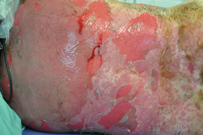
A 44-year-old woman presented to the emergency department with a 3-day history of a progressive rash, fever, malaise and mucosal ulceration. She met the diagnosis of toxic epidermal necrolysis (TEN) based on the following criteria: bullae and desquamation affecting about 84% of the body surface (Box 1 and Box 2), buccal and vaginal ulceration, a positive Nikolsky sign (this is a useful sign in bullous skin diseases and can be demonstrated by rubbing the skin surface, which will blister within a few minutes if the sign is positive), fever, tachycardia and mild hypotension. She also had abnormal results of liver function tests: bilirubin level, 69 µmol/L (reference interval [RI], < 20 µmol/L); alkaline phosphatase level, 180 U/L (RI, 25–100 U/L); γ-glutamyl transferase level, 499 U/L (RI, < 30 U/L); alanine aminotransferase level, 1730 U/L (RI, < 30 U/L); and aspartate aminotransferase level, 638 U/L (RI, < 30 U/L). She had a white cell count of 4.3 × 109/L (RI, 4.0–10.0 × 109/L) and a raised C-reactive protein level of 53.1 mg/L (RI, < 5 mg/L).

The patient was immediately transferred to the burns unit and managed with nanocrystalline silver dressings, intravenous immunoglobulin, aggressive fluid and electrolyte balance therapy, analgesia and intravenous antibiotics. She was discharged home on Day 15.

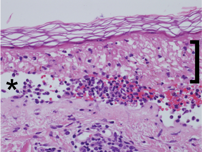
Histopathological examination (Box 3) showed extensive epidermal necrosis and subepidermal clefting with a sparse superficial perivascular infiltrate of lymphocytes, occasional neutrophils and eosinophils, and exocytosis of cells into the epidermis. Results of staining for immunofluorescence were negative. This was consistent with the clinical diagnosis of TEN, and the possibility of pemphigus vulgaris was excluded.

The patient’s only recent exposure to medications included 150–200 µg of thyroxine sodium daily for 19 years and 2.5 mg of indapamide daily for 6 months. The patient underwent a computed tomography (CT) neck scan with the contrast medium iopamidol about 4 weeks before the development of symptoms. The patient had recently (3 weeks before onset of rash) stopped taking a herbal “liver cleanser”. She had been taking this intermittently for 2 months. She had no recent travel history or vaccinations.

The patient recalled having a previous CT scan of the neck before her surgery 17 years ago. Unfortunately, however, any records of this had been destroyed.

The patient’s past medical history was notable for mild stable hypertension, hypothyroidism and a benign mixed salivary gland tumour electively excised 17 years earlier.

Discussion

Toxic epidermal necrolysis (TEN), or Lyell syndrome, is a rare and life-threatening severe systemic condition associated with dramatic cutaneous sloughing of up to 100% of the body surface area. The incidence of TEN is two cases per million person-years.1 It is characterised by necrosis and subsequent detachment of the epidermis from the dermis in more than 30% the body surface. If not treated and managed promptly, the consequences can be fatal; patients are vulnerable to infections and sepsis leading to death. The mortality associated with TEN is high, at 30%–40%.2

At the other end of the spectrum, and more common, are mild-to-moderate skin reactions to contrast media (CM). These include, in increasing severity, lichenoid reaction, erythema multiforme and Stevens–Johnson syndrome.3 Patients at risk of late skin reactions are those with a previous history of CM reactions.3,4

TEN is attributed to medications in 80% of cases.1 The most commonly associated medications include sulfonamides, penicillin and other antibiotics, anticonvulsants, oxicam nonsteroidal anti-inflammatory drugs, allopurinol and corticosteroids.5 TEN commonly occurs 1–3 weeks after the start of therapy. Other triggers include infections, malignancy and vaccination.1

The dermatological reactions caused by CM can be classified as early or late reactions. Early reactions occur soon after injection of the contrast medium, and late reactions occur within a week. The incidence of late adverse reactions is 2%.3,6 They commonly present as maculopapular erythema, angioedema and urticaria. Evidence to date suggests that late reactions are more common with non-ionic CM, in particular dimers,3 despite non-ionic CM being touted as having fewer adverse reactions. Late reaction incidence with non-ionic CM varies between 8% and 71%.3

Iodinated CM can be divided into ionic and non-ionic contrast medium. The ionicity pertains to the osmolality the CM create in blood; ionic CM create higher osmolality leading to CM reaction. The move from the use of ionic CM to non-ionic CM was based on the need for an agent with fewer adverse effects and equal or slightly improved diagnostic efficacy.7 The morbidity and mortality associated with non-ionic CM were less than for ionic CM.8 Although non-ionic CM have these advantages, ionic CM are still in use today. A recent study highlighted that although non-ionic CM are the best tolerated in the early phase, they are associated with a higher level of adverse effects such as late skin reactions.8 Iopamidol is a non-ionic contrast medium.

There have been several cases of TEN caused by CM reported in the literature. Commonly, the cases have involved repeated exposure or sensitisation to the CM in the cardiac catheter laboratory over a period of days to even years.911 TEN occurs with subsequent exposures to the CM administered. In our case report, there was a history of prior CT scan of the neck; however, records of the scan are no longer held by the radiologist to verify the date and contrast medium used.

TEN has also been shown to be caused by gastrointestinal oral CM.12

Only two published cases of TEN have been attributed to the administration of iopamidol. The first case is of a young boy with subsequent exposure to iopamidol.10 The case was not biopsy-proven TEN, but was based on clinical diagnosis. The second case is that of a patient who underwent intravenous urography for investigation of systemic lupus erythematosus with renal involvement.13 The patient died despite intensive care and support. Our case report would be the third reported case of TEN caused by iopamidol.

Although it is difficult to be sure that iopamidol was responsible for the development of TEN in our patient, it is highly likely to be the cause. She had been on indapamide for a period and had been taking the herbal “liver cleanser” intermittently. One recent case of herbal medicines and TEN has been reported.14 However, as noted by the author, it was difficult to determine the causative agent.

It is important to be aware of the risk of CM and to think twice about the necessity of CM in imaging. Although rare, life-threatening adverse effects such as TEN should lead to reconsideration of contrast dyes, as patients may suffer unnecessarily or lose their lives.

1 Bullae and desquamation of back

2 Desquamation of both feet

3 Skin punch biopsy sample showing extensive epidermal necrosis (bracket) and subepidermal clefting (asterisk) with mixed inflammatory infiltrate and negative results of staining for immunofluorescence

Missing malaria? Potential obstacles to diagnosis and hypnozoite eradication

Poor specimen collection and limited availability of primaquine may be putting patients at risk

Recently, one of us experienced an episode of probable malaria on returning from fieldwork in the Solomon Islands. Although a clinical diagnosis of malaria was made, and the illness responded to empirical therapy with artemether–lumefantrine (Riamet, Novartis), a laboratory diagnosis was not achieved.

Suspected malaria in travellers who have returned to Australia from overseas will present without notice and, owing to the often severe nature of this illness, will require immediate attention. This may occur in localities where personal consultation with an infectious diseases physician is not possible. Primaquine for the eradication of malarial hypnozoites from the liver may not be readily available. In this article, we aim to provide brief expert guidance on the diagnosis of malaria, the use of primaquine for eradication therapy and the implications of the limited availability of this treatment in Australia.

Patients presenting with fever should be questioned about their travel history. Clinicians should be mindful that malarial relapse (Plasmodium vivax and P. ovale) or recrudescence (P. malariae) may occur months, or even years, after primary infection. Further, relapse may be the first symptomatic presentation.1 Therefore, any patient with pyrexia and a history of travel to an endemic area in the past 3 years might be considered as potentially having malaria.2

Initial investigation

Clinical suspicion should be raised in patients who demonstrate specific symptoms associated with the disease, such as relapsing fever, rigors or chills. Note that immune-naive people may not always present with the typical cyclical fevers of malaria.2,3 Unless a separate, simultaneous, pathological process is present (such as concurrent dengue fever, other infections or a non-infectious cause), the presence of localised symptoms, a rash, or the onset of symptoms within the prepatent period (7 days) after initial travel to an endemic area may assist in excluding a diagnosis of malaria.3

Laboratory investigation of patients who potentially have malaria requires blood collected in EDTA anticoagulant tubes immediately on presentation. Both thick and thin film microscopy should be requested. As morphological changes in parasites will develop within hours, blood should be delivered to the laboratory within an hour of collection. Immunodiffusion-based rapid diagnostic antigen tests should also be performed if available, but these tests do not supplant microscopy.4 Currently, there is no consensus regarding the correct timing and number of specimens required to exclude a diagnosis of malaria. It appears that a single collection is often sufficient for diagnosis.3 However, further specimen collections taken shortly after the onset of febrile paroxysms may be necessary for the detection of P. falciparum malaria, as this species is sequestered in the deeper microvasculature at other times during its life cycle.2,3

Returned travellers who have used malaria prophylaxis may occasionally still acquire malaria, even when they strictly adhere to the dosage regimen.1 In such cases, the parasitaemia is often very low, requiring multiple blood collections for diagnosis, but individuals with little or no prior exposure will still be significantly unwell. Very rarely, malaria may be acquired during short stays in endemic areas; for example, during airport stopovers.5

The role of PCR

Polymerase chain reaction (PCR) testing represents a more recent and highly efficacious method for the detection and speciation of malaria in febrile patients. Nevertheless, specimen collection during an afebrile period may lead to a false-negative PCR test result. Due to its expense and limited availability, PCR testing is currently restricted to confirmation and speciation, or cases where a malaria diagnosis is strongly suspected but microscopy and antigen testing are negative.

Primaquine

Infection with relapsing species of malaria (P. vivax and P. ovale) requires eradication of hypnozoites from the patient’s liver using primaquine. P. ovale malaria requires half the dose of primaquine used in cases of P. vivax. Some strains of P. vivax acquired in the South Pacific and South-East Asia may need higher doses of primaquine for eradication.6 Tests for glucose-6-phosphate dehydrogenase deficiency should be performed on all patients before primaquine therapy, in order to avoid potentially life-threatening oxidative events in enzyme-deficient individuals. Currently, primaquine is erratically available in hospital pharmacies and may not be stocked at all in smaller, regional facilities. Also, it cannot be accessed under the Pharmaceutical Benefits Scheme, despite being indicated in Australian therapeutic guidelines.6 These factors limit its availability to hospitals and community pharmacies. For example, when malaria presents and is treated in general practice, the limited availability of primaquine could result in this important therapy not being administered, especially in regional, rural and isolated areas.

In summary, given increasing rates of travel to endemic areas by Australians, clinicians may be faced with a case of malaria at any time. Hence, it is important that they have the correct specimen-collection and treatment protocols at hand. Primaquine should be available through the Pharmaceutical Benefits Scheme to patients treated in a community setting.

The impact on vulnerable Australians if the Government removes the Medicare bulk billing incentive items

GP services – bulk billing rate is 82.2%.  

Metropolitan patients will have to pay at least $6.25 when they see their GP.

Rural patients will have to pay at least $9.25 when they see their GP.


Pathology services – bulk billing rate is 87.7%.  

Metropolitan patients will have to pay at least $6 for their tests.

Rural patients will have to pay at least $9.10 for pathology tests.


Diagnostic imaging services – bulk billing rate is 76%.  

Metropolitan patients will have to pay at least $6 for imaging.

Rural patients will have to pay at least $9.10 for imaging.


Commonwealth Medicare funding cut by least $632m per annum for the most vulnerable people in the Australian community.

Collaboration between the coroner and emergency physicians: efforts to improve outcomes from aortic dissection

Following the recommendations in 2006 of the Victorian Parliament Law Reform Committee,1 the Coroners Act 1985 (Vic) was amended. The revised Coroners Act 2008 (Vic)2 included “prevention”, to explicitly recognise the coroner’s role in public health and safety. The Law Reform Committee report identified the need for a multidisciplinary team to assist coroners to fulfil their prevention mandate. The Coroners Prevention Unit (CPU), within the Coroners Court of Victoria (CCV), was established in 2008, comprising personnel from medicine, nursing, law, public health and social sciences. The CPU reviews cases to identify prevention opportunities and assess the adequacy of health care diagnosis and treatment proximate to death. The CCV annual report for 2011–12 shows that about 10% of deaths reported to the coroner were referred to the CPU, including those resulting from suicide, homicide and unintentional injury, and those that occurred in a health care setting. The CPU reviews statements from family, friends and witnesses, medical records, forensic reports, statements from clinicians and expert opinions, before preparing advice regarding identified risks and protective factors to the coroner, who may then make recommendations for government and non-government organisations with or without an inquest. The overriding aim is to identify prevention opportunities, particularly any system changes that might prevent future similar deaths.

The death of an older woman from acute aortic dissection (AAD) at home, following discharge from a hospital emergency department (ED) after investigation for chest pain, led the coroner to review similar cases over recent years. After considering identification and review by the CPU of previous and subsequent deaths, expert opinion, and advice about the medicolegal challenges posed by such cases,3 the coroner concluded that a dialogue with Victorian emergency physicians (EPs) might enable opportunities for prevention to be better diffused.

Through this article, we aim to improve clinicians’ understanding of coronial processes and increase awareness of diagnostic difficulties in AAD by reporting the case, investigation and the roundtable discussion convened by the coroner between representatives of the CCV and Victorian EPs.

Case report

A 74-year-old woman woke after midnight with severe, sharp, left scapular pain radiating to both jaws, with nausea and sweating. She had a history of gout and hypertension, and was taking warfarin, allopurinol, telmisartan, felodipine and atenolol. Her condition was initially assessed by paramedics as possible ischaemic chest pain, although thoracic aortic dissection was recorded as a second possibility. The patient was monitored and an intravenous line inserted. Aspirin was withheld because of possible thoracic AAD. A mobile intensive care ambulance team continued management. An electrocardiogram (ECG) showed atrial fibrillation (heart rate, 44 beats/min) with T-wave inversion in lead I. Aspirin 300 mg and morphine in aliquots to 15 mg were administered, and the patient arrived in the ED at 01:07, 49 minutes after pain onset.

In the ED, a triage category 2 was allocated. Triage assessment noted chest pain starting in the scapular region and radiating to the jaw, but now also radiating all over the patient’s body. Nursing observations were a pulse rate of 45 beats/min, blood pressure of 175/79 mmHg, and no pain from 18 minutes after arrival in the ED. The doctor assessed the patient 103 minutes after arrival, noting sudden sharp pain (score, 10/10) in the left upper back and radiating to the jaw, with nausea. ECG showed atrial fibrillation with old inferior ST segment changes; troponin level was 0.02 µg/mL (reference interval, < 0.03 µg/mL); and international normalised ratio was 2.3. The diagnosis was musculoskeletal pain. A chest x-ray showed a markedly enlarged heart, and the repeat troponin level was 0.03 µg/mL. The patient was discharged at 07:30 for an outpatient stress ECG, arranged through her general practitioner.

At 13:00 that day, an ambulance was called when the patient was found collapsed and unresponsive at home. Despite resuscitation attempts, she was pronounced deceased. The death was reported to the coroner. The autopsy carried out at the Victorian Institute of Forensic Medicine showed “a posterior inferior wall tear of the aorta around the arch … [and] a second tear … just above the aortic valve(s)”.

The coroner referred the case to the CPU, which recommended statements from treating clinicians and expert emergency medicine opinion be obtained. The expert found a failure to convey the concerns of the first paramedic crew about the possibility of AAD to hospital staff, and that AAD had not been seriously considered by treating clinicians in the ED, despite the suggestive presentation. The expert believed that the outcome may have been different if surgery could have been organised before rupture, while acknowledging time constraints.

Coroner’s round table

After the inquest, the coroner convened a roundtable discussion on AAD, inviting senior EPs from Melbourne metropolitan hospitals. Seventeen EPs and directors of emergency medicine assembled at the CCV in August 2013, with the coroner, the coroner’s registrar, two EPs and two nurses from the CPU, and representatives of the Police Coronial Support Unit. A descriptive statistical overview and case summary was circulated before the meeting, outlining the frequency of reported deaths from AAD, with health service contact within 3 months of death, from 2010 to 2012.

The round table was chaired by a CPU EP (G A J), who requested participants’ cooperation in reaching conclusions about improving case detection of AAD in Victoria. It was emphasised that the purpose was to gather expertise, without intention to find fault or blame, in a safe environment where any comment or contribution would be welcomed. The coroner described the circumstances, the problems with case detection, and the hope that the combined expertise and experience of the group might identify system changes to improve outcomes. This was followed by an overview of AAD by the EP who had provided expert opinion in the case, along with recommendations.

Robust discussion followed. It was acknowledged that it is imperative for clinicians not to miss the vastly more common condition, ischaemic heart disease, also a potentially lethal disease, and that there are sensitive, readily available diagnostic tests that reliably enable exclusion of that diagnosis. While AAD is mostly excluded by computed tomography (CT) scanning, it was noted that CT is not always easily available and is potentially harmful due to radiation and risks from contrast media. Therefore, although investigating and excluding AAD more often with CT scanning might improve detection, this might be counterproductive in producing more harm in the long term, given the rarity of the disease.

While many people with AAD present without classical features, making detection difficult, it was agreed that the diagnosis is often simply not considered, and this is where preventive measures may be particularly valuable.

Important system issues identified included seniority of staff assessing people presenting with chest pain, and the use of chest pain and acute coronary syndrome (ACS) care pathways. Supervision has largely been provided through the development of emergency medicine as a specialty in Australia, but there may still be opportunities to improve the degree of supervision, particularly in smaller hospitals and after hours. The group concurred that ACS care pathways may be having a negative effect on detection of AAD, noting the importance of exclusion of other serious diseases, including AAD, both at the beginning and the end of such pathways. Senior clinician review before discharge, with consideration of rare serious causes of chest pain, was recommended as a routine component.

Similarly, implementation of time-based performance targets in Australian EDs through the National Emergency Access Target may be compromising the care of people with AAD and other uncommon diseases.4,5 Imperatives to discharge patients within 4 hours may limit opportunities for considered reflection of unusual causes of presentation. While ED targets aim to improve overall hospital bed management to reduce overcrowding in EDs, they may create problems, such as inadequate time to reflect on rare or complex presentations and delays to timely inpatient review, resulting from the rapid transit of ED patients to hospital wards. ED physicians present agreed that ED targets should not be allowed to affect patient-centred care and decision making, with greater use of short-stay medicine wards to allow time for diagnostic consideration and investigation.

Clinical features of importance included focused history taking in the diagnosis of AAD.6,7 Risk factors should be sought; in particular, a history of hypertension, Marfan syndrome or other connective tissue disease, atherosclerotic heart disease, and cardiovascular surgery, especially aortic valve or aneurysm repair. History should focus on onset, severity and nature of pain. Pain is typically sudden, severe and maximal at onset, often described as worst-ever, often in the back or chest, and severe and sudden enough to wake someone from sleep. Pain may radiate to the neck, throat, jaw or back. However, although these features may suggest AAD, their absence does not exclude the diagnosis, and when no other reasonable diagnosis is found, AAD should be considered and excluded.

It was noted that AAD was similar in some of these features to subarachnoid haemorrhage (SAH), albeit in a different site. It was suggested that EPs should teach that AAD is “the SAH of chest pain”. In the assessment of headache, junior doctors now generally present to senior clinicians first by reporting whether red flags for SAH were present, before continuing to case details. Participants suggested that this should become routine in chest pain assessment, that there should be a cultural change in EDs so that junior staff would start presenting by reporting red flags for AAD, and making a decision on CT scanning early. Thus, if a patient has risk factors and has developed very sudden onset of suggestive pain, then AAD should be considered and investigated alongside testing for ACS using ECG and troponin assay.

Examination should include comparison of blood pressure in each arm; a significant differential suggests AAD, although its absence is not sensitive enough to exclude the diagnosis. A new aortic regurgitation murmur is strongly suggestive. Neurological deficits in people with chest pain suggest AAD involving the carotid arteries and should prompt AAD exclusion.

Preliminary testing for AAD normally includes a chest x-ray and tests to exclude ACS, which may also be abnormal in AAD. A widened mediastinum on chest x-ray should never be dismissed, as it strongly suggests AAD. Additionally, D-dimer testing may be helpful; virtually all patients with AAD have an elevated D-dimer level.8 A meta-analysis concluded that a negative D-dimer result can identify patients not requiring imaging, given its sensitivity of 97% and negative predictive value of 96% at a level of < 500 ng/mL.9 An elevated D-dimer level can also raise the possibility of pulmonary embolism, which may present similarly, and this differential diagnosis may also need to be considered.

Overall, the group considered that careful risk assessment is required, using these clinical features, giving due weight to the assessment of paramedics and nursing staff. Patients at significant risk should be reviewed by a senior clinician, and the risks and benefits of CT considered. Barriers to timely access to CT scanning — the imaging modality of choice,10 particularly after hours — should be removed if possible. Some sites have access to potentially less harmful investigations such as magnetic resonance aortography or transoesophageal echocardiography.

However, the group noted that risk assessment for AAD cannot occur without the clinician having at some point considered its possibility, and that this may be where the greatest preventive gains can be made. Education programs run by Victorian hospitals and curricula of training organisations such as the Australasian College for Emergency Medicine need to highlight the importance of diagnosis of AAD, particularly vigilance for suggestive features in people with chest or back pain. Previous education has succeeded in bringing the exclusion of SAH and bowel infarction to the centre of discussions about people presenting with headache or abdominal pain, and a similar education effort is required with AAD in relation to chest or back pain.

The group suggested further research. In particular, emergency clinicians may consider developing a risk score for AAD, incorporating the main clinical features suggestive of AAD (history, examination findings, chest x-ray, D-dimer level), in line with multisociety guidelines, emphasising the importance of estimating a pretest probability,11 where a certain score prompts a CT scan. It was suggested that the Victorian Department of Health Emergency Care Improvement and Innovation Clinical Network may assist in development and validation of such a score, which may be sensitive enough to rule out clinically many suspected cases of AAD and would ensure that the condition is considered more frequently.

The meeting lasted 2.5 hours; those attending felt that it had been very important, practically and symbolically. The coroner had important consensus information on which to base findings (the inquest finding is available at http://bit.ly/1nnpH9V), and many EPs resolved to take the recommendations to departmental education sessions. Recommendations from the meeting were drafted, and a lay summary prepared for the family of the patient. Some months later, the chair of the meeting presented to an annual meeting of ED clinicians on how coronial information can enhance clinical practice in AAD detection. The State Coroner suggested further dissemination of these key recommendations via journal publication, to reach the widest possible target audience and help close the prevention loop. Symbolically, the round table represented an important collaboration between the CCV and the medical profession.

Conclusion

Many clinicians have felt some apprehension in their interaction with the CCV, often triggered by their own involvement in death investigations. This innovative interaction between the CCV and emergency clinicians provided an opportunity for focused dialogue in an atmosphere of collegiality and mutual assistance. Both the CCV and clinicians are likely to benefit from the experience, and further forums are planned with EPs and other medical specialists depending on the circumstances of the case. Public health and the wellbeing of families of the deceased are likely to be enhanced by such an approach.

Advice to dump prostate test could ‘cost lives’

Australian specialists have launched a rare broadside at their Canadian colleagues over suggestions that a widely used test for prostate cancer should be dropped.

As public consultations on new national draft prostate cancer testing guidelines is about to commence, the Urological Society of Australia and New Zealand has warned that revised Canadian guidelines issued late last month could “ultimately cost lives”.

The Canadian Task Force on Preventive Health Care, in a report published in the Canadian Medical Association Journal, concluded that the “available evidence does not conclusively show that PSA [Prostate Specific Antigen] screening will reduce prostate cancer mortality, but it clearly shows an elevated risk of harm”.

The recommendation has come amid claims that PSA screening has led to over-diagnosis of prostate cancer, causing many men to undergo biopsies and other procedures that can have serious side effects, including incontinence.

But Urological Society President-elect Professor Mark Frydenberg said the Society rejected the Canadian Task Force’s interpretation of the data.

“While we acknowledge there have been issues with over-diagnosis of prostate cancer in the past, we believe these recommendations go too far and may lead to delayed diagnosis and increased mortality,” Professor Frydenberg said.

He said that although the PSA blood test was “imperfect, it still remains the best chance of catching prostate cancer in time, and long-term international studies confirm that this simple blood test can reduce the risk of prostate cancer deaths by 21 per cent”.

Professor Frydenberg said the Canadian interpretation did not take into account recent improvements in surgical techniques, nor the fact that 45-year-old men with a PSA level in the top 5 to 10 per cent comprised 50 per cent of subsequent prostate cancer fatalities – making them the ideal group to test.

“We don’t want to return to the old days when men suffered and died unnecessarily, but we need to be smarter about how we use the test so that men are not harmed by unnecessary interventions,” he said, adding that urologists sought to tailor treatment to eliminate unnecessary procedures.

Associate Professor Shomik Sengupta, of the Society’s Genitourinary Oncology Special Advisory Group, said new diagnostic tools, such as multiparametric MRI scans, were being used to help rule out cancer in men with elevated PSA, helping reduce the number of biopsies, while improved techniques reduced the chances of complications like infection when biopsies were required.

In addition, Professor Sengupta said, where a cancer is found to be of low risk, it is often managed through active surveillance rather than surgery.

He said currently 36 per cent of Australian men with low risk prostate cancer were subject to active surveillance, with little to no impact on their quality of life.

“[The Society] is concerned the Canadian publication appears to place greater emphasis on the potential risk and harms of PSA testing, based on low-level evidence, than on the potential benefits based on stronger evidence,” Professor Sengupta said.

New draft guidelines on prostate cancer testing are due to be released by the National Health and Medical Research Council for public consultation on 4 December.

Adrian Rollins


Equivalence of outcomes for rural and metropolitan patients with metastatic colorectal cancer in South Australia

Metastatic colorectal cancer (mCRC) is the fourth most common cause of cancer death in Australia.1 The past 15 years have seen improved outcomes in patients with mCRC, largely due to increased chemotherapeutic and biological treatment options and widespread adoption of liver resection for liver-limited mCRC.2 These improvements have led to an increase in reported median survival from 12 to 24 months since 1995. Despite these advances, patients with unresectable mCRC usually die from the disease, with 5-year overall survival of about 15%.2 Initial treatment for mCRC involves combination chemotherapy or single-agent therapy. Survival is improved in patients who ultimately receive all three active chemotherapy drugs (oxaliplatin, irinotecan and a fluoropyrimidine)3 and have access to biological agents, such as bevacizumab.2

Australia’s geographical challenges (large land area and low population density) contribute to difficulties in service provision and disparity of cancer outcomes.4 Some authors have suggested the observed higher death rate among Australia’s rural population is the result of a double disadvantage: higher exposure to health hazards and poorer access to health services.5,6 There is a complex interplay between remoteness of residence and other known causes of poor cancer outcome, including unequal exposure to environmental risk factors,5 less participation in cancer screening programs,79 delayed diagnosis,10 socioeconomic disadvantage,4,11 and higher proportions of disadvantaged groups such as Indigenous Australians.12 Despite these factors, an Australian study of patients with rectal cancer found that increasing distance between place of residence and a radiotherapy centre was independently associated with inferior survival.6 A recent analysis of cancer outcomes using population mortality data found that reductions in the cancer death rate between 2001 and 2010 were largely confined to the metropolitan population, with an estimated 8878 excess cancer deaths in regional and remote Australia, including 750 CRC deaths.13

Remoteness poses practical difficulties that may lead patients with cancer and their clinicians to make choices based on the need for travel, or because of perceived toxicity risks of different regimens. Population studies have shown that rural patients have reduced rates of radical surgery,9 less adjuvant radiotherapy,14 delays in commencing adjuvant chemotherapy15 and reduced clinical trial participation.16 Rural cancer patients can also face a significant financial and travel burden.17

Rural patients in South Australia have historically had limited access to regional oncology services, as population numbers outside metropolitan Adelaide are insufficient to support onsite oncologists. Until recently, this has meant that most chemotherapy is delivered in Adelaide, reflecting a more centralised service than in Australia’s eastern states. An effort is currently being made to shift to more rural chemotherapy delivery and an expanded visiting oncology service.18

In this study, we used the South Australian Clinical Registry for Metastatic Colorectal Cancer (SA mCRC registry) to investigate disparity in outcomes and treatment delivery for rural patients with mCRC compared with their metropolitan counterparts.

Methods

The SA mCRC registry is a state-wide population-based database of all patients diagnosed with synchronous or metachronous mCRC since February 2006. Previous registry-based analyses have led to the description of important associations of patient subgroups and outcomes.1921 Core data include age, sex, demographics, tumour site, histological type, differentiation and metastatic sites. Treatment data consist of surgical procedures, chemotherapy (including targeted therapy), radiotherapy, radiofrequency ablation, and selective internal radiation therapy. The date and cause of death for each patient in the registry is obtained through medical records review and electronic linkage with state death records. Approval for this study was granted by the SA Health Human Research Ethics Committee.

For this study, we included data collected between 2 February 2006 and 28 May 2012. We compared the oncological and surgical management (primarily metastasectomy) and survival of metropolitan versus rural patients. Based on the accepted registry definitions, patients residing in metropolitan Adelaide (postcodes 5000–5174) were designated the “city” cohort, with all other patients (postcodes 5201–5799) in the “rural” cohort. Patient characteristics, use of chemotherapy across first, second and third lines of treatment, choice of first-line chemotherapy, hepatic resection rates and survival were analysed and compared between the city and rural patient cohorts.

All analyses were undertaken using Stata version 11 (StataCorp). Overall survival (OS) analysis was done using conventional Kaplan–Meier methods. Survival was calculated from the date of diagnosis of stage IV disease to the date of death, with a final censoring date of 28 May 2012. The log-rank test of equality was used for comparisons. OS was used as the end point because this outcome measure was available in the registry data and to avoid misclassification of cause of death in disease-specific survival.

Results

Patient characteristics

Data from 2289 patients, including 624 rural patients (27.3%), were available for analysis (Box 1). There was a higher proportion of male patients in the rural than the city cohort (62.7% v 53.6%; P < 0.001). The colon was the primary site of malignancy in a higher proportion of city than rural patients (75.7% v 71.5%; P = 0.04). Kirsten rat sarcoma viral oncogene homolog (KRAS) mutation testing was performed in around 14% of patients in both cohorts, and the proportion of KRAS exon 2 wild-type tumours was not significantly different between rural and city cohorts (59.8% v 59.7%; P = 0.96). Clinical trial participation did not differ significantly between the cohorts (7.1% v 9.2%; P = 0.10).

Treatment

Chemotherapy

First-line chemotherapy was administered in 58.3% of rural patients, compared with 56.0% of city patients (P = 0.32) (Box 2). As a percentage of patients who received any chemotherapy, rates of second-line (22.5% v 23.3%; P = 0.78) and third-line (9.3% v 10.1%; P = 0.69) chemotherapy administration were also similar between rural and city cohorts. There were differences between the cohorts in the type of first-line treatment: rural patients had less use of combination chemotherapy (59.9% v 67.4%; P = 0.01) and biological agents (16.8% v 23.7%; P = 0.007) than city patients, though numerically these differences were small. When an oxaliplatin combination was prescribed, the oral prodrug of 5-fluorouracil, capecitabine, was used more frequently in rural patients than city patients (22.9% v 8.4%; P < 0.001). Only 21 rural patients (5.8%), and no city patients, received their first dose of first-line chemotherapy in a rural chemotherapy centre.

Non-chemotherapy

Adoption of any of the non-chemotherapy treatment modalities did not differ significantly by place of residence (Box 3). Of note, there was no significant difference in rates of hepatic metastasectomy between city and rural cohorts (13.7% v 11.5%; P = 0.17). Pulmonary metastasectomy rates were higher in city patients (3.2% v 2.1%; P = 0.10), but total numbers were small.

Survival

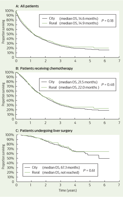
Among all patients, the median OS was 14.6 months for city patients and 14.9 months for rural patients (P = 0.18) (Box 4, A). Among patients receiving chemotherapy (with or without metastasectomy), the median OS was 21.5 months for city patients and 22.0 months for rural patients (= 0.48) (Box 4, B). For patients undergoing liver metastasectomy, the median OS was 67.3 months for city patients and was not reached in rural patients (P = 0.61) (Box 4, C).

Discussion

Our results demonstrate that rural patients with mCRC in SA receive comparable treatment and have equivalent survival to their metropolitan counterparts. In particular, patients in rural areas are treated with equivalent rates of potentially curative metastasectomy and chemotherapy, two key determinants of length of survival. These are the first Australian data specifically analysing rates of chemotherapy in rural patients with mCRC, and they suggest the excess colon cancer mortality seen in rural patients relates to factors other than access to treatment in the metastatic setting.

While there were no significant differences between the cohorts in rates of patients receiving chemotherapy across all lines of treatment, rural patients received less first-line combination chemotherapy, increased use of capecitabine and reduced use of biological agents in the first line than city patients.

First-line combination chemotherapy with intravenous infusional 5-fluorouracil, folinic acid and oxaliplatin (FOLFOX) has equivalent efficacy to oral capecitabine and oxaliplatin (XELOX).22 The choice between the two regimens is based on differing toxicities and practical considerations. FOLFOX requires a central venous catheter (CVC) and a second visit to a chemotherapy day centre every fortnight for ambulatory pump disconnection. XELOX has the advantages of single 3-weekly clinic visits and no CVC, but compliance with twice-daily chemotherapy tablets and potentially higher rates of symptomatic toxicity (hand–foot syndrome and diarrhoea) are limitations. The higher use of XELOX among rural patients reflects the relative practical benefits of this regimen where travel distances and access to nursing staff trained in CVC management are important considerations. The potential for toxicity of XELOX requires careful patient education and system approaches to enable early recognition and intervention in the event of severe toxicity among rural, often isolated, patients. Early follow-up telephone calls by a nurse practitioner or telemedicine consultations are potential strategies to provide this important aspect of care to rural patients.23,24

We observed a small but significant reduction in the rate of biological agents used in first-line therapy for rural patients, mostly due to reduced bevacizumab prescribing. It is possible clinicians were reluctant to “intensify” therapy in rural patients due to a lack of supervision or access to health care, particularly given risks of haemorrhage. It is also possible this small difference reflects a chance finding. The pattern of bevacizumab prescribing has evolved over the period captured in the registry, and an updated analysis of patients diagnosed since 2010 may provide further insights.

The equivalent rate of attempted curative metastasectomy in rural mCRC patients compared with city patients is reassuring, given this approach provides the only option for long-term survival in mCRC. The survival curves of patients undergoing liver metastasectomy showed a survival plateau at 5 years of 50% or greater for both city and rural patients (Box 4, C). This compares favourably with other modern surgical case series, with reported 5-year survival of 32%–47% after liver resection.25

Delivery of specialised health care services for rural Australians requires policymakers to carefully balance the merits of a centralised versus a decentralised system, with unique consideration for each region. For example, no regional centres in SA have a population sufficient to support a full-time resident medical oncologist and are instead serviced by a visiting (fly-in fly-out) oncologist. Limited infrastructure and staff training have also largely prevented widespread administration of chemotherapy in regional centres. Highlighting this point, we found that only 5.8% of rural patients receiving chemotherapy received their first cycle in a rural treatment centre. The SA Statewide Cancer Control Plan 2011–2015 lists the establishment of regional cancer services and chemotherapy centres as a key future direction to optimise care for rural cancer patients.18 Unfortunately, no publications have assessed outcomes of rural patients with mCRC treated in other regions of Australia, particularly in the eastern states where regional oncology services are common. While our analysis supports equivalent survival outcomes for rural patients treated within SA’s largely centralised service, the practical, social and economic advantages of regional cancer centres remain an important consideration not captured in our study. Given this, we consider that our findings highlight the positive outcomes achieved through high-quality, specialised care, rather than suggest that current regional services in Australia should also adopt a centralised approach.

As our analysis dichotomised patients into city and rural cohorts, it does not provide outcome information based on the degree of remoteness. Despite this limitation, chemotherapy and surgical treatment were almost entirely delivered in Adelaide, and thus our analysis appropriately distinguishes those patients who had to travel to access oncological care. The possibility of inadequate registry ascertainment of rural cases of mCRC also poses a possible limitation. However, we are confident this is not a source of bias, as the registry collects information from all histopathology reports in SA, which are processed centrally in Adelaide. An important limitation of our study is that we report only on mCRC, and stage I–III disease is not included. The impact of treatment differences in early-stage CRC (eg, quality and timeliness of surgery, use of adjuvant chemotherapy) on overall survival of patients with mCRC cannot be determined in this analysis. Reassuringly, however, about two-thirds of mCRC cases in both cohorts were synchronous (ie, no prior early-stage disease), suggesting this is unlikely to limit our conclusions. Further, the equivalent rates of synchronous diagnosis in rural and urban patients may suggest there was no major delay in diagnosis of rural patients.

Although higher cancer incidence and poorer outcomes have been consistently demonstrated for rural cancer patients in Australia, we found equivalent treatment patterns and survival for rural patients diagnosed with mCRC in SA since 2006 compared with their metropolitan counterparts. This confirms optimal treatment of rural patients results in equivalent outcomes to metropolitan patients, irrespective of disadvantage. Further, it suggests previously demonstrated disparate outcomes may be due to factors such as higher incidence of CRC as a result of burden of risk factors and potentially reduced screening participation, rather than treatment factors once mCRC has been diagnosed. Targeting these factors is likely to provide the greatest impact on reducing the excess cancer burden for rural Australians.

1 Patient characteristics, by city versus rural residence (n = 2289)*

Characteristic

City

Rural

P


No. (%) of patients

1665 (72.7%)

624 (27.3%)

Median age (range), years

73 (17–105)

72 (31–100)

0.11

Sex


Male

893 (53.6%)

391 (62.7%)

< 0.001

Female

772 (46.4%)

233 (37.3%)


Primary site


Colon

1260 (75.7%)

446 (71.5%)

0.04

Rectum

405 (24.3%)

178 (28.5%)


Synchronous disease

1070 (64.3%)

407 (65.2%)

0.67

Site of metastasis


Liver only

665 (39.9%)

226 (36.2%)

0.10

Lung only

128 (7.7%)

45 (7.2%)

0.70

Liver and lung only

178 (10.7%)

65 (10.4%)

0.85

All other sites

694 (41.7%)

290 (46.5%)

0.13

> 3 metastatic sites

138 (8.2%)

54 (8.7%)

0.38

KRAS testing

243 (14.6%)

87 (13.9%)

0.77

KRAS wild-type

145 (59.7%)

52 (59.8%)

0.96

Clinical trial participation

154 (9.2%)

44 (7.1%)

0.10


KRAS = Kirsten rat sarcoma viral oncogene homolog. * Data are number (%) of patients unless otherwise indicated. † P values calculated using χ2 tests.

2 Frequency of first-line, second-line and third-line chemotherapy, and regimens, by city versus rural residence


First-line treatment


Second-line treatment


Third-line treatment


Regimen

City

Rural

P

City

Rural

P

City

Rural

P


Total

933 (56.0%)

364 (58.3%)

0.32

217 (23.3%)*

82 (22.5%)*

0.78

94 (10.1%)*

34 (9.3%)*

0.69

Single-agent chemotherapy

271 (29.0%)

118 (32.4%)

0.23

58 (26.7%)

18 (22.0%)

0.40

21 (22.3%)

7 (20.6%)

0.83

Capecitabine

202

82

0.30

24

4


8

0


5-FU

58

31

0.29

3

3


4

1


Irinotecan

11

3


31

11


9

6


Oxaliplatin

0

2


Combination chemotherapy

629 (67.4%)

218 (59.9%)

0.01

115 (53.0%)

44 (53.7%)

0.92

49 (52.1%)

17 (50.0%)

0.83

FOLFOX

491

146

0.001

21

8


14

2


XELOX

53

50

< 0.001

15

4


8

2


FOLFIRI

76

18

0.12

62

26


15

7


XELIRI

1

0


1

2


2

2


MMC–5-FU or capecitabine

8

4


16

4


10

4


Other

33 (3.5%)

28 (7.7%)


44 (20.3%)

20 (24.4%)


24 (25.5%)

10 (29.4%)


Biological agent

221 (23.7%)

61 (16.8%)

0.007

97 (44.7%)

30 (36.6%)

0.21

72 (76.6%)

34 (100%)

0.003

Bevacizumab

185

52


60

22


16

14


EGFR mAb

15

5


26

8


52

19


Other

21

4


11

1


4

1


5-FU = 5-fluorouracil. FOLFOX = folinic acid–5-FU–oxaliplatin. XELOX = capecitabine–oxaliplatin. FOLFIRI = folinic acid–5-FU–irinotecan. XELIRI = capecitabine–irinotecan. MMC = mitomycin C. EGFR mAB = epidermal growth factor receptor monoclonal antibody. * Total rates of second-line and third-line chemotherapy use are expressed as a percentage of patients who received any chemotherapy. † Includes use of raltitrexed and MMC (as single agent and combination).

3 Frequency of non-chemotherapy treatments, by city versus rural residence

Treatment

City (n = 1665)

Rural (n = 624)

P


Lung resection

53 (3.2%)

13 (2.1%)

0.10

Hepatic resection

228 (13.7%)

72 (11.5%)

0.17

Surgery*

858 (51.5%)

345 (55.3%)

0.11

Ablation

12 (0.7%)

3 (0.5%)

0.53

Selective internal radiation therapy

10 (0.6%)

8 (1.3%)

0.10

Radiotherapy

299 (18.0%)

132 (21.2%)

0.08


* Includes resection of colorectal primary cancer.

4 Overall survival (OS) in city versus rural patients

Should general practitioners order troponin tests?

Cardiac troponin I and T are the preferred biomarkers for assessing myocardial injury. Understanding the pathobiology of troponin and the timing of troponin testing is fundamental to the clinical utility of these biomarkers, as troponin and its kinetics are central to the universal definition of acute myocardial infarction (AMI).1 Troponin levels become elevated in serum within a few hours of an AMI, and they remain elevated for up to 7–10 days.2 However, numerous other conditions may elevate troponin levels, so it remains essential that the results of troponin tests be interpreted with clinical findings and electrocardiography results.3 The dynamics of troponin levels (rise and/or fall over time) help distinguish AMI from non-AMI conditions, thus serial troponin testing is the standard approach recommended for assessing patients with suspected acute coronary syndrome (ACS).4 In this article, we explore troponin testing in general practice, including problems faced by laboratories that offer testing in this context.

The absolutist stance

One stance on this topic is that general practitioners should never order a troponin test. The basis of this argument is that the only widely accepted clinical indication for measurement of troponin levels is suspected ACS, which should prompt referral to hospital based on clinical and electrocardiography findings without recourse to troponin testing. Supporting this argument is that serial troponin testing is unrealistic in most general practice settings, and opens the question of how a patient should be monitored while the results are awaited.

One argument against this absolutist stance is that it is an oversimplification. Further investigation and management depends on the degree of suspicion for ACS, and timing of the presentation may obviate the need for serial testing. Chest pain is a challenging symptom and the prevalence of unstable angina or AMI in general practice is low, in the order of fewer than 5% of patients with chest pain.5 Atypical presentations of AMI, such as in young people,6 people with diabetes and older people, are a perennial concern. GPs have been shown to be fairly accurate in assessing chest pain clinically as due to coronary artery disease, but not accurate enough to safely exclude it.7 A system of estimating pretest probability of ACS, or risk of short-term complications, is an attractive approach. Clinical decision-making rules and pretest probability tables have been developed to assist with this process in general practice8,9 and, while some risk stratification tools may be more relevant to doctors in emergency departments, they are potentially useful to GPs.10,11 How troponin testing might fit into risk stratification in general practice is not entirely clear.

How well do GPs understand troponin tests?

Audits from New Zealand suggest that GPs have a generally sufficient understanding of the use of troponin tests in primary care.12,13 Knowledge of false-negative results (eg, due to sampling too soon after symptoms) appears to be better than knowledge of false-positive results (eg, due to non-AMI causes of raised troponin levels). Most GPs would refer high-risk patients without troponin testing, but a small proportion of GPs would defer hospitalisation while waiting for the troponin result (mostly for patients with an intermediate probability of AMI).

Why do GPs request troponin tests?

Our experience suggests that GPs mainly request troponin tests to rule out AMI in one of two situations. The first situation is patients who had symptoms in the preceding days but for whom symptoms have resolved (also the experience of others13,14). One expert has suggested that this may be a justifiable use of troponin testing in primary care15 and troponin testing is suggested in National Institute for Health and Care Excellence (NICE) primary care guidelines in pain-free patients who had chest pain more than 72 hours earlier.9 The second situation is patients who have atypical symptoms and/or a low likelihood of ACS, in whom troponin testing appears to cover the residual clinical uncertainty. Unexpectedly positive troponin results occasionally occur in such situations, which may otherwise not be detected.

How do GPs currently request troponin tests?

Most requests for troponin testing from general practice are requests for a single test, not serial testing.16 This begs the question of whether ordering a single troponin test is an appropriate strategy. Given our understanding of troponin kinetics, a single negative troponin test result a certain time after symptom onset could be clinically useful in ruling out AMI (ie, in “late presenters”). The suggested time frame varies between publications, but is usually in the order of 6–9 hours4,17 with the caveat that the time of symptom onset can be unreliable. Local experts have suggested that a single troponin test 12 hours after resolution of suggestive symptoms (with a normal electrocardiogram and no high-risk features) is useful for this purpose.11 With the so-called high-sensitivity troponin assays, this window may decrease: in an emergency department setting, an undetectable (ie, not merely negative) troponin value obtained from a high-sensitivity troponin assay at presentation has been shown to have a very high negative predictive value for a subsequent diagnosis of AMI,18 but this strategy is experimental. The safest rule of thumb is that a single negative test result for troponin does not exclude AMI in a patient with current or very recent symptoms, nor does it exclude clinically significant coronary artery disease.

Conditions associated with chronic troponin elevation

As most GP requests for troponin testing are for a single test, conditions associated with chronic, non-AMI elevation of troponin levels present a problem. Examples include chronic cardiac failure and chronic kidney disease (CKD). A positive result from a single troponin test could be misleading because it might reflect the underlying chronic disease and not AMI. The prevalence of positive troponin test results (defined as above the 99th percentile of the general population) in CKD depends on the stage of the CKD (positive results are more likely during more advanced stages) and on the troponin assay used. This is exemplified by a recent study of asymptomatic patients who had CKD but were not on dialysis. The prevalence of a positive troponin result (for the whole cohort) was 68% when a high-sensitivity troponin T assay was used, 38% when a high-sensitivity troponin I assay was used, and 16% and 8% for troponin T and I, respectively, when contemporary (“less sensitive”) assays were used.19 Despite the high rates of positive troponin results in this study, a negative troponin result from a sample taken at an appropriate time is useful for ruling out AMI in patients who have CKD, but at the considerable disadvantage of reduced positive predictive value, with the attendant risk of unnecessary hospitalisation. Clinical assessment of the acute event in such patients becomes all the more important if this is to be avoided.

Logistics of troponin testing for outpatients

Offering troponin testing in the community is logistically complex and there is a lack of formal guidance for laboratories in this area. Guidelines on management of ACS recommend that a troponin test result should be available within 60 minutes of blood being drawn and, if not, that point-of-care testing should be available.4 This is aimed at hospital-based laboratories and is not a realistic target for large private pathology networks that may test hundreds of community samples per day at variable geographical distances from large networks of collection centres and general practices. So what is the solution? Accept the longer turnaround times and promote judicious use of troponin tests by GPs? Longer turnaround times may be acceptable if testing is largely confined to patients who have a low pretest probability, or low risk, of AMI. If so, what is a reasonable turnaround time for community samples — three hours? Six? At the other extreme is rigorous pursuit of fast turnaround times to meet the apparent clinical need in the community, probably with the help of point-of-care testing, although there are questions about the performance of point-of-care troponin assays.20 The solution is probably a compromise between the two. The only guidelines that provide advice on this are the NICE guidelines, which state that troponin testing can be undertaken in general practice “providing timely results can be obtained” but do not elaborate on what “timely” means.9

After-hours elevated troponin levels can be problematic for all concerned. For example, when samples are taken late in the afternoon, results might not be available until after clinic hours. A common policy is to treat positive troponin test results as “critical results” and to notify the requesting doctor or a representative (such as a locum GP service, if nominated). In the event that a doctor cannot be found to take the result, which is not uncommon in our experience, laboratory staff (usually pathologists) phone patients directly and advise that hospitalisation is the safest course of action. But when a patient cannot be contacted, laboratory staff face a dilemma: can the result wait until office hours, or should emergency services be arranged? We are aware of anecdotal cases in which after-hours notifications of high troponin levels to patients at home have probably contributed to their early survival — but this raises the question of whether such patients are better served by referral to hospital in the first instance. A published coroner’s case touches on these important issues for both GPs and pathologists.21

Conclusion

We suggest that GPs should have a high threshold for requesting troponin testing and carefully assess risk before ordering troponin tests. Positive troponin test results usually change the course of management, but the time frame in which the result becomes available must be balanced against the risk of delay in diagnosis and therapy. A troponin test should not be requested unless a GP is certain that a robust process is in place by which they can be contacted, day or night, if the result is positive. There is an obvious need for further education, research and inclusion of this topic in future clinical guidelines. Our suggestions for using, or not using, troponin tests in general practice are summarised in the Box.

Suggestions for using, or not using, troponin tests in general practice

The default position

  • The default position for patients who have symptoms suggestive of acute coronary syndrome is hospitalisation without prior troponin testing.

Using a single troponin test

  • It is reasonable to use a single troponin test in general practice to exclude the possibility of acute myocardial infarction (AMI) in asymptomatic patients whose symptoms (typical or otherwise) resolved at least 12 hours prior, so long as they have no high-risk features and a normal electrocardiogram.11
  • A single troponin test may also be useful to investigate an otherwise unexplained creatine kinase elevation.

Using serial troponin tests

  • In patients presenting to general practice within 12 hours of symptom onset who are at low risk of AMI and/or have atypical symptoms, and for whom troponin testing is being considered, serial testing is advised.
  • In patients with conditions that are associated with a high prevalence of positive troponin test results, such as chronic kidney disease, a single test can be misleading. Serial testing may be required to resolve clinical uncertainty.
  • Serial testing is most appropriately performed in hospital. The safety of serial testing in outpatient settings has not been established.

Skin rash, a kidney mass and a family mystery dating back to World War II

This case of hereditary leiomyomatosis and renal cell cancer in a young man illustrates the importance of considering a hereditary basis for renal cancer in a young patient, and highlights how targeted therapy underpins modern personalised medicine in renal oncology.

Clinical record

A 25-year-old white man presented with a year-long history of malaise, a palpable right loin mass and a painless nodular rash over his back of indeterminate duration. Computed tomography (CT) showed a right renal mass with para-aortic and retroperitoneal lymphadenopathy. The patient underwent radical nephrectomy and para-aortic lymphadenectomy.

Examination of the kidney showed a 14 cm tumour centred in the renal medulla (Box, A). The tumour cells were organised in papillary fronds and featured abundant eosinophilic cytoplasm, large nuclei and prominent inclusion-like eosinophilic nucleoli (Box, B). There was widespread vascular space invasion and involvement of the renal sinus fat and hilar vein. All four para-aortic lymph nodes contained metastatic disease.

The patient’s longstanding skin rash (Box, C) was reviewed and confirmed on biopsy to represent multiple cutaneous leiomyomata.1 The patient’s mother was found to have had multiple cutaneous and uterine leiomyomata. The death certificate of his maternal grandfather stated that he died of metastatic renal carcinoma at the age of 44. He had returned from active service in World War II with a mysterious and longstanding rash, the pattern of which was consistent with cutaneous leiomyomatosis. On this basis a provisional diagnosis of hereditary leiomyomatosis and renal cell cancer (HLRCC) was made.1

After the patient underwent genetic counselling and provided informed consent, blood was drawn for molecular analysis of the fumarate hydratase (FH) gene locus by Sanger sequencing and multiplex ligation-dependent probe amplification assay (MLPA). MLPA showed complete deletion of the FH gene in one allele, confirmed by comparative genomic hybridisation on an Agilent custom oligonucleotide array (Agilent Technologies Inc) to be a 0.5 Mb deletion including FH and OPN3. No further cascade genetic testing has been undertaken in this family to date.

The patient’s postoperative recovery was uneventful. Fluorodeoxyglucose positron emission tomography 6 months after surgery showed multiple hepatic and left flank chest wall subcutaneous metastatic deposits. There was no evidence of recurrence in the nephrectomy bed. First-line therapy with sunitinib (50 mg daily, orally) was commenced and well tolerated.

Eighteen months after the patient’s surgery, CT imaging showed a reduction in the volume of the hepatic tumour and complete regression of the previously identified extra-hepatic disease. Subsequent planned subtotal hepatectomy was aborted due to the size and location of hepatic disease apparent only at exploratory laparotomy. Therapy was changed to everolimus (10 mg daily, orally). This resulted in symptomatic anaemia requiring transfusion. A restaging CT scan performed 8 months after commencement of therapy with everolimus showed progression in the size of the hepatic metastatic deposits.

Treatment was then changed to combination therapy with bevacizumab (15 mg/kg intravenous infusion every 3 weeks) and erlotinib (150 mg daily, orally) based on the treatment arm of a current National Cancer Institute phase II clinical trial (NCI-10-C-0114). The disease was stable for 16 months after commencement of the combination regimen, with the patient able to return to full-time employment. However he subsequently developed progressive disease and, despite a brief trial of sorafenib, died with widespread metastases 64 months after his initial presentation.

Discussion

At least 4% of renal cancers are hereditary, and there are known associations with germline mutations of VHL, c-MET, SDHx and FLCN loci and constitutional translocations of chromosome 3.2 Therefore, the possibility of hereditary disease should always be considered in people who present with renal carcinoma at a young age.

HLRCC is an autosomal dominant syndrome characterised by an inherited predisposition to cutaneous and uterine leiomyomata and renal cancer. Although multigenerational families with cutaneous and uterine leiomyomata were first described in the 1950s,3 the syndromic association with renal cancer was not appreciated until 2001 when two Finnish kindreds with a predisposition for cutaneous and uterine leiomyomata and early onset renal cancers were reported.4 Renal tumours occurred in four members of the index family in their 30s; all tumours were unilateral solitary lesions that had metastasised at the time of diagnosis, and displayed papillary architecture on histological examination. Using genome-wide linkage analysis, it was possible to map the genetic abnormality to the long arm of chromosome 1 (1q24). Subsequent studies have shown that this locus encodes FH, an enzyme involved in oxidative respiration through the Krebs cycle.5,6 The gene appears to function as a tumour suppressor in patients with HLRCC, in keeping with Knudson’s two-hit hypothesis.7

The distinctive histological features of HLRCC renal tumours first described in 20014 have recently been corroborated in a review of 40 tumours resected from members of HLRCC-affected families. It was primarily that we recognised this distinct morphology (a papillary architecture with strikingly prominent nucleoli)1 that subsequently allowed us to make a definitive diagnosis by molecular confirmation in this case.

Given the rarity of HLRCC, no consensus diagnostic criteria currently exist for the condition. The presence of cutaneous leiomyomata correlates strongly with the presence of germline mutations in the FH gene.8 Cutaneous leiomyomata tend to occur more commonly in women, are typically sensitive to cold or abrasion, and develop in the second and fourth decades of life as intradermal lesions with a disseminated or segmental distribution.6 Although uterine leiomyoma is a common incidental finding in adulthood, the presence of multiple large symptomatic uterine leiomyomata further increases the likelihood of HLRCC. The histology of these cutaneous and uterine smooth muscle tumours is typically bland. Although leiomyosarcomas have been described in patients with FH mutations, this clinical association is rare and appears predominantly confined to people of Finnish origin.9 In addition to renal tumours with distinctive HLRCC papillary morphology, collecting duct carcinomas have also been described in association with HLRCC.10

More than 120 families have been reported with presumed pathogenic mutations in the FH gene, with mutations being predominantly missense, nonsense and frameshift changes. No correlation between genotype and phenotype has been reported: mutations that result in reduced FH activity are anticipated to produce a similar phenotype irrespective of mutation class.6 Two whole-gene deletions, which were 2.4 Mb and 1.9 Mb in size, were identified in one cohort of patients, although no phenotype data was given.5 One other whole-gene deletion was recently reported, with three affected family members having a combination of cutaneous and uterine leiomyomata, but no renal tumours.11

In-vitro studies suggest that FH-mutated renal tumours and von Hippel–Lindau (VHL) renal tumours share a common tumorigenic pathway, via dysregulation of hypoxia-inducible factor (HIF).12 The VHL protein couples changes in oxygen availability to gene expression through the regulation of HIF. Inactivation of the VHL gene results in increased HIF activity which, through increased expression of vascular endothelial growth factor (VEGF), platelet-derived growth factor ß (PDGFß) and transforming growth factor α (TGFα), plays a critical role in tumorigenesis. Because of the similar downstream effects of FH and VHL mutations, these factors represent rational therapeutic targets in HLRCC.

Sunitinib is a small-molecule multitargeted tyrosine kinase inhibitor that acts on VEGF and PDGFß in the context of renal cancers, while bevacizumab is a humanised monoclonal antibody targeting VEGF. Everolimus inhibits the mammalian target of rapamycin (mTOR), which is involved in HIF regulation.13 The use of these targeted therapies in advanced renal cell carcinoma has resulted in significant gains in progression-free survival compared with cytokine therapy or placebo, providing proof of principle for exploiting HIF-dependent pathways therapeutically. Inhibition of the epidermal growth factor receptor, a receptor tyrosine kinase through which TGFα acts, is being explored as a means to overcome TGFα overexpression. Both antibodies and small-molecule inhibitors, such as erlotinib, are under investigation.14

In summary, this case highlights the importance of recognising the unique pathological and clinical features of hereditary renal cancer. Not only does this facilitate early detection and genetic counselling in family members, in this age of personalised medicine such recognition may also serve to guide therapy.

Anatomical and histological features of the patient’s renal cancer, and a photograph showing his skin rash


A: A 14 cm tumour in the renal medulla. B: Haematoxylin and eosin stained section of the renal tumour showing cells organised in papillary fronds with abundant eosinophilic cytoplasm, large nuclei and prominent inclusion-like eosinophilic nucleoli. C: The patient’s skin rash, which was confirmed on biopsy to represent multiple cutaneous leiomyomata.

Medical claims for diagnostic imaging and pathology provided to patients in public hospital emergency departments

Diagnostic imaging and pathologist specialist members should be aware they are legally responsible for all services claimed under Medicare that are billed under their provider number or in their name, even if the billing was done by hospital administration.

Pathology and diagnostic imaging services for patients in public hospital emergency departments are covered by Australian Government funding arrangements and are not eligible for Medicare benefits. 

Emergency patients are to be treated as public patients until a clinical decision to admit has been made and the patient has elected to be admitted as a private patient.

More information about this is available from the Medicare website:  https://www.medicareaustralia.gov.au/provider/business/audits/public-hospital-emergency-depts.jsp

The AMA provides the following advice to members about the use of provider numbers in public hospitals:

·         where medical services claimed against Medicare are being rendered in public hospitals under a medical practitioner’s name and billing provider number, the practitioner must be made fully aware of, and be prepared to accept responsibility for, that billing;

·         where services claimed are being rendered in a public hospital, medical practitioners should seek a written guarantee from the hospital that the arrangement is not in breach of the relevant Australian Health Care Agreement; and

·         public hospitals must provide doctors with full records of all medical accounts raised in their name.

If you believe your provider number may have been used in ways that contravene the relevant provisions in the Health Insurance Act, the AMA recommends you contact your State AMA Office. As this issue affects employment contracts, State AMAs will be able to coordinate representation for affected members.

The AMA will keep abreast of Medicare compliance matters through its participation on the DHS Compliance Working Group.

Code opens window on conduct

In vitro diagnostic technology companies must clearly qualify any advertised claim that their products are safe, under updated ethical rules adopted by the peak industry association IVD Australia.

Companies that manufacture and supply in vitro diagnostics have also been told any hospitality provided to health professionals should be “limited”, and any gifts given must be modest.

The recommendations are made in the 2nd edition of the IVD Australia Code of Conduct, which was formally adopted in October last year following an extensive review process involving submissions from both internal and external stakeholders.

The Code aims to establish minimum standards of behaviour for the industry to help it regulate itself, including the conduct of relationships with health care professionals.

Among issues addressed in the revised code was a clearer definition of what constitutes a gift (providing product samples is not considered to be gift-giving), as well as what information and terminology should be included in ads, (the term “safe” should not be used unless clearly qualified, and the term “new” may only be used in the first 12 months of promotion).

In a new addition to the code, member companies are advised that any promotion of a product, directly or indirectly, through social media is considered advertising, and should be subject to the same rules as other forms of promotion.

Where a company commissions an article, it must be clearly and prominently identified, and any claims made must be referenced.

Where firms engage health care professionals as consultants, they are advised that any payments made must be “reasonable”, and sales and promotional meetings must be conducted in labs, training institutions, medical facilities or other appropriate venues, with only modest hospitality costs to be incurred.

In instances where health professionals own a material or significant interest in a member company, the Code advises that any conflict of interest be managed in a way to avoid compromising public trust, and health professionals are advised to disclose any such interest to a patient when recommending a product marketed by a firm in which their interest is held.

A copy of the code can be viewed at: http://www.ivd.org.au/about-us/code-of-conduct/

Adrian Rollins