×

Eliciting and responding to patient histories of abuse and trauma: challenges for medical education

Toward trauma-informed medical education

Traumatic experiences such as childhood abuse, family violence, elder abuse and combat exposure influence both physical and mental health, health-related behaviour, and the ways in which patients interact with medical practitioners.1,2 Despite greater knowledge of the pervasive sequelae of psychological trauma, the implications for medical practice and for medical education are not well articulated. Many doctors lack confidence and remain ill-informed or avoidant when dealing with patients’ psychological trauma.3,4 The consequences of this include non-recognition of somatisation and of psychiatric disorders, delay in instituting proper treatment, and costs to the patient and health care system of unnecessary investigations and treatments.5,6 Here, we discuss why and how we should better train doctors to elicit and respond to patient histories of trauma.

High prevalence of trauma and its clinical sequelae

The lifetime prevalence of exposure to traumatic events is high (74.9% in Australian adults).7 Most people who experience trauma do not develop mental illness; however, trauma and abuse are substantial contributors to the burden of mental and physical ill health. The risk of post-traumatic stress disorder after trauma is about 10%,8 but childhood abuse and neglect in combination with later life stress contribute to the development of mental illnesses as diverse as psychoses, depression, eating disorders and addictions, as well as a range of physical illnesses.2,9 There are also clear associations between past trauma and abnormal illness behaviour10 and, related to this, increased health care use.6 These sequelae of patient trauma pervade all medical specialties and also dentistry.1

Incorporating teaching about trauma in medical curricula: key issues

Although there are several studies that describe training interventions for specific forms of trauma,3,11,12 there is little in the literature on current practices in medical education, either in Australia or elsewhere. The diversity in general structure, content and methods of medical curricula as outlined by the Australian Medical Council (AMC) probably extends to trauma-relevant components.13 Despite this diversity, it is possible to offer initial considerations for trauma-informed education. We focus on six interrelated aspects: communication skills; knowledge of the health effects of trauma and abuse; knowledge about the effects of trauma and abuse disclosures on doctors and other health professionals; specific knowledge relevant to different medical specialities and settings; teaching formats and methods; and the need for a staged, incremental, integrated program, structured to achieve continuity between undergraduate, prevocational and specialist phases.

Communication skills curricula13 in pre-clinical and clinical phases afford opportunities for trauma-specific education, but are also relevant to junior hospital and specialty training. Common issues that need to be addressed include the personal discomfort many doctors experience asking about trauma and abuse; when not to screen for or discuss trauma; and when to seek advice from senior colleagues, as overconfidence can be harmful, leading to patient distress and even re-traumatisation. Communication skills education should extend to discussion of services relevant to different forms of trauma, such as social work, refuges, police and the courts. Here, it would be valuable for medical students to visit these settings or meet with workers from them.

Training needs to be realistic in that doctors often work in settings that are not conducive to asking about trauma, such as busy emergency departments, hospital wards that lack privacy, and overloaded outpatient clinics, often with a lack of psychiatric support. However, these realities should not be a pretext for avoiding clinically competent trauma-informed practice. We do not propose that doctors become trauma therapists; rather that they become competent in empathically recognising and eliciting information about trauma, and at effective referral of patients to relevant services, including psychology and psychiatry.

Exposure to patients who have experienced trauma or abuse evokes a range of psychological reactions in students and trainees, from normal discomfort and distress through to vicarious traumatisation.3,4,14 In addition, there may be unhelpful, if not harmful, responses: doctors may adopt an avoidant “don’t ask, don’t tell” style; over-investigate; refer patients to other clinicians; exhibit stigmatising attitudes toward patients; or become over-involved.4 Course content on these issues could be introduced early and developed further during clinical and postgraduate phases.

There are many opportunities pre-clinically to learn about the effects of abuse and trauma on human development, including the short and long term effects on the brain and behaviour. Relevant knowledge can be taught within sections of the curriculum devoted to neuroscience, cognitive science and population health. In addition, medical humanities have a powerful capacity to expand our knowledge and understanding of diverse human experiences, including trauma, and to foster empathy. Every clinical specialty that medical students and trainees encounter in hospital training brings opportunities to learn specialty-specific trauma knowledge and skills. For example, rotations in paediatrics and in obstetrics and gynaecology are opportunities for teaching about child abuse and about family violence.

Trauma is an everyday part of clinical discourse within psychiatry and a key dimension of academic and clinical learning in psychiatry rotations. However, trauma-informed education is relevant to all clinical specialties. Relevant specific knowledge encompasses common clinical presentations of trauma in those specialties; how trauma-relevant inquiry can be embedded within the specialty-specific clinical interview; clinical, social and legal services relevant to various forms of abuse; and legal requirements regarding mandatory reporting.

Some medical students and trainees have their own experiences of trauma, including childhood trauma and abuse, but also vicarious trauma stemming from clinical encounters, such as witnessing horrific physical injury or disfigurement. Post-traumatic stress disorder in medical practitioners is often unrecognised.14 Such experiences may increase or may impair empathic capacity to engage with traumatised patients.

Although the AMC standards discuss the stressful and traumatic nature of medical work and provide recommendations about availability of counselling, peer support and other measures, they construe the reality of trauma as external to the core business of medical education.13 Instead, we propose that learning about the emotional impacts of clinical work should be core medical education, to be dealt with in lectures, tutorials, simulations — that is, a range of appropriate, complementary educational methods, as is done for other topics. In addition, curricula should include safe, confidential, non-coercive opportunities for experiential learning in small groups, allowing participants to reflect on and share their own emotional reactions to patients and understand how these reactions can shape their clinical practice. Ideally, some teaching should be conducted jointly with students from other disciplines, notably nursing.

The AMC standards stress the centrality of clinical clerkships in the development of clinical competence and judgement.13 We agree, but when it comes to trauma-informed clinical teaching, there are several entailments. Teaching about trauma has to become a routine, everyday feature of clinical teaching in wards and clinics, not something outsourced by referral to psychiatry or social work. Clinician teachers in all specialties need to acquire the skills to do such teaching and to act as role models. Given the limited evidence base, it is premature to recommend a mix or staging of methods, and this should be a focus of future research and curriculum innovation.

The medical education literature is marked by separate discourses on differing forms of trauma. For example, education regarding intimate partner violence has been extensively explored and excellent curricula have been implemented.3,13,14 However, patients have often experienced several concurrent or sequential traumas; and the clinical sequelae of different traumas have many similarities, demand similar clinical skills (albeit allied with different bodies of specific knowledge), and thus present similar challenges for medical education. Valuable educational synergies are likely if the currently disparate, unconnected trauma-relevant elements in medical curricula are integrated.

These considerations point to the need for the creative design and evaluation of staged, incremental, integrated programs, structured to achieve continuity between undergraduate, pre-vocational and specialist phases of medical education. We do not propose removing the various trauma-specific educational components from medical curricula and replacing them with some form of generic trauma education. We do propose, however, examining creatively how they may be better integrated to become mutually reinforcing.

Conclusion

Trauma-informed health care is an invaluable concept which we propose should extend to trauma-informed medical education.15 Although the arguments for trauma-informed medical education are compelling, new lines of educational research will be needed to guide curriculum design and build on the small body of work already available.3,4,11,12,16 It is likely that if doctors of all kinds have the knowledge, skills and attitudes to deal competently with abuse and trauma, we can expect improvements in patient care and health service costs, and in the health and wellbeing of medical practitioners. These possibilities deserve empirical study. As well as becoming better clinicians, medical students and trainees will also become better teachers and role models and, as they move into more senior and leadership roles, advocates for competent trauma-informed medical care.

Towards an integrated model for child and family services in central Australia

An innovative model for the delivery of child and family services

The Central Australian Aboriginal Congress is a large Aboriginal community-controlled health service based in Alice Springs in the Northern Territory. Since the 1970s, Congress has developed a comprehensive model of primary health care delivering evidence-based services on a foundation of cultural appropriateness.

In recent years, the community-elected Congress Board has focused on improving the developmental outcomes of Aboriginal children. This has led to the development of an innovative model for the delivery of child and family services, based on the belief that the best way to “close the gap” is to make sure it is not created in the first place.

Early childhood development

It is well established that social and environmental influences in early childhood shape health and wellbeing outcomes across the life course. Adverse childhood experiences are correlated with a wide range of physical health problems and with increased levels of depression, suicide attempts, sexually transmitted infections, smoking and alcoholism.1

The pathways for these effects are complex; however, we know that during the first few years of life, the interactions between genetics, environment and experience have a dramatic impact on brain development. During this critical period, children need stimulation and positive relationships with caregivers to develop the neural systems crucial for adult functioning.2

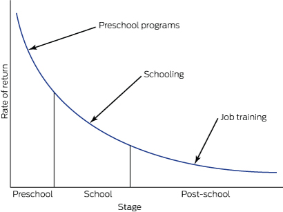
This evidence indicates that we should not wait to intervene until a child is ready for school at around 5 years of age. By this stage, children have passed many developmental gateways for language acquisition, self-regulation and cognitive function, and their developmental trajectories are set. Of course, developmentally challenged children must be provided with appropriate services during their school years and later in life, but such interventions require increasing amounts of resources (Box 1)3 and produce diminishing returns as the child gets older.4

Governments and policy makers have now widely recognised the importance of investing in the early years.5 The Organisation for Economic Co-operation and Development has advised that investing in early childhood is the most important measure that Australia can take to grow the economy and be competitive in the future.6

Successful programs

There are well evidenced programs for young children and their families that significantly improve health, educational and social outcomes throughout the life course, and which are highly cost-effective compared with later interventions (or with doing nothing, which is the most expensive option).

Congress has taken particular interest in two preventive programs that have been successfully implemented in disadvantaged communities overseas. These are the Nurse–Family Partnership (NFP) and the Abecedarian approach to educational day care. Both programs work with caregivers and children before developmental problems arise, providing children with the stimulation, quality relationships and access to the services they need for healthy development.

The NFP is a program of nurse home visitation which begins during pregnancy and continues until the child’s second birthday. Pioneered in the work of Olds and colleagues,7 trained nurses use a structured program to address personal and child health, quality of caregiving for the infant, maternal life course development and social support. Special attention is given to establishing a safe, nurturing and enriched parent–infant relationship. Over many years of working with low income, socially disadvantaged families in the United States, the NFP has achieved improvements in women’s prenatal health8 and reductions in child abuse and neglect, maternal use of welfare, substance misuse, and contact with the criminal justice system.9 Children who participated in the program showed long term benefits, such as reduced antisocial behaviour and substance misuse during young adulthood.10

The Abecedarian approach provides a centre-based preventive program for children who are at high risk of developmental delay. It has three main elements — learning games, conversational reading and enriched caregiving — with a priority on language acquisition. The approach has been rigorously evaluated, and longitudinal studies that followed children into adulthood found that participants did better at school; gained more years of education; had better employment outcomes;4 showed reduced rates of smoking, drug use and teen pregnancies and led a more active lifestyle.11 There is also evidence of a significantly lower prevalence of risk factors for cardiovascular and metabolic disease for participants (particularly men) when they reach their mid-30s.12 These effects work against the social gradient, with children from more disadvantaged environments benefitting the most,13 which makes the approach a potentially powerful contributor to social equity.

The situation in central Australia

While the cultures and histories of Aboriginal communities in central Australia make them unique, they share many characteristics with communities in which these programs have been effective.

In particular, many Aboriginal children in and around Alice Springs grow up in an environment marked by poverty, substance misuse and lack of responsive care, with low levels of formal education and school attendance coupled with economic marginalisation and social exclusion.

This is reflected in figures from the Australian Early Development Census (AEDC), which show that by the time they start school, 43% of Indigenous children in the Alice Springs region are vulnerable on two or more of five developmental domains. This is six times the rate for non-Indigenous children (7%) (NT AEDC Manager, Early Childhood Education and Care, NT Department of Education, personal communication, 1 April 2016).

Responding to the developmental needs of Aboriginal children

The Congress integrated model for child and family services is the culmination of its efforts to develop an innovative service response to these challenges based on the best available evidence (Box 2).The key elements of the model include nurse home visitation through the Australian Nursing Family Partnership Program (ANFPP); the Preschool Readiness Program (PRP); the Healthy Kids Clinic; family support services, such as Targeted Family Support and Intensive Family Support; and the Child Health Outreach Program. Congress also runs a childcare centre.

As part of this model, Congress has delivered two short-term programs drawing on the Abecedarian approach. The first was an intensive intervention run through the PRP in 2011 and 2012.14 In the second, Congress is collaborating with the University of Melbourne to assess the impact of a limited implementation of the Abecedarian approach with children attending the Congress childcare centre.

The importance of integration

The Congress model is founded on a long term population health approach that is expected to deliver results in health and wellbeing across the life course. Integration of services under a single provider is the key to achieving this potentially transformative change, enabling children and families to be referred seamlessly to the services that best meet their needs. Such integration is now recognised as a crucial reform needed to increase the cost-effectiveness of services and improve access and outcomes for children and their families.15

The advantages of the Congress integrated model are that:

  • it supports a consistent approach to screening, allowing children and families to be referred to the programs that best meet their needs;

  • it allows internal efficiencies between programs to enhance services, thus making a better use of available resources;

  • it is built on the existing relationships that Congress has with Aboriginal families in Alice Springs through the delivery of culturally appropriate primary health care;

  • it allows a common evidence-based approach, modified to meet the cultural and social needs of our clients;

  • it may provide secondary gains for other health programs (eg, working with client families on healthy lifestyles or addiction problems); and

  • it encourages partnerships with researchers to evaluate progress and with other service providers for follow-up of clients.

Promising early results

This integrated model for child and family services may take many years to show all its benefits. Nevertheless, the early signs are promising.

While maintaining engagement with any disadvantaged population is a challenge, the ANFPP shows a high level of client acceptance, largely due to the inclusion of Aboriginal community workers alongside the nurses. This is reflected in good retention rates: the attrition rate before the child reaches 1 year of age is 44.1%, lower than in the overseas implementations where it is 49.5%. Moreover, a preliminary analysis shows an infant mortality rate of 8.3 per 1000 live births for the 240 infants whose mothers have been on the Congress program, which compares favourably with the NT rate of 13.7 infant deaths per 1000 live births. While these small numbers must be interpreted with caution, they are consistent with the reductions in infant mortality demonstrated in randomised control trials in the US.16

The PRP, which incorporated the Abecedarian approach, also showed positive results even with a limited program delivery. This included developmental gains in expressive language and social skills,17 higher preschool attendance rates and improvements in confidence and school readiness.15

While the data from the collaboration between Congress and the University of Melbourne to implement the Abecedarian approach at the Congress childcare centre are not yet available for publication, an early analysis suggests that they will also show significant benefits in children’s language acquisition and attention.

Conclusions

The integrated model implemented by Congress is already yielding some important lessons on addressing early childhood development in Aboriginal Australia.

First, there is a need for an evidence-based approach adapted to local social and cultural conditions. This requires fidelity to the original program design allied with the local knowledge that Aboriginal community-controlled health services such as Congress have built up over the years. We contrast this approach of responsible innovation with reckless innovation, which ignores what has already been achieved and proceeds on the basis of little or no evidence.

Second, there are the benefits of integrated solutions before school age being provided through the primary health care sector where possible. It is this sector which, through its delivery of antenatal and perinatal care, establishes supportive relationships with mothers, families and children in the period from conception to 3 years of age. Thereafter, the education sector should continue to take responsibility for preschool and primary education.

Box 1 –
Rates of return for human capital investment for disadvantaged children


Modified from Heckman and Masterov3

Box 2 –
Central Australian Aboriginal Congress integrated model for child and family services

Description

Primary prevention*


Secondary prevention


Child focus

Carer focus

Child focus

Carer focus


Centre based

Most work is done at a centre where a child or families come in to access service

Abecedarian educational day care; immunisations; child health checks; developmental screening

Health advice to parents in clinic (eg, nutrition, brushing teeth, toilet training)

Child-centred play therapy; therapeutic day care; Preschool Readiness Program; antibiotics

Filial therapy; circle of security; parenting advice/programs; parent support groups

Home visitation

Most work is done in the homes of families where staff outreach to children and families

Mobile play groups

Nurse home visitation; families as first teachers (home visiting learning activities)

Child Health Outreach Program; ear mopping

Targeted Family Support; Intensive Family Support; case management models for children at risk; Parents under Pressure


* The primary prevention targets children with no current problems, but who are at risk of developing them — the identified risk is usually based on low socio-economic status or maternal education level. † The secondary prevention targets children with current problems identified early in life when they are most likely to respond to intervention and before the problems get worse — it is determined by screening or referral to services.

[Editorial] Substance use in young people

Last week the American Academy of Paediatrics published new guidance for clinicians treating adolescents and children who misuse substances, including alcohol, marijuana, and tobacco. The report focuses on the public health impact of substance use on this high-risk population group, and recommends a cycle of substance use screening, brief intervention, and referral to treatment (known as SBIRT).

AMA Awards

President’s Award

Dr Paul Bauert OAM and Dr Graeme Killer AO

Two doctors, one a passionate advocate for the disadvantaged and the other a pioneering force in the care of military veterans, have been recognised with the prestigious AMA President’s Award for their outstanding contributions to the care of their fellow Australians.

Dr Paul Bauert, the Director of Paediatrics at Royal Darwin Hospital, has fought for better care for Indigenous Australians for more than 30 years. More recently, he has taken up the battle for children in immigration detention.

Dr Bauert arrived in Darwin in 1977 as an intern, intending to stay for a year or two. In his words: “I’m still here, still passionate about children’s health and what makes good health and good healthcare possible for all children and their families. I believe I may well have the best job on the planet.”

Dr Graeme Killer, a Vietnam veteran, spent 23 years in the RAAF before becoming principal medial adviser to the Department of Veterans’ Affairs. Over the next 25 years, he pioneered major improvements in the care of veterans, including the Coordinated Veterans’ Care project.

Dr Killer has overseen a series of ground-breaking research studies into the health of veterans, including Gulf War veterans, atomic blast veterans, submariners, and the F-111 Deseal and Reseal program. He was also instrumental in turning around the veterans’ health care system from earlier prejudicial attitudes towards psychological suffering.

Dr Bauert and Dr Killer were presented with their awards by outgoing AMA President, Professor Brian Owler, at the AMA National Conference Gala Dinner.

Excellence in Healthcare Award

The Excellence in Healthcare Award this year recognised a 20-year partnership devoted to advancing Aboriginal health in the Northern Territory.

Associate Professor John Boffa and Central Australian Aboriginal Congress CEO Donna Ah Chee were presented with the Award for their contribution to reducing harms of alcohol and improving early childhood outcomes for Aboriginal children.

Associate Professor Boffa has worked in Aboriginal primary care services for more than 25 years, and moved to the Northern Territory after graduating in medicine from Monash University.

As a GP and the Chief Medical Officer of Public Health at the Central Australian Aboriginal Congress, he has devoted his career to changing alcohol use patterns in Indigenous communities, with campaigns such as ‘Beat the Grog’ and ‘Thirsty Thursday’.

Ms Ah Chee grew up on the far north coast of New South Wales and moved to Alice Springs in 1987. With a firm belief that education is the key pathway to wellbeing and health, she is committed to eradicating the educational disadvantage afflicting Indigenous people.

Between them, the pair have initiated major and highly significant reforms in not only addressing alcohol and other drugs, but in collaborating and overcoming many cross-cultural sensitivities in working in Aboriginal health care.

Their service model on alcohol and drug treatment resulted in a major alcohol treatment service being funded within an Aboriginal community controlled health service.

AMA Woman in Medicine Award

An emergency physician whose pioneering work has led to significant reductions in staph infections in patients is the AMA Woman in Medicine Award recipient for 2016.

Associate Professor Diana Egerton-Warburton has made a major contribution to emergency medicine and public health through her work as Director of Emergency Research and Innovation at Monash Medical Centre Emergency Department, and as Adjunct Senior Lecturer at Monash University.

Her just say no to the just-in-case cannula has yielded real change in practice and has cut staff infections in patients, while her Enough is Enough: Emergency Department Clinicians Action on Reducing Alcohol Harm project developed a phone app that allows clinicians to identify hazardous drinkers and offer them a brief intervention and referral if required.

Associate Professor Egerton-Warburton has been passionate about tackling alcohol harm, from violence against medical staff in hospitals to domestic violence and street brawls.

She championed the first bi-annual meeting on public health and emergency medicine in Australia and established the Australasian College of Emergency Medicine’s alcohol harm in emergency departments program.

In addition, she has developed countless resources for emergency departments to facilitate management of pandemic influenza and heatwave health, and has authored more than 30 peer-reviewed publications.

Professor Owler said Associate Professor Egerton-Warburton’s tireless work striving for high standards in emergency departments for patients and her unrelenting passion to improve public health made her a deserving winner of the Award.

AMA Doctor in Training of the Year Award

Trainee neurosurgeon Dr Ruth Mitchell has been named the inaugural AMA Doctor in Training of the Year in recognition of her passion for tackling bullying and sexual harassment in the medical profession.

Dr Mitchell, who was a panellist in the Bullying and Harassment policy session at National Conference, is in her second year of her PhD at the University of Melbourne, and is a neurosurgery registrar at the Royal Melbourne Hospital.

Presenting the award, Professor Owler said Dr Mitchell had played a pivotal role in reducing workplace bullying and harassment in the medical profession and was a tireless advocate for doctors’ wellbeing and high quality care.

MJA/MDA National Prize for Excellence in Medical Research

A study examining the impact of a widely-criticised ABC TV documentary on statin use won the award for best research article published in the Medical Journal of Australia in 2015.

Researchers from the University of Sydney, University of NSW and Australian National University found that tens of thousands of Australians stopped or reduced their use of cholesterol-lowering drugs following the documentary’s airing, with potentially fatal consequences.

In 2013, the science program Catalyst aired a two-part series that described statins as “toxic” and suggested the link between cholesterol and heart disease was a myth.

The researchers found that in the eight months after program was broadcast, there were 504,180 fewer dispensings of statins, affecting more than 60,000 people and potentially leading to as many as 2900 preventable heart attacks and strokes.

AMA/ACOSH National Tobacco Scoreboard Award and Dirty Ashtray

The Commonwealth Government won the AMA/ACOSH National Tobacco Scoreboard Award for doing the most to combat smoking and tobacco use, while the Northern Territory Government won the Dirty Ashtray Award for doing the least.

The Commonwealth was commended for its continuing commitment to tobacco control, including plain packaging and excise increases, but still only received a B grade for its efforts.

The Northern Territory received an E grade for lagging behind all other jurisdictions in banning smoking from pubs, clubs, and dining areas, and for a lack of action on education programs.

State Media Awards

Best Lobby Campaign

AMA NSW won the Best Lobby Campaign award for its long-running campaign to improve clinician engagement in public hospitals.

The campaign started after the Garling Inquiry in 2008, which identified the breakdown of trust between public hospital doctors and their managers as an impediment to good, safe patient care.

It led to a world-first agreement between the NSW Government and doctors, signed in February 2015 by Health Minister Jillian Skinner, AMA NSW and the Australian Salaried Medical Officers’ Federation NSW, to embed clinician engagement in the culture of the public hospital system, and to formally measure how well doctors are engaged in the decision-making processes.

Best Public Health Campaign

AMA NSW also took home the Best Public Health Campaign award for its innovative education campaign on sunscreen use and storage.

The campaign drew on new research which found that many Australians do not realise that sunscreen can lose up to 40 per cent of its effectiveness if exposed to temperatures above 25 degrees Celsius.

The campaign received an unexpected boost with the release of survey results showing that one in three medical students admitted to sunbaking to tan, despite knowing the cancer risk.

Best State Publication

AMA WA won the highly competitive Best State Publication award for its revamped Medicus members’ magazine.

The 80-page publication provides a mix of special features, clinical commentaries, cover articles and opinion pieces to reflect the concerns and interests of WA’s medical community and beyond.

The judges said that with its eye-catching covers, Medicus made an immediate impact on readers.

Most Innovative Use of Website or New Media

AMA WA won the award for its Buildit portal, a mechanism for matching trainee doctors with research projects and supervisors.

The judges described Buildit as taking the DNA of a dating app and applying it to the functional research requirements of doctors in training, allowing for opportunities that may have otherwise been missed.

National Advocacy Award

AMA Victoria won the National Advocacy Award for its courage and tenacity in tackling bullying, discrimination and harassment within the medical profession.

AMA Victoria sought the views and concerns of its members, and made submissions to both the Royal Australasian College of Surgeons’ inquiry and the Victorian Auditor-General’s audit of bullying, harassment and discrimination within state public hospitals.

The judges said that tackling a challenge within your own profession was a particularly difficult task, especially in the glare of public scrutiny, making the AMA Victoria campaign a standout.

Maria Hawthorne

Margaret Anne Burgess

A remarkable paediatrician who pioneered research into vaccines and vaccine-preventable diseases in Australia

Margaret Burgess (nee Menser, 1937–) was born in Sydney, and it is Sydney alone that can claim credit for her education and the achievements of a career spanning, unusually, laboratory, clinical and population-based research. Her contributions to medicine are of international importance to our understanding of congenital rubella and its prevention, and to clinical and public health aspects of the control of vaccine-preventable disease more generally. With more than 250 published articles and 20 book chapters, and an Order of Australia for services to public health, Margaret can rightly be considered a pre-eminent paediatric researcher of her generation.

Margaret’s intellectual and personal qualities first became evident as captain of Fort Street Girls High School in 1954. She went on to study medicine at the University of Sydney, where she was active in the University Women’s Union. On graduating in 1961, she won the Dagmar Berne Prize for first place among women candidates and was first in the year in surgery. The link with Dagmar Berne, who was the first woman to enrol in medicine in Australia in 1885, but who needed to transfer to London to evade obstruction to her graduation in Sydney, is apposite. In 1964, Margaret became the first woman appointed as a medical or surgical registrar at the Royal Prince Alfred Hospital (RPAH) since the end of World War II, and had to deal with a number of obstacles on her path — but more of this later.

Margaret Burgess, Director of the National Centre for Immunisation Research and Surveillance of Vaccine-Preventable Diseases, 2003.

Her inclination towards paediatrics and child health began when she was a medical student. This was prompted first by the tragic but expected death of her sister Lynette, after surgery for cyanotic congenital heart disease, and second by the inspirational teaching of Australia’s first Professor of Paediatrics, Sir Lorimer Dods. He encouraged her to stay at RPAH to study for membership of the Royal Australasian College of Physicians (MRACP), as there was no separate paediatric examination, and Margaret qualified as MRACP in 1965. She immediately began research work with Lorimer Dods at the Children’s Medical Research Foundation (CMRF) and began a lifelong association with the Royal Alexandra Hospital for Children.

The year 1965 was a propitious one to commence a paediatric research career. There was a rubella outbreak in Sydney, which followed epidemics in the United States. The large number of newborns in the US with severe sequelae, such as blindness, deafness and congenital heart disease, together with the isolation of the rubella virus in 1962, galvanised efforts to develop a vaccine to prevent rubella infection. Sydney already had a strong association with rubella. In 1941 ophthalmologist (later Sir) Norman Gregg had noted that mothers of infants with cataract frequently gave a history consistent with rubella (then believed to be no more than a mild illness with rash) during their pregnancy. Thus, Margaret could draw on a cohort of congenital rubella survivors for study, starting with those from 1941.

Her first article on the late sequelae and associations of rubella appeared in The Lancet in 1966, and described “renal artery stenosis in the rubella syndrome”.1 After publishing seven additional articles in The Lancet, including landmarks such as the isolation of the rubella virus from the lens of a child with cataract,2 Margaret, with medical school friend Jill Forrest and husband John Burgess, reported the propensity of congenital rubella survivors to develop diabetes.3 This important article was published in 1971, the year she was awarded her Doctor of Medicine (MD) degree.

During this period of great research productivity, Margaret married endocrinologist John Austin Burgess in 1968, and had given birth to their two sons, Michael (1970) and David (1972). David was delivered at home after an unexpectedly rapid labour while his father was, by his own account, “still looking for the textbook”. Careers for married women, and certainly for those with children, were a novelty in the Australia of the 1960s. For Margaret, time out after the birth of her sons was limited to accumulated leave.

Margaret with a congenital rubella patient at the Royal Alexandra Hospital for Children, 1967.

In 1968, an opportunity arose — which combined Margaret’s awareness of the needs of deaf children through her congenital rubella work and of the needs of professional women — to advocate for a childcare centre at the University of Sydney. The Shepherd Centre for education of the deaf (named after orthopaedic surgeon Bruce Shepherd and his wife Annette who had two deaf children), was being planned, and there was room on the ground floor for a childcare centre. An appointment was arranged to the see the University Registrar, who listened to the four women in the delegation with studied disinterest before declaring “I see no more reason why the University should provide a childcare centre than a petrol station”. This kindled both anger and — in the tradition of Dagmar Berne — a steely determination to overcome the insolence of office. Margaret and her co-conspirators were put in touch with the wife of Vice-Chancellor (Sir) Bruce Williams (who had five daughters) and the childcare centre was born in 1970.

Later in the 1970s, while her children were still young, the next phase of Margaret’s career began — conducting clinical trials of rubella vaccine allied with epidemiological and public health aspects of vaccines and vaccination programs. Her work on the rubella vaccine expanded over the next 30 years to include vaccines against measles, mumps, pertussis, varicella, hepatitis B and rotavirus, to name only some. In 1978, Margaret published a trial of a new rubella vaccine in schoolgirls and seroprevalence studies in The Medical Journal of Australia, and in 1984, she reported the impact of rubella vaccine in Australia in The Lancet,4 all while working on a busy roster in clinical oncology. There were competing approaches to rubella vaccination at the time. Australia, like the United Kingdom, gave rubella vaccine only to girls in early high school whereas the United States gave rubella vaccine to young children. Later, it became apparent that both strategies in both sexes were needed to eliminate the indigenous transmission of rubella, thus minimising risks to the developing fetus.5

Eliminating measles — an even greater challenge than rubella — was tackled through the national Measles Control Campaign (MCC) of 1998, which provided combined measles, mumps and rubella (MMR) vaccine to all Australian primary school children.6 The MCC had been preceded in 1997 by the Immunise Australia Program or “Seven Point Plan”, which had as one of its seven points the establishment of a National Centre for Immunisation Research and Surveillance of Vaccine-Preventable Diseases (NCIRS). Margaret led the successful tender, becoming the founding Director of NCIRS in August 1997 and Professor of Paediatrics and Preventive Medicine at the University of Sydney in 1998. This late recognition reflects the slow ascension of women to higher academic appointments of the time — Margaret had attained the rank of Clinical Associate Professor only in 1992, despite academic achievements many years previously that today would justify full professorial appointment.

As a key component of the bid for the NCIRS, Margaret, in collaboration with Lyn Gilbert at the Institute of Clinical Pathology and Medical Research, proposed the now regular national serosurvey, which examines age-specific prevalence of protective levels of antibodies to vaccine-preventable diseases at a population level. The national serosurvey proved to be a critical component of the evaluation of the MCC, demonstrating its success in increasing population immunity.5 This evidence later underpinned the declaration of elimination of indigenous transmission of measles and rubella in Australia. Margaret’s tenure as Director of NCIRS from 1997 to 2003 coincided with an enormous expansion of immunisation programs in Australia, with national funding for the purchase of vaccines increasing more than tenfold. Margaret played a large role in paving the way for the addition of varicella vaccine (in 2005) and rotavirus vaccine (in 2007) to the National Immunisation Program.

Less well known is Margaret’s vision in drawing attention to the need for strengthened monitoring and reporting of vaccine safety in the 1990s,7 a time when this was not a popular cause. She emphasised the special importance of safety monitoring during national campaigns — groundwork that proved vital when Australia had challenges relating to the safety of vaccines against human papillomavirus in 2007 and influenza in 2010.

Despite (or perhaps aided by) her personal modesty, others have recognised the calibre of Margaret’s achievements. In Australia, she was awarded the Queen Elizabeth II Silver Jubilee Medal for services to the community in 1997 and the Howard Williams Medal of the Royal Australasian College of Physicians in 2006. Internationally, she was invited to join the Strategic Advisory Group of Experts (SAGE) of the World Health Organization’s Department of Vaccines and Biologicals in 2001, and was awarded an Order of Australia in 2003 for her services to public health in Australia and overseas, particularly through providing policy advice to government and research into vaccine-preventable diseases. In his foreword to the NCIRS biennial report in 2006, Sir Gustav Nossal commented that Margaret’s “legacy is evident underlying the productivity of NCIRS through its team spirit, flexibility of work practices and mutual supportiveness, which takes a long time to nurture”.

Perhaps the greatest testimony to Margaret’s personal and professional qualities is the universal regard she engendered in so many who were in positions elevated and humble, academic and at the coalface. Those treated to dinner at their Killara home remember with enormous affection the hospitality and unfailing courtesy of both Margaret and her late husband John, who sadly passed away in 2010. A giant photograph of Margaret and John adorned billboards outside the Chris O’Brien Lighthouse at RPAH during its construction. Margaret is actively engaged in the lives of her twin grand-daughters, Eleanor and Jacqueline, and although steadfastly maintaining she is retired, still reads The Lancet and New England Journal of Medicine every week. She leaves a lasting legacy to the fields of paediatrics, immunisation and public health. As a role model, to borrow the words of Kathryn North, a paediatrician who trained at the Royal Alexandra Hospital for Children and who is now Chair of the National Health and Medical Research Council Research Committee: “Margaret is a fabulous mentor — she leads by example, she listens, she cares. She has been an amazing role model for so many. I want to be just like her when I grow up”.

[Editorial] Learning Together to improve children’s health

Bringing general practitioner (GP) and paediatric trainees together for shared training could improve children’s health care says a new position paper published jointly by the UK’s Royal College of Paediatrics and Child Health (RCPCH) and the Royal College of General Practitioners (RCGP) on May 13, 2016. “Despite the high quality of specialist GP and paediatric training in the UK, children still face relatively poor health outcomes when compared to other comparable countries”, said David Evans of the RCPCH.

Increasing incidence of type 2 diabetes in Indigenous and non-Indigenous children in Western Australia, 1990–2012

An increase in the incidence of childhood type 2 diabetes (T2D) has been reported in several populations worldwide, including Australia, with the highest risk being observed in children of Indigenous descent.13 In Western Australia, children throughout the state who are diagnosed with T2D are managed by a single multidisciplinary team at Princess Margaret Hospital, WA’s only tertiary paediatric hospital. In this study, we aimed to determine the incidence and incidence rate trends of childhood T2D in Indigenous and non-Indigenous children in WA.

We undertook a retrospective population-based cohort study of children aged less than 17 years who were diagnosed with T2D in WA between 1990 and 2012, inclusive. Data were obtained from the previously described Western Australian Children’s Diabetes Database.3 T2D was diagnosed according to current guidelines, based on both clinical and laboratory findings.4 Patients identifying themselves as being of Aboriginal and/or Torres Strait Islander descent were considered to be of Indigenous descent.

Incidence rates were calculated by age, sex and Indigenous status, per 100 000 person-years at risk, using cases of T2D as the numerator and population data obtained from the Australian Bureau of Statistics as the denominator. Incidence rate trends were analysed using Poisson regression with Stata version 13 (StataCorp).

The study was approved by the WA Health Department Human Research Ethics Committee.

Between 1990 and 2012, 135 eligible cases of T2D were identified, with a mean age at diagnosis of 13.3 years (SD, 2.0 years). Of these cases, 61% (82/135) were in girls, and 56% (76/135) were in children of Indigenous descent. At diagnosis, the mean body mass index Z score was 2.0 (SD, 0.6), with 12% of children being classified as overweight and 61% obese. Their mean glycated haemoglobin (HbA1c) level at diagnosis was 9.0% (SD, 2.8%) compared with 7.7% (SD, 2.5%) 1 year after diagnosis.

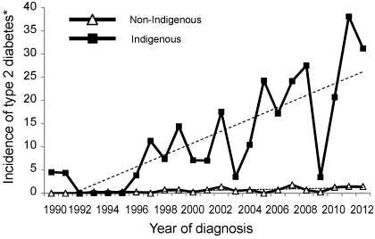
The overall mean incidence of T2D was 1.3 per 100 000 person-years (95% CI, 1.1–1.6 per 100 000 person-years), increasing from 0.2 per 100 000 person-years in 1990 to 3.1 per 100 000 person-years in 2012. The mean incidence in Indigenous children was 12.6 per 100 000 person-years (95% CI, 10.0–15.8 per 100 000 person-years) compared with 0.6 per 100 000 person-years (95% CI, 0.5–0.8 per 100 000 person-years) in non-Indigenous children. Between 1990 and 2012, the incidence increased from 4.5 to 31.1 per 100 000 person-years in Indigenous children, and from 0 to 1.4 per 100 000 person-years in non-Indigenous children (Box). The mean annual rate of increase in incidence over this period was 12.5% per year (95% CI, 8.0–17.0%) in Indigenous children and 10.9% per year (95% CI, 6.1–16.0%) in non-Indigenous children.

This population-based study provides further evidence of an increasing incidence of diagnosed childhood T2D in WA.1 Although a 20-fold higher mean incidence was observed in Indigenous children compared with non-Indigenous children, both groups had similarly high annual rates of increase. As childhood T2D may not present acutely, and population-screening programs are not routine in Australia, the incidence observed in this study is likely an underestimation of the true incidence. Furthermore, as diabetes-related complications occur early in youth with T2D,5 while the disease remains undiagnosed, diabetes-related complications may develop before clinical presentation.

The continued increase in childhood T2D reported in this study highlights the need for early diagnosis and screening for diabetes-related complications in youth at risk of developing the disease.

Box –
Incidence of type 2 diabetes in children aged < 17 years in Western Australia (1990–2012), by Indigenous status


* Per 100 000 person-years at risk.

Vaccination objection rates haven’t changed: study

Despite media reports to the contrary, the overall level of vaccination objection has remained largely unchanged since 2001.

Research published in the Medical Journal of Australia looked at the trends in registered vaccination objection and estimated the contribution of unregistered objection to incomplete vaccination among Australian children.

Dr Frank Beard and colleagues from the National Centre for Immunisation Research and Surveillance at The Children’s Hospital at Westmead and the University of Sydney found that registered objectors affecting children from 1 – 6 had increased from 1.1% in 2002 to 2.0% in 2013.

However the proportion of children with incomplete vaccinations but no objection recorded declined during this period.

Related: Doctors get carrot, anti-vax parents the stick, in immunisation boost

The authors also found that more than half of the 2.4% of children with no vaccinations recorded were born overseas.

It’s suggested that most of these children are likely to be vaccinated however they haven’t been recorded on the Australian Childhood Immunisation Register.

“We recommend that primary care clinicians pay close attention to ensuring that the vaccination history of overseas-born children is correctly recorded in the ACIR,” the authors urged.

Related: Punishing families not the way to boost vaccination rates

The authors estimate 1.3% of children were incompletely vaccinated due to unregistered parenting vaccination objection. In total, an estimation of 3.3% of children were affected by registered or unregistered objection.

A 2001 survey found that 2.5-3.0% of children had parents who had registered an objection, suggesting “that there has been little change in the overall impact of vaccination objection since 2001”.

The authors urged GPs to be on the lookout for appropriate catch up opportunities for under vaccinated children.

Latest news:

Increase in late diagnosed developmental dysplasia of the hip in South Australia: risk factors, proposed solutions

Early detection and treatment of developmental dysplasia of the hip (DDH) in newborns is important because late diagnosis (later than 3 months of age) is associated with a significant risk of poorer outcomes. This includes increased likelihood of surgery, more invasive surgical procedures, longer hospital stays, and early osteoarthritis of the hip, as well as increased health care costs.1,2

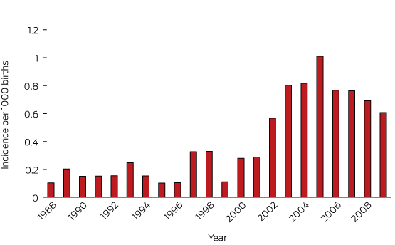
We previously reported a worrying increase in the number of infants diagnosed between 3 and 18 months of age with DDH in South Australia; prospective data showed an incidence of late diagnosed DDH of greater than 0.7 cases per 1000 live births, or around 15 cases each year.3 This contrasts with the low rate of 0.22 per 1000 live births, or four to five cases per year, during the period 1988–2003,4 and has occurred despite continued routine physical examination screening of all neonatal hips. Reports from New South Wales5 and Western Australia6 confirm that the increased incidence of late diagnosed DDH is not limited to SA, but has been observed nationally.

The aim of our study was to determine whether there are identifiable perinatal risk factors associated with late DDH in babies born in SA since 2003, and to review the treatment required for patients with late DDH. Additionally, we provide a discussion of current screening practices and discuss strategies to redress the increased incidence of late DDH in Australia.

Methods

The South Australian Birth Defects Register (SABDR) has received notifications since 1986 of all cases of DDH diagnosed during the first 5 years of life. Notifications are received from a wide range of health professionals and treatment facilities, and are mandatory under the SA Health Care Act.

All diagnoses of DDH in children born in SA from 2003 to 2009 and notified to SABDR were identified for this study. Notifications for a given birth year are not considered complete until all children have achieved their fifth birthday, allowing for a late diagnosis. Consequently, all patients in our series were followed up for a minimum of 5 years after birth, with data collection ceasing at the end of 2014.

Late DDH was defined as an initial diagnosis of DDH at or after 3 months of age. Our methods were identical with those of a published SA study that reported data from the same institution for 1988–2003,4 and there were no changes in reporting practices. Incidence data from the earlier and current studies were combined to produce an incidence graph of late DDH for 1988–2009.

Cases were linked with the Pregnancy Outcome Statistics Unit of the SA Department of Health, which also collects details on mother and baby as a legislative requirement. These data include congenital abnormalities diagnosed at birth, as well as socio-demographic and clinical information.

Teratological hip dislocations and patients with an SABDR notification of significant congenital or genetic anomaly at any time were excluded.

Statistical methods

Likelihood ratio χ2 tests, Fisher exact tests, and logistic regression analysis were used to examine possible significant predictors at the univariable level; predictors with a trend to significance (P < 0.1) were then included in a multivariable logistic regression model, using backward selection. Statistical significance was defined as P < 0.05.

Ethics approval

Approval for the study protocol was granted by the Women’s and Children’s Health Network Human Research Ethics Committee (HREC/13/WCHN/68).

Results

Among babies born between 2003 and 2009, 902 cases of DDH were notified in SA. Twenty-four cases were excluded from our analysis; the timing of the diagnosis in five patients could not be confirmed, and in 19 cases a significant genetic disorder was subsequently diagnosed. Of the remaining 878 children, 777 were diagnosed before 3 months of age, and 101 children were diagnosed at or after 3 months of age. The incidence of late DDH in babies born between 2003 and 2009 was therefore 0.77 per 1000 live births; late diagnosed DDH represented 11.5% of all cases of DDH.

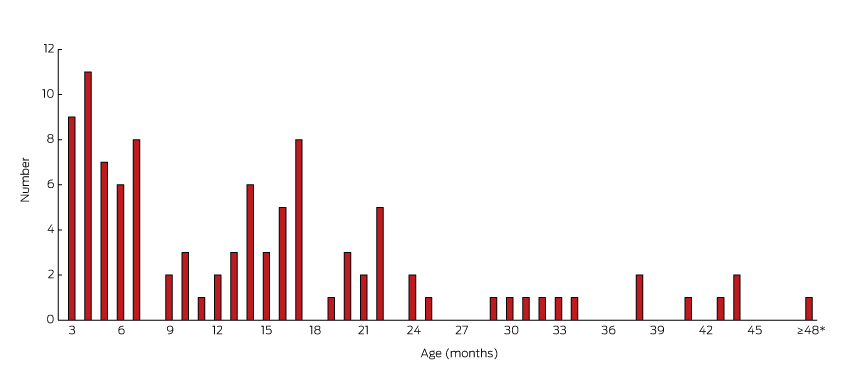
The incidence of late DDH by year of birth ranged between 0.6 and 1.0 per 1000 live births during 2003–2009. Box 1 combines the current data with previously published SA data to show the trend in late diagnosed DDH cases in babies born from 1988 to 2009. The oldest child was diagnosed at 4 years and 5 months of age; 52% of late diagnosed children were over 12 months of age (Box 2).

The results of univariable and multivariable analysis of risk factors associated with late DDH are summarised in Box 3. Significant factors at the univariable level were rural birth (odds ratio [OR], 2.65; 95% confidence interval (CI), 1.49–4.70; P = 0.001), being the second child (v being the first-born: OR, 2.05; CI, 1.33–3.17; P = 0.001), and female sex (OR, 2.06; CI, 1.07–3.94; P = 0.03). Breech presentation was protective against late diagnosis DDH (OR, 0.22; CI, 0.10–0.45; P < 0.001).

Significant perinatal risk factors (multivariable analysis) were birth in a rural hospital (v metropolitan public hospital: OR, 2.47; CI, 1.37–4.46; P = 0.003), and being the second child (v being the first-born: OR, 1.69; CI, 1.08; P = 0.023). Breech presentation was highly significant as a protective factor when compared with cephalic presentation (OR, 0.25; CI, 0.12–0.54; P < 0.001).

There was a trend to a shorter hospital stay for late DDH than for children with an early diagnosis of DDH (P = 0.075), and a significantly shorter hospital stay when compared with the entire population of live births (P = 0.038).

There was no association between either maternal age or maternal ethnic group and late DDH; 91% of babies with late DDH and 93% of babies with early DDH were born to mothers of European descent.

Thirty-one of the 101 children diagnosed with late DDH were successfully treated with splints alone. A further 25 had a successful closed reduction under general anaesthesia, with or without percutaneous adductor tenotomy, between 3.5 and 22 months (mean age, 9.7 months; standard deviation, 5.1 months). An open surgical procedure was required for 44 children. Seven patients required a second open reduction to achieve a reduced hip, and two patients underwent a third open reduction. Thirty-seven pelvic and 15 femoral osteotomies were performed.

Discussion

The incidence of DDH diagnosed at or after 3 months of age in babies born in SA between 2003 and 2009 was 0.77 per 1000 live births, comprising 11.5% of all DDH diagnoses. This has increased from 0.22 per 1000 live births, or 3.5% of all DDH cases, for babies born 1988–2003.4 There has been only a minor increase in the overall incidence of DDH, from 6.4 per 1000 live births for 1988–2003 to 6.8 per 1000 live births for 2003–2009.

Fifty-two of the 101 late DDH cases born between 2003 and 2009 were children of walking age. This compares with NSW data showing a tenfold increase in hip dislocations diagnosed in walking age children5 and a WA report of 17 patients diagnosed between the ages of 6 months and 5 years during 2010.6

Late diagnosed DDH is considered a significant public health issue because late diagnosis and treatment is associated with a lower likelihood of success, higher rates of surgery, increased complications (such as avascular necrosis of the femoral head1,2), and increased health care costs, including those associated with hospital admission, general anaesthesia, and the fact that 44% of such patients require major operative procedures.

The increased incidence of late DDH in Australia has occurred despite an ongoing clinical screening program that involves physical examination of all babies at birth, at 6 weeks, and at 3, 6 and 12 months of age, as well as increasing use of selective ultrasound screening.4,5 This approach has been used for patients with known risk factors for DDH, such as breech presentation and a positive family history, and may be used where the clinical examination is inconclusive.4,7 The American Academy of Pediatrics recommends routine ultrasound screening at 6 weeks of age for girls born in the breech position, with optional screening for breech boys, as well as for girls with a family history of DDH.7 Our results support previous findings8 that selective ultrasound screening fails to prevent the occurrence of late presenting DDH patients, as the majority of babies requiring intervention are not identified by the current criteria.

It is noteworthy that breech presentation was protective, a finding that corroborates previous SA data.4 One could speculate that this known risk factor for DDH increases the physician’s awareness of the potential for dysplasia and promotes extra care when examining the baby. Another risk factor for late DDH was being the second child born to a mother. Clinicians should remain mindful that DDH can occur in any child, and late presentations occur in children without traditional risk factors.

An increase in the number of late DDH diagnoses has also been observed in the United Kingdom, North America and France;811 it has been attributed to external factors, such as swaddling, as well as to reduced clinical expertise in hip examination, and the lack of repeated hip checks of children until walking age.8

A systematic review has cast doubt on the value of screening for DDH in general, citing a lack of evidence for benefits in long term functional outcomes, as well as the risk of overtreating.12 This highlights the challenges when making recommendations for screening, with differences in definitions, treatment protocols and outcome measures making conclusions difficult. DDH encompasses a broad spectrum of conditions, including neonatal clinical instability, early ultrasound dysplasia,12 and acetabular dysplasia in adolescence or adulthood.13

Our study has several limitations. It was a retrospective analysis of data collected prospectively by the Pregnancy Outcome Statistics Unit, so that other potential risk factors described in the literature, including family history, could not be analysed. The analysis of the diagnosis of dysplasia and its treatment was retrospectively performed by retrieving medical records. The severity of DDH was not graded, but all late diagnosed DDH hips were considered dislocated by the treating clinician. Our study was limited to South Australian data, and the incidence and risk factors in other states may be different.

The low published incidence rate of late DDH in SA over a number of years provides evidence that our historical screening program was successful.4 The reasons for the current increase in late diagnosed DDH in SA and elsewhere are likely to be multifactorial.

In the first instance, there is a need for greater awareness of the problem. Although there has been significant concern within the paediatric–orthopaedic community about an increase in late DDH presentations,3,5,6 awareness of this trend may not be widespread among other practitioners involved in hip screening and early child care. Web-based advocacy groups have been formed to promote awareness of hip dysplasia and to provide educational material and family support. These include the International Hip Dysplasia Institute14 and Australian patient advocacy groups.15,16

Most cases of DDH in SA are diagnosed clinically, and a high quality clinical hip examination of newborns by a competent examiner remains a powerful tool. However, an early neonatal hip examination that finds nothing abnormal does not always preclude DDH at a later follow-up.17 Early discharge from hospital is a risk factor for late DDH in SA; previous research found a higher incidence when mother and child were discharged less than 4 days after the birth.4 Our study found an association when data were analysed continuously rather than categorically, possibly because a greater number of mothers are now discharged less than 4 days after the birth. A potential reason underlying this risk factor is the reduced opportunity to examine a compliant, relaxed baby before they leave hospital.

Peaks in late DDH diagnoses occur at ages 3 and 6 months (Box 2) in association with standard timings for baby hip examinations, providing further evidence of the value of repeated clinical examination until walking age.9 Increased clinician awareness of the importance of repeated hip checks is required.

Possible reasons for the increased risk of late DDH in babies born in the country include the lack of resources to ensure the availability of appropriate screening checks and reduced clinical examination skills in practitioners who routinely examine fewer baby hips than practitioners in busy metropolitan centres. Online resources, such as the education module developed by the Royal Children’s Hospital in Melbourne,18 may be particularly helpful for rural practitioners involved in neonatal care. General practitioners and other relevant health professionals should focus on a dedicated hip examination as part of their general assessment of rural babies. High quality physical examination by a trained practitioner remains the best tool for reducing the incidence of late DDH.

There is abundant epidemiological evidence for the negative effects on hip development of wrapping, swaddling or carrying susceptible babies with hips tightly adducted and extended,10,19 as well as confirmatory animal studies.20,21 There is growing concern among the orthopaedic fraternity in North America, the UK and Australia that a resurgence in the popularity of swaddling, including the increased use of “swaddling cocoons” (which force the lower limbs into extension), places children at risk of late diagnosed DDH.3,10,19 This increased popularity has occurred following promotion of swaddling as a technique for settling babies and for reducing the risk of sudden infant death syndrome.19 Although swaddling is more common in certain cultures,10 changes in immigration trends in Australia are unlikely to account significantly for the increase in late DDH cases; in our study, only nine babies with late DDH were born to non-European mothers. Conversely, childcare practices in some cultures that encourage flexion and abduction of newborn hips, such as baby-wearing, are associated with low rates of DDH.22 Advice regarding healthy hip swaddling, as promoted by the International Hip Dysplasia Institute,14 should be provided to all new parents and practices, including advice about carrying babies with hips flexed and abducted.

The increase in late DDH cases in Australia and abroad contrasts with the experience of centres that practise universal ultrasound screening of all neonates.1,2 Evidence for the value of universal ultrasound screening includes reduced rates of surgery, hospitalisation, and late diagnoses in screened children.1,2 A variety of ultrasound methods have been successfully employed for screening, and, although variation in interpretation has been reported, this tends to occur with degrees of dysplasia of lesser clinical relevance.23 Arguments against universal ultrasound screening include variability in technique and reporting, increased follow-up, risk of overtreatment, and the acknowledgement that many abnormalities detected by ultrasound resolve spontaneously, particularly in the presence of a normal clinical examination.24 Additionally, late dysplasia can still occur in patients with a normal early screening ultrasound.25 In SA, the previously low published rates of late DDH could be considered as evidence against the need for universal ultrasound screening, but further investigation is warranted if late DDH rates remain high despite the adoption of other proposed strategies, including avoiding lower limb wrapping.

In Japan, a systematic program of public education that aimed to eliminate the use of traditional swaddling, together with education of medical practitioners and the introduction of a clinical screening program, was successful in reducing the incidence of infantile hip dysplasia from 5–6% to less than 0.4%.21 We can therefore be optimistic that the recent increase in the incidence of late diagnosed DDH can be reversed in SA and other Australian states by increasing awareness and education, and by reducing childcare practices that may be detrimental to hip development.

Box 1 –
Incidence of late diagnosed cases of developmental dysplasia of the hip in babies born 1988–2009 in South Australia

Box 2 –
Age of children (born 1988–2009 in South Australia) diagnosed with late developmental dysplasia of the hip

Box 3 –
Univariable and multivariable analysis of risk factors associated with late developmental dysplasia (DDH) of the hip in babies born 1988–2009 in South Australia

Characteristic

Late DDH

Early DDH

Unadjusted odds ratio (95% CI)

P

Adjusted odds ratio* (95% CI)

P


Number

101

777

Mother’s ethnic background

European descent

92

725

1.00

Other

9

52

1.36 (0.65–2.86)

0.411

Hospital category

Country hospital

23

93

2.65 (1.49–4.70)

0.001

2.47 (1.37–4.46)

0.003

Home birth

1

1

Public metropolitan hospital

35

375

1.00

1.00

Private metropolitan hospital

42

308

1.46 (0.91–2.35)

0.116

1.41 (0.87–2.29)

0.163

Sex

Female

90

621

2.06 (1.07–3.94)

0.03

1.75 (0.90–3.40)

0.098

Male

11

156

1.00

1.00

Parity

First-born

49

462

1.00

1.00

Second-born

45

207

2.05 (1.32–3.17)

0.001

1.69 (1.08–2.66)

0.023

Third-born

7

75

0.88 (0.38–2.02)

0.762

0.62 (0.25–1.53)

0.3

Fourth-born or later

0

33

Presentation

Cephalic

92

547

1.00

1.00

Breech

8

220

0.22 (0.10–0.45)

< 0.001

0.25 (0.12–0.54)

< 0.001

Other

1

8

0.74 (0.09–6.01)

0.781

0.99 (0.12–8.16)

0.989

Unknown

0

2

Delivery

Spontaneous vaginal

47

291

1.00

Assisted vaginal

16

95

1.04 (0.56–1.92)

0.893

Breech

0

12

Elective caesarean delivery

20

193

0.64 (0.37–1.12)

0.116

Emergency caesarean delivery

18

186

0.60 (0.34–1.06)

0.08

Post-natal days in hospital

< 4 days

36

225

1.00

≥ 4 days

65

552

0.74 (0.48–1.14)

0.168

Baby weight

< 2500 g

6

24

1.92 (0.76–4.83)

0.166

2500–3999 g

87

668

1.00

≥ 4000 g

8

85

0.72 (0.34–1.54)

0.401

Gestation

< 37 weeks

4

39

0.77 (0.27–2.22)

0.636

37–41 weeks

97

734

1.00

≥ 42 weeks

0

4

Maternal age

< 25 years

12

98

1.20 (0.57–2.52)

0.636

25–29 years

22

215

1.00

30–34 years

44

280

1.54 (8.90–2.64)

0.121

≥ 35 years

23

184

1.22 (0.66–2.26)

0.525


* Multivariable logistic regression model including predictors with a trend to significance (P < 0.1), using backward selection.

Cord blood vitamin D and the risk of acute lower respiratory infection in Indigenous infants in the Northern Territory

One fifth of Indigenous infants born in the Northern Territory are hospitalised with an acute lower respiratory infection (ALRI) during their first year of life.1 Several international studies have reported an inverse relationship between cord blood vitamin D levels and infant respiratory infections.24 As exposure to sunshine is the most important influence on vitamin D status, there has been little consideration of the relationship between vitamin D status and disease in the tropical north of Australia.

Vitamin D is produced in the skin after exposure to sunlight. Subsequent hydroxylation in the liver yields the dominant circulating vitamin D metabolite, 25(OH)D3. The discovery that vitamin D receptors are widely distributed throughout human tissues and that several cell types, including those of the immune system, can synthesise the active vitamin D metabolite (1,25(OH)2D) from 25(OH)D3 has prompted renewed interest in the role of vitamin D. Vitamin D is required for innate (antimicrobial peptide production) and adaptive (favours response by Th2 effector T cells) immune responses.5 These may be particularly important in the respiratory tract of the developing infant, and perhaps relevant to the relationship between cord blood vitamin D levels and the risk of respiratory infection.24

Circulating 25(OH)D3 and the less common 25(OH)D2 are together referred to as 25(OH)D. In the United States, vitamin D deficiency is defined as a serum 25(OH)D level under 50 nmol/L, and vitamin D insufficiency as levels of 50–75 nmol/L.6 In Australia, 25(OH)D levels of 50 nmol or more are considered sufficient, although higher levels are regarded as optimal.7

Neonates and breastfed infants rely almost exclusively on maternal vitamin D.8,9 According to national population surveys, the prevalence of low 25(OH)D levels (< 50 nmol/L) in women during pregnancy varies from 10% among women in south-east Queensland10 to more than 80% in dark-skinned and/or veiled women in Melbourne, Victoria.11 In Far North Queensland, a small study of pregnant women at mid-gestation (93 non-Indigenous and 23 Indigenous women) found that only eight (7%) had 25(OH)D values under 75 nmol/L.12 While little is known about the vitamin D status of pregnant Indigenous women and children, dark skin is a risk factor for low vitamin D levels,7 and our recently published data indicate that about 40% of hospitalised Indigenous infants in the NT (median age, 7 months) had 25(OH)D3 levels below 75 nmol/L.13

The aims of our study were to describe the natural history of vitamin D status from the third trimester of pregnancy to infancy (age 7 months), and to determine whether low vitamin D levels at birth (cord blood 25(OH)D3) were associated with an increased risk of ALRI hospitalisation during the first year of life.

Methods

Participants and study design

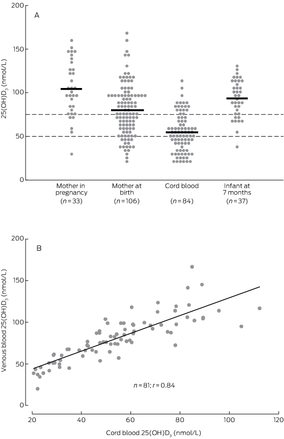
From our randomised controlled trial of maternal pneumococcal vaccination (PneuMum; ClinicalTrials.gov NCT00714064), we established a cohort of 109 Indigenous mother–infant pairs from the Top End of the Northern Territory, in regions serviced exclusively by Royal Darwin Hospital. Participants were recruited from 2006 to 2011, and followed over several visits from the third trimester of pregnancy until the infant was 7 months old. Within this cohort, blood was available from 33 mothers during the third trimester of pregnancy (< 36 weeks), from 106 mothers at birth, from 84 cord specimens (< 72 hours after birth), and from 37 infants at age 7 months. Vitamin D levels were measured in each of these blood samples to assess temporal trends in vitamin D status during the birth period, and to establish the exposure of interest (cord blood vitamin D status) before ascertaining the primary outcome, ALRI hospitalisation before 12 months of age.

Vitamin D measurements

Serum 25(OH)D3 and 25(OH)D2 levels were measured using isotope dilution–liquid chromatography–tandem mass spectrometry (ID-LC-MS/MS), as described previously.13,14 Low, medium and high commercial controls (UTAK Laboratories) were used to monitor assay precision. Sample identity was concealed during testing. As 25(OH)D2 levels were undetectable or negligible in all specimens, we defined 25(OH)D3 levels below 75 nmol/L as vitamin D insufficiency,6 and below 50 nmol/L as vitamin D deficiency.7

ALRI hospitalisations

Infant ALRI hospitalisations during the first 12 months of life were identified by International Classification of Diseases, 10th revision, Australian modification (ICD-10-AM) codes recorded during admission to Royal Darwin Hospital (J09–J22, A37–A37.9).15 Hospital and study data were linked via the Hospital Registration Number, common to each dataset and unique to each infant. Diagnoses made during the birth admission (ICD-10-AM, Z37.0–Z39.2) and related admissions within 7 days of birth were excluded from the analysis.

Analysis

Vitamin D levels are reported for all available blood samples at each time point. Participant characteristics were assessed according to cord blood vitamin D status categories (< 50 nmol/L, 50–74 nmol/L, ≥ 75 nmol/L) to assess potential confounders of the exposure. The Fisher exact test (proportional data) and Kruskal–Wallis test (continuous data) were used to assess differences between groups. The primary analysis was a comparison of cord blood 25(OH)D3 levels in infants who were subsequently hospitalised with an ALRI with those of infants who were not. Student t tests were used to compare the normally distributed vitamin D data; P < 0.05 (two-tailed) was defined as statistically significant. With 84 cord blood samples and assuming that 20% of infants would be hospitalised with an ALRI1 and that mean cord blood 25(OH)D3 levels for healthy infants ranged between 50 and 75 nmol/L (standard deviation, 25 nmol/L), our analysis had 80% power to detect a difference in 25(OH)D3 levels (between those of infants who were hospitalised for ALRI and of those who were not) of 20 nmol/L.

Ethics approval

The study was approved by the Human Research Ethics Committee of the NT Department of Health and by the Menzies School of Health Research (HREC 05/52, HREC-2012-1882). Written consent was obtained for access to each child’s medical records and the analysis of their blood samples.

Results

Participant characteristics

In general, participant characteristics were similar across the cord blood 25(OH)D3 categories, except that remote dwelling was associated with lower cord blood 25(OH)D3 levels (Box 1). The median maternal age at recruitment was 24 years, and almost half (43%) reported smoking during pregnancy. Uptake of the influenza vaccine during pregnancy was low (14%). Most infants (91%) had received three doses of the pneumococcal conjugate vaccine (PCV; 7-valent or 10-valent plus Haemophilus influenzae protein D) by 12 months of age.

Vitamin D levels

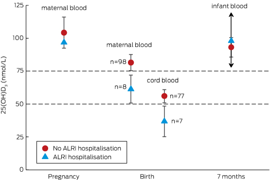
As assessed in maternal venous blood, the prevalence of vitamin D insufficiency was 21% (7 of 33) during the third trimester (median gestation time, 32 weeks; range, 28–36 weeks) and 45% (48 of 106) at birth (median gestation time, 39 weeks; range, 34–41 weeks) (Box 2). In cord blood (median gestation time, 39 weeks; range, 36–41 weeks), the prevalence of vitamin D insufficiency was 80% (67 of 84); 44% (37 of 84) had 25(OH)D3 levels below 50 nmol/L, and 10% (8 of 84) below 25 nmol/L. The prevalence of vitamin D insufficiency among infants at the 7 month visit (median age, 7.1 months; range, 6.6–8.1 months) was 22% (8 of 37).

Considering all samples (unmatched), the relative difference in mean 25(OH)D3 levels between maternal venous blood during the third trimester and at birth was 23% (104 nmol/L v 80 nmol/L) and between maternal venous and cord blood levels at birth 33% (80 nmol/L v 54 nmol/L) (Box 2; Box 3A). Overall, there was a 48% relative difference in 25(OH)D3 levels between mothers’ levels during the third trimester and those of cord blood. This trend in relative difference was similar in matched samples (data not shown). At birth, the 25(OH)D3 concentrations of the 81 matched maternal venous and cord blood samples exhibited a linear correlation (r = 0.84; P < 0.001; Box 3B).

Vitamin D levels in urban and remote participants

The mean 25(OH)D3 concentration was lower in remote than in urban participants during pregnancy, at birth, and at infant age 7 months (Box 4). The relative difference in 25(OH)D3 concentration between maternal blood in the third trimester and cord blood in remote participants was 57% (87 nmol/L v 37 nmol/L), compared with 46% in urban participants (108 nmol/L v 58 nmol/L). The cord blood 25(OH)D3 concentrations of all 14 remote infants were below 75 nmol/L; 86% (12 of 14) were under 50 nmol/L and 14% (2 of 14) were under 25 nmol/L.

Vitamin D and ALRI hospitalisation

Of the 84 infants for whom cord blood samples were available, seven (8%) were hospitalised with an ALRI during their first 12 months of life; the median age at initial admission was 5.3 months (range, 1.9–7.5 months). In our primary analysis (Box 5), the mean cord blood 25(OH)D3 concentration in these seven infants was 37 nmol/L (95% CI, 25–48 nmol/L), compared with 56 nmol/L (95% CI, 51–61 nmol/L) for the 77 infants not hospitalised for an ALRI (P = 0.025). Mean 25(OH)D3 levels in maternal venous blood at birth were similarly lower in the mothers of infants subsequently hospitalised with an ALRI than in the mothers of those not hospitalised for an ALRI (Box 5).

ALRI among urban and remote participants

The proportion of remotely dwelling infants who were hospitalised with an ALRI (4 of 14, 29%) was higher than for urban infants (3 of 70, 4%; P = 0.013). The low number of ALRI hospitalisations was insufficient for a model including remote dwelling as a confounding factor.

Discussion

This is the first study to longitudinally assess vitamin D levels in pregnant Indigenous mothers and their infants. We found that the mean 25(OH)D3 level in cord blood was about half (48%) that of maternal blood during the third trimester of pregnancy (about 7 weeks earlier), a difference due equally to a decline in maternal levels late in pregnancy and to a gradient across the placenta. We also found that the 25(OH)D3 concentration was less than 75 nmol/L in 80% of cord blood samples, and that the mean cord blood 25(OH)D3 concentration was lower in infants who were subsequently hospitalised with an ALRI than in those who were not (37 nmol/L v 56 nmol/L; P = 0.025). This comparison of cord blood vitamin D levels according to ALRI hospitalisation outcome should be interpreted with caution, however, given the small number of ALRI hospitalisations (seven) and an inability to adequately investigate potential confounders, such as remote dwelling. This characteristic was associated with both lower cord blood vitamin D levels and a higher proportion of ALRI hospitalisations, and may have independently caused both low vitamin D levels and increased risk of ALRI hospitalisation. Further, this study did not measure specific factors known to influence vitamin D status, such as skin pigmentation, time spent outdoors, and diet. Despite the limitations of this study, our findings warrant further investigation.

Physiological changes in vitamin D metabolism occur during pregnancy to support the increased calcium demands of the fetus, but the specific mechanisms are not fully understood. Levels of vitamin D-binding protein and the active vitamin D metabolite, 1,25(OH)2D, increase steadily during pregnancy, while concentrations of 25(OH)D3 generally remain stable.16,17 In our study, maternal 25(OH)D3 concentrations fell both late in pregnancy (by 23%) and across the placenta (by 33%). The observed difference in 25(OH)D3 concentrations between venous (maternal) and cord blood at birth is consistent with other studies;18 however, few have specifically characterised 25(OH)D3 levels late in pregnancy. In 2003, a small study of 20 Hungarian women found no difference in mean maternal 25(OH)D3 levels between 22–24 weeks’ gestation and birth,19 while a study of 14 healthy French women found a 20% decline in mean 25(OH)D3 levels between 36 weeks’ gestation (46.8 nmol/L) and birth (37.4 nmol/L).20 As there is little seasonal variation in 25(OH)D3 levels in the tropical NT,13 the drop in late pregnancy may reflect natural progression, perhaps related to maternal–fetal immune tolerance or neonatal immune development,21 increased calcium demands of the growing fetus,22 or the emergence of risk factors, such as increased body mass index or more time spent indoors.7

The cord blood vitamin D data in our study suggest that 80% of infants were born with vitamin D insufficiency (< 75 nmol/L), 44% with mild deficiency (< 50 nmol/L), and 10% with moderate deficiency (< 25 nmol/L). The significance of these definitions for the neonate, however, is unclear, and more work is needed to define vitamin D reference ranges in cord blood. Vitamin D status was generally normal by infant age 7 months, the next sampling point.

Concordant with the trends in our data, a recent trial of maternal-plus-infant vitamin D supplementation for the prevention of deficiency among New Zealand infants8 showed that mean 25(OH)D levels in the placebo group were higher in mothers at 36 weeks’ gestation (50 nmol/L) than in cord blood (33 nmol/L), and that infant levels steadily increased through ages 2 (50 nmol/L), 4 (75 nmol/L) and 6 months (78 nmol/L). As the authors did not report maternal 25(OH)D levels at birth, it was not possible to determine whether the drop between 36 weeks’ gestation and cord blood was the result of a maternal decline or of the placental differential. Compliant daily maternal (from 27 weeks’ gestation to birth) and infant supplementation (from birth to age 6 months) at low (1000 and 400 IU/day respectively) and high doses (2000 and 800 IU/day respectively) increased mean cord blood 25(OH)D3 levels to 60 nmol/L and 65 nmol/L respectively (v 33 nmol/L in placebo-treated participants); mean infant 25(OH)D3 levels of 75 nmol/L or more were maintained at each of the 2, 4 and 6 months sampling points.

Among the participants who contributed a cord blood sample to our study, there were fewer infant ALRI hospitalisations in the first 12 months of life (8%) than predicted by historical NT data (22%).1 As ALRI hospitalisation rates are highest among remote and Central Australian infants,1 this difference was probably caused by the over-representation of Top End and urban infants in our cohort (Top End, 100%; urban, 83%) compared with the NT-wide historical data (Top End, 71%, urban, 39%). Nevertheless, cord blood 25(OH)D3 concentrations were lower in infants who were subsequently hospitalised with an ALRI than in those who were not. We could not reliably adjust our analysis to account for remote dwelling as a confounder because of the low number of ALRI hospitalisations, but our unadjusted findings are consistent with those of several other studies. In the Netherlands, cord blood 25(OH)D concentration was lower in infants who developed a respiratory syncytial virus-associated ALRI during their first 12 months than in controls (65 nmol/L v 84 nmol/L; P = 0.009);3 in New Zealand, lower cord blood 25(OH)D concentration was associated with an increased risk of any respiratory infection by 3 months of age (odds ratios, 1.00 for ≥ 75 nmol/L; 1.39 for 25–74 nmol/L; 2.16 for < 25 nmol/L).2 In a Korean study, 90% of 525 cord blood samples tested had 25(OH)D levels below 75 nmol/L, and reduced cord blood 25(OH)D concentration was strongly associated with increased risk of acute nasopharyngitis during the first 6 months of life.4 In their recent randomised controlled trial of vitamin D supplementation, Grant and colleagues23 audited health care visits by children (to age 18 months) as a secondary outcome; infants in the high dose group (87%; 66 of 76) but not the low dose supplementation group (95%; 76 of 80) had significantly fewer health care presentations for acute respiratory infections than infants in the placebo group (99%; 79 of 80).

Although not all studies support an inverse association between vitamin D levels and ALRI risk, a supplementation strategy beginning in the third trimester of pregnancy may be useful in preventing both vitamin D insufficiency and subsequent acute respiratory infections in Indigenous neonates in the NT. Acute respiratory infections are endemic in remote Indigenous communities because of factors such as overcrowding and exposure to tobacco smoke.24 However, it is less obvious why remote participants in our study had lower 25(OH)D3 levels during pregnancy and at birth, or why the relative difference in mean concentrations in maternal blood at 30–36 weeks’ gestation and in cord blood was greater than in their urban counterparts (remote, –57% v urban, –48%). Similar vitamin D levels in urban and remote infants at age 7 months suggest that the negative influence of remoteness on vitamin D levels was confined to the mothers. As the climate and time spent outdoors are likely to be similar for urban and remote participants, risk factors other than exposure to sunlight require further investigation. Interestingly, vitamin D has also been found to be a negative acute phase reactant that is depleted after an inflammatory insult.25 Lower vitamin D levels seen among remote participants may therefore be the result, rather than a cause, of their high burden of infection.

Conclusions

While only one in five Indigenous mothers had 25(OH)D3 levels below 75 nmol/L midway through the third trimester of their pregnancy, four in five cord bloods tested had lower levels as the result of declining 25(OH)D3 levels late in pregnancy and differences in levels across the placenta. The significance of low cord blood 25(OH)D3 concentrations is unclear, but the seven infants hospitalised with an ALRI during their first 12 months of life had significantly lower levels than those not hospitalised with an ALRI. Our findings, in conjunction with emerging international data, support the need for further longitudinal studies and for randomised controlled trials of vitamin D supplementation for the prevention of infant ALRI.

Box 1 –
Participant characteristics, by cord blood 25(OH)D3 status

Total

Cord blood 25(OH)D3 levels (nmol/L)


P

< 50

50–74

≥ 75


Number

84

37

30

17

Maternal characteristics

Median maternal age, years (range)

24 (17–37)

25 (17–37)

23 (17–33)

26 (17–33)

0.197

Household occupancy, people (range)

4 (1–11)

5 (2–11)

4 (1–11)

4 (3–10)

0.117

Remote community residence

14 (17%)

12 (32%)

2 (7%)

0

0.003

Smoker

36 (43%)

19 (51%)

8 (27%)

9 (53%)

0.086

Influenza vaccine during pregnancy

12 (14%)

6 (16%)

5 (17%)

1 (6%)

0.613

Infant characteristics: at birth

Boys

47 (56%)

20 (54%)

17 (57%)

10 (59%)

0.960

Low birth weight (< 2500 g)

2 (2%)

1 (3%)

0

1 (6%)

0.491

Premature (< 37 weeks)

1 (1%)

1 (3%)

0

0

1.000

Special or intensive care admission

9 (11%)

4 (11%)

4 (13%)

4 (6%)

0.901

Infant characteristics: after the birth

Exclusively breastfed

1 month after the birth

48 (47%)

18 (49%)

18 (60%)

12 (71%)

0.290

2 months after the birth

34 (40%)

14 (38%)

12 (40%)

8 (47%)

0.807

7 months after the birth

31 (37%)

16 (43%)

9 (30%)

6 (35%)

0.536

Mother smoking*

1 month after the birth

35 (46%)

19 (61%)

7 (25%)

9 (53%)

0.016

2 months after the birth

31 (45%)

15 (56%)

9 (32%)

7 (50%)

0.196

7 months after the birth

38 (55%)

17 (57%)

13 (50%)

8 (62%)

0.856

Vaccination

2 doses of PCV by 7 months

57 (68%)

25 (68%)

22 (73%)

10 (59%)

0.595

3 doses of PCV by 12 months

68 (91%)

31 (89%)

24 (96%)

13 (87%)

0.591

23vPPV during pregnancy

24 (29%)

8 (22%)

9 (30%)

7 (41%)

0.322

23vPPV at birth

29 (35%)

11 (30%)

14 (47%)

4 (24%)

0.221


23vPPV = 23-valent pneumococcal polysaccharide vaccine; PCV = pneumococcal conjugate vaccine (the 7-valent pneumococcal conjugate vaccine [7vPCV] or the 10-valent pneumococcal Haemophilus influenzae protein D conjugate vaccine [10vPHID-CV]). All figures are numbers of individuals and column percentages unless otherwise indicated. Data were compared across categories using the Fisher exact test for proportional data and the Kruskal–Wallis test for continuous data. * Smoking data were unavailable for eight mothers at 1 month and for 15 mothers at 2 and 7 months post partum.

Box 2 –
Serum vitamin D (25(OH)D3) levels measured during pregnancy, at birth, and in the infant at age 7 months

Visit

Blood sample

n

Median age (range)

25(OH)D3 levels (nmol/L)


Mean (95% CI)

Relative difference*

< 50

50–74

≥ 75


Pregnancy

Maternal

33

32 weeks (28–36 weeks)

104 (93–115)

Base

1 (3%)

6 (18%)

26 (79%)

Birth

Maternal

106

39 weeks (34–41 weeks)

80 (74–86)

−23%

18 (17%)

30 (28%)

58 (55%)

Birth

Cord

84

39 weeks (36–41 weeks)

54 (50–59)

−48%

37 (44%)

30 (36%)

17 (20%)

7 months

Infant

37

7.1 months (6.6–8.1 months)

93 (86–101)

−10%

1 (3%)

7 (19%)

29 (78%)


* Compared with maternal vitamin D blood concentration during pregnancy. † Gestational age. ‡ Infant age.

Box 3 –
(A) Maternal and infant vitamin D levels.* (B) Correlation between 84 matched maternal venous and cord blood vitamin D measurements at birth


* Dashed lines indicate reference 25(OH)D3 values for vitamin D deficiency (< 50 nmol/L) and insufficiency (< 75 nmol/L).

Box 4 –
Vitamin D levels (nmol/L) for urban and remote dwelling participants

Visit

Blood sample

Urban


Remote


n

Median age (range)

Mean 25(OH)D3 levels (95% CI)

Relative difference* (%)

n

Median age (range)

Mean 25(OH)D3 levels (95% CI)

Relative difference* (%)


Pregnancy

Maternal

26

33 weeks (30–36)

108 (95–122)

Base

7

32 weeks (28–35)

87 (68–107)

Base

Birth

Maternal

85

39 weeks (35–41)

86 (79–92)

−23%

21

39 weeks (34–41)

57 (49–66)

−34%

Birth

Cord

70

39 weeks (36–41)

58 (53–63)

−46%

14

39 weeks (37–41)

37 (30–43)

−57%

7 months

Infant

33

7.1 months (6.8–8.1)

94 (86–101)

−13%

4

7.1 months (6.6–8.1)

90 (56–124)

+3%


* Compared with maternal vitamin D blood concentration during pregnancy. † Gestational age. ‡ Infant age.

Box 5 –
Mean vitamin D levels during pregnancy, at birth, and in the infant at age 7 months, according to infant hospitalisation with an acute lower respiratory infection (ALRI) during the first 12 months of life


Only one maternal vitamin D measurement during the third trimester of pregnancy was associated with an infant ALRI hospitalisation, so that there is no confidence interval for the open triangle. Only two infants with vitamin D measurements at 7 months were hospitalised with an ALRI, so that the upper and lower confidence boundaries around the open triangle are very wide (exceeding the graph scale), as indicated by the arrows.