×

Decreasing prevalence of Trichuris trichiura (whipworm) in the Northern Territory from 2002 to 2012

Trichuris trichiura (whipworm) is a soil-transmitted helminth (STH) endemic to areas with a tropical climate. Infection occurs after the soil-residing egg of T. trichiura is ingested.1 Eggs are expelled in the faeces of infected hosts and continue this cycle after a period of maturation in the soil.1 An estimated 600–800 million people are infected with T. trichiura worldwide and this infection is estimated to cause the loss of 1.6–6.4 million disability-adjusted life-years.1,2 T. trichiura is the most prevalent helminth in many countries surveyed.35 Heavy infections (> 10 000 eggs per gram of faeces) are associated with anaemia, malnutrition, the trichuris dysentery syndrome and rectal prolapse.68

The Northern Territory has a population of about 232 000 in a geographic area of 1 200 000 km2.9 Thirty per cent of the population is Indigenous and 80% of Indigenous residents live in remote locations.9 T. trichiura is one of three STHs that are endemic in the NT; the other two are Ancylostoma duodenale (hookworm) and Strongyloides stercoralis. A 1997 prevalence study found T. trichiura to be the commonest STH, with 25% of adults in a remote Indigenous community infected;10 no data on T. trichiura in the NT have been reported since. T. trichiura infection is difficult to treat and even more difficult to target within a deworming program. The Central Australian Rural Practitioners Association (CARPA) treatment manual currently recommends albendazole (400 mg) on 3 consecutive days for treatment of proven T. trichiura infection.11 This regimen has been correlated with a 50% cure rate.12 Since 2005, CARPA has recommended a single dose of albendazole (400 mg) for all children aged 6 months to 16 years as part of a community children’s deworming program. Empirical deworming is recommended before and after the wet season, or coinciding with child health assessments or school-age screening.11 Pregnant women are not targeted within this program. However, when pregnancy does occur within the target group, deworming with pyrantel is recommended.11

Under these deworming protocols, it has been reported that the prevalence of hookworm in the NT declined dramatically in the past 11 years and may be heading toward eradication.13 Our aim in this study was to describe the population at risk, disease associations and temporal trends of T. trichiura infections over the same 11 years.

Methods

In September 2013, we conducted a retrospective observational analysis of consecutive, microbiologically confirmed cases of T. trichiura infection in the NT between 1 January 2002 and 31 December 2012. Ethics approval for the study was obtained from the Human Research Ethics Committee of the NT Department of Health and Menzies School of Health Research (HREC-2013-1978).

Cases were identified from the NT Government pathology information system, Labtrak, which covers all NT Government health care facilities including five hospitals, two correctional centres and over 50 remote clinics. Previous STH studies13 have shown that the public NT laboratories identified 94% of all documented STHs, compared with 6% by other pathology service providers. Cases were diagnosed by examination of faeces specimens for T. trichiura eggs, other STHs and parasites by wet mount microscopy and a concentration method.14 Egg counts were not performed.

Infections were linked to NT Government electronic databases by means of medical record numbers to obtain data on age, sex, Indigenous status, place of residence, haemoglobin level and eosinophil count. Anaemia was defined as a haemoglobin level of ≤ 110 g/L and eosinophilia as an eosinophil count of ≥ 0.5 × 109/L.

Statistical analysis

Data were entered into a Microsoft Excel 2007 database (Microsoft Corporation) and analysed with Stata statistical software (version 13; Statacorp). Results are presented as medians and interquartile ranges (IQRs) for non-normally distributed parameters. The estimated prevalence rates in the NT for each year were expressed as cases per 100 000 Indigenous population per year with 95% confidence intervals. Indigenous population by age and percentage Indigenous population in the NT were obtained from Australian Bureau of Statistics data.15 Bivariate analyses were performed using the χ2 or Fisher exact test if expected frequencies were less than 5. For non-parametric data, the Mann–Whitney U test was used, with P values of < 0.05 considered significant.

Results

There were 417 episodes of T. trichiura infection diagnosed in 400 patients from a total 63 668 faecal samples tested between 1 January 2002 and 31 December 2012. About 85% of these were from hospital inpatients admitted to Royal Darwin Hospital, usually for reasons other than T. trichiura infection. Thirteen patients were screened as part of a prisoner health check, 11 patients were living in the community and the remainder were inpatients of an NT Government health care facility (Royal Darwin Hospital, Alice Springs Hospital, Katherine District Hospital, Tennant Creek Hospital or Gove District Hospital) at the time of T. trichiura detection. The preponderance of inpatient samples comes about because community clinics are likely to send samples to private rather than government laboratories, and transport of samples from remote communities can be problematic.

Patients were considered to have had a repeat episode of infection if T. trichiura was detected at least 6 months after the first diagnosed episode (median duration between infections, 23.7 months; range, 7.8–100.9 months). Thirteen patients (3%) had two infections and two patients (0.5%) had three infections. Forty-three episodes of repeat T. trichiura infections that were detected in 33 patients within 6 months of the initially detected infection were deemed to be the same episode of infection and were excluded.

The demographic and laboratory parameters of patients with T. trichiura infection are shown in Box 1. Most infections were in children aged under 17 years (239; 59.8%), with 175 (43.8%) in children younger than 5 years. The median age of those infected was 8 years (IQR, 3–36 years). The vast majority of infections were in Indigenous patients (381 [95.3%] compared with 10 [2.5%] in non-Indigenous patients). Ethnicity was unknown for nine patients. Boys were more likely to be infected than girls (P < 0.001) and women were more likely to be infected than men (P < 0.001).

Haemoglobin levels and eosinophil counts were available for 356 and 345 of the 400 patients, respectively; 143 (40.2%) patients had anaemia and 178 (51.6%) had eosinophilia. After excluding episodes of T. trichiura infection where patients had co-infection with another STH, 115 patients (39.2%) had anaemia and 139 (34.8%) had eosinophilia. There were 112 children (46.9%) and 48 adults (29.8%) who had co-infection with at least one other faecal parasite (P = 0.001).

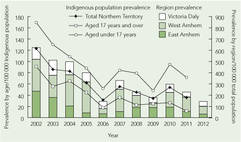
The period prevalence of T. trichiura infection (Box 2; and Appendix 1 (PDF)) decreased from 123.1 (95% CI, 94.8–151.3) cases per 100 000 Indigenous population in 2002 to 35.8 (95% CI, 21.8–49.9) cases per 100 000 Indigenous population in 2011. This downward trend was documented for both children and adults (Box 2). Most cases occurred in patients who had lived in one of three remote Top End NT locations, Victoria Daly, East Arnhem Land and West Arnhem Land (Appendix 2 (PDF)). The number of faecal microscopy samples tested each year was relatively constant, with a median of 5764 samples (range, 5276–6527 samples) per year. We were unable to obtain accurate data on numbers of doses of albendazole dispensed for the community children’s deworming program over our study period.

Discussion

Our study shows that T. trichiura in the NT predominantly affects Indigenous patients from remote Top End communities. Children had the highest prevalence of infection across all the years in our study period. Among children, boys had a statistically significant higher proportion of infections. Conversely, women had a statistically significant higher proportion of infections. It is likely that adult women have higher rates of infection because they care for and live in closer proximity to infected children who contaminate the nearby environment. The difference we observed between the prevalence in boys and girls may reflect greater soil exposure among boys.

We found a strong association between T. trichiura infection, anaemia and eosinophilia, and this association persisted when coinfection with other STHs was excluded. The precise contribution that T. trichiura makes towards anaemia is difficult to ascertain, as infection occurs in populations with a high level of intestinal parasite co-infection, socioeconomic disadvantage and nutritional deficiencies.6

Our data show a consistent reduction in microbiologically diagnosed T. trichiura infections in the NT over the 11 years from 2002 to 2012. Despite this reduction, a significant percentage of infections (59.8%) continued to be diagnosed within the community children’s deworming program target population of children less than 17 years of age. This finding is most likely explained by the reduced efficacy of single-dose albendazole in T. trichiura infection compared with other STHs12,16 and to the rapid reinfection rates seen with T. trichiura.4 Similar disparities in reduction of infection with hookworm and T. trichiura in response to mass deworming campaigns have been observed elsewhere.17 Despite these poor cure rates, egg reduction rates of over 80%12 do occur with single-dose albendazole and this may be sufficient to protect our population against the morbidity seen in high-intensity infections. Interventions to improve sanitation are very effective in reducing the prevalence of T. trichiura infections,18 and level of maternal education, access to latrines, household wealth indexes and remoteness are important risk factors for infection.19

Our retrospective study has several limitations. Systematic sampling from the community was not undertaken, so the true prevalence rates are undoubtedly much higher than the laboratory-diagnosed rates found in our study population. Notably, all but 24 patients were inpatients of a NT Government health facility, reflecting a selection bias towards patients with acute illness and comorbid conditions. Furthermore, without a control group, the association of T. trichiura infection with anaemia cannot be further analysed. T. trichiura egg counts were not performed, so anaemia and eosinophilia correlates with the intensity of infection could not be determined.

We have shown a reducing T. trichiura infection rate in the NT over the 11 year period of our study. A large number of infections continue to be diagnosed in the community children’s deworming program target population. With the move towards eradication of hookworm in the NT, our data raise the question of whether the deworming program should be adapted to improve efficacy against T. trichiura (400 mg albendazole daily for 3 days). More importantly our study supports increasing the focus on health education, healthy living practices and essential housing infrastructure. These factors are deficient in remote Indigenous Australian communities20 and greatly impact on the prevalence of all STH infections.3,18,19

1 Demographic and laboratory parameters of 400 patients* with Trichuris trichiura infection,
Northern Territory, January 2002 to December 2012

Age group


Parameter

All

< 17 years

≥ 17 years

P


Number

400 (100%)

239 (59.8%)

161 (40.3%)

< 0.001

Sex

< 0.001

Male

205 (51.3%)

141 (59.0%)

64 (39.8%)

Female

195 (48.8%)

98 (41.0%)

97 (60.2%)

Indigenous status

< 0.001

Indigenous

381 (95.3%)

236 (98.7%)

145 (90.1%)

Non-Indigenous

10 (2.5%)

3 (1.3%)

7 (4.3%)

Unknown

9 (2.3%)

0 (0)

9 (5.6%)

Median haemoglobin level
(g/L [IQR]))

114 (104–125)

114 (105–123)

113 (95–130)

Anaemia

143 (40.2%)

78 (32.6%)

65 (40.4%)

0.14

Median eosinophil count
(× 109/L [IQR])

0.5 (0.1–1.0)

0.5 (0.1–1.2)

0.5 (0.1–0.9)

Eosinophilia§

178 (51.6%)

104 (43.5%)

74 (46.0%)

0.87

Polyparasitism

160 (40.0%)

112 (46.9%)

48 (29.8%)

0.001

Episodes with no STH coinfection

Number

333 (83.3%)

205 (85.8%)

128 (79.5%)

0.10

Median haemoglobin level
(g/L [IQR])

114 (104–125)

114 (105–124)

114 (98–132)

Anaemia**

115 (39.2%)

66 (36.9%)

49 (43.0%)

0.30

Median eosinophil count
(× 109/L [IQR])

0.4 (0.1–0.9)

0.4 (0.1–1.0)

0.5 (0.1–0.8)

Eosinophilia

139 (48.9%)

82 (47.7%)

57 (50.9%)

0.60


IQR = interquartile range. STH = soil-transmitted helminth.
* Data from the 17 episodes of repeat infection were excluded from the analysis. Aboriginal or Torres Strait Islander; Indigenous status was not available for nine patients aged ≥ 17 years. Anaemia defined as a haemoglobin level ≤ 110 g/L; data available for 356 patients (211 aged < 17 years, 145 aged ≥ 17 years). § Eosinophilia defined as an eosinophil count ≥ 0.5 × 109/L; data available for 345 patients (203 aged < 17 years, 142 aged ≥ 17 years). Defined as detection of at least one other intestinal parasite (Ancylostoma duodenale, Strongyloides stercoralis, Cryptosporidium spp, Giardia lamblia, Hymenolepis nana, Isospora spp, Blastocystis hominis in high numbers). ** Data available for 293 patients (179 aged < 17 years, 114 aged ≥ 17 years). Data available for 284 patients (172 aged < 17 years; 112 aged ≥ 17 years).

2 Prevalence of Trichuris trichiura infections in the Northern Territory in 2002–2012 by age and region

Nutrition in schools — outdated guidelines need updating

To the Editor: The National Healthy School Canteens (NHSC) project commenced in 2008 to help provide guidelines for healthier food and drink choices in Australian schools. At their core, the guidelines seek to restrict the availability of poor food choices by encouraging the preferential availability of healthy options. These guidelines should ensure the translation of health research and national health curriculum into practice. However,
the current NHSC guidelines are inadequate and fall short of their aims as they rate foods only on energy, fat and sodium, and disregard the sugar content of commercially available foods.

The initial decision to disregard sugar as a criterion for rating foods available in school canteens was intentional. The New South Wales government website states that sugar content was not included “To keep the criteria as simple as possible and to ensure that foods containing naturally occurring sugars . . . were not disadvantaged”.1 Surprisingly, sugar content is not even a criterion for assessing “sugar-sweetened drinks”.2

Excess sugar consumption is associated with type 2 diabetes and obesity. This has been reported in both human and non-human studies and is evidenced by outcomes including fatty liver, impaired glucose tolerance and increased site-specific adiposity. It is of little surprise then, that the most recent update to the Australian dietary guidelines in February of this year included a revision of the recommendation about foods and drinks containing added sugars (Guideline 3).3

Before February 2013, national dietary advice was to consume only moderate amounts of food and drinks with added sugar. This has since
been revised to advise their intake be limited. This shifts foods and drinks containing added sugar into the same category as foods high in fat, salt or alcohol. There is now an immediate need to review policies borne from the outdated national guidelines (such as the NHSC) to include “added sugar” as criteria for assessing the suitability of foods, just as fat and sodium currently are. Until then, the healthy development of our children and hence future health of our nation is at risk.

First aid for burns: too little, too late and often wrong

To the Editor: The Australian and New Zealand Burns Association (ANZBA) defines adequate first aid for acute burns as 20 minutes of cold running water within the first 3 hours of a burn injury.1 Despite first aid campaigns, inappropriate and inadequate first aid treatment for burns continues to occur.2

Following Sydney Children’s Hospitals Network Human Research Ethics Committee approval, we performed a retrospective analysis of the first aid received by 4368 children who presented to the Burns Unit at The Children’s Hospital at Westmead between 1 January 2008 and 31 December 2012.

Nearly a third of children (34% of inpatients and 30% of outpatients) received inadequate, inappropriate or no first aid, irrespective of the size of
the burn. Inadequate first aid included cold compresses or wet wraps in 414 children (9.5%) and Burnaid (Rye Pharmaceuticals) in 238 (5.4%). Inappropriate first aid included ice in 227 children (5.2%), with a wide range of bathroom products, foods, creams and oils used in most of the remaining patients. In 144 children (3.3%), the first aid received was not documented.

Products prescribed by general practitioners, ambulance officers or obtained from pharmacies, such as silver sulfadiazine cream, antiseptics and antibiotic ointments, were often used (455 children; 10.4%). Of the 70 children (1.6%) who were treated with bathroom products, toothpaste was the most common. Of the 49 children (1.1%) who had food applied, dairy products, such as yoghurt, were the most widespread. Of the 19 children (0.4%) who received oils, plant oils, such as tea tree oil, were the most frequent. Alternative treatment from a Chinese medicine practitioner or a “witch doctor” was used in 13 children (0.3%) (Box).

Twenty minutes of cool running water has been proven to be
the most effective in reducing progression of burn depth and time
to re-epithelisation.3 Unfortunately, products such as ice and toothpaste, which may have adverse effects, continue to be used on acute burns.4 While most children in our study eventually received appropriate first aid, 31.1% did not. There remains a need to educate health practitioners and the wider community about appropriate first aid for burns.

Summary of products used as first aid for burns in 4368 children presenting to the Burns Unit at The Children’s Hospital at Westmead

First aid type

No. of children (% of total)


Adequacy of first aid

Adequate

2866 (65.6%)

Inadequate, inappropriate or no first aid

1358 (31.1%)

Not documented

144 (3.3%)

Type of inappropriate first aid

Cold compresses, ice and other

Cool compresses or wet wraps

414 (9.5%)

Ice

227 (5.2%)

Other

39 (0.9%)

Total

680 (15.6%)

Creams, gels or ointments

Burnaid

238 (5.4%)

Antiseptic or antibiotic

98 (2.2%)

Other cream, gel or ointment

52 (1.2%)

Plant-based cream, gel or ointment

52 (1.2%)

Nappy cream

5 (0.1%)

Petroleum-based ointment

4 (0.1%)

Steroid cream

4 (0.1%)

Animal-based cream, gel or ointment

2 (0.0%)

Total

455 (10.4%)

Bathroom products

Toothpaste

67 (1.5%)

Talc powder

2 (0.0%)

After shave

1 (0.0%)

Total

70 (1.6%)

Foods

Dairy products

22 (0.5%)

Egg

6 (0.1%)

Fruit or vegetables

5 (0.1%)

Honey

5 (0.1%)

Spices, salt or sugar

4 (0.1%)

Soy sauce

3 (0.1%)

Drinks (non-water)

2 (0.0%)

Flour

2 (0.0%)

Total

49 (1.1%)

Oils

Plant oils

14 (0.3%)

Oil (other)

4 (0.1%)

Animal oils

1 (0.0%)

Total

19 (0.4%)

Alternative medicine

Chinese medicine

12 (0.3%)

“Witch doctor”

1 (0.0%)

Total

13 (0.3%)

The power of 13

A fragile life opens a new dimension to family love

Some months ago one of my daughters-in-law, Tania, announced at the dinner table that she was expecting again, but with a tone that caused me to look up, then to my son, and back and forth. Something was not right. “There is something wrong. They say he has trisomy 13. Do you know much about that?” I am a paediatrician who has specialised in care of very sick babies. I know a lot about trisomies, but how to share that knowledge? With tears, I am afraid.

Trisomy 13 means there are three copies of the 13th largest chromosome in every cell of the body. There should only be two copies. The extra information delivered by the third copy interferes with the development of the baby, particularly the brain. But the face, the heart, the limbs and fingers — almost everything — will be abnormal, and life will be restricted to hours if the baby is born alive. Chromosome 13 is bigger than chromosome 21, whose trisomy results in Down syndrome. The greater the amount of genetic misinformation, the greater the disability. Babies with trisomy 13 are, therefore, much more disabled than those with trisomy 21.

In Tania’s case the diagnosis was made at around 14 weeks of gestation, after it had been suggested by ultrasound abnormalities and confirmed by amniocentesis. In all my 40 years of caring for sick children I had never observed a mother who had not elected to terminate a pregnancy affected by trisomy 13, and I awaited the inevitable statement. I imagined (but what male can know) something of the pressures on her. I had observed mothers struggling with grief, misplaced guilt and the questions of whether it would be unfair to continue the pregnancy. What would its effect be on her five other children, on her relationship with the father, on herself? There was silence in the dining room which seemed to go on for ever. My opinion was awaited. Lord God, what do I say next? What do I do now?

But Tania had no intention of terminating the pregnancy. She carried to term, held the boy, Mitchell Darcy, for 18 and a half hours until he died, and took 4 days to be able to leave him behind and go home from the hospital. She continued to weep but gathered strength to go on to deliver a eulogy beside the little white casket at the funeral several days later. Before a congregation stilled with emotion, she articulated something of what we had sensed during the pregnancy — something new, something wondrous though painful. As if turning a gem to reflect yet more displays of colour, she confirmed a new dimension to a mother’s love, brought a new dimension to family life and, for me, as a Christian, a fresh dimension of the value of imperfect, transient life. Perhaps He really does care about the fallen sparrows?

While Tania never seriously considered termination, she confessed in the eulogy to have felt “numbness, terror, pain, deep confusion and heartache all at the same time” after being told the diagnosis. She decided she would give the baby as much life, as much of her love, and as much family fellowship as she could muster. She declared to the child at the funeral, “I tried to give you a life inside me. I ate lots of ice cream, so you could taste it. I sat in the sun, so you could feel the warmth of the rays. I religiously took my vitamins, to help you grow. I sang to you. I talked to you. I talked with you and told you stories about your family. I explained that all that noise was your big brothers and sister, who were so excited about your arrival. I used to lie in bed and cuddle Daddy, knowing that you were in the middle being touched by us both.

“As the day of your birth grew nearer, I was torn. I wanted to meet you, to hold you in my arms and kiss you all over. But the knowledge your birth would create problems for you, that it would be the start of your having to breathe on your own and sustain yourself, tore me to pieces.”

The child was born alive in April 2013 by elective caesarean section because of previous similar deliveries. Tania was conscious. The father was present. The baby was limp and blue but breathed, and was able to be introduced to his family.

Tania eulogised, “There was so much love in the [delivery] room. I had the privilege of holding you on my skin for 18 and a half hours. Thank you for trying your hardest to stay with us. I hope you felt and knew, while you grew inside me, and when you were born and with us, that you were and still are loved as unconditionally, as completely, and as fiercely in that short time as anyone could hope to be loved in a lifetime. One moment you were in my arms . . . I was holding your hand, and then in a heartbeat you were in the hands of God. I pray that when I walk through death’s portal, Mitchell Darcy Whitehall will be the first face that I shall see, running towards me with your arms spread wide saying, ‘Look, Mummy is home’ ”.

The other children, four boys from 3 to 8 years and a little girl aged 2 had all been “in” on the pregnancy. They had chosen the child’s name. They had counted the weeks. They had felt the baby moving and observed its growth. Of course, they did not understand but were informed Mitchell would not be staying with us for long: he would be going “home” because he was not really made for this world. There was, however, a painful side to this because they had observed a Grandma going home not long before, and she had been part of their lives. How can you explain these things to children?

Somehow, they seemed to comprehend and when the day of the funeral arrived, with solemnity and with not one punch or poke, they dressed in their new clothes, went to the car, filed into the church and proceeded to the front. For small boys used to chasing footballs, they were an intuitive “honour guard” for their brother, however it cannot be said that the 2-year-old girl departed from her usual gay and exploratory self. Mercifully, she skipped from soul to soul, from row to row, warming hearts in her wake.

During all this my son maintained his usual silence on intensely personal matters, and it was hard to know what he was thinking. He had had the job of driving in to the hospital to bring Mitchell out to the funeral parlour and there had been concern about how long it was taking. Much later I learned he had taken the boy to visit the island and little beach at La Perouse where the child’s siblings loved to play.

As the family is living with us, my wife and I had also been drawn in to the saga. I was invited to translate various reports and comment on any “progress” of which, of course, there was none in the physical world. Once (why only once?) we prayed together around the dinner table.

As Tania was to have a caesarean and our son would be with her, my wife and I were to take turns minding the children in the hospital, so they could meet Mitchell as soon as possible after delivery. We all got up at about 5.00 am to travel into Sydney, as Tania was allegedly first on the list but, inevitably, she slid down to an afternoon operation. In the meantime, I took turns walking the children up and down the long corridors.

In the foyer a stall had been erected and “pink lady” volunteers were selling toys and stuffed animals. This spectacle inspired a buying frenzy in my charges and, at that stage, I was prepared to part with any amount of money for a bit of peace. It dawned on me, however, that the frenzy had developed a kind of cerebral edge: there was discussion, selection, reselection and no argument. I noticed each selection was different: a different animal, a different colour. The 3-year-old settled on a stuffed caterpillar, as long as him, as thick as his leg, segmented brightly with all the colours of the rainbow, and adorned with bright buttons for eyes and a wide stitched, slightly skewed grin. “Do you really want that, Gus?” I enquired. “It is not for me”, he explained, perhaps defensively, “It’s for Mitchell”. As if by some common inner compulsion, they had all, without prior discussion, rushed for gifts for the unborn. I was astonished. More was to come.

When Mitchell was born and Mum was ready, we were all introduced, one by one, to the new family member. Those who were bearing gifts waited patiently in line. For Gus, the line thing is not popular. But he waited, silently, in turn, then proffered his caterpillar version of gold, frankincense and myrrh. I was shaken and had nothing to offer but a grandfather’s sorrow.

A couple of nights later, I was minding all the children for some reason on my own. I heard sobbing in the night. It was an older boy sobbing for his now dead brother. I prayed with him. The little girl woke. More tears. Everyone was upset. I could not leave the children’s room (they sleep in bunks) and there was no room in a child’s bunk for an extra grandfather and small girl. So I found a blanket and slept on the floor, near the boys with the girl on my arm, thanking Him in awe of the power of love.

In my speech at the funeral, I was able to thank Tania for what she had brought to us all: new insights into the value of life, into the contributions that can be made to the Kingdom in a mere 18 and a half hours of breathing, into a new dimension of family love based on a suffering Son. What a privilege Mitchell Darcy has been. He might have ended many months before as relatively unnoticed (by all but his parents?), like many other imperfect sparrows. Instead, he really did become “of greater value”.

Why I don’t mind working nightshift anymore

A newborn child’s critical illness changes his mother’s perspective on her own medical career

On a cold Monday night in late winter, I pulled on my scrubs, packed my lunch and crept quietly into the small, cosy room my children share. My daughter was lying rumpled and skewiff in her big bed, covers thrown back, her little nappy-clad bottom in the air. I moved to the cot where my son was safe and snuggly in his baby sleeping bag, little hands balled into tight fists by his face, fair head turned to one side, breath soft and rhythmic. Then I kissed my husband, patted our dogs and went to work, staying up all night to treat other people’s families while mine slept. I was back on the grindstone of the emergency department registrar roster, where nightshift is as inevitable as breathing.

Six months earlier, late in the afternoon of his due date, my son Tom entered the world and took his first breath. But when the warm slippery bundle was placed upon my chest, he simply lay, still and blue, his initial mew of surprise followed by silence. The paediatrician went to work, suctioning his trachea before gently inflating his lungs with tiny, quick bursts of the toy-like resuscitator. Oxygenated blood now circulating again, Tom gave a few tentative cries then settled into steady, if slightly fast, respirations. The crisis had passed and we all relaxed a little. Except my husband, the only non-health professional in the room, who still had the sick feeling of something being very wrong. His human intuition turned out to be more accurate than the accumulated knowledge and experience of the rest of us.

The next time I saw him, my now 2-hour-old son was lying prone, grunting, breathing too fast, the muscles retracting deep between his ribs as he desperately tried to move air into his little lungs. The thick, toxic meconium sludge was clogging his air passages, delicate alveoli tissue becoming progressively more inflamed and waterlogged. A weakened patch of his right lung had overexpanded and perforated, the growing pneumothorax further compromising his efforts to breathe. The special care nurses were quiet and serious. Perhaps still unsure of the situation and my role within it, I asked to see his cord blood gas test results. They were not reassuring. My boy was sick.

Events progressed. A solemn talk with the paediatrician. The wait while he was intubated and “plumbed” for transfer. A midnight departure for elsewhere. I was not well enough to accompany him, but my husband and I agreed that he should leave my side to be with our new son. To advocate for him, to hold his little hand so he never felt alone, and, we both felt but didn’t articulate, to ferociously fight off death if it circled. That the separation felt so unnatural as to be outside the laws of physics didn’t concern me. If Tom were to survive, he would need more than cuddles and his mother’s breast.

Born elsewhere in place or history, Tom would have died in his first 24 hours. And if our son had died that night, he would have been simply one of the almost 2 million babies who die on the day they are born each year. Our personal story, however, would have changed profoundly, sadness and loss seeping through to wash the colour out of our days. Despite our beautiful daughter to hold close, and the possibility of more children to come, we would have known much of what is known to the brave parents who have to live with empty arms.

But Tom survived. After 5 days on extracorporeal membrane oxygenation, another 7 days ventilated and a total of 3 weeks in hospital, we strapped our tiny bundle into his capsule and, driving slowly and carefully, took him home.

There are many reasons why Tom didn’t die during that first night. The paediatrician on call recognised his early deterioration and organised a timely transfer. The retrieval system worked as it should, and Tom was taken to the right hospital at the right time. The retrieval staff maintained his standard of care en route, and his receiving hospital was a paediatric centre of excellence that therefore had very good extracorporeal membrane oxygenation facilities.

But Tom also survived because caring and experienced clinicians left their homes and families at bedtime and went to work. There, they made difficult judgement calls and performed challenging procedures at hours during which they could have been sleeping undisturbed had they chosen another career.

And that is why I don’t mind working nightshift anymore. Because it would be an honour to be the alert and skilled doctor whose actions overnight changed the course of someone else’s personal story. Because I have a hell of a lot of paying it forward to do. And because, at the end of my shift, I go home and I see my Tom.

Circadian rhythm disorders among adolescents: assessment and treatment options

Lethargics are to be laid in the light, and exposed to the rays of the sun for the disease is gloom.

Aretaeus of Cappadocia, celebrated Greek physician, 1st century CE

Circadian rhythm and the biological clock

Biologically, the timing and duration of sleep are regulated by two interacting systems — the homoeostatic sleep drive (process S) and the circadian system (process C).1 Process S assumes that the longer one stays awake, the more pressure there is to fall asleep. Once asleep, this pressure dissipates until a homoeostatic equilibrium is achieved. Process C regulates the timing of sleep by controlling periods of biological activity and inactivity throughout the day. These peaks and troughs in biological functioning are known as circadian rhythms and run for slightly longer than 24 hours in humans.2 Circadian rhythms are generated by the central nervous system pacemaker, the hypothalamic suprachiasmatic nucleus (SCN), sometimes called the body clock. The SCN regulates the rhythmicity of many biological processes, such as temperature and hormone release, and is responsible for synchronising these processes to each other and to the external environment.3 For all terrestrial vertebrates, evening light phase delays and morning light phase advances the biological clock. This daily resetting is how the SCN is synchronised to the 24-hour light–dark cycle and to a multitude of internal rhythms at the level of organs, tissues, cells and genes. In regard to the sleep–wake cycle, the SCN uses external cues such as light, activity and food intake (in some species) to synchronise the timing of sleep to the 24-hour cycle of the social environment. Misalignment between the circadian system and the external environment, where sleep occurs outside societal norms, leads to a circadian rhythm sleep disorder. Only delayed sleep phase disorder (DSPD) and advanced sleep phase disorder are discussed in this article; other circadian rhythm sleep disorders are described elsewhere.4

Common circadian rhythm sleep disorders

DSPD is commonly found in teenagers and young adults (average age of onset, 20 years), with the pattern developing in adolescence.4,5 Sleep onset is delayed by 3–6 hours compared with conventional times (10–11 pm).6 Once sleep is attained, it is normal in length and quality but is delayed, resulting in social and often psychological difficulties. DSPD develops due to an interaction of a delay in the intrinsic circadian rhythm and poor sleep hygiene (staying up increasingly late and often using social networking).

Non-24-hour sleep–wake syndrome (also known as free-running disorder) is where the circadian clock loses synchrony to the day–night cycle and free runs, with sleep onset and wake times occurring progressively later each day. Social and environmental time cues are essentially ineffective and the pattern temporarily moves in and out of phase with societal norms. Sleep onset times may be shifted by 7 hours or more across a week. DSPD is uncommon in the general population but is found in people who are visually impaired, former rotating shift workers and some chronic fatigue/fibromyalgia sufferers.

Advanced sleep phase disorder is uncommon in adolescence, although it may manifest secondary to anxiety and depression. Sleep onset occurs early in the evening (7–9 pm), despite efforts to achieve a later bedtime. Sleep quality is typically normal but duration is often curtailed as a result of early morning waking (2–5 am). Staying in bed until the desired waking time will fragment sleep and may be misdiagnosed as irregular sleep–wake pattern.

Presentation of DSPD

DSPD is relatively common in adolescents and young adults, with a prevalence of 7%–16%, and represents 10% of individuals diagnosed with chronic insomnia disorder in sleep clinics.4 Individuals with DSPD may have an extended circadian cycle of 24.75 hours or longer.3 The major sleep period is therefore delayed, with wake times set intractably late, leading to a propensity to fall asleep later and get up later until there is relative pattern.

When forced to be out of bed at conventional wake-up times, adolescents with DSPD continually experience a short sleep duration and feel permanently jetlagged. This may mask the true nature of the problem, resulting in a diagnosis of psychophysiological insomnia (PPI; also known as sleep-onset insomnia) rather than a circadian rhythm sleep disorder. Adolescents may present to a general practitioner with a history of taking “hours” to get to sleep and being extremely difficult to wake in the morning for school, university or work. They are usually accompanied by a very frustrated parent who may also describe himself or herself as a “night owl”. Exploring family history is important. Adolescents may be withdrawn, indicating an underlying depression often comorbid with DSPD.7 Anxiety symptoms may also be present. The refusal to go to bed when the rest of the family do may be misinterpreted as an adolescent behavioural issue and not a genuine sleep problem. Misunderstandings from both perspectives will negatively impact on family dynamics.

An interaction between PPI and DSPD is not uncommon in adolescence, often stemming from unrealistic parental expectations. Expecting adolescents to fall asleep immediately after being mentally active with homework in the bedroom is unrealistic. The bed in that room has become a psychological reinforcement associated with heightened mental arousal and not sleeping. Time spent on the computer in the bedroom late in the evening playing video games and social messaging has a potentially similar outcome.8

Research indicates that mean optimal daytime alertness in adolescents requires a 9-hour sleep.9 This is rarely achieved, with most students cumulatively sleep-deprived as school weekdays progress,10 negatively impacting on academic performance and psychological health,11 with the added potential of motor vehicle accidents in teenage drivers.12 Restoring the correct timing, enabling sleep for daytime functioning and safety, is paramount.

Treatment of DSPD

There is a paucity of studies examining treatment of DSPD. Few have examined combinations of treatments, and some have focused only on the effects of manipulating sleep timing in healthy sleepers.13,14

DSPD may be treated by:

  • a chronotherapeutic regimen: changing the timing of sleep onset to progressively delay (send forward) sleep onset until it matches a more conventional time;

  • photic factors: bright light therapy;

  • chronobiotic administration: use of a phase-shifter such as melatonin;

  • non-photic factors and healthy sleep parameters: timing of exercise; diet; limiting the use of social media; improving mood.

Tips for assessing and treating DSPD in adolescents are provided in Box 1.

Chronotherapeutic regimen

A raster plot (a graphic representation of sleep–wake patterns) or actigraphy (using a device resembling a wristwatch, which measures movement via an accelerometer to infer sleep/wakefulness from rest/activity cycles) are essential for recording sleep patterns over time.16 Once the current delayed sleep times are established, sleep/bedtime is progressively delayed (moved later and later), usually by 3 hours every 2 days or longer, until sleep onset time moves around the clock to reach the desired bedtime (around 10–11.30 pm).6 Exposure to post-sleep morning light (natural or artificial or a combination) is used to anchor sleep phase to the new, desired time. Sleep and temperature need to be in tandem to maintain this new desired sleep time (Box 2). This is a difficult treatment to implement, as it requires considerable planning, time away from usual daytime activities, specialist input and considerable family support.

Bright light therapy

For the whole of the animal kingdom, irrespective of whether the species is nocturnally or diurnally active, evening light exposure delays the clock while morning light phase advances it. Bright light therapy for DSPD must always be given after the core temperature minimum, which occurs 2–3 hours before wake-up time (Box 2). The body clock is then reset every day. At certain latitudes and seasons, natural exposure to dawn/dusk sunlight is not available and bright artificial light can be substituted to maintain a normal circadian phase. Bright light therapy at the appropriate post-sleep phase drives the sleeping times earlier, back to the desired bedtime (Box 3, B). Light intensity, spectrum, duration and distance from the source are crucial variables. Studies have shown the light intensity required to successfully advance the circadian phase is typically between 2500 and 10 000 lux.17 However, when bright light therapy is used in combination with another therapy, such as cognitive behaviour therapy, as little as 1000 lux exposure is successful.18 Retinal cells in the lower part of the eye sending information to the SCN are tuned to the blue-green end of the spectrum, and this wavelength appears more efficacious than full-spectrum lighting.19

Melatonin

A chronobiotic is a chemical substance capable of therapeutically re-entraining short-term dissociated or long-term desynchronised circadian rhythms, or prophylactically preventing disruption following environmental insult.20 Melatonin is the most researched chronobiotic in terrestrial non-seasonal breeding vertebrates. Human endogenous melatonin levels start to rise about 2 hours before natural sleep onset and peak about 5 hours later (Box 2).

About 40% of overnight core temperature decline during natural sleep is caused by the endogenous release of melatonin, which increases peripheral temperature.19,21 Time of day of melatonin administration is the critical variable with dose being second. Melatonin is administered at the reverse time of day to bright light therapy; ie, evening melatonin advances the sleep–wake cycle while evening light delays it (Box 3, A).

It is important to distinguish between the use of melatonin as a soporific (a weak hypnotic) for PPI15 and its use as a chronobiotic for treating DSPD. In the treatment of PPI, exogenous melatonin administration works best when taken 2 hours before the desired bedtime. When taken for DSPD, it may need to be administered 4–6 hours before the current sleep onset time and be moved progressively earlier as sleep onset moves earlier.22 A soporific effect may occur in the very early evening, with potential driving-safety consequences.

A combination of morning bright light therapy (after core temperature minimum) and evening melatonin can be an ideal treatment regimen. Compared with chronotherapy alone, this approach is more practical and manageable, owing to its shorter implementation period (10–20 days).13

Melatonin: safety issues

Despite assurance from studies,23 there are concerns recommending administration of high doses of melatonin. Circulating endogenous melatonin levels are very high in childhood and decline precipitously at puberty, hence melatonin was speculated but not substantiated to be the pubertal hormone.24 The importance of this rapid natural decline of endogenous levels in early adolescence is unknown, and supplementing high dosages of exogenous melatonin has not been systematically researched. Although the liver is very efficient in clearing circulating levels of melatonin, with a half-life of 45–60 minutes (Box 4), a small dose of 0.3–0.5 mg was found to be as effective as 3 mg for advancing sleep onset.22,25 In the absence of data, the lowest effective dose of 1 mg is recommended (compounding pharmacies).

Where sleep onset is 2 am, we suggest that melatonin be given, for example, at 8.30 pm (ie, 5.5 hours before) for four to five nights, at 8 pm for four to five nights, then slowly working back (7.30 pm, 7 pm, 6.30 pm) until an earlier, desired sleep onset time of 11 pm–12 am is achieved. Once this sleep onset time is established, the individual can be maintained on 0.5 mg of melatonin 2 hours before expected sleep onset (eg, 9.30–10 pm), which will then enhance the natural rise in the melatonin curve. The current prescribing norm of 3 mg (effective for jet lag in adults) and 9 mg doses needs more research and is not recommended for adolescents. Parental supervision is needed to ensure adherence.

Prolonged-release melatonin is thought to mimic the natural endogenous release profile, phase-advance sleep and improve sleep-maintenance insomnia when used as treatment for primary insomnia in older people (> 55 years).26 Research has found the 2 mg melatonin dose subjectively improved sleep quality and morning and evening alertness in that population.26 Anecdotally, it has been used in children and adolescents; however, until there are more research data it would be prudent not to use this medication in adolescents.

Agomelatine, currently marketed as an antidepressant, is a melatonin analogue with phase-advancing properties in rodents (as S 20098)27 and humans.28 Theoretically, agomelatine may be beneficial in older adolescents who have DSPD plus depression, since circadian changes can be associated with major depression.7 It is not the absolute delay in sleep but changes to the phase angle (timing) of sleep relative to other internal changes (onset of endogenous melatonin release relative to sleep phase) that appear crucial in the onset of depression.

Non-photic and extrinsic factors

DSPD can be exacerbated by extrinsic factors, such as use of social media (ie, electronic devices), diet, timing of exercise, and depression and anxiety. Good sleep habits or sleep hygiene are behavioural practices that result in good sleep quality and sufficient sleep duration, and prevent daytime sleepiness.29

Limiting use of technology in the bedroom, particularly in the hour before desired sleep time

The alerting effect of media is strongest when light is predominantly emitted within a blue spectrum.30 Watching television, texting and using a computer or electronic tablet device are associated with delayed sleep onset and poorer sleep quality.8,31,32

Establishing regular sleep patterns

Adolescents tend to sleep longer on weekends to compensate for sleep deprivation incurred over the week. If a catch-up sleep of 1–2 hours (9 am) is required, it is better for this to occur on a Saturday morning. Sunday morning get-up time needs to be 8 am, a mid point between Saturday sleep in time and the necessary Monday morning get-up time of 7 am. Some health professionals advocate adjusting the get-up time to include weekends but we believe a balance between resetting sleep and repaying sleep debt is important.

Caffeine and energy-dense foods before desired
sleep time

Caffeine is a stimulant. The standard measure of one cup of espresso coffee (85 mg caffeine) can last 4 hours after consumption and longer.33 Energy-dense foods, such as those high in sugar content, stimulate the digestive and endocrine system, producing an alerting effect.

Exercise too close to sleep time

In general, regular exercise is a good way to promote sleep and good health. Exercise can delay sleep in young adults if undertaken at usual sleep onset time, and prolonged aerobic exercise even a few hours earlier can maintain high body temperature, increasing alertness and interfering with evening “wind down”.34

Treatment for depression and anxiety

Depression is common in DSPD. If symptoms of depression are present or develop later, it is imperative to treat to reduce exacerbation or a reduction in treatment response to DSPD.7 Sleep anxiety is commonly associated with long periods of lying in bed waiting for sleep onset in DSPD.

Conclusion

DSPD is a circadian rhythm sleep disorder that is most commonly seen in adolescents and needs to be differentiated from insomnia. Sleep diaries or actigraphy illustrating consistently delayed sleep onset and waking with normal (when unrestricted) sleep duration confirm the diagnosis. Many individuals with DSPD feel permanently jetlagged, which impacts on academic performance and has safety ramifications. Awareness and education are important components of the treatment plan, with care being taken to identify the core body temperature minimum. Without this, the effects of DSPD will be exacerbated and the individual is unlikely to respond to treatment. A combination of chronotherapeutic strategies (bright light therapy and melatonin) and behavioural management appears to be the most effective treatment.

1 Tips for assessing and treating delayed sleep phase disorder (DSPD) in adolescents presenting with severe sleep onset insomnia

  • Establish the patient’s full family history — ask about sleep onset difficulties in other family members

  • Establish whether there is a history of sleep onset difficulties as a child/adolescent. Is there a history of napping after school and difficulty getting up for school in the morning?

  • Establish a DSPD diagnosis based on a 2-week diary in the form of a raster plot or actigraphy

  • Refer the patient to a sleep clinic with circadian rhythm specialists where possible

  • Refer to a good reference manual — eg, Wirz-Justice et al15

  • Consider a chronotherapeutic regimen for school holidays if there is considerable family support

  • Establish possible core temperature minimum (2–2.5 h before most usual getting up time)
  • Encourage light exposure (outside or artificial light for at least 40 min) after the minimum core temperature time
  • Consider carefully timed administration of a low dose of melatonin at 1 mg 4–6 hours before prescribed bedtimes
  • Once desired sleep onset time is established, maintain a dose of 0.5 mg of melatonin 2 h before expected sleep onset Have realistic expectations — an individual successfully treated for DSPD is still likely to prefer a later sleep onset time

2 Relationship between endogenous melatonin release,
24-hour sleep–wake cycle and core temperature

DLMO = dim light melatonin onset. M+ = melatonin onset. M2 = melatonin off. Tmax = core temperature maximum. Tmin = core temperature minimum. A. Optimal sleep onset for 7 h total sleep time (TST) is well down the descending limb of the core temperature rhythm and wake up time about 2–2.5 h after Tmin. B. For an 8 h TST, natural wake-up time would be about 3 h after Tmin. DLMO occurs about 2 h before sleep onset and 40% of the fall in core temperature is due to melatonin release. From the perspective of a teenager with a “normal” melatonin profile, there is no reason to expect that exogenous melatonin administration can drop core temperature any lower and thereby increase TST (in contrast to older people with low melatonin, reduced circadian amplitude and often fragmented sleep).

3 Phase–response curve in relation to melatonin administration and light exposure, along with how to instigate bright light therapy

A. Phase–response curve in a normally entrained individual for melatonin (3 mg) administration over 3 consecutive days compared with bright light. Evening light phase delays the human clock while morning light phase advances. Early evening melatonin phase advances the clock while morning administration modestly delays phase. Source: Barion and Zee;13 redrawn with permission. Original data derived from Littner et al16 and Gooley.17 B. Schematic diagram of “morning” bright light therapy in a delayed sleep phase disorder patient with sleep onset at about 0300 h and natural wake-up time at 1100 h. Full-spectrum bright light exposure is moved earlier and earlier every 2 days (in this example) until the target bedtime is achieved. The decision on how often to advance light exposure is made from the advancing sleep onsets recorded daily in raster plots. If pre-sleep melatonin is administered to achieve a similar result, it would be taken earlier and earlier as sleep onset advances over successive days.

4 Natural and exogenous melatonin profiles

A. Endogenous plasma melatonin profile (pg/mL) of an adult male. Source: Norman TR, Armstrong SM; unpublished data, 1986; redrawn with permission. B. Plasma melatonin profile (ng/mL) of another adult male after ingestion of 5 mg melatonin capsule during daytime hours. Note the efficient clearing of circulating melatonin by the liver within
a 40 min window, but despite this efficiency, the persistence of aphysiological levels (1300 pg) 4 hours postingestion. Source: Short and Armstrong;20 redrawn with permission.

Consistently high incidence of diabetic ketoacidosis in children with newly diagnosed type 1 diabetes

To the Editor: Data from the tertiary paediatric hospitals in Brisbane (Royal Children’s and Mater Children’s Hospitals) support Claessen and colleagues’ letter.1

A total of 1091 children aged < 18 years were initially admitted from 1 January 2001 to 31 December 2011 with a new diagnosis of type 1 diabetes (T1D) (Box). Diabetic ketoacidosis (DKA) was defined as venous pH < 7.3 or serum bicarbonate level < 15 mmol/L in association with hyperglycaemia and ketoacidosis. Severity was defined as mild (pH 7.2 to < 7.3, or serum bicarbonate level 10 mmol/L to < 15 mmol/L), moderate (pH 7.1 to < 7.2, or serum bicarbonate 5 mmol/L to < 10 mmol/L) and severe (pH < 7.1, or serum bicarbonate < 5 mmol/L). Overall, 348 of 1091 children (31.9%; 95% CI, 29.1%–34.7%) presented with DKA over the 11 years studied. Initial analysis of trend suggested that the proportion of DKA was increasing over the period (χ2 test for trend, P = 0.005). However, when the 119 children whose DKA status was not recorded were excluded, this trend was no longer significant (P = 0.296), suggesting that the trend observed was a result of case ascertainment bias. To further assess this bias we analysed the period from 1 January 2006 to 31 December 2011 (which had minimal patients with DKA status not recorded), and there was no change in the trend of DKA presentations (P = 0.272).

A recent Australian study aimed at increasing awareness of T1D resulted in a decreased number of children presenting with DKA in the intervention region (15/40 to 4/29; P < 0.03), while in the control region there was no significant reduction in the rate of DKA over the same period (46/123 to 49/127).2

We confirm the high rates of DKA in children first admitted with a diagnosis of T1D to tertiary paediatric hospitals in Brisbane, and this has not decreased over
the past decade. Given that DKA is associated with significant morbidity and mortality, combined with recent evidence that improved awareness of T1D can decrease DKA rates at presentation, it seems appropriate to initiate public awareness campaigns on a larger scale.3

Diabetic ketoacidosis (DKA) status and severity in children first admitted to hospital with a diagnosis of type 1 diabetes

Research using autologous cord blood — time for a policy change

It is now well established that type 1 diabetes is a chronic, multifactorial disease that results from autoimmune-mediated destruction of pancreatic β cells. However, no intervention has successfully prevented the disease to date. Recently, reinfusion of autologous umbilical cord blood has been proposed as a novel preventive therapy and is the focus of an Australian Phase I trial, the Cord Reinfusion in Diabetes (CoRD) pilot study (https://www.anzctr.org.au/Trial/Registration/TrialReview.aspx?id=363694). However, the use of publicly stored cord blood for research in Australia is currently limited by policy that restricts its use to recognised indications, including allogeneic haematopoietic stem cell transplantation for oncological, haematological, genetic and immunological disorders. There are also specific ethical issues associated with the collection and storage of cord blood, including storage (public v private), informed consent (from whom, when and how?), ownership (does it belong to the child or the parent?), access (exclusive autologous v allogeneic use) and the principle of beneficence.1

A substantial body of research in recent years has been directed towards prevention of type 1 diabetes. Primary prevention has largely targeted putative environmental risk factors such as early introduction of docosahexaenoeic acid or dietary cow milk protein.2 The latter is being investigated in the international randomised controlled Trial to Reduce the Incidence of Type 1 Diabetes in the Genetically at Risk (TRIGR).3 In contrast, secondary prevention trials have targeted immunomodulation through interventions such as nicotinamide and oral or parenteral insulin. While previous trials have failed to demonstrate significant therapeutic benefit,2 and evidence-based guidelines state that no interventions are recommended for use in clinical practice to delay or prevent the onset of type 1 diabetes,4 the role of intranasal insulin is under investigation through the Type 1 Diabetes Prevention Trial.5 Nevertheless, there is a compelling argument to explore novel approaches to the prevention of type 1 diabetes.

An alternative preventive strategy is to modify the regulatory components of the immune system — in particular, Foxp3+ regulatory T cells (Tregs). There is emerging evidence in animal models and in humans to suggest that the loss of normal immunological self-tolerance in type 1 diabetes, a crucial step in its pathogenesis, may be attributable to the failure of Tregs.6 The specific Treg abnormalities involved in type 1 diabetes are yet to be fully elucidated, but may include defects in Treg number and function, and increased resistance to regulation by effector T cells.6

Umbilical cord blood is rich in Tregs, which become functionally suppressive on antigen stimulation,7 and is also a source of haematopoietic and pluripotent stem cells.8 Thus, there is a strong scientific rationale behind the potential for cord blood to prevent or delay the onset of type 1 diabetes. The CoRD trial will examine whether autologous cord blood infusion can prevent type 1 diabetes in high-risk children with serum antibodies to multiple β-cell antigens. In parallel, the trial will study the immunological effects of cord blood infusion. This is the first time such a trial will be undertaken for the prevention of type 1 diabetes in humans, although studies have used autologous cord blood after the onset of type 1 diabetes. In a Phase I trial involving 24 children (median age, 5.1 years) with type 1 diabetes,9 infusion of autologous cord blood after a median diabetes duration of 3 months was associated with a transient increase in total and naive Tregs at 6 and 9 months, respectively. No adverse events were observed. Nevertheless, the intervention did not preserve β-cell function, as C-peptide levels decreased after infusion. However, the time after diagnosis at which the infusion was given may have been important; a rapid loss of β-cell mass has been frequently observed, and this decline may have occurred before the infusion was given. In a pilot study of 15 individuals (median age, 29 years) with type 1 diabetes,10 circulating lymphocytes were cocultured with allogeneic cord blood-derived stem cells and subsequently reintroduced into the circulation. There was a significant improvement in mean fasting C-peptide levels and a reduction in glycated haemoglobin levels and daily insulin requirements, in parallel with an increase in Tregs. The procedure was well tolerated, with no adverse events. These two studies suggest that cord blood may increase the frequency of Tregs in people with type 1 diabetes and may therefore induce immune tolerance. Whether cord blood has the same effect among people with prediabetes is unknown.

There are several fundamental methodological issues that must be addressed in the development of trials such as CoRD, which involve autologous cord blood. Studies that have demonstrated either no or minimal adverse effects in the use of autologous cord blood have involved small study samples.9,10 While the safety of autologous cord blood may also be inferred from the known safety of allogeneic cord blood, further data are required, particularly in the paediatric population. Rates of microbial contamination are low (< 5% in privately banked samples), although such samples are generally not suitable for use. In addition, samples with low total nucleated cell counts may be ineffective;11 however, private banks specify a lower limit of 108 total nucleated cells for storage, thereby reducing the likelihood of inadequate samples being collected.

Despite the clear need for well designed trials to examine the specific therapeutic applications of cord blood, there are important differences in the ways in which public and private banks collect, store and provide access to cord blood, which could affect potential research. Public banks store donated cord blood units for allogeneic use, with around 3000 units stored per year in Australia (about 1% of live births). The rate of collection in public banks is dependent on available funding and only a few hospitals participate in collection nationally. In contrast, private banks provide storage for personal and familial use, for a fee. The storage rate in private banks is around 4000 units per year (Mark Kirkland, Cord Blood Bank Director, Cell Care Australia, personal communication), and growth is estimated at 12%–15% per annum.12 Globally, over a million cord blood units have been stored in private banks. Nevertheless, the chance of using a privately stored cord blood sample is less than 0.01%.13 Although a number of potential therapeutic indications for autologous cord blood have been proposed — such as cerebral palsy, hypoxic–ischaemic encephalopathy,14 congenital hydrocephalus and stroke15 — there are few published data. The number of published clinical trials using autologous cord blood is limited; however, there are 14 ongoing trials registered on ClinicalTrials.gov using both publicly and privately stored cord blood.16

The expansion of cord blood trials, along with high consumer demand for storage, places pressure on regulatory bodies to develop and adapt policies to meet these needs. Although the regulatory framework surrounding cord blood banking in Australia has undergone significant development, issues remain regarding access to publicly donated cord blood. In particular, there is no clear guideline that addresses degifting and use of publicly stored cord blood for autologous reinfusion beyond recognised indications. At present, the use of publicly banked cord blood is essentially limited to well researched and established applications, particularly for haematopoietic reconstitution, and does not extend to research purposes (Anthony Montague, National Cord Blood Network Operations Manager, Australian Bone Marrow Donor Registry, personal communication). These processes are, however, currently under review. Beyond being a policy issue, this raises deeper ethical questions regarding the rights of public donors to access their donated cord blood and equity between public donors and those who privately bank cord blood, particularly as the private industry continues to expand.17,18

While the future applicability of cord blood-based therapeutics, including prevention of type 1 diabetes, is at present unclear, this is an emerging area of research. An evidence base is clearly needed in response to the burgeoning interest in the community for storage of cord blood. However, important questions regarding the storage and use of publicly donated cord blood remain unanswered. Should cord blood banks be permitted to degift altruistically donated samples to enable participation in research? Will novel therapeutic uses for cord blood lead to changes to public cord blood banking policy? Given the likelihood of future cord blood-based clinical trials, the existing framework of cord blood banking policy must be reviewed to meet the needs that will be posed by such research, which may lead the way to expanding novel uses of cord blood.

Utility of auscultatory screening for detecting rheumatic heart disease in high-risk children in Australia’s Northern Territory

Rheumatic heart disease (RHD), the long-term sequel of acute rheumatic fever, is a leading cause of heart disease in children in low- and middle-income countries.1 Poverty and overcrowding are known risk factors for RHD,2 and with improvements in socioeconomic conditions, the disease has essentially disappeared from industrialised countries, with the exceptions of the Indigenous populations of Australia and New Zealand.3 Indigenous Australians continue to experience among the highest rates in the world, with an acute rheumatic fever incidence of up to 380 per 100 000 children aged 5–14 years, and an estimated RHD prevalence of 8.5 per 1000 children in this age group.4 A recent government report shows that young Indigenous Australians (< 35 years) in the Northern Territory have a 122-fold greater prevalence of RHD than non-Indigenous Australians.5

In populations with high prevalence, RHD satisfies many of the criteria for a disease to be deemed suitable for screening,6 and RHD has long been a target of public health screening internationally. Cardiac auscultation was the traditional approach,7 but with the evolution of portable echocardiography there has been increasing interest in echocardiographic screening for RHD.815 In the echocardiographic era, a new category of RHD has been recognised: “subclinical RHD”, defined as structural or functional changes consistent with RHD evident on an echocardiogram in the absence of a pathological cardiac murmur.6 By definition, it is not possible to identify children who have subclinical RHD using auscultatory screening alone, and published data consistently show that auscultation is considerably less sensitive than echocardiography, missing up to 90% of cases of RHD in some studies.8 Also of concern is the high false-positive rate associated with auscultation, resulting in many children undergoing further unnecessary diagnostic evaluation.9,16

Auscultatory screening for RHD commenced in the NT in 1997 and is still used today. Cardiac auscultation is performed by primary care doctors on schoolchildren aged 10 and 15 years who live in remote Indigenous communities; those with a cardiac murmur are referred for echocardiography.17 The NT is the only jurisdiction in Australia with a formal RHD screening program.

As part of a large echocardiographic screening study undertaken in northern Australia, we performed cardiac auscultation on a subset of schoolchildren in remote Indigenous communities in the NT and compared clinical findings with echocardiographic findings. We aimed to establish whether cardiac auscultation is an appropriate tool for RHD screening to identify children who should be referred for echocardiography.

Methods

Setting and participants

Our study was conducted in 12 rural and remote communities in Central Australia and the Top End of the NT between September 2008 and June 2010. Children aged 5–15 years, identified by school enrolment records, were eligible to participate. These children were a subset of a larger group of children, from 17 communities in Northern Australia, who had echocardiography performed for a larger study. Nurse and doctor auscultators were present during visits to the 12 communities, and all the children in these communities who were participating in the larger study were eligible to participate in the auscultation component.

Written informed consent was obtained from parents and guardians, and written assent was obtained from children aged ≥ 13 years before they took part. Ethics approval was obtained from the Human Research Ethics Committee of the Northern Territory Department of Health and Community Services, and the Central Australian Human Research Ethics Committee.

Echocardiography

All children had a screening echocardiogram performed by an experienced cardiac sonographer using a Vivid e (GE Healthcare) portable cardiovascular ultrasound machine. Sonographers were blinded to the auscultators’ findings and to the clinical history of the children. Screening echocardiograms were performed according to an abbreviated protocol, previously used in Tonga and Fiji,9,16 that focused on the mitral and aortic valves, but would also enable detection of significant congenital lesions. If a potential abnormality was detected, a complete echocardiogram was performed.

Echocardiograms were recorded to DVD and reported offsite by a pool of 14 cardiologists who were blinded to the clinical findings. Detailed data about the mitral and aortic valves were entered into an electronic database.

Children were classified as having definite or borderline RHD according to the 2012 World Heart Federation (WHF) criteria for the echocardiographic diagnosis of RHD.18 This was done by extracting each individual echocardiographic feature, as objectively measured and recorded by reporting cardiologists, and combining features to determine whether WHF definitions were met. Children were also classified as having pathological mitral regurgitation or pathological aortic regurgitation according to these criteria.

Cardiac auscultation

Children underwent auscultation performed by a nurse and a doctor who were blinded to the sonographers’ findings, each others’ findings and to the clinical history of the children. Auscultation was performed by nurses with varying levels of experience and doctors of different specialties (including general practitioners, paediatricians and cardiologists). It was completed with children supine and sitting, in a quiet room where possible. The diaphragm and bell of the stethoscope were used at the apex and axilla, lower left sternal edge, upper left sternal edge and upper right sternal edge. The nurses and doctors who performed auscultation were asked to comment on the presence or absence of a murmur. The doctors were further asked to specify whether a murmur was “innocent”, “suspicious” or “pathological”. Suspicious and pathological murmurs were classified as “significant” murmurs. This enabled assessment of three screening approaches: one-stage auscultation by a nurse to detect any murmur; one-stage auscultation by a doctor to detect any significant murmur; and two-stage auscultation, with the first stage to detect any murmur by a nurse and the second stage to detect which of these was significant by a doctor.

Analysis

Statistical analysis was performed using Stata statistical package version 12.1 (StataCorp). Sensitivity, specificity, positive predictive value (PPV) and negative predictive value (NPV) were calculated for each screening approach.

Results

A total of 1986 NT children had a screening echocardiogram as part of the larger study, of whom 1015 had auscultation performed by a doctor and a nurse; 960 (94.6%) were Indigenous and 498 were girls (49.1%). The mean age was 9.3 years (SD, 2.5 years), and the median body mass index was 15.6 kg/m2 (interquartile range, 14.4–17.8 kg/m2). Children who had an echocardiogram but did not undergo auscultation were slightly older (mean age, 9.7 years), but were otherwise comparable based on sex and body mass index.

Echocardiographic findings

Thirty-four children (3.3%) had abnormalities identified on their echocardiogram. Fifteen (1.5%) of them had definite RHD, 9 (0.9%) had borderline RHD (including two who also had small atrial septal defects), and 10 (1.0%) had isolated congenital anomalies: ventricular septal defect (two), atrial septal defect (one), mitral valve prolapse (two), patent ductus arteriosus (two), dilated aortic root (two) and complex congenital heart disease (one). Of the 24 children with RHD, 14 had pathological mitral regurgitation, six had pathological aortic regurgitation, and one child had both.

Clinical findings

One-stage auscultation

A cardiac murmur (significant or not) was heard by nurses in 263 children (25.9%), by doctors in 257 children (25.3%), and by a doctor and a nurse in 137 children (13.5%). Compared with echocardiogram, one-stage auscultation to detect any murmur by a doctor or a nurse had a sensitivity of less than 50%, a specificity of about 75%, and a positive predictive value (PPV) of less than 10% (Box 1). Asking doctors to decide which murmurs were pathological or suspicious increased the specificity from 75.1% to 92.2%, but further dropped the sensitivity to 20.6%. The breakdown of medical specialists and their auscultation findings are presented in Box 2.

Two-stage auscultation

Only 52% (137/263) of the murmurs heard by nurses were also heard by doctors. Of these, 57 were considered pathological or suspicious. Using two-stage auscultation, 28 children with abnormalities were missed (sensitivity, 17.6%), and six children with abnormalities were correctly identified (PPV, 10.5%). This approach had a specificity of 94.8%.

Discussion

Our study confirms that cardiac auscultation has poor sensitivity, despite moderately high specificity, for detecting RHD and other cardiac abnormalities evident on echocardiograms, regardless of the experience of the examiner. More than 50% of children with abnormal echocardiography results did not have a murmur detected, and more than 90% of murmurs heard were false positives. The observed high NPVs and low PPVs are expected in a low-prevalence disease such as RHD, and are consistent with the results of previous studies (Box 3). Our findings highlight the paramount importance of sensitivity in determining the utility of auscultation as a screening test for RHD.

The current approach to screening for RHD in the NT is one-stage doctor auscultation by a GP, with referral of any child with a murmur for an echocardiogram.17 Program reports suggest that cardiac murmurs are heard in about 10% of those screened,19 but few data regarding follow-up and clinical outcomes for these children are available. In a detailed report on RHD screening in Central Australia during 2009, 67 of 1095 children who were screened (6.1%) had a murmur and were referred for echocardiography. One year later, only 38 of them had had their echocardiogram, of whom four had abnormalities (two RHD, two non-RHD abnormalities).19 This prevalence of RHD (2 per 1000 children) is considerably lower than expected in the Central Australian population and suggests that some disease went undetected. In addition, the fact that nearly half of referred children had not had their echocardiogram 12 months later also highlights difficulties with the current approach.

According to the current NT screening model (one-stage doctor auscultation), 257 children in our study would have been referred for echocardiogram, with only 13 of them having abnormalities (eight with RHD, five with congenital heart disease). A high false-positive rate has important implications for screening programs, to both the individual and the health system. In the NT, limited paediatric cardiology services exist, and waiting times for echocardiography can be long. Such high false-positive rates would result in a substantial increase in referral of children to tertiary services for further evaluation, and would risk overburdening already-stretched paediatric cardiology services with children who do not have heart disease.

Of greatest concern, however, is that using the current approach to RHD screening, 16 of 24 children with RHD (10 with definite RHD, six with borderline RHD) would have been missed. While there is uncertainty about the significance of the borderline RHD category, the WHF recommends that all children meeting echocardiographic criteria for definite RHD be started on secondary prophylaxis.18 In our study, the 10 children who met these criteria but did not have murmur detected by one-stage doctor auscultation would not have had further evaluation and would not have commenced secondary antibiotic prophylaxis, leaving them at high risk of acute rheumatic fever recurrences and further valve damage.

The prognosis of RHD is best if secondary prophylaxis with long-acting intramuscular penicillin is commenced when the disease is mild; continuous adherence to treatment with penicillin can result in valve damage being halted or reversed.2022 It is therefore imperative that the test used to screen for RHD is highly sensitive, so that children with the earliest stage of disease, who stand to gain the most from the only currently available preventive treatment, are identified.

It is widely accepted that echocardiography is more sensitive than auscultation. While there has been much discussion about echocardiographic definitions of RHD, including concerns about specificity, it is hoped that the publication of the WHF diagnostic criteria will minimise false-positive results. Whether echocardiographic screening for RHD is appropriate, feasible and cost-effective will vary between settings, and remains a topic of vigorous debate.6,2325 A cost-effectiveness analysis of our data is underway and will contribute to our ultimate recommendations about the future of echocardiographic screening in Indigenous Australian children who are at high risk of RHD.

A limitation of this study is that auscultation was carried out by several different doctors and nurses, potentially leading to high interobserver variation. Similarly, the screening environment varied between communities, and the conditions under which auscultation was performed (eg, in a quiet room) were not the same for all participants. However, we believe that these limitations reflect the day-to-day reality of health care service provision in the participating communities, allowing valid extrapolation of our results to the current school screening procedure in the NT and many other settings.

We conclude that cardiac auscultation is not an effective method of RHD screening, regardless of the expertise of the auscultator. The risk of missing more than 50% of children with RHD, and the risk of overburdening cardiology services with false positives, preclude recommendation of one-stage or two-stage auscultation as a rational approach to RHD screening. We recommend that cardiac auscultation no longer be used to screen for RHD in high-risk schoolchildren in Australia.

1 Comparison of auscultation findings with echocardiographic findings for 1015 children from rural and remote parts of the Northern Territory,
2008–2010

Auscultation approach

No. of children
with abnormalities* (n = 34)

No. of children
without abnormalities
(n = 981)

Sensitivity
(95% CI)

Specificity
(95% CI)

PPV
(95% CI)

NPV
(95% CI)

AUC
(95% CI)


One stage, by nurse

Any murmur

16

247

47.1%
(29.8%–64.9%)

74.8%
(72.0%–77.5%)

6.1%
(3.5%–9.7%)

97.6%
(96.2%–98.6%)

0.61 
(0.52–0.70)

No murmur

18

734

One stage, by doctor

Any murmur

13

244

38.2%
(22.2%–56.4%)

75.1%
(72.3%–77.8%)

5.1%
(2.7%–8.5%)

97.2%
(95.8%–98.3%)

0.57 
(0.48–0.65)

No murmur

21§

737

One stage, by doctor

Significant murmur

7

77

20.6%
(8.7%–37.9%)

92.2%
(90.3%–93.8%)

8.3%
(3.4%–16.4%)

97.1%
(95.8%–98.1%)

0.56 
(0.49–0.63)

No significant murmur

27

904

Two stage**

Significant murmur

6

51

17.6%
(6.8%–34.5%)

94.8%
(93.2%–96.1%)

10.5%
(4.0%–21.5%)

97.1%
(95.8%–98.1%)

0.56 
(0.50–0.63)

No significant murmur

28

930


PPV = positive predictive value. NPV = negative predictive value. AUC = area under the receiver operating characteristic curve. * Definite or borderline rheumatic heart disease and congenital abnormalities detected on echocardiogram; there was no difference in the findings when only definite rheumatic heart disease and congenital abnormalities were considered true cases (data not shown). AUC is a measure of overall test accuracy; 0.5 indicates zero discrimination, and values approaching 1.0 indicate high sensitivity and specificity. Includes 8 children with rheumatic heart disease (5 definite, 3 borderline) and 5 with congenital heart disease. § Includes 16 children with rheumatic heart disease (10 definite, 6 borderline) and 5 with congenital heart disease. Includes 20 pathological and 64 suspicious cardiac murmurs. ** By a nurse to identify any murmur, then by a doctor to identify significant murmur.

2 Comparison of one-stage doctor auscultation findings with echocardiographic findings, by specialty of doctors who performed auscultation, for children from rural and remote parts of the Northern Territory, 2008–2010

No. of children who
underwent auscultation

No. of children with abnormalities*

No. (%) of children with any murmur

No. (%) of children with significant murmur

Sensitivity

Specificity


General practitioner

157

8

33 (21.0%)

14 (8.9%)

12.5%

91.3%

Paediatrician

637

17

159 (25.0%)

48 (7.5%)

17.7%

92.7%

Cardiologist

106

4

37 (34.9%)

2 (1.9%)

0

98.0%

Physician

45

2

14 (31.1%)

7 (15.6%)

100.0%

88.4%

Resident medical officer

70

3

14 (20.0%)

13 (18.6%)

33.3%

82.1%

Any doctor

1015

34

257 (25.3%)

84 (8.3%)

20.6%

92.2%


* Definite or borderline rheumatic heart disease and congenital abnormalities detected on echocardiogram. Comparison of doctor identification of significant cardiac murmur with any abnormality detected on echocardiogram.

3 Comparison of auscultation findings with echocardiographic findings in three large rheumatic heart disease screening studies

Country (auscultator)


Mozambique
(physician)8

Tonga
(medical student)9

Tonga
(paediatrician)9

Fiji
(paediatrician)16


No. of children who underwent auscultation

2170

980

980

3462

No. of children who underwent echocardiography

2170

980

980

331

No. of children with abnormalities detected on echocardiogram

71

140

140

41

No. (%) of children with any murmur

456 (21%)

964 (98%)

779 (79%)

889 (26%)

No. (%) of children with significant murmur

91 (4%)

NA

358 (37%)

359 (10%)

Sensitivity*

14%

96%

46%

NA

Specificity*

96%

1%

65%

NA

Positive predictive value*

11%

14%

18%

14%

Negative predictive value*

97%

69%

88%

NA


NA = not applicable. * Comparison of significant murmurs (where reported) with any abnormality (rheumatic heart disease and congenital heart disease) detected on echocardiogram; echocardiographic definitions of rheumatic heart disease varied slightly between studies.

Seeking asylum: health and human rights in Australia

To the Editor: We congratulate Newman for her editorial advocating a humane response to the health needs of asylum seekers in Australia.1 Recently, we have been involved in the care of two such vulnerable children, referred by non-specialist doctors at a remote detention centre and transported to Perth, Western Australia, for emergency management and paediatric subspecialist input. These cases illustrate some of the unique medical and psychological problems affecting this population, and the potential risks associated with inadequate provision of health services.

The first child, a 19-month-old boy, presented with marasmus after a prolonged period of food insecurity and concurrent acute Campylobacter jejuni enteritis. He was treated with oral azithromycin and nutritional supplements. Electrolyte supplementation and multivitamins were administered to obviate complications of refeeding.

The second child, a 2-year-old boy, presented with multiple medical problems including pulmonary tuberculosis, severe impetigo (methicillin-resistant Staphylococcus aureus and Streptococcus pyogenes), extensive tinea capitis with multiple kerions, lymphadenopathy, malnutrition, failure to thrive, and developmental regression secondary to transit trauma. He was treated with nutritional support, trimethoprim–sulfamethoxazole, terbinafine, rifampicin, isoniazid, ethambutol and pyrazinamide.

The families of both children suffered significant psychological trauma during transit to Australian shores, compounded by the uncertainty associated with their detention status. Additionally, both families were initially separated, with the sick children and their mothers transferred to Perth for medical care, and their fathers and siblings remaining in the remote detention centre, before being reunited.

The psychological trauma of detention has resulted in increased rates of post-traumatic stress disorder (PTSD) among the population presenting to our tertiary paediatric refugee health clinic. The prevalence of PTSD features was remarkably low in children seen during 2006–2008 (nightmares in 25/1026 [2.4%]),2 but in a cohort of 200 children presenting in 2011 and 2012, 18 (9.0%) were considered to have PTSD, based on the presence of at least one symptom from each category of re-experiencing, avoidance and hyperarousal.3

Many factors may influence rates of PTSD diagnosis, including shifts in the demographics and associated experiences of the refugee population. Asylum seekers detained in Australia, previously rarely encountered at our clinic, represented 12.5% (25/200) of patients in the 2011–2012 cohort. Children in this cohort who had experienced detention in Australia were significantly more likely to present with features of PTSD than children who were resettled under the humanitarian entrant program (11/25 [44.0%] v 7/175 [4.0%]; P = 0.0001) (our unpublished data).

The true burden of medical and psychological disease among children in detention remains unknown, as access to specialist services is limited to those with acute or life-threatening manifestations. As Newman highlights,1 improved services and ongoing advocacy are required to deal with known problems and to gain a truer picture of the extent of health problems in this group.