×

Ocular biomarkers for neurodegenerative and systemic disease

The eye is a readily accessible window to the brain and the retina has been proven to reveal presymptomatic evidence of brain disorders and systemic diseases, including Alzheimer disease, stroke and diabetes. Here we describe three approaches we are taking to examine the utility of retinal imaging for age-onset diseases.

Alzheimer disease develops slowly, with “plaques” of amyloid-β building up in the brain 15–20 years before memory symptoms and clinical diagnosis. With United States biotech company NeuroVision Imaging, we have been investigating whether similar plaques deposit in the retina. Researchers in the Australian Imaging, Biomarkers and Lifestyle Flagship Study of Ageing (aibl.csiro.au) are running a trial that involves highlighting amyloid plaques in the retina using oral dosing of curcumin, a natural ingredient which gives the spice turmeric its fluorescent yellow colour. The study builds on previous work that found changes to vision and to the retinal blood vessels in Alzheimer disease.1 We believe that retinal imaging may hold a key to early screening for disease and to monitoring interventions aimed at preclinical disease before irreversible brain damage occurs.

Vascular risk factors appear years before stroke and may also influence the likelihood of recurrent stroke. Another CSIRO study is embedding retinal vascular photography in a hospital stroke ward setting. The aims are to investigate the feasibility and utility of ward-based retinal photography as a tool to screen for retinal signs of disease, which may improve diagnosis of the aetiology of acute ischaemic and haemorrhagic stroke. The study may also facilitate appropriate preventive treatments, and improve the prediction of risk of recurrent stroke and other major vascular events.

CSIRO is also a partner in the Remote-I telemedicine project, which supports remote reporting, through a web-based, fully automated disease grading and clinical decision support system for eye diseases, such as diabetic retinopathy and age-related macular degeneration. It is now widely used in Australia and in China.

Children’s protective eyewear: the challenges and the way forward

Children’s eye injuries prompt calls for increased adoption of eye protection for children at risk

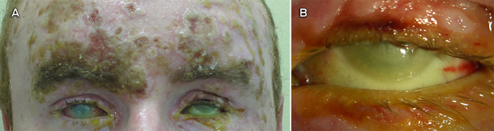
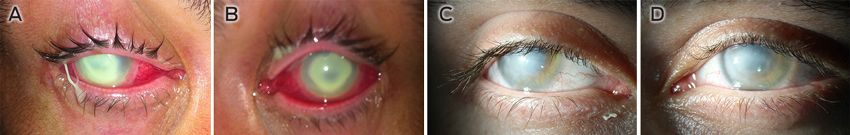
Ocular injuries are common in childhood, and their aetiology and epidemiology are well documented.1,2 Internationally, 20%–59% of all ocular trauma occurs in children (male to female ratio, 3.6 : 1), with 12%–14% of cases resulting in severe monocular visual impairment or blindness.2,3 In 2009, the Australian Institute of Health and Welfare identified that people aged < 19 years represented 15.6% of eye-related emergency department presentations between 1999 and 2006.4 Most eye injuries in children occur at home (76%), with the remainder occurring during sport and other recreational activity.2,3,5 In a recent New South Wales study, open globe injuries accounted for 40% of ocular trauma in children; of these, 48% occurred at home and involved common household objects.6 In 2000, 2.4 million eye injuries in the United States were related to sporting activity; 43% in children aged < 15 years and 8% in children aged < 5 years.7 Retrospective studies in Australia have shown that eye injuries from sporting activities accounted for 10% of severe ocular trauma in children, with permanent visual damage occurring in 27% of these cases.1 However, there is a lack of detailed information regarding the nature and incidence of children’s sporting eye injuries.

Due to their developing physical coordination, limited ability to detect risks inherent in the environment and vulnerable facial morphology, children are at higher risk of ocular trauma compared with adults of working age.5 Moreover, given that a child’s visual development continues from birth until 7–8 years of age, visual outcomes following trauma in children are worse than adults, affecting their subsequent career and social opportunities as adults.2 By adopting simple protective measures, such as using eye protectors when necessary, 90% of ocular injury is preventable.8

Outcomes of intervention in eye protection

A decline in paediatric ocular injuries in some sports, including lacrosse, field hockey, ice hockey and hurling, has been attributed to the adoption of mandatory protective eyewear in Canada, the US and Ireland. In the US, children’s eye injuries in lacrosse dropped by 84% following the introduction of mandatory eye protection in 2010.9 Key to effective implementation of eye-protection programs has been the development of product standards and policy statements by supporting organisations such as the American Academy of Ophthalmologyand American Academy of Pediatrics.5,7,9 In Australia, Squash Australia has set down strict guidelines for use of eye protectors in children (regulation 42, http://www.squash.org.au/sqaus/regulations_policies/regulations.htm [accessed Mar 2014]), but no studies have been conducted to review the effectiveness of this intervention.

Hurdles to effective use of protective eyewear in children

Significant research has been undertaken on the aetiology and management of eye injuries in children worldwide.3,5,7 However, there is less emphasis on research, public policy development and promotion of protective eyewear to reduce the incidence of eye trauma and vision loss. Occupational adult eye injuries have been found to decrease significantly with the use of eye protection.10 Little information is available in the scientific and grey (public domain) literature regarding protective eyewear in children; information for the general public is similarly limited.

Studies have shown that despite an awareness among adults, caregivers and children (particularly 15–18-year-olds) of the need for children’s eye protection, the rate of use remains low at about 19%.11 Parents and peers are major influences on attitudes to the use of eye protectors, with media not playing an important role. Deterrents to using appropriate eye protection include discomfort, poor visibility, unsuitable material, cosmetic appeal, availability, cost, storage and accessibility, no formal education on eye protection, and the perception that eye protectors are unnecessary.11

Regular (dress optical) spectacles are not an adequate substitute for eye protectors and can pose an additional danger due to the nature of the lens materials and the frame design.12 Because of its superior impact resistance, polycarbonate is the material of choice for prescription eye-protector lenses, but higher cost and the perception of its reduced scratch-resistance inhibit its uptake.13

Strategies to reduce ocular trauma in children

To address the large gaps in research and public policy regarding children’s eye protection, a multilevel approach is required to influence change in risk-reduction behaviour. Development, promotion and use of eye protection for children can be achieved through education and standards and policies.

Any approach to reduce the incidence of eye injuries should attempt to remove or limit hazards. Keeping potentially hazardous chemicals such as dishwashing liquid, toilet cleaners, paint, superglue and sprays out of young children’s reach, and purchasing toys that are appropriate for age and do not have sharp or projecting edges, can help protect children’s eyes.5 While it is impractical to suggest that a child wear eye protectors all the time, as most eye injuries in children occur at home, children’s activity should be supervised by a responsible adult, particularly when the child is exposed to potentially hazardous household items such as scissors, knives and other sharp objects.

Children, parents and caregivers, teachers, coaches and sports venue operators need to be reminded of the risks involved with particular sporting activities and the type of eye protectors that are available. Children who are at higher risk of eye injury — such as those who have experienced ocular surgery, trauma or disease, and functionally one-eyed individuals — should be encouraged to wear eye protection for all medium to high-risk activities and avoid sports or activities where no adequate eye protection is available.7 Health campaigns are essential to promote eye protection, using mass media to highlight the problems of eye damage in terms of blindness and its long-term impact. The support of professional organisations, local and national eminent people, eye-protection manufacturers and policymakers is important. Doctors and health professionals also play a key role in increasing public awareness and developing positive attitudes towards eye protection.

Standards in Australia currently focus on occupational more than domestic and sports eye protection. Internationally, there is a lack of standards and policies specifically for children’s eye protection. Of 43 spectacle and eye-protection standards for occupational and recreational requirements in the UK, US, Canada, Europe and Australia, only 21 allow for the specific needs of children. Most of the limited number of policies developed to prevent children’s eye injuries have focused on sports eye-protection requirements. It is imperative that policies and standards reinforce the needs of children who are functionally one-eyed and those at higher risk of ocular trauma to ensure that their vision is adequately protected.

Development of more advanced materials like polyurethane for occupational and sports eye protection has allowed for improved comfort and fit for adults. Children’s eye protection is yet to benefit from many of these advances. The optimal eye protector allows clear, distortion-free vision with a lens that does not fog, in a frame that is stylish and comfortable with adequate coverage and impact resistance. This highlights the need for spectacle designers to develop eye-protection solutions for children that meet standards requirements as well as addressing other issues to maximise compliance and use.

The way forward

To enable adequate measurement of the impact of interventions, the lack of detailed eye-injury data in Australia will need to be addressed. Long-term follow-up studies are required to improve our understanding of the nature and incidence of children’s eye injuries. Critical to the sustainability of any injury-prevention program is the ability to measure behaviour change following an intervention, to improve and develop more effective programs. Health education regarding eye protection should not stop with awareness campaigns but must be an ongoing process — awareness is highest soon after a campaign, after which it diminishes.

An improved understanding of the reasons for non-compliance with existing eye-protection recommendations will enable increased success of eye-protection programs. Importantly, we need an understanding of the current knowledge, influences and societal practices regarding ocular injuries, as well as the perception of risk, adequacy and subsequent use of eye protectors before and after an intervention.

Gaps in the current standards for children’s eye protection provide an important opportunity for a change in policies, recommendations and legislation, as well as for gaining support from relevant individuals and bodies. We have identified the need for specific standards to protect functionally one-eyed children and those at higher risk of eye injury; to determine when dress optical spectacles should be replaced with prescription eye protectors; and to identify which sports and recreational activities pose a medium to high risk of eye injury and require that participants use eye protection.

Severe alkali burns from beer line cleaners warrant mandatory safety guidelines

To the Editor: Two patients at our tertiary referral eye hospital had received severe alkali burns to the face and eyes while cleaning beer lines. In hotels, pubs and clubs, beer lines are cleaned weekly using strong alkaline solutions at high pressure. In an accident, alkali released under high pressure can produce blinding damage.

Our patients were young, typical of workers at these venues, and their injuries have had major impacts on their lives. One incident involved the face (60%–80% of facial skin) and both eyes (grade IV1) of a 23-year-old man (Box) using a commercially available beer line cleaner (potassium hydroxide; pH, 14). His eyes were immediately irrigated with water. His initial visual acuity was hand movements in the right eye and light perception in the left eye. He had bilateral corneal opacification and ocular ischaemia with hypotony in the left eye. He was treated according to burns protocol,1 but a non-healing right corneal ulcer developed, requiring multiple operations. Bilateral reconstructive eyelid surgery was also needed.2 His vision is currently light perception and no light perception with hypotony, respectively, in his right and left eyes. He requires further surgery to preserve his remaining vision, as well as ongoing psychological and social support.

The second patient received a grade IV injury to her left eye in 2006. Multiple operations were needed to heal the ocular surface, along with psychological and social support. After 4 years of treatment, her final visual acuity was light perception. She returned to limited work late in 2010.

Alkali injuries to the eye are devastating as they cause liquefactive necrosis and pass rapidly through the cornea to the eye’s internal structures.3 Damage to the stem cells of the ocular surface and intraocular structures produces permanent, difficult-to-treat and often blinding ocular disease. First aid should include copious ocular irrigation with water before referral to an emergency department.

Neither patient was wearing safety glasses. We found there are no mandatory safety guidelines in Australia (http://www.safeworkaustralia.gov.au), the United Kingdom (http://www.hse.gov.uk) or the United States (http://www.osha.gov), and perhaps worldwide, for beer line cleaning. Although the product information recommends use of safety equipment, this is not enforced and the equipment is usually not sufficient. Most workplaces provide safety glasses, but these offer suboptimal protection from a splash injury. Non-vented safety goggles are needed for adequate protection and should be worn throughout the cleaning procedure, from set-up to clean-up. We suggest that mandatory guidelines are indicated, as although such injuries are uncommon, they are severe and debilitating in the working-age group, with significant costs to the individual, health system and society.

Severe facial and ocular burns soon after injury


A: Image shows the limbal and scleral ischaemia of the left eye and bilateral opaque corneas. The sunken appearance of the left eye, indicative of hypotony, is a very poor prognostic sign. B: A close-up view of the left eye, showing the cloudy cornea and limbal ischaemia.

Severe bilateral Pseudomonas keratitis exacerbated by prolonged contact lens wear

Clinical record

A 28-year-old man with a 4-day history of bilateral ocular pain and a 2-day history of bilateral vision loss and purulent discharge was referred by a local hospital to our tertiary hospital. At the time of symptom onset, he had been wearing prescription monthly wear silicone hydrogel contact lenses (for correction of myopia) while swimming in lakes in Banlung in north-east Cambodia. The pain had worsened over 24 hours and the patient thought he had removed his contact lenses. He was prescribed topical neomycin 0.3% to use 2-hourly and topical moxifloxacin 0.5% to use three times a day in both eyes, and advised to return to Australia for follow-up. He had no prior medical problems.

On examination, both contact lenses were still in situ and the patient had only light perception. There was conjunctival and ciliary injection, with peripheral thinning of both corneas, especially in the inferior areas (Figure, A and B). Both corneas were completely white and opaque, owing to dense corneal infiltrations involving the whole cornea. Subtotal epithelial defects were seen with 1.5 mm hypopyon in both eyes.

The contact lenses were removed immediately. Both eyes were then treated empirically with hourly topical cephazolin 5% and gentamicin 1.5%, topical prednisolone acetate 1% six times a day and topical cyclopentolate 1% three times a day. Preservative-free lubricant (white soft paraffin [57.3% w/w] plus liquid paraffin [42.5% w/w] ointment) was administered four times a day. The patient was also started on intravenous meropenem 2 g three times a day, oral ciprofloxacin 750 mg twice a day, oral ascorbic acid 500 mg twice a day and oral doxycycline 100 mg once a day.

On Day 2, the organism grown from corneal scrapes from both eyes and the contact lenses was identified as Pseudomonas aeruginosa. The patient had lost his contact lens case and solution while in Cambodia. On Day 4, disc diffusion revealed that the organism was susceptible to ciprofloxacin rather than cephazolin. Topical cephazolin 5% was replaced with topical ciprofloxacin 0.3%.

From Day 20, lubricant was used hourly along with 0.3% tobramycin ointment twice a day. Bilateral temporal tarsorrhaphy was performed on Day 20 to promote healing. The patient was started on oral prednisolone therapy on Day 22 to help reduce inflammation associated with the bulging and melting of the corneas. Topical antibiotic therapy was tapered down as he began to respond well.

On Day 53, the tarsorrhaphy was opened as the epithelium had completely healed. Scars involving almost the whole surface areas of both corneas remained. On Day 74, there was extensive scarring and neovascularisation of both corneas (Figure, C and D). On Day 97, the patient’s unaided visual acuity was counting fingers in the right eye at 75 cm and counting fingers in the left eye at 50 cm, which improved with pinhole testing to 6/24-1 and 6/36, respectively.

A: Right eye at presentation. B: Left eye at presentation. C: Right eye at Day 74. D: Left eye at Day 74.

Pseudomonas aeruginosa is a gram-negative bacterium that is ubiquitous in the environment. It is found particularly in tropical areas, so lakes and rainforests in north-east Cambodia would be ideal places to come in contact with it.13

P. aeruginosa is usually susceptible to aminoglycosides and fluoroquinolones.4,5 Ofloxacin eye drops are effective in over 90% of bacterial contact lens-associated infections, are relatively non-toxic and do not need to be manufactured in a pharmacy, so could be used as first-line therapy for suspected microbial keratitis in contact lens wearers.2,4

Contact lens wear has led to a large rise in cases of Pseudomonas keratitis,6 owing to adherence of the bacterium to contact lens materials such as silicone hydrogel and the bacterium’s resistance to disinfectants.1 Also, the quite static environment on the posterior side of a contact lens enables organisms to stay on the ocular surface for a long period and be protected from host defence mechanisms, and thereby replicate more easily.1,7

It is commonly believed that for microbial keratitis to occur there needs to be an injury to the cornea so that the organism can pass through the epithelium and into the corneal stroma.6,8 However, studies in rats have found that P. aeruginosa forms biofilms on the posterior surface of contact lenses and subsequently causes severe microbial keratitis without prior injury to the corneal epithelium.1 Endophthalmitis is also a risk if the organisms are present in sufficient quantities, as they can penetrate intact epithelium and endothelium.1,7 Risk factor analysis has found that microbial keratitis disease load is decreased by 60%–70% by avoiding night-time wear of lenses.9 Other research suggests a 20 times increased risk of microbial keratitis for patients who wear contact lenses while sleeping compared with those who wear daily disposable contact lenses.2,4 Our patient had worn his lenses for at least 4 nights from when his symptoms began. The release of metalloproteinases from the P. aeruginosa is likely to have caused the rapid progression of the infection and subsequent corneal melting.

Bilateral Pseudomonas keratitis is rare, but this case shows how wearing contact lenses in a high-risk area and poor hygiene can lead to very severe bilateral microbial keratitis. Daily wear disposable lenses have been found to be the safest contact lenses, with the lowest rates of associated microbial keratitis, as compared with daily wear lenses that are put in solution overnight.2,4

After over 3 months of intense therapy, our patient is able to count fingers at less than 1 metre. He will almost certainly require corneal graft transplantation (which has its own risks) in 6–9 months for any chance to go back to his baseline functioning. We strongly advise that contact lenses be removed before swimming in lakes and that they be removed immediately if the wearer develops a sore eye.

Lessons from practice

  • In any contact lens wearer who presents with a sore eye, first check whether the contact lenses have been removed and remove them immediately if still in situ.
  • In suspected Pseudomonas keratitis, fluoroquinolones and aminoglycoside topical antibiotics are generally very effective.
  • Removing contact lenses before swimming in lakes, especially in tropical climates, is strongly advised.
  • Daily wear disposable contact lenses have been shown to be associated with lower rates of microbial keratitis, and would be ideal for people who wear contact lenses while travelling.

Eye injuries and tasers

To the Editor: Taser (TASER International) injuries have been topical in the news media. This provides an important reminder of the possible traumatic sequelae associated with the use of electronic control devices.

A taser is a battery-powered unit that uses a nitrogen cartridge to propel two darts on a 7 m copper wire.1 Each dart consists of a 4 mm harpoon-like barbed electrode on a 13 mm × 1 mm shaft (Box), deployed at 18 m/s from a distance of 3–6 m. Increasingly, tasers are being used by police in every state of Australia to subdue violent people.

When the deployed darts attach to a target individual’s skin or clothing, a current of up to 50 000 volts is released for a period of up to 5 s, depending on the skin’s resistance (which varies based on fat content, thickness, cleanliness and body chemistry).2 The mechanical impact of the barbs, combined with the subsequent voltage released, represents a considerable hazard to eyes, genitalia and large blood vessels in the neck.2

Essentially, the eyeball is a liquid-filled globe with a wall thickness < 1 mm, making it particularly susceptible to electrical damage. TASER International states that “serious injury, including permanent vision loss” can result from barb contact with the eye.3 Our literature search found seven case reports and one review of ocular damage relating to taser use.1,2,48 In five cases, penetration of the globe was reported;1,4,5,7,8 in three cases, entry through the lids made this difficult to determine without ophthalmic surgical examination.1,2,7 Ocular damage associated with taser use includes mydriasis, iritis, macular cysts, lid lacerations, cataracts, retinal detachment, optic neuritis, vitreous haemorrhage and globe penetration.6 Damage may be thermal or mechanical,6 with visual outcome ranging from final visual acuity of 6/9 to total vision loss and enucleation.1,2,4,5,7,8

As taser use increases, medical staff need to be aware of the implications of both the impact energy and the electrical damage associated with taser deployment. As in the management of a barbed fishhook penetrating the eye or ocular area, a taser barb should not be removed at the scene but should be immobilised (eg, by covering it with a foam or paper cup) until appropriate ophthalmic surgical removal is possible.1

Taser dart, showing dimensions (mm)

Vision screening in preschoolers: the New South Wales Statewide Eyesight Preschooler Screening program

Vision assessment is an important component of preventive health in childhood. Prevalence estimates for visual disorders in Australian children suggest rates of about 2% for amblyopia (reduced visual acuity in one or both eyes with no pathological cause),1 up to 7.3% for strabismus, and between 1% and 14.7% for refractive error.2 Many of these disorders can only be diagnosed through a monocular visual acuity screen and cannot be identified by family history, vision surveillance or observation of the child’s behaviour or appearance alone. Optimal treatment outcomes for childhood vision disorders are achieved with early detection and treatment, preferably before school entry.3 Failure to detect and treat vision disorders during childhood may lead to permanent loss of vision.4

There is evidence that a visual acuity test at about 4 years of age is best practice for preventing and treating vision disorders.2,5,6 Screening before school entry is ideal to optimise treatment outcomes1 and maximise compliance with treatment.6 All Australian states and territories offer some form of vision surveillance or screening to children: some recommend screening for all children before school entry, while others only screen children who are at risk or have a concern with their vision.6 However, child vision surveillance and screening typically rely on the child attending regular health checks, and systems that rely on parental vigilance are known to have variable participation rates.1,6

In New South Wales, vision surveillance is recommended as part of regular child health checks, as documented in the NSW Personal Health Record. From 2008, this was formalised as the Statewide Eyesight Preschooler Screening (StEPS) program, a free, universal vision screening program for 4-year-old children. Rather than relying on health check attendance, the StEPS program actively identifies and targets all 4-year-old children in NSW through preschools, childcare and other children’s services. Children attending the service who are 5 years old and have not previously received a StEPS screen are also eligible for screening.

The program is administered by local health districts (LHDs) — eight in the Sydney metropolitan region and seven covering regional and rural NSW. Ideally, LHDs attempt to offer vision screening to 100% of 4-year-old children but, as it may not be possible to reach all children, a minimum target of 90% has been set.7 Each LHD employs a StEPS Coordinator or assigns a staff member to oversee local implementation of the program. The coordinators are responsible for ensuring maximum program coverage and equity of access, and offer assistance to families to ensure that children identified through screening as having a possible vision disorder receive appropriate referral and diagnostic assessment. They also monitor and report on screening outcomes using data provided by LHDs.

Our aim was to evaluate the StEPS program to assess initial screening, referral and diagnosis rates for the target population.

Methods

We obtained de-identified data, collected between 1 July 2010 and 31 June 2011, from the LHD StEPS databases. Consent was sought from parents or carers for screening, and vision screening tests were conducted by StEPS vision screening staff who are trained and employed by each LHD. Data items collected on consent forms and at the time of screening were: demographic data; Indigenous status; personal and family history of vision disorders; vision screening results for each eye; result of screening (pass, borderline pass, type of referral); and other observations by the screener that were indications for referral, such as abnormal head posture or external eye abnormalities. In the event of a referral, data on the outcome or diagnosis after further investigation were also retrieved. Target population estimates for the study period were based on population projections using 2006 Census data8 provided to us by the Statewide Services Development Branch of the NSW Ministry of Health in collaboration with the NSW Department of Planning. As this evaluation was an analysis of routinely collected program data for the monitoring and quality improvement of health service delivery, ethics approval was not required.

All screening and referral was done according to StEPS protocols. The StEPS referral pathway is shown in Box 1. If a consent form was not returned, a minimum of two follow-up screening offers were made. If a child was absent on the day of screening, parents or carers were asked to have their child’s vision assessed through their child and family health centre or general practitioner. Children were screened using a 6 m linear chart (or 3 m chart where a distance of 6 m was not available). Children passed the screening test if their visual acuity was 6/9 or above in each eye. A score of 6/9-1 or 6/9-2 in one or both eyes was regarded as a borderline pass, and parents or carers were advised to retest in 12 months. Children were referred to a GP or eye health professional for follow-up: if they scored less than 6/9-2 but better than 6/18 in one or both eyes; if there were obvious external eye abnormalities or the child’s vision could not be assessed on the day of screening (eg, if the child was unwell or distracted); or if they scored 6/18 or less in one or both eyes — this was regarded as a high-priority referral, and parents were advised to have the children urgently undergo further testing.Completeness of data on diagnostic outcomes after referral from the StEPS screening program varied by LHD. Combined results from the two LHDs (which comprised one area health service at the time of the study) with the most complete data for the study period were analysed in more detail to enable better evaluation of outcomes.

Results

Of 91 324 eligible 4-year-olds in NSW during the study period, 80 328 (88.0%) were offered screening. Of these, the parents or carers of 71 081 children (88.5%) accepted and provided consent for screening. Of those for whom consent was obtained, 65 834 children (72.1% of the eligible population) were screened. The remaining children for whom consent was received but who were not screened were absent on the day of screening. Of 74 249 parents or carers who returned consent forms, 1600 (2.2%) indicated they were declining because their child had already received a vision screen. Of children who were screened, 2568 (3.9%) identified as being of Aboriginal and/or Torres Strait Islander origin.

Outcomes of the testing were that 52 870 children (80.3%) passed the screening test, 6405 (9.7%) received a borderline pass, and 6421 (9.8%) were referred to their GP or eye health professional for further assessment. The group referred for further assessment comprised 3867 children (5.9%) with routine referrals, 1425 (2.2%) with high-priority referrals, and 1129 (1.7%) who were unable to be assessed on the day of screening. Screening results were missing for 138 children (0.2%), most likely due to inaccurate data entry.

The two LHDs (one metropolitan and one rural) with the most complete follow-up data for the study period achieved an average follow-up rate of 95.7% (1231/1286). These two LHDs are considered representative of metropolitan and rural LHDs in NSW, with an average referral rate of 9.9% of children (1286/12 977) screened. Box 2 shows the primary diagnoses for children followed up after screening in these two LHDs. Of 304 children with high-priority referrals, 28.0% were later diagnosed with amblyopia, 44.1% were prescribed glasses, and 5.6% were diagnosed with other vision disorders, including cataract, ptosis, glaucoma and nystagmus. Among 847 children with routine referrals after screening, 5.9% were later diagnosed with amblyopia, 32.0% were prescribed glasses, and 14.8% were diagnosed with other vision disorders. Some of the children who were referred because they could not be assessed were also diagnosed with vision disorders: for example, 4.4% were prescribed glasses and 25.2% required monitoring and later review.

In addition to vision disorders, anecdotal evidence from the two LHDs indicated that some children who were referred because they could not be assessed had other diagnoses such as developmental delay.

Only a small proportion of children who were referred for further investigation after screening were found to have no visual abnormality (Box 2). Follow-up data were available for 285 of the 304 high-priority referrals (93.8%) and, among these, there were only seven children in whom no abnormality was found. Therefore, 278 of 285 children (97.5%) for whom follow-up data were available required either treatment or review at a later date. For routine referrals, 704 of 779 children (90.4%) who received a follow-up assessment required either treatment for a vision problem or review at a later date.

Discussion

Data from this evaluation indicate that the StEPS program is close to achieving its aim of offering vision screening to at least 90% of 4-year-old children in NSW. The high proportion of parents being offered and consenting to screening for their children indicates a high level of acceptance of screening in this population. Our results suggest the program can also be successfully offered to specific groups of children, such as those from Aboriginal and Torres Strait Islander backgrounds. We found that the proportion of screened children who identified as Aboriginal and/or Torres Strait Islander (3.9%) was similar to the estimated percentage of all 4-year-old Aboriginal and/or Torres Strait Islander children in NSW.9

Although many jurisdictions worldwide screen children’s vision at or after the time of school entry, only a few, such as Sweden and parts of Canada, undertake universal visual acuity screening before school entry.5 Follow-up data indicate that preschool screening programs reduce the incidence of vision disorders,10 with one cohort study finding a 45% reduction in the prevalence of amblyopia among 7-year-olds who received preschool screening, compared with those who did not.11 Although reviews of the literature have found a lack of evidence of effectiveness for preschool vision screening programs, due to a lack of well designed randomised controlled trials,2,12,13 expert opinion suggests that the value of early intervention is sufficient that screening programs should be implemented on the strength of existing evidence.1,6

Our data support the likelihood of considerable benefits for children receiving a preschool vision screen. For the 6421 children we identified as requiring further assessment after screening, early detection and treatment may have prevented loss of vision from conditions such as anisometropic amblyopia,4 which was diagnosed in 25% of children with high-priority referrals. This potentially serious disorder is not detectable by vision surveillance alone, and failure to treat it at a young age may lead to permanent loss of vision.14 Treatments for amblyopia, such as patching, are not only more effective during the preschool years, they are also more appropriate, as patching at school may lead to bullying.6 A significant proportion of children referred for investigation in our study were also prescribed glasses before entering school.

Before the implementation of the StEPS program, vision assessment in preschool children in NSW relied on voluntary attendance at the 4-year-old child health check, for which there is some evidence that attendance is poor, particularly among the most socioeconomically disadvantaged groups.15 We found that only 2% of parents declined consent because their child had previously had a vision screen. In addition, vision surveillance conducted during health checks does not necessarily include a monocular visual acuity screen.6 Thus, without universal preschool screening, many children with vision problems are likely to remain undiagnosed, and their vision uncorrected, into their school years. Preschool screening is preferable to school screening, as the critical window for intervention may otherwise be lost.6

The StEPS program aims to make the best possible treatment readily available to the screened population by offering assistance to families to ensure that children receive appropriate diagnostic assessment and referral. The StEPS Coordinator may provide a referral either to a GP, who can then refer to an eye health professional as appropriate, or directly to one of the dedicated tertiary paediatric ophthalmic outpatient clinics that have been established in NSW for children referred via the program. Maximising positive treatment outcomes after early diagnosis is an important feature of screening programs.16 We found that only 1.7% of children were unable to be assessed on the day of screening, which demonstrates the appropriateness of this screening in our target group. Use of this test for screening allows more costly, invasive and time-consuming investigations to be reserved for those identified to be at higher risk. Our data indicate that among children referred from the StEPS program for further investigation, only a small proportion (2.3% of high-priority and 8.9% of routine referrals) had no abnormality diagnosed, suggesting this screening test is accurate and that the referral criteria in the StEPS program are appropriate.

There are three important limitations of this study. First, because children who tested negative for vision disorders during screening were not followed up as part of StEPS to determine their true disease status, we were not able to calculate sensitivity or specificity of the screening test. A second limitation is incompleteness of our data on vision disorders that are diagnosed after referral from the StEPS program. Although each LHD is required to follow up visual outcomes for children who are referred for further assessment, this has proven difficult in many districts because of the diversity of services that provide follow-up. The StEPS policy directive is currently being revised to ensure consistency of diagnostic outcome reporting from all LHDs, and accuracy and completeness of outcome data have significantly improved since mid 2012. Future evaluation studies will benefit from this more complete outcome data measured over a longer period. Finally, this evaluation could not measure the cost–benefit ratio of screening, in financial terms and with respect to the costs, risks and benefits for individual children.16 Initial funding of the program amounted to about $14 million over 4 years for both the StEPS program and the paediatric ophthalmic outpatient clinics. Our evaluation focused only on screening and diagnostic outcomes, but the cost-effectiveness of the StEPS program is an important area for future research. Although the program would ideally exist as part of a universal comprehensive health screening program before school entry, vision screening is considered of sufficient importance that the StEPS program should be conducted in isolation in the meantime.

In conclusion, the StEPS program is conducting visual acuity testing in a high proportion of 4-year-olds in NSW. Prompt referral and optimal treatment of common visual disorders may prevent potentially permanent loss of vision in a significant number of children. The StEPS model may be useful for other jurisdictions in Australia and internationally considering implementation of similar programs.

1 New South Wales Statewide Eyesight Preschooler Screening program referral pathway


LHD = local health district. GP = general practitioner.

2 Primary diagnoses or other outcomes for preschoolers referred for further investigation after screening in the New South Wales Statewide Eyesight Preschooler Screening Program in two local health districts, July 2010 to June 2011

Primary diagnosis or other outcome

High-priority referrals (n = 304)

Routine referrals
(n = 847)

Referred due to inability
to assess (n = 135)


Diagnoses

Amblyopia

Anisometropic amblyopia

76 (25.0%)

33 (3.9%)

1 (0.7%)

Strabismic amblyopia

9 (3.0%)

17 (2.0%)

0

Total amblyopia

85 (28.0%)

50 (5.9%)

1 (0.7%)

Prescribed glasses

Refractive error

109 (35.9%)

242 (28.6%)

6 (4.4%)

Anisometropia

25 (8.2%)

29 (3.4%)

0

Total prescribed glasses

134 (44.1%)

271 (32.0%)

6 (4.4%)

Other vision disorders

17 (5.6%)

125 (14.8%)

5 (3.7%)

Strabismus or squint

0

59 (7.0%)

0

Total visual abnormality diagnoses

236 (77.6%)

505 (59.6%)

12 (8.9%)

Other outcomes

No visual abnormality detected

7 (2.3%)

75 (8.9%)

18 (13.3%)

Review and monitoring indicated

18 (5.9%)

197 (23.3%)

34 (25.2%)

Already under the care of an eye health professional

24 (7.9%)

2 (0.2%)

2 (1.5%)

Referral not followed up by parent or carer

11 (3.6%)

48 (5.7%)

42 (31.1%)

Lost to follow-up

8 (2.6%)

20 (2.4%)

27 (20.0%)

The influence of Australian eye banking practices on corneal graft survival

Corneal transplantation enables the restoration of sight to people with corneas damaged by disease or trauma. In Australia, as in many countries, donor corneas are preserved between retrieval and transplantation in several ways and for variable periods in licensed eye banks.

It has been recommended that corneas be retrieved within 12 hours of donor death,1 but there is no consensus on the acceptable maximum interval, and this time limit is sometimes stretched.2 In Australia, after retrieval and processing within a 24-hour period, corneas are currently preserved in an eye bank in one of three ways: in tissue-culture medium at 4ºC, in organ culture at 30–37ºC, or occasionally as a whole enucleated globe in a moist pot at 4ºC. Storage time depends on the preservation method but corneas will generally be transplanted as soon as practicable, to limit the inevitable deterioration that occurs after death and during storage.3

Although Australia’s five eye banks are able to collect sufficient donor tissue to meet the nation’s needs, they are not located close to all transplant centres. Tasmania sources virtually all its donor tissue from the Australian mainland, and corneas may be transported between other states, and occasionally between Australia and New Zealand, to cover local shortfalls in availability or to meet an emergency demand. When surgery is performed at a distance from the cornea procurement site, the tissue is transported by air freight. Potential issues associated with transportation include the additional handling involved, variations in temperature and pressure during transit, shaking, and extended donor death-to-transplantation time.46

Using data from a national registry of corneal graft outcomes, we examined the impact of methods of corneal preservation and air transport on subsequent corneal graft survival.

Methods

Australian eye banks licensed by the Therapeutic Goods Administration are located in Brisbane, Sydney, Melbourne, Adelaide and Perth. The banks request consent for corneal donation and subsequently retrieve, evaluate, preserve and distribute human corneas for transplantation.

The Australian Corneal Graft Registry follows the progress of all corneal grafts performed in Australia. Established in May 1985, it collects information on recipients, donors, eye bank practices, surgical procedure, subsequent management, complications, graft survival, and visual outcomes after the graft. Yearly follow-up is requested until graft failure, recipient death or loss to follow-up. De-identified and amalgamated registry analyses are used to inform clinical practice and to identify risk factors for poor outcomes. The registry’s operations are approved by the Southern Adelaide Clinical Human Research Ethics Committee and are carried out in accordance with the Declaration of Helsinki.

At the census date for this study (July 2012), 24 984 corneal grafts had been registered (Box 1). We analysed data for penetrating (full-thickness) and lamellar (partial-thickness) grafts, but not limbal (stem cell) grafts. Lamellar grafts were further categorised into traditional (peripheral or patch) lamellar, deep anterior lamellar and endothelial keratoplasty. Methods of data collection have been published elsewhere.7

Statistical analysis

To identify potential risk factors for graft failure, we performed univariate analysis for 19 254 corneal grafts in 15 160 patients for which at least one follow-up had been recorded by the census date (“followed grafts”). Graft failure was reported by follow-up surgeons when the graft was no longer achieving the function for which it was performed (eg, functional vision). We examined the distribution across variables for grafts that had been followed and those that had not, to determine potential biases in the study population.

We performed Kaplan–Meier survival analysis8 using SPPS version 18 (SPSS Inc), with significance set at P < 0.05 (Mantel–Cox log-rank χ2 statistic), to examine the influence of: time from death to enucleation (≤ 3 hours, 4–6 hours, 7–9 hours, 10–12 hours, > 12 hours); time from death to storage (≤ 6 hours, 7–12 hours, 13–18 hours, 19–24 hours, > 24 hours); time from enucleation to graft (≤ 48 hours, 49–96 hours, 97–144 hours, > 144 hours); storage method used (Optisol [Bausch and Lomb], organ culture, moist pot, superseded media); and whether or not the cornea had been transported interstate by air. Superseded media included McCarey-Kaufman medium, K-Sol (Bausch and Lomb) and Dexsol (Bausch and Lomb). Where fewer than 20 grafts were followed for a variable (Appendix 1), these grafts were excluded from analyses relating to the variable for that type of graft. For variables with just two groups, univariate analyses were not conducted if one group had insufficient numbers. No other exclusions were applied.

Indication for corneal transplantation has previously been found to be a highly significant predictor of graft survival in the registry,9 with keratoplasties performed for keratoconus exhibiting good survival rates compared with all other indications. Furthermore, although most corneal grafts performed in Australia are elective, a small proportion are performed on an emergency basis (eg, for a penetrating eye injury or herpetic perforation) and such grafts tend to be at high risk of subsequent failure.9 We therefore considered that indication for graft (emergency, keratoconus or other) might be a confounder in our analysis, particularly as corneas transported by air and those stored in moist pots in recent years are more likely to have been used in emergency procedures. Where appropriate, we examined the effect that graft era (1985–1995 v 1996 onwards) may have on survival, as eye banking techniques have changed over time.

We conducted multivariate Cox proportional hazards regression analyses10,11 using Stata version 11 (StataCorp) to determine independent risk factors for survival of each type of graft. As some patients had received more than one graft, the data were clustered by recipient, using the Breslow method for ties, to control for intergraft or intereye dependence.12 Analyses were performed in a backwards stepwise manner, initially including all variables found to be significant in the univariate analysis. Where appropriate, variables were treated as time-variant. The least significantly contributing variable was removed until all variables made a significant (P < 0.05) independent contribution to the model. Some follow-up records were missing data for one or more of the variables of interest. At each stage of the regression, records with complete data for all of the included variables were analysed. Hazard ratios (HRs), adjusted for clustering and the impact of other variables in the model, were used to compare survival across categorical variables. The first level of each variable was used as the referent.

Results

Eye bank practices for followed and not-yet-followed grafts are reported in Appendix 2. Equivalent proportions of corneas in each cohort had been transported by air freight. There were similar distributions of indications for grafts across the two groups. For followed grafts, the main indications for each type of graft are shown in Box 2.

Univariate analyses of survival for each type of graft, stratified by eye bank variable, are shown in Box 3. Storage method significantly influenced survival of traditional lamellar and endothelial grafts (P < 0.001). For endothelial grafts, corneas stored in organ culture fared significantly worse than those stored in Optisol (Box 4). Interstate air freight transportation of the donor cornea affected survival of penetrating grafts and traditional lamellar grafts (P < 0.001) (Box 3 and Box 5). Death-to-enucleation time exerted a significant influence on the survival of deep anterior lamellar grafts (Box 6) and penetrating grafts, and enucleation-to-graft time also significantly affected the survival of penetrating grafts (Box 3). Death-to-storage time exerted no significant influence on graft survival. Indication for the graft had a significant impact on survival for penetrating and traditional lamellar grafts, and graft era significantly affected survival of traditional lamellar grafts (all P < 0.001).

In the multivariate model (Box 7), interstate transportation by air freight and indication for graft were significant contributors to outcomes of penetrating grafts (χ2 = 7863.75, P < 0.001). These variables, along with storage method, were also significant contributors to outcomes of traditional lamellar grafts (χ2 = 65.51, P < 0.001). For both penetrating and traditional lamellar grafts, transport of the donor cornea by air freight and high-risk indication for the graft were significant risk factors for failure. Penetrating grafts performed for keratoconus had significantly better survival than those performed for other indications, but this was not the case for traditional lamellar grafts. For traditional lamellar grafts, corneas that had been stored in moist pots fared better than did those stored in Optisol.

Discussion

Unlike many other factors that are known to influence corneal graft survival, eye banking practices are amenable to change. In line with previous reports, we found that, once the influence of other significant variables was taken into account, donor death-to-enucleation time, enucleation-to-graft time, and storage time of a cornea in the eye bank did not exert a significant influence on the probability of graft survival for most graft types. While a significant effect of death-to-enucleation time on survival of deep anterior lamellar grafts was found, no specific relationship between the two that could inform best practice was apparent. Most analysed grafts were conducted with donor tissue that was retrieved, stored and transplanted within nationally recommended time frames.13 Our findings add to the evidence that operating within these guidelines protects corneal donor tissue from detrimental postmortem changes.

For some graft types, we found that the method of cornea preservation influenced subsequent corneal graft survival. Optisol, a tissue culture medium to which antibiotics, dehydrating agents, ATP precursors and vitamins have been added, is one of the latest alternative cold-storage techniques.1,14,15 It gradually replaced other now superseded media in Australia over about 10 years from 1990. Corneas are stored in Optisol at 4ºC for a recommended maximum of 14 days.16 Moist pot storage1,17 involves the entire eye being placed in a container kept at 4ºC. It is limited by rapid deterioration in the viability of the cornea after 24–48 hours18 and is now seldom used. In recent years, there has been a shift in some parts of Australia towards preserving donor corneas in organ culture, as is the practice in Europe.14,17 Corneas are stored at 30–37ºC, with bovine calf serum and antibiotics including antifungal agents added to the tissue culture medium, for up to 4 weeks.14,19 This shift was reflected in the grafts registered at the time of this study, with 9% and 87% of all grafts performed from 2000 onwards using corneas stored in organ culture and Optisol, respectively, compared with 26% and 72%, respectively, for grafts performed from only 2010 onwards (data not shown).

In confirmation of previous studies, we found no significant difference in penetrating graft survival between corneas stored in Optisol or organ culture.5,15,16 Corneas used for traditional lamellar procedures exhibited significantly better survival when stored in moist pots than in Optisol. We speculate that the greater swelling of the corneal stroma that occurs in Optisol-stored corneas may render wound apposition during lamellar surgery more difficult. This suggests that different storage techniques may be warranted, depending on graft technique. The small number of traditional lamellar grafts being performed these days and the reduced length of viability for corneas stored in moist pots mean that this finding is unlikely to influence clinical practice. Our finding that for endothelial grafts, corneas stored in organ culture fared worse than those stored in Optisol may suggest a negative impact of organ culture preservation on endothelial cell viability. However, a similar difference was not found for penetrating grafts, which also involve the endothelial layer. Possibly there are other variables not related to the eye bank that are influencing this result, and further exploration is warranted. Overall, the choice of corneal storage medium may affect graft survival, and this effect appears to vary for different types of keratoplasty.

A significant difference was apparent between the outcomes of grafts involving corneas that had been sent interstate by air compared with those retrieved and used locally. Transport of donor corneas by air within Australia was associated with poorer survival of penetrating and traditional lamellar grafts, and the HRs indicated a clinically important impact. Furthermore, the multivariate model also included recipient indication for graft, which shows that differences in survival cannot be explained by transported corneas being used more often in emergency procedures. For penetrating grafts, this effect varied over time: there was an initial reduction in survival, but equivalent levels were seen after the first 12 years, with equal median survival.

The impact of international air travel on the outcomes of penetrating keratoplasty has been reported previously.4,6,20 One study examined the changes in corneal endothelial cell morphology that occurred in corneas between leaving the United States and arriving in Taiwan, and evaluated outcomes achieved after transplantation of these corneas.4 Obvious transport-related morphological changes were observed and by 4 years after the graft, nearly half the grafts had failed. Despite this, the authors concluded that the surgical success rate was not influenced by the air transport. With no comparison group, it cannot be determined whether this failure rate was unusual. A second study, from Israel, evaluated the outcomes of grafts involving local donors and those for which corneas were procured from the US.6 The authors concluded that air transport had not compromised the success of the corneal transplants. In a third study, no significant differences in the likelihood of survival, primary graft failure, rejection or infection were found between corneas imported from the US and those sourced locally in Japan.20 All these studies were small, with limited follow-up periods.

Our finding that air transport of stored donor corneas was associated with reduced corneal graft survival was unexpected. Nevertheless, given the size of the dataset, the inclusion of data from multiple eye banks working within the same guidelines, and the extended follow-up available for penetrating and traditional lamellar grafts, we consider the result to be robust. So what might be the cause of the deleterious influence of air transport within Australia on human donor corneas? Australian eye banks have undertaken validation of temperature control in shipping containers under various simulated and actual conditions, and report that temperature fluctuations are small. Shaking, pressure changes, and rapid acceleration and deceleration may play a role.4,5,21,22 Although our study has identified an association between air freight of donor corneas and subsequent poor outcomes after transplantation, we cannot identify the cause. Further modification of corneal transport containers is difficult without a clearer understanding of the underlying cause of the problem.

Our findings highlight the need for continued evaluation of the impact of eye bank practices on corneal graft survival. Transportation of donor corneas around the world has increased in recent years, as some countries retrieve more corneas than others. A balance needs to be struck between duplication of services with attendant additional expense, appropriate use of a scarce human resource so that wastage is minimised, and the needs of recipients and their surgeons. Air freight of some corneas will always be necessary. However, in Australia, where sufficient donor tissue is generally available within each state, efforts to avoid domestic air transportation of corneal tissue may be warranted. Furthermore, we suggest that centralisation of donor cornea retrieval services may not be in the best interests of the community, at least until the transport of human donor corneas can be improved.

1 Numbers of patients and corneal grafts registered and followed, by graft type

Patients


Grafts


Graft type

Registered

Followed

Registered

Lost before
follow-up

Followed

Failed*

Recipient has died

Lost after follow-up

Still being followed

Not yet
followed

Still active in registry


Penetrating

16 491

13 673 
(82.91%)

21 303

1896 
(8.90%)

17 301 (81.21%)

4173 (19.59%)

4896 (22.98%)

6142 (28.83%)

3337 (15.66%)

2106 
(9.89%)

5443 (25.55%)

Lamellar

Endothelial

1415

644 
(45.51%)

1697

47 
(2.76%)

738 (43.49%)

216 (12.73%)

28 
(1.65%)

48 
(2.83%)

451 (26.58%)

912 
(53.74%)

1363 (80.32%)

Traditional lamellar

1083

857 
(79.13%)

1191

161 
(13.52%)

939 (78.84%)

223 (18.72%)

265 (22.25%)

390 (32.75%)

121 
(10.16%)

91 
(7.94%)

212 (17.80%)

Deep anterior lamellar

639

273 
(42.72%)

677

94 
(13.88%)

276 (40.77%)

30 
(4.43%)


(0.59%)

58 
(8.57%)

186 (27.47%)

307 
(45.35%)

493 (72.82%)

Limbal (not analysed)

104

88 
(84.61%)

116

10 
(8.62%)

98 (84.48%)

47 (40.52%)

26 
(22.41%)

28 
(24.14%)


(5.17%)


(6.90%)

14 
(12.07%)

Total in registry

19 163

15 248 
(79.57%)

24 984

2208 (8.84%)

19 352 (77.46%)

4689 (18.77%)

5219 (20.89%)

6666 (26.68%)

4101 (16.41%)

3424 
(13.70%)

7525 (30.12%)

Total analysed

19 059

15 160 
(79.54%)

24 868

2198 (8.84%)

19 254 (77.42%)

4642 (18.67%)

5193 (20.88%)

6638 (26.69%)

4095 (16.47%)

3416 
(13.74%)

7511 (30.20%)


* Figures for failed grafts may also include grafts for which the recipient is known to have died, if this occurred after the graft had failed. Grafts that were still surviving when the recipient died are not treated as failed grafts but rather censored at the time of death. Peripheral or patch.

2 Main indication for followed grafts as specified by surgeon, by graft type (n = 19 254)

Main indication for graft

Penetrating

Traditional lamellar

Deep anterior lamellar

Endothelial


Keratoconus

5213 (30.13%)

70 (7.45%)

199 (72.10%)

Bullous keratopathy

3935 (22.74%)

11 (1.17%)

0

215 (29.13%)

Failed previous graft

3585 (20.72%)

141 (15.02%)

6 (2.17%)

136 (18.43%)

Corneal dystrophies

1900 (10.98%)

13 (1.38%)

6 (2.17%)

365 (49.46%)

Corneal scars and opacities

391 (2.26%)

28 (2.98%)

6 (2.17%)

0

Non-herpetic corneal ulcers

338 (1.95%)

121 (12.89%)

2 (0.72%)

0

Herpetic eye disease

738 (4.27%)

37 (3.94%)

18 (6.52%)

1 (0.14%)

Accidental injury

395 (2.28%)

118 (12.57%)

7 (2.54%)

13 (1.76%)

Other*

806 (4.66%)

400 (42.60%)

32 (11.59%)

8 (1.08%)

Total

17 301

939

276

738


* For example: corneal degenerations, congenital abnormalities, descemetoceles, iridocorneal endothelial syndrome, metabolic deposits.

3 Univariate analyses for influence of eye bank practices and related variables on corneal graft survival,
by graft type

Penetrating


Traditional lamellar


Deep anterior lamellar


Endothelial


Variable

χ2

P

χ2

P

χ2

P

χ2

P


Storage method

6.25

0.10

19.71

< 0.001

0.81

0.37

20.67

< 0.001

Transport by air freight

23.19

< 0.001

13.73

< 0.001

na

na

1.44

0.23

Death-to-enucleation time

9.91

0.04

1.56

0.82

10.67

0.03

7.87

0.10

Death-to-storage time

3.85

0.43

1.03

0.91

4.92

0.30

5.24

0.26

Enucleation-to-graft time

9.06

0.03

1.85

0.60

2.57

0.46

0.81

0.85

Indication for graft

1597.95

< 0.001

49.42

< 0.001

3.62

0.16

na

na

Graft era

1.31

0.25

7.37

< 0.001

na

na

na

na


na = not applicable to the type of graft.

4 Kaplan–Meier plot of survival of endothelial grafts performed with donor corneas stored in Optisol or organ culture*

* P < 0.001, Peto log-rank statistic.

5 Kaplan–Meier plot of survival of penetrating grafts (A) and traditional lamellar grafts (B) performed with donor corneas transported by air compared with those retrieved and used locally*

No air transport = corneas retrieved and used within the same state. Air transport = cornea retrieved in one state and transported by air freight for use in a different state. * P < 0.001, Peto log-rank statistic.

6 Kaplan–Meier plot of survival of deep anterior lamellar grafts performed with donor corneas enucleated at various times after donor death*

* P = 0.031, Peto log-rank statistic.

7 Multivariate analysis for influence of corneal transport and storage and indication for graft on corneal graft survival, for penetrating and traditional lamellar grafts

Variable

Number of grafts

Hazard ratio (95% CI)

P

Global P


Penetrating grafts

Transported by air freight*

No

14 684

1.00

Yes

919

1.44 (1.21–1.73)

0.001

Indication for graft*

Not keratoconus or high-risk

10 265

1.00

< 0.001

Keratoconus

4611

0.16 (0.13–0.19)

< 0.001

High-risk indication

727

2.53 (2.10–3.04)

< 0.001

Traditional lamellar grafts

Transported by air freight

No

813

1.00

Yes

64

1.69 (1.03–2.78)

0.038

Storage media

Optisol

233

1.00

0.047

Moist pot

480

0.61 (0.41–0.91)

0.016

Superseded media

164

0.63 (0.39–1.02)

0.059

Indication for graft*

Not keratoconus or high-risk

600

1.00

< 0.001

Keratoconus

65

0.66 (0.32–1.33)

0.247

High-risk indication

212

3.15 (2.16–4.61)

< 0.001


* Analyses adjusted for interaction with time for both variables in the penetrating graft model and for indication for graft in the traditional lamellar graft model. Includes endophthalmitis, corneal melt, perforation, trauma or accidental injury, and burn, sometimes in combination with another indication.

Antivascular endothelial growth factor treatments for neovascular age-related macular degeneration save sight, but does everyone get treated?

To the Editor: Neovascular
age-related macular degeneration (NVAMD) is the most common cause of blindness in Australia.1 Current treatment to prevent further deterioration in vision, and an improvement in some, involves
the antivascular endothelial growth factor (anti-VEGF) agents ranibizumab (Lucentis, listed on
the Pharmaceutical Benefits Scheme [PBS] since August 2007) or bevacizumab (Avastin, used off-label for NVAMD).2

The Australian Macular Degeneration Foundation estimated the annual number of new cases
of NVAMD to be 20 734 in 2010 
in Australia, based on incidence figures from the Blue Mountains
Eye Study, based on an Australian population.3

We used PBS data to investigate people accessing treatment for the first time (first prescription for ranibizumab) between 2008 and 2010, and found that 7501 people in 2008, 6979 people in 2009 and 6623 people in 2010 were treated with ranibizumab (indicated by a first and subsequent prescriptions).

Considering the predicted new cases of NVAMD at around 20 000 annually, there is a large gap between people receiving treatment and people in need of treatment. Let us assume that a further 20% of people access treatment under the Repatriation PBS (based on PBS item statistics), and an additional 20% are treated with bevacizumab off-label (based on a 10% observed figure in the largest Australian NVAMD treatment registry, the Fight Retinal Blindness! Project [Mark Gillies, professor of clinical ophthalmology and eye health, University of Sydney, personal communication, 24 November 2012]). These are both intentional overestimates, but they still leave a gap of more than 5000 new cases per year which may be
in need of treatment but are not treated.

Factors which may discourage patients from accessing and accepting treatment include availability, cost because of gap fees and the need for ongoing monitoring and treatment. This involves intraocular injections which may
be perceived as uncomfortable. However, side effects are few, and virtually no patient discontinues treatment due to these or a perceived discomfort.4

Blindness comes with a considerable loss of quality of life, increased morbidity and mortality, higher rates of institutionalisation and increased personal and community cost. In Denmark,
anti-VEGF treatment has been shown to halve the rate of legal blindness caused by NVAMD.5
Thus it is important to reduce barriers to anti-VEGF treatment
for NVAMD in Australia.