×

Survey identifies key dementia research priorities

The National Health and Medical Research Council (NHMRC) National Institute for Dementia Research has identified diagnosis, prevention and interventions to reduce risk as the key priorities for dementia research.

Australian scientists are world leaders in dementia research and, over the coming decades, would have found treatments for dementia. However, this timeline is too slow for the economic and social pressures of an ageing population. Therefore, the federal government moved to accelerate research discoveries with a $200 million investment to boost dementia research in the 2014–15 Budget.

A survey and interviews of consumers (people with dementia, families, personal carers), researchers (new and established), medical practitioners and aged care providers identified three top priorities.

For professional care providers, senior investigators, researchers and medical practitioners, the priority is to identify effective interventions to reduce the risk and prevent the incidence of dementia.

For people with dementia, timely, accurate and supported diagnosis is paramount, with prevention the second priority — a reminder of the importance of accurate and sensitive diagnosis and support for people with dementia and their families.

For personal carers, the priority is to develop effective interventions to support their opportunity and capacity to provide care, which are currently quite variable; again, prevention is their second priority.

The priorities form a key element of the government’s commitment to supporting dementia research. The NHMRC National Institute for Dementia Research will target, coordinate and translate dementia research, guided by the priorities.

The NHMRC report states: “The priority for prevention indicates a consensus and confidence that research into risk and preventative factors will have an impact in reducing the incidence of dementia and is the first priority for Australian research.”

The NHMRC Dementia Research and Translation Priority Setting Project was prepared by ThinkPlace consultants and released on 27 January 2015 at http://www.nhmrc.gov.au.

An unusual neurological complication from a garden-variety organism: post-melioidosis parkinsonism

We report the first case of acute parkinsonism following disseminated melioidosis with multiorgan abscesses in a 62-year-old man. After 1 month of treatment with levodopa, the parkinsonism resolved completely. Melioidosis should be considered as a possible cause for parkinsonism in endemic areas.

A 62-year-old man presented to our tertiary hospital’s emergency department with a 4-week history of fever associated with lethargy and constitutional symptoms. For 9 days before admission, he had been vomiting two to three times per day. He had longstanding diabetes and hypertension and worked for the local city council as a truck driver, transporting water to local gardens and public areas. About 2 months previously, he had sustained an abrasion on his left foot that had healed completely at time of presentation.

On initial assessment, he had a blood pressure of 129/78 mmHg, a heart rate of 111 beats/min, an SpO2 of 96% in room air, and a respiratory rate of 16 breaths/min. He was clinically dehydrated and his body temperature was 38.8°C. His abdomen was soft and non-tender, with hepatomegaly of two fingers’ breadth. Respiratory examination revealed left basal lung crepitations. Results of the clinical assessment, including cardiovascular and neurological examinations, were otherwise normal.

The patient’s initial blood investigations revealed an elevated random blood glucose level (11.6 mmol/L; reference interval [RI], 4.4–6.1 mmol/L) and white cell count (12.1 × 109/L; RI, 4.0–11.0 × 109/L) with neutrophilia (93%). His haemoglobin level was low (117 g/L; RI, 130–170 g/L). His sodium concentration was low (115 mmol/L; RI, 135–145 mmol/L) and his potassium concentration was normal (3.5 mmol/L; RI, 3.5–5.0 mmol/L). His creatinine level was low (45 µmol/L; RI, 70–104 µmol/L) and C-reactive protein level was elevated (116.2 mg/L; RI, 0–100 mg/L). Platelet count (240 × 109/L; RI, 150–400 × 109/L) and urea levels (2.9 mmol/L; RI, 2.5–6.7 mmol/L) were normal. Urine analysis, including culture and sensitivity tests, yielded normal results. Leptospirosis IgG and IgM test results were negative. Results of serological testing for hepatitis B, hepatitis C, syphilis and HIV were negative. However, blood culture tested positive for Burkholderia pseudomallei.

A computed tomography (CT) scan of the thorax, abdomen and pelvis showed right pleural effusion and liver and prostate abscesses (Box). The patient was diagnosed with disseminated melioidosis with multiorgan abscesses, and he was started on intravenous imipenem for a planned duration of 6 weeks. Supportive therapy with intravenous normal saline was instituted to resolve his dehydration. Therapeutic drainage of the liver abscess and right pleural effusion was performed under ultrasound guidance.

The patient’s condition responded well to treatment, showing clinical improvement after 3 days. He became afebrile, and his blood parameters normalised with a gradual increase in his serum sodium level to 122 mmol/L over 3 days.

However, on Day 7 of admission, he started feeling weak, requiring help to ambulate. He was noted to be slow in his movements and in answering questions, with slurred speech. He complained that his upper limbs and trunk felt stiff. There were multiple new skin abscesses on his forehead. On neurological examination, he was alert, with negative Kernig’s and Brudzinski’s signs. Results of cranial nerve examination were normal. There was generalised rigidity of the neck, trunk and limbs. He had mask-like facies, bradykinesia and bradyphrenia, with monotonous speech and fine resting tremor of both hands. Medical Research Council muscle power grading of all four limbs was 4/5 with normal reflexes. Sensations were otherwise normal, and he had no cerebellar signs. He had not been given any antidopaminergic medications.

A CT scan and magnetic resonance imaging (MRI) of the brain was normal. A lumbar puncture revealed clear cerebrospinal fluid (CSF) with a cell count of 20 cells/mm3, predominantly neutrophils (RI, < 5 cells/mm3, predominantly lymphocytes). His total CSF protein level was 405 mg/L (RI, 150–450 mg/L), and CSF glucose level was 3.1 mmol/L (RI, 2.8–4.2 mmol/L) with a CSF to blood glucose ratio of > 0.5 (RI, > 0.5). CSF Ziehl–Neelsen smear and polymerase chain reaction results were negative for tuberculosis. The CSF culture was negative for B. pseudomallei.

A diagnosis of parkinsonism secondary to melioidosis was made after excluding other causes of parkinsonism, including drug-induced parkinsonism and extrapontine myelinolysis. Extrapontine myelinolysis was unlikely in our patient as the correction of hyponatraemia was gradual, and there were no supportive MRI changes.

The patient was treated symptomatically with levodopa/benserazide 50/12.5 mg twice daily for a month. The intravenous antibiotic for melioidosis was continued. After 1 month, his parkinsonism symptoms resolved and his antiparkinson medication was stopped. An ultrasound of his abdomen showed resolution of the abscess. Repeated blood culture showed no growth. He was subsequently discharged after a 1.5-month stay, and prescribed oral co-trimoxazole (trimethoprim–sulfamethoxazole 320/1600 mg) 12-hourly and oral doxycycline (100 mg 12-hourly) for 3 months.

Discussion

To our knowledge, this is the first reported case of parkinsonism secondary to melioidosis. Melioidosis is an infection caused by B. pseudomallei, a gram-negative bacterium transmitted through direct skin contact with contaminated soil. It is endemic in the Asia–Pacific region, with a reported incidence of 4.4 per 100 000 person-years in north-eastern Thailand and 50.2 per 100 000 person-years in the Top End of the Northern Territory.1,2

Neurological complications of melioidosis are rare. In the Darwin Prospective Melioidosis Study, only 14 of 540 patients (3%) developed neurological complications following melioidosis over a 20-year study period.2,3 The clinical features reported include unilateral limb weakness, cerebellar signs, brainstem signs and flaccid paraparesis.2,4 Parkinsonism and extrapyramidal signs have not been reported in previous case series.

Various infective organisms have been reported to cause post-infectious parkinsonism, including dengue virus,5 Japanese encephalitis B virus,6 West Nile virus,7 encephalitis lethargica8 and Streptococcus species.9 It is postulated that infective organisms can cause parkinsonism by three different mechanisms.

The most widely accepted mechanism is via direct infiltration of the causative organism into the central nervous system. Patients usually have pathological changes on imaging of the central nervous system and abnormal CSF findings. In the Darwin Melioidosis Prospective Study, of the 14 patients who developed neurological complications, 10 had meningoencephalitis, two had myelitis and two had cerebral abscesses.2 All were noted on MRI to have abnormal T2-weighted hyperintensities and had abnormal results of CSF analysis, with mononuclear pleocytosis and elevated protein levels.2

The second mechanism involves endotoxin lipopolysaccharide released from the gram-negative bacterial cell wall causing damage to the blood–brain barrier. There is subsequent microglia and macrophage activation, as well as the release of cytokines and oxygen radicals. This results in dopaminergic neurone damage. This pathophysiological mechanism has been postulated as a possible model for development of Parkinson disease based on animal studies.10

The final possible mechanism involves the development of antibasal ganglia antibodies with resultant insult to the basal ganglia. Antibasal ganglia antibodies are commonly implicated in many movement disorders, including chorea and tics.11 Acute parkinsonism with antibasal ganglia antibodies following streptococcal infection has been reported.9

Our patient did not have MRI changes to suggest a pathophysiological mechanism of direct invasion of the infective organism into the central nervous system. Although the CSF culture was negative and the protein level was normal, there was pleocytosis with predominant neutrophils, suggesting an ongoing inflammatory process in the central nervous system.

The onset of parkinsonism was delayed and developed when the patient was recovering from the bacteraemia, as evidenced by improving blood indices and vital signs. This suggests, at least in our patient, that the most probable mechanism for the parkinsonism was an immune-mediated process, either by liposaccharide endotoxins or antibasal ganglia antibodies. Unfortunately, we do not have a facility to test for antibasal ganglia antibodies at our centre.

The recommended treatment for neurological melioidosis includes parenteral ceftazidime or a carbapenem for 6 to 8 weeks, followed by maintenance treatment with oral doxycycline or co-trimoxazole.12 However, to date, there is no standard guideline for managing post-infectious parkinsonism. Previous cases of post-infectious parkinsonism were treated symptomatically with levodopa and other anti-parkinson agents.13 Evidence for immunotherapy for post-infectious parkinsonism is anecdotal at best.5,13

In conclusion, parkinsonism could be a neurological complication of melioidosis. Despite its rarity, melioidosis should be considered as a differential diagnosis of parkinsonism, particularly in endemic areas. In our case, the pathophysiological mechanism appears to be secondary to immunological response rather than direct CNS infiltration. Little is known about the treatment of post-infectious parkinsonism. However, at least in our patient, it was self-limiting and responded well to symptomatic treatment.

Abscesses due to melioidosis in a 62-year-old man


Liver (A) and prostate (B) abscesses.

Infliximab therapy in two cases of severe neurotuberculosis paradoxical reaction

Clinical record

Patient 1

A 60-year-old HIV-negative woman presented with a week’s history of fever, vomiting and confusion, followed by progressive personality change. On admission, she was noted to have urinary retention, left oculomotor nerve palsy and an upgoing right plantar response. A magnetic resonance image (MRI) of the brain showed leptomeningeal enhancement with gyral swelling and subtle cortical T2 signal hyperintensity in the right frontal lobe, suggesting meningoencephalitis. Cerebrospinal fluid (CSF) cultures grew fully susceptible Mycobacterium tuberculosis. Antituberculous therapy was started with isoniazid, rifampicin, ethambutol and pyrazinamide, plus dexamethasone. CSF cultures tested negative by Week 1. Over the next month, she had ongoing fevers and fluctuating conscious state. High CSF pressures necessitated ventriculoperitoneal (VP) shunting. An MRI 3 months into therapy showed numerous granulomas, microabscesses and infarcts. Her condition failed to improve with a further course of dexamethasone, and an MRI at 5 months showed increasing size and number of granulomas, with worsening oedema and midline shift (Figure 1, A). She was given a trial of three doses of infliximab 10 mg/kg, 1 month apart, resulting in marked improvement in neurological status and radiological findings (Figure 1, B). She regained movement of her limbs, opened her eyes spontaneously and was able to articulate a few words. After completing 2 months of four-drug therapy, she received isoniazid and rifampicin for 10 months, with ongoing improvement. She was left with mild cognitive deficit and required some assistance with activities of daily living.

Patient 2

A 32-year-old HIV-negative woman presented with delirium and back pain. A chest radiograph suggested miliary tuberculosis. A computed tomography brain scan was unremarkable. Results of CSF molecular testing were positive for M. tuberculosis complex, and cultures from CSF, blood and a laryngeal swab grew fully susceptible M. tuberculosis. Isoniazid, rifampicin, ethambutol, pyrazinamide and prednisolone 50 mg were commenced. One month into therapy, she developed headache in the context of weaning from prednisolone. An MRI showed multiple rim-enhancing nodules in the CSF spaces, with leptomeningeal enhancement and enhancing lesions in the right cerebellum and hemipons. CSF cultures tested negative.

The prednisolone dose was increased to 60 mg, with little response, then converted to dexamethasone 12 mg/day. One month later, while steroid tapering, she developed diplopia. An MRI showed worsening tuberculomas with increasing oedema (Figure 2, A). Dexamethasone was reinitiated at 12 mg/day. Three months into therapy, she developed obstructive hydrocephalus requiring VP shunting. Over the following weeks, she developed peripheral visual field loss. After 4 months of tuberculosis therapy, a trial of infliximab 5 mg/kg was initiated. The steroid dose was tapered over the next week without worsening of symptoms, and she was discharged. One month later, an MRI showed moderate improvement (Figure 2, B). Two further doses of infliximab were given over the subsequent 6 weeks, with complete resolution of visual symptoms. She completed 2 months of four-drug therapy, followed by 10 months of isoniazid and rifampicin. The course was complicated by a seizure at Month 8, necessitating antiepileptic therapy, but she made an otherwise full neurological recovery.

A paradoxical reaction (PR) in tuberculosis (TB) is the worsening of disease after starting TB therapy, usually despite microbiological response. It may represent an inflammatory response to the release of antigen from dying bacilli.1 Such disease exacerbation has also been observed in people with HIV when antiretrovirals are started,2 and in individuals with TB when tumour necrosis factor alpha (TNF-α) antagonists are discontinued.3 A PR may manifest with new pulmonary lesions or lymphadenopathy3 and can be life-threatening, especially in patients with neurotuberculosis.1 Management involves high-dose corticosteroids, but in intractable cases success has been reported with TNF-α blockade.1,35

Our two patients had severe neurotuberculosis PRs unresponsive to dexamethasone, which abated after administration of the anti-TNF-α antibody infliximab. Before these two cases, there was only one report of therapeutic use of infliximab for TB PR in an individual without prior history of TNF-α antagonist use.1 Our two cases add weight to this approach being safe and effective in patients with steroid-refractory TB PR. The previously reported patient had steroid-refractory neurotuberculosis that did not respond to a trial of cyclophosphamide. Radiological and neurological parameters improved only after infliximab was given.1 In that case and ours, cultures tested negative soon after antituberculous therapy was started, suggesting that ongoing disease was due to an immunologically mediated PR rather than inadequate microbiological control.

These cases highlight the potentially devastating effects of central nervous system (CNS) TB, which, despite contemporary therapeutic approaches, still results in permanent disability or death in half of those treated.6 Much of this morbidity can be attributed to the inflammatory response. A key inflammatory cytokine is TNF-α, which plays an integral role in granuloma formation to contain TB infection. However, in mouse models of neurotuberculosis, TNF-α has been shown to increase blood–brain barrier permeability, resulting in increased CSF leukocytosis and CNS inflammation.7

Attenuation of the inflammatory response with routine administration of corticosteroids in patients with neurotuberculosis has been shown to reduce mortality.6 However, in cases of PR, outcomes are often poor despite steroids. There is growing evidence that medications with anti-TNF-α activity may have a role in controlling this inflammatory response, without compromising microbiological response.1,35

Thalidomide, a potent TNF-α inhibitor, was administered to two patients with steroid-refractory neurotuberculosis, with apparent improvement.8 While also showing promise in rabbit models and a small pilot study, it was poorly tolerated and failed to show clinical benefit when used as adjunctive therapy for childhood TB meningitis in a randomised trial.9

There are accumulating data on the role of the anti-TNF-α monoclonal antibodies infliximab and adalimumab and the soluble TNF-α receptor etanercept. They have potent anti-inflammatory properties and are well tolerated, but have been associated with increased risk of TB in those taking them for autoimmune conditions.10 TB developing in patients receiving TNF-α antagonists is more likely to be extrapulmonary or disseminated,11 and early reports suggested that it was more refractory to treatment.12 However, as experience with these agents grew, it became apparent that the poor response could be a PR to the TNF-α antagonist withdrawal. As the immunosuppressive effect of the TNF-α antagonist wanes, the recovering immune system can generate an intense inflammatory reaction against mycobacterial antigens. Two patients with steroid-refractory disease were successfully treated with reintroduction of the offending TNF-α antagonist.4,5

Our report supports an additional role for TNF-α inhibition in severe PRs in immunocompetent individuals. Given that TNF-α antagonists appear to be safe in TB PR, further studies of their role in management are warranted.

Lessons from practice

  • Central nervous system tuberculosis remains a potentially devastating disease that, despite contemporary therapeutic approaches, still results in permanent disability or death in half of those treated.
  • A paradoxical reaction is an inflammatory reaction that can cause disease progression and complications after initiation of antituberculous therapy.
  • High-dose corticosteroids are recommended but if these are ineffective, there is mounting evidence for the use of tumour necrosis factor alpha antagonists such as infliximab.

An outbreak of enterovirus 71 in metropolitan Sydney: enhanced surveillance and lessons learnt

Enterovirus infections, although commonly asymptomatic, may also be associated with a wide range of clinical diseases including hand, foot and mouth disease (HFMD), herpangina, aseptic meningitis and acute flaccid paralysis.1 Transmission of enteroviruses can occur directly by the faecal–oral route, from contaminated environmental sources, or by respiratory droplet transmission.1

Human enterovirus 71 (EV71) is a major cause of HFMD worldwide and, in the past 15 years, has caused large outbreaks in South-East Asia associated with severe neurological disease and deaths.2 Patients with severe and fatal cases of EV71 infection have usually been diagnosed with meningitis, encephalomyelitis or brainstem encephalitis associated with systemic features such as cardiopulmonary compromise and myocarditis.3,4 Large outbreaks of EV71 infection have been reported in Victoria (1986), Perth (1999) and Sydney (2000–2001), and all outbreaks included cases of patients with severe neurological disease.57 Enterovirus infections (apart from poliomyelitis) are not notifiable in New South Wales.

In early March 2013, paediatricians practising in the northern beaches area of Sydney alerted their public health unit (PHU) to an increase in the number of young children presenting with severe neurological manifestations of enterovirus infection. The Sydney Children’s Hospital in the Sydney suburb of Randwick confirmed EV71 infection in two of these cases and suspected infection in others. The PHU interviewed parents of 16 children with suspected cases, but identified no point sources of infection. The PHU issued alerts to clinicians and the local community, and the Sydney Children’s Hospitals Network circulated advice to clinical staff on diagnosing and managing patients with suspected neurological complications of enterovirus infection.

Also in March 2013, New South Wales Health issued a statewide media release and alerts to general practitioners, emergency departments (EDs), paediatricians and neurologists, and established an enhanced surveillance system focusing on children with severe enterovirus infection. The aims of this surveillance were to describe the extent of the outbreak and the clinical features of cases to aid in their identification and management. Here, we report on the findings of this surveillance.

Methods

The enhanced enterovirus surveillance system had two arms. The first used the existing NSW Public Health Real-time Emergency Department Surveillance System (PHREDSS) to monitor ED presentations and admissions from 14 April to 2 June 2013 of children aged less than 10 years with a provisional diagnosis of HFMD or “meningitis or encephalitis”. The resulting data were compared with historical data retrospectively available from PHREDSS, which collects information on all visits to 59 NSW EDs, and includes about 84% of ED activity in the state.8

The second arm was enhanced surveillance established at the Sydney Children’s Hospitals Network (Sydney Children’s Hospital at Randwick and The Children’s Hospital at Westmead). Cases were defined as children aged under 10 years admitted from 1 January to 2 June 2013 with suspected or confirmed enterovirus infections and with neurological diagnoses (meningitis, encephalitis, meningoencephalitis, acute flaccid paralysis or transverse myelitis).

Cases of suspected EV71 infection for the period 1 January to 13 April 2013 were identified retrospectively through review of daily hospital admission lists. Only demographic and laboratory testing data were collected for these patients.

From 14 April to 2 June 2013, hospital admission lists were reviewed daily by designated clinical staff. Clinical features, treatment and final diagnosis were gathered for all suspected cases of EV71 infection. Respiratory, stool and/or cerebrospinal fluid samples were tested for enterovirus with standard nucleic acid test kits. A subset of samples that tested positive were referred to either of the two state enterovirus reference laboratories for EV71 typing. Both suspected and confirmed cases were included in the analysis. Presence of the virus in samples from non-sterile sites might indicate coincidental carriage, but in samples from sterile sites, the viral load is often low.911

Results

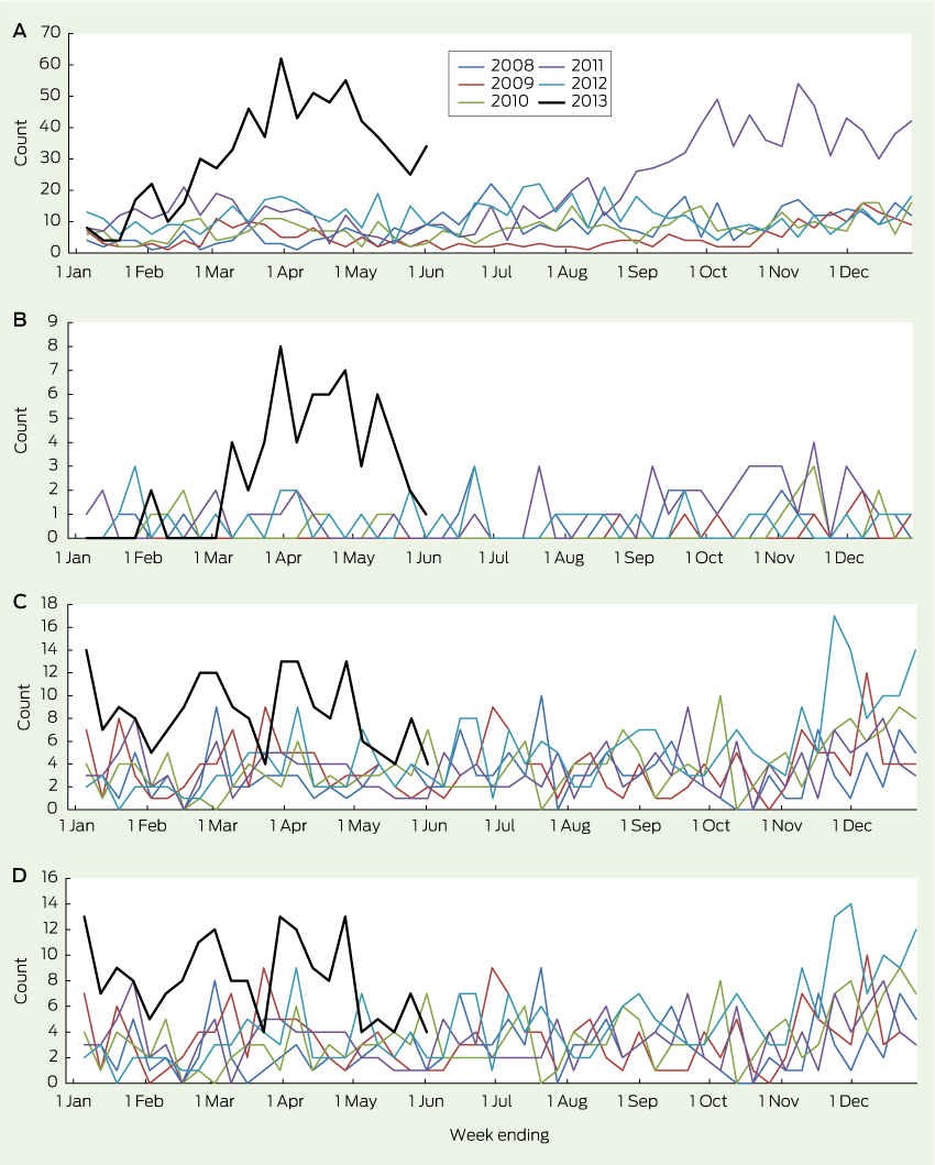
Presentations to EDs for HFMD in children aged under 10 years began to rise in February 2013, peaked in late March, then remained above the usual range through to June (Box 1, A). This was temporally associated with a sharp rise in the number of children with HFMD who required hospital admission (Box 1, B). Presentations to EDs for HFMD also rose in the final quarter of 2011, but without an increase in resulting admissions, indicating that the outbreak of 2013 may have been associated with more severe illness.

The number of ED presentations of children with “meningitis and encephalitis” began to increase in mid November 2012 and remained above the usual range until June 2013 (Box 1, C). Most of these children were admitted, consistent with historical trends (Box 1, D).

Between 1 January and 2 June 2013, 119 cases of suspected EV71 infection were identified in the Sydney Children’s Hospitals Network. Suspected cases were evenly distributed between the two hospitals (53% at Sydney Children’s Hospital at Randwick and 47% at The Children’s Hospital at Westmead). The median age of infected children was 19 months (range, 1 month to 10 years) and 47% were female.

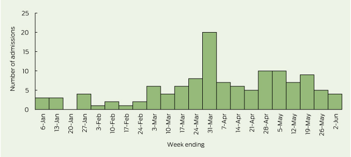
The weekly number of suspected cases rose sharply at the beginning of March 2013, and peaked at 20 cases in the final week of that month (Box 2). A second, smaller rise was noted in late April and May 2013.

Although cases of suspected EV71 infection were widely spread within the Sydney metropolitan area, Sydney’s northern beaches, central parts of Western Sydney and the eastern suburbs experienced more intense activity. Most suspected cases in the March 2013 peak came from the coastal areas of Sydney, particularly the northern beaches and eastern suburbs. During April and May, the number of suspected cases coming from the eastern suburbs remained stable, while the numbers from western Sydney increased and from Sydney’s northern beaches declined sharply.

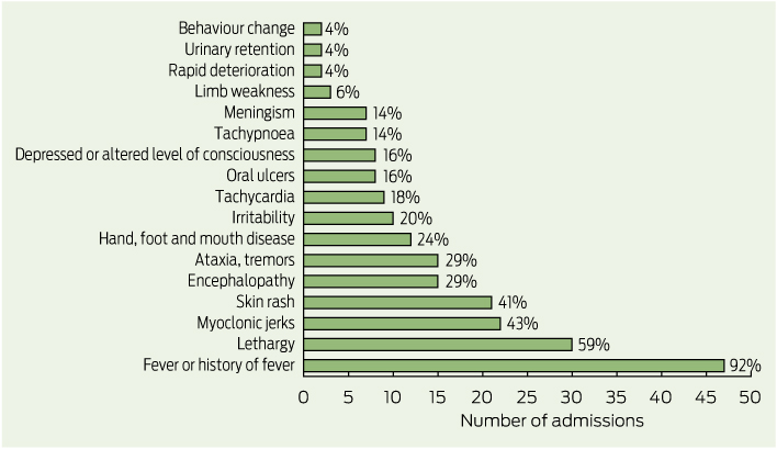
Case forms were completed for all 50 children with suspected EV71 infection who presented between 14 April and 2 June 2013. The most common presenting clinical features were fever (47 children), lethargy (30 children), myoclonic jerks (22 children) and skin rash (21 children) (Box 3). Only 12 children (24%) presented with signs or symptoms of HFMD. The average length of hospital stay was 4.2 days. Five of the 50 children were admitted to an intensive care unit (ICU) and three of these required intubation. The average length of stay in the ICU was 2.4 days. Two children with suspected EV71 infection were treated with intravenous immunoglobulin and six received corticosteroids. The most common diagnoses at discharge were meningitis (15 children), encephalomyelitis (10 children), myoclonus (seven children) and viral meningitis (six children).

Of the 50 children with suspected EV71 infections and completed case forms, 45 tested positive for enterovirus. Infections were typed for 37 of these, and 18 cases were confirmed to be EV71. Other enteroviruses identified included coxsackieviruses (11 children) and echoviruses (four children). Two children had both EV71 and a coxsackievirus, and two had both coxsackieviruses and echoviruses.

Discussion

EV71 emerged as an important cause of severe neurological disease in young Sydney children during the first half of 2013. Activity peaked in March 2013. The focus of the outbreak moved from Sydney’s northern beaches area to its eastern and western suburbs over several weeks. Myoclonic jerks were a relatively common feature of severe infection.

A number of countries in Asia have implemented HFMD surveillance programs in the past 15 years.2,12 HFMD and enterovirus infection are not notifiable diseases in NSW, as most infections are asymptomatic or mild, and many patients are unlikely to see a doctor. This means that notification does not provide a mechanism for developing meaningful patient-based interventions to interrupt transmission. Population-based disease-control measures focus instead on personal hygiene and sanitation. The effect of social-distancing measures, including the closure of childcare facilities,13 has been questioned given the limited evidence for its efficacy, the unquantified but likely substantial socioeconomic costs, and the risk of prolonging the epidemic.1

Two previous studies have suggested that EV71 epidemic activity has followed the introduction of circulating genotypes from Asia into Australia.14,15 EV71 was detected in samples from five patients with acute flaccid paralysis and other patients with suspected enterovirus infections in early 2013, and phylogenetic analysis showed homology with the EV71 C4a subgenogroup circulating in Asia, which was associated with severe neurological complications.16

Our report has important limitations. First, the use of syndromic ED surveillance has uncertain sensitivity and specificity for enterovirus infections. The positive predictive value of syndromic diagnoses would, however, be expected to rise during recognised outbreaks. Second, enhanced surveillance focused on two sentinel children’s hospitals. Other infected patients are likely to have presented to other hospitals in and outside Sydney. Third, before enhanced surveillance, further typing of samples testing positive for enterovirus was not routine practice, so many samples collected between 1 January and 14 April were not typed. Fourth, our description of suspected cases included some patients who were not confirmed to have EV71 infection, so may not truly represent the severe outcomes of the infection.

The continued escalation of EV71 epidemics in Asia, and evidence of the introduction of the EV71 strain into Australia from Asia suggest that enterovirus may continue to be a public health problem here. The results of a Phase III clinical trial of a vaccine were recently published,17 but vaccines have limitations.18 Other vaccines are being developed, and perhaps EV71 will become vaccine-preventable. The use of routinely collected ED data appears to be a useful and efficient method for monitoring enterovirus infections, including the more severe outcomes associated with EV71 epidemics.8

1 Total weekly counts of emergency department presentations (A) and admissions (B) for hand, foot and mouth disease, and emergency department presentations (C) and admissions (D) for “meningitis or encephalitis” for children aged less than 10 years at 59 New South Wales hospitals — 2013 up to 2 June (black line) compared with each of the 5 previous years

2 Weekly number of children aged less than 10 years admitted to the Sydney Children’s Hospitals Network between 1 January and 2 June 2013 with clinical features of meningitis, encephalitis, meningoencephalitis, acute flaccid paralysis or transverse myelitis and suspected or confirmed enterovirus infection

3 Reported presenting clinical features for suspected or confirmed cases of enterovirus 71 infection in children aged less than 10 years admitted to the Sydney Children’s Hospitals Network, 14 April to 2 June, 2013

Ocular biomarkers for neurodegenerative and systemic disease

The eye is a readily accessible window to the brain and the retina has been proven to reveal presymptomatic evidence of brain disorders and systemic diseases, including Alzheimer disease, stroke and diabetes. Here we describe three approaches we are taking to examine the utility of retinal imaging for age-onset diseases.

Alzheimer disease develops slowly, with “plaques” of amyloid-β building up in the brain 15–20 years before memory symptoms and clinical diagnosis. With United States biotech company NeuroVision Imaging, we have been investigating whether similar plaques deposit in the retina. Researchers in the Australian Imaging, Biomarkers and Lifestyle Flagship Study of Ageing (aibl.csiro.au) are running a trial that involves highlighting amyloid plaques in the retina using oral dosing of curcumin, a natural ingredient which gives the spice turmeric its fluorescent yellow colour. The study builds on previous work that found changes to vision and to the retinal blood vessels in Alzheimer disease.1 We believe that retinal imaging may hold a key to early screening for disease and to monitoring interventions aimed at preclinical disease before irreversible brain damage occurs.

Vascular risk factors appear years before stroke and may also influence the likelihood of recurrent stroke. Another CSIRO study is embedding retinal vascular photography in a hospital stroke ward setting. The aims are to investigate the feasibility and utility of ward-based retinal photography as a tool to screen for retinal signs of disease, which may improve diagnosis of the aetiology of acute ischaemic and haemorrhagic stroke. The study may also facilitate appropriate preventive treatments, and improve the prediction of risk of recurrent stroke and other major vascular events.

CSIRO is also a partner in the Remote-I telemedicine project, which supports remote reporting, through a web-based, fully automated disease grading and clinical decision support system for eye diseases, such as diabetic retinopathy and age-related macular degeneration. It is now widely used in Australia and in China.

Telehealth for motor neurone disease

To the Editor: Telehealth is an expanding area with emerging evidence of use in the management of motor neurone disease (MND) and home mechanical ventilation.13 Here, we report our experience with the use of telehealth for managing MND in Queensland.

MND is a devastating disease with affected patients having an average life expectancy of 2–3 years. Progressive muscle weakness leads to the loss of speech and ability to swallow, inability to use arms and legs and, eventually, respiratory failure. Patients are usually reviewed in specialised outpatient clinics at major Australian tertiary hospitals.4 However, as the disease advances, MND patients find it difficult to attend these clinics because of advanced disability.

As in other centres in Australia, in Brisbane, the Royal Brisbane and Women’s Hospital (RBWH) and, separately, The Prince Charles Hospital (TPCH) have multidisciplinary MND clinics that involve medical, nursing and allied health staff. Since 2008, a monthly MND telehealth clinic has been conducted (using a dedicated bandwidth, average 768 kbps) for patients across Queensland and northern New South Wales. Thirty-eight patients have been seen, an average of three times at 3–4-monthly intervals. This has meant that the patients have been followed for about 12 months beyond the last tertiary hospital visit. The average driving distance from Brisbane per telehealth event was 612 km (range, 158–1824 km). The local service was based in hospitals or community health services, with multiple-site login to the RBWH (MND clinical nurse consultant, neurologist and palliative care physician) and TPCH (sleep physician and sleep nurse).

The major problems addressed at telehealth consultations were symptom-based, dominated by the respiratory and palliative concerns. Practical support was provided for general practitioners for managing symptoms (such as excess saliva, immobility and communication problems), the use and withdrawal of non-invasive ventilation, and end-of-life support.

The advantages of telehealth over traditional outpatient clinics include the continued support by personnel who are experienced in MND. Skype and other technologies would allow even greater access, but have issues of confidentiality and security. Telehealth may also reduce the sense of hopelessness that MND patients feel: there is something intangibly positive about finishing a consultation with “so should we make an appointment for 3 months time?”

Use of secondary stroke prevention medicines in Australia: national trends, 2003-2009

Individuals diagnosed with transient ischaemic attack (TIA) or ischaemic stroke are at high risk of recurrent vascular events.1,2 Current Australian guidelines recommend continued use of antihypertensive, antithrombotic and lipid-lowering medicines after TIA or ischaemic stroke to reduce the risk of a recurrent ischaemic event, unless contraindications exist.3

In Australia, a national audit is conducted every 2 years to assess the quality of acute stroke care, including use of secondary stroke prevention medicines at the time of hospital discharge.4 However, few studies have examined use of secondary stroke prevention medicines after discharge from hospital.5,6 Of those available, none have assessed changes in use of these medicines at the national level, and it is unclear whether use has increased since the release of Australia’s first stroke management guidelines in 2003. Consequently, the aim of this study was to examine national trends in the use of secondary stroke prevention medicines by TIA and ischaemic stroke survivors to determine whether use has increased over time.

Methods

A retrospective observational study was conducted using data from the Australian Government Department of Veterans’ Affairs (DVA) administrative health claims database. The database contains details of all hospital and pharmaceutical claims subsidised by DVA for Australian veterans and their eligible dependents. At the end of 2009, the treatment population consisted of 263 433 veterans.7

Patients discharged alive after an episode of care for TIA (identified by International classification of diseases, 10th revision, Australian modification [ICD-10-AM] codes G45.0, G45.1, G45.2, G45.8 and G45.9) or ischaemic stroke (code I63) between 1 January 2000 and 31 December 2009 were eligible for inclusion. They were eligible for subsidisation of all health services by the DVA. We assessed consecutive hospital claims after each TIA or ischaemic stroke claim up to 30 June 2010, as patients may have multiple claims recorded for treatment of the same event.8 Data rules established in consultation with clinicians8 were used to link stroke-related separations and determine final discharge dates.

The proportion of patients using secondary stroke prevention medicines was determined monthly, commencing in January 2003. Each month, the cohort included all patients aged ≥ 65 years who had had a previous episode of care for TIA or ischaemic stroke recorded between 1 January 2000 and the month under study. Patients with a previous episode of care for both TIA and ischaemic stroke were eligible for inclusion in both cohorts. Patients were included each month until either their death or the end of the study period.

To determine the number of patients dispensed recommended medicines each month, all claims for antihypertensives (identified by the World Health Organization Anatomical Therapeutic Chemical classification codes C02, C03, C07, C08 and C09 [excluding C08EX02, perhexiline]), antithrombotics (code B01A [excluding B01AD, thrombolytics]) and lipid-lowering medicines (code C10) between 1 July 2002 (to include medications taken at, but dispensed before, study commencement) and 31 December 2009 were extracted. As dosage information is not available from the database, prescription durations were used as a measure of duration of use of each medicine. The prescription durations were calculated from the DVA pharmaceutical claims dataset and represent the time in which 75% of prescriptions for an individual item were refilled. Use of each of the three classes of medicine, combined treatment with antihypertensive and antithrombotic therapy, and treatment with a combination of all three classes of medicine were determined for all patients still living each month.

Prevalence of use each month (January 2003 to December 2009) was age and sex standardised using the DVA population in January 2003 to account for changes in population characteristics over time. After standardisation, Poisson regression models with generalised estimating equations were used to test for trends in medicine use, using an autoregressive working correlation matrix to adjust for serial correlation. The regression models compared the rate of medicine use in 1 year with the rate in the previous year to test for linear trends between 2003 and 2009. Separate models were used for each treatment and diagnosis combination. All analyses were performed using SAS version 9.4 (SAS Institute).

This study was approved by the University of South Australia and DVA human research ethics committees.

Results

A total of 19 019 patients were included in our analysis. Of these, 403 patients (2.1%) were included in both disease cohorts for at least 1 month during the study period. The characteristics of those included at the start and end of the study are described in Box 1.

Significant increases in use of each class of secondary stroke prevention medicine occurred during the study period (Box 2, Box 3). There was also an increase in the total number of guideline-recommended medicines taken by survivors (Box 2, Box 4) with a near doubling in prevalence of the combined use of all three recommended medicines.

Discussion

This is the first Australian study to examine national trends in the use of secondary stroke prevention medicines among patients with a previous TIA or ischaemic stroke. The median duration of time patients had spent in the cohort was 1.3 years in January 2003 and 3.4 years in December 2009, meaning trends are reflective of use among the prevalent population, rather than among patients with a recent event. Increased use observed in this study suggests practice is moving towards guideline recommendations. Despite this, only half of the population were dispensed medicines from all three recommended classes in December 2009, suggesting there may be opportunity to further increase use of these medicines among the older population.

These findings are consistent with results from international studies conducted within the general practice population over a similar period.9,10 A large study from the United Kingdom showed use of antihypertensives in the year after a first stroke increased from approximately 50% to 70% between 1999 and 2008, antiplatelet use increased from 60% to 75%, and use of lipid-lowering therapy increased from 15% to 80%.10 Large increases in use of lipid-lowering therapy were also shown in a Danish population-based study, with use among ischaemic stroke survivors increasing from 40% to 65% between 2004 and 2010.11

Along with the release and dissemination of national stroke guidelines (which were regularly updated during the study period), other quality use of medicines initiatives may have contributed to the increased use observed in this study. Stroke-specific sections were included in yearly editions of Australia’s national formulary (the Australian medicines handbook) and updated versions of Therapeutic guidelines (neurology). Evidence-based stroke prevention and management was also reviewed in Australian prescriber12 and targeted by National Prescribing Service initiatives.13 The study population and their general practitioners would have received information about antithrombotics through the Veterans’ Medicines Advice and Therapeutics Education Services (Veterans’ MATES) program during the study period. Additional factors likely to have had an impact on the use of lipid-lowering medicines include the publication of a landmark trial14 and changes to eligibility criteria for subsidisation of these medicines through Australia’s national pharmaceutical subsidy scheme15 during 2006.

Factors influencing the use of secondary stroke prevention medicines in older populations are complex, and may be related to lack of awareness of guideline recommendations, prescriber-related factors (such as concern about the lack of evidence to guide secondary prevention among older patients and potential harms of treatment) or patients’ preferences.16 Although there may be room for improvement in use of these medicines, our results reflect use among all survivors, as we lacked clinical information necessary to exclude those with treatment contraindications or previous adverse reactions. We do not expect all patients could be dispensed each medicine, as some older patients may be unsuitable for treatment on entering the cohort. For others, treatment priorities may change over time,17 and medicines for secondary prevention (such as lipid-lowering therapy) may be withdrawn during the late stages of life, or in those with severe physical impairment or cognitive deficit.18 The number of older patients ineligible for treatment may be significant. In a study assessing antithrombotic use by older patients with acute ischaemic stroke, more than one-third were excluded from the analysis, owing to contraindications or refusal of treatment at discharge.19 Trends in antithrombotic use observed in our study may be further underestimated, as aspirin can be purchased without a prescription in Australia (although patients included in this study had access to subsidised aspirin via prescription).

This study used hospital claims data to determine whether patients had a TIA or ischaemic stroke. To minimise selection bias, patients were selected using primary diagnosis codes and those with an unspecified stroke (ICD-10-AM code I64) were not included. There is high adherence to Australian standards for ICD-10-AM coding,20 and 95% of patients with a primary diagnosis code for stroke were correctly coded in a recent Australian audit.21

We expect that use of recommended medicines by patients included in this study is indicative of use by older Australians previously hospitalised for TIA or ischaemic stroke. Age-specific comparisons show veterans without a service-related disability and the general Australian population have similar use of pharmaceuticals, hospital services and GP visits.22 However, changes in medicine use observed in this study may not be generalisable to patients managed solely in the community setting, and without assessment of clinical records it is not known if treatment targets were attained.

The increased use of secondary stroke prevention medicines shown between 2003 and 2009 in this large cohort of older Australians with a previous TIA or ischaemic stroke is consistent with Australian stroke guideline recommendations and initiatives to support quality use of medicines during the study period.

1 Characteristics of patients who were included at the start and end of the study

Characteristics, by disease cohort

Jan 2003

Dec 2009


Transient ischaemic attack


No. of patients

2765

5242

Age (years), median (IQR)

81.0 (78.3–84.4)

86.8 (84.1–89.5)

No. of men (%)

1761 (63.7%)

2716 (51.8%)

Time in cohort (years), median (IQR)

1.3 (0.6–2.0)

3.5 (1.6–6.1)

Ischaemic stroke


No. of patients

2493

4302

Age (years), median (IQR)

80.9 (78.0–84.2)

86.6 (84.1–89.2)

No. of men (%)

1609 (64.5%)

2376 (55.2%)

Time in cohort (years), median (IQR)

1.3 (0.6–2.1)

3.3 (1.5–5.8)

2 Changes in use of secondary stroke prevention medicines by transient ischaemic attack and ischaemic stroke survivors between 2003 and 2009


Standardised monthly rate of use (per 100 patients)


Medicines, by disease cohort

Jan 2003

Dec 2009

Standardised rate ratio (95% CI)

Average annual % change

P


Transient ischaemic attack


Antihypertensive

72.5

78.2

1.016 (1.015–1.016)

+1.6%

< 0.001

Antithrombotic

70.4

74.0

1.013 (1.011–1.014)

+1.3%

< 0.001

Lipid-lowering

33.5

58.0

1.087 (1.084–1.091)

+8.7%

< 0.001

Antihypertensive + antithrombotic

55.9

63.0

1.025 (1.023–1.027)

+2.5%

< 0.001

Antihypertensive + antithrombotic + lipid-lowering

24.4

43.0

1.094 (1.088–1.101)

+9.4%

< 0.001

Ischaemic stroke


Antihypertensive

73.3

81.1

1.019 (1.019–1.020)

+1.9%

< 0.001

Antithrombotic

74.2

80.4

1.014 (1.013–1.015)

+1.4%

< 0.001

Lipid-lowering

36.8

64.8

1.088 (1.087–1.090)

+8.8%

< 0.001

Antihypertensive + antithrombotic

59.4

70.2

1.027 (1.025–1.028)

+2.7%

< 0.001

Antihypertensive + antithrombotic + lipid-lowering

26.9

52.3

1.102 (1.098–1.106)

+10.2%

< 0.001

3 Trends in monthly use of secondary stroke prevention medicines by patients previously hospitalised with a transient ischaemic attack or ischaemic stroke

4 Trends in monthly use of combination therapy by patients previously hospitalised with a transient ischaemic attack or ischaemic stroke

Time to reconsider steroid injections in the spine?

To the Editor: In their review, Harris and Buchbinder call for a ban on all spinal injections of steroids.1 These injections are not a singular procedure; they differ by indications, technique, and the evidence that serves each.

For the treatment of low back pain, no evidence supports the use of epidural steroids, and intra-articular injections of steroids are clearly no more effective than sham therapy.2 Disavowing this treatment is, therefore, justified. Ironically, the treatment with the greatest success rate and longest duration of success for chronic low back pain is injection of normal saline.3

For the treatment of radicular pain, multiple studies have shown that epidural injections of steroids
are no more effective than sham treatment.4,5 One study showed marginal superiority over placebo at 3 weeks,6 but by 30 days, the number needed to treat is 100.7 Yet, the Faculty of Pain Medicine still endorses this intervention.8

The evidence is different for transforaminal (TF) injection of steroids. The success rate of TF injection of steroids is significantly greater than that of TF injection
of bupivacaine or saline, or intramuscular injection of steroids
or saline.9 Furthermore, TF steroid therapy has a surgery-sparing effect and is cost-effective.10 A blanket ban on steroid injections would deny patients with radicular pain the
only proven alternative to surgery.

The baby’s bathwater is distinctly polluted, but within it is a gem. Before throwing it out, the water needs to be carefully filtered.

Time to reconsider steroid injections in the spine?

To the Editor: We thank Harris and Buchbinder for their focus on interventional pain procedures.1 However, their description of the procedure they discuss has not been standard practice in pain medicine
in Australia for many years.

They misrepresent the Medicare Benefits Schedule (MBS) number 39013 to solely include facet joint injections, whereas it includes local anaesthetic medial branch blocks, which are an evidence-based diagnostic test for posterior elements as a source of spinal pain, and can
be therapeutic in their own right.2

The number needed to treat (NNT) is the number of patients that need
to be treated for one to benefit compared with a placebo or sham in a clinical trial. The ideal NNT is one. After a positive medial branch block response, radiofrequency medial branch neurotomies — a procedure with an NNT ranging from two patients3 to 4.4 patients4 treated
for one patient to receive effective
relief of spinal pain — creates a “therapeutic window” in which ongoing spinal rehabilitation
can occur.

From the 35 000 MBS number 39013 procedures performed in 2012, we can infer that fewer than 0.5%
of Australians with spinal pain have facet joint injections and medial branch blocks in a 12-month period.

Contrary to the comments of
Harris and Buchbinder, lumbar transforaminal epidural steroid injections are effective for the treatment of radicular pain associated with disc protrusion, with an NNT of 2.7,5 and in conjunction with active pain strategies may forestall spinal surgery.

Before impeaching facet joint injections and medial branch blocks, and thereby medial branch neurotomies, as well as lumbar transforaminal epidural steroid injections, Harris and Buchbinder should consider:

  • interprofessional patient-centred approaches are key;

  • pharmacological management is often ineffective;

  • their view does not reflect the current practice of Australian pain medicine physicians;

  • these procedures help people struggling to continue in social roles and maintain quality of life, so they help to reduce the economic impact
    of spinal pain on Australian society.

We support education to improve evidence-based practice of interventional procedures.