×

First report of Lyme neuroborreliosis in a returned Australian traveller

Lyme borreliosis is a tick-borne zoonosis endemic in many parts of the world. We report the first case of Lyme neuroborreliosis in an Australian traveller returning from an endemic area. The diagnosis should be considered in patients with chronic meningoencephalitis and a history of travel to an endemic area.

Clinical record

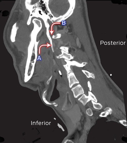
A 58-year-old woman of European ancestry presented to a rural hospital in New South Wales in May 2014 with an 8-month history of worsening motor instability, confusion and bilateral occipital headaches associated with photophobia, lethargy and somnolence. The patient was from Geraldton in Western Australia and was staying for 2 weeks with her family in NSW. Her symptoms had started 1 month after returning from Lithuania, where she had spent 3 weeks. A tick had bitten her in the pubic hairline during a trip to a pine forest 30 km from Vilnius. One month later, the patient developed two circular non-pruritic rashes on her right thigh (distal to the site of the tick bite) and lower leg, each about 30 mm in diameter; they resolved after 2 weeks without specific intervention. She had experienced continuing headaches, lethargy and a self-limiting episode of diplopia that prompted her to see her general practitioner. Computerised tomography and magnetic resonance imaging of the brain performed before she presented to the hospital showed nothing abnormal.

The patient’s past medical history included hypertension that was well controlled with irbesartan. On presentation, she was afebrile. There were no focal neurological signs or papilloedema. Cardiovascular, respiratory and gastrointestinal parameters were within normal limits. There were no rashes on her extremities, nor evidence of synovitis in her joints.

The results of a full blood examination, urea and electrolyte assessments and liver function tests were within normal limits. The cerebrospinal fluid (CSF) white cell count was 377 × 106/L (reference interval [RI], 0–5 × 106/L), consisting purely of monocytes (ie, no polymorphonuclear leukocytes). No organisms were identified by Gram staining; the results of acid-fast bacilli staining were also negative. CSF biochemical parameters were markedly abnormal, with elevated protein levels (1.93 g/L; RI, 0.15–0.45 g/L) and reduced glucose levels (1.3 mmol/L; RI, 2.8–4.4 mmol/L). Cryptococcal antigen was not detected by lateral flow assay, and polymerase chain reaction (PCR) analysis was negative for herpes simplex viruses 1 and 2, enterovirus, and Mycobacterium tuberculosis. Antibody to syphilis treponema and to HIV was not detected.

Given the clinical presentation and the history of travel to an area where Lyme disease is endemic, serological testing for Borrelia in both serum and CSF was requested, and treatment with ceftriaxone (4 g daily) was commenced, with a presumptive diagnosis of Lyme neuroborreliosis.

Serological screening was performed at the Institute of Clinical Pathology and Medical Research, Westmead (Sydney), with an enzyme immunoassay (EIA) for IgG against recombinant antigens from Borrelia burgdorferi sensu stricto strain B31, B. afzelii and B. garinii (NovaLisa Borrelia burgdorferi IgG, NovaTec Immundiagnostica GmbH, Germany). The signal to cut-off ratio in this assay was 6.83 for serum and 5.57 for CSF (ratios less than 0.9 are considered negative).

To confirm these results, western immunoblotting was undertaken using a modification of the polyacrylamide gel electrophoresis (PAGE) method described by Dressler and colleagues.1 Sonicated B. burgdorferi strain 297 (at 0.5 mg/mL) and B. afzelli ATCC 51567 (at 1.0 mg/mL) were separately applied to precast SDS-PAGE gels (ExcelGel SDS Homogeneous 12.5, GE Healthcare Life Sciences, Sweden). The serum of our patient showed IgG responses to two B. burgdorferi antigens (molecular weights: 41, 58 kDa) and five B. afzelii antigens (22, 39, 41, 58, 83 kDa). IgG to the same antigens, as well as to a sixth B. afzelii antigen (45 kDa), was detected in her CSF. The criteria of the United States Centers for Disease Control and Prevention (CDC) stipulate that five or more specific IgG bands are required for a positive serological result.2

The patient returned to Geraldton, where she completed two weeks of treatment with intravenous ceftriaxone (4 g daily). The cellular and biochemical parameters of CSF collected 3 weeks after completion of treatment had improved: the white cell count was 45 × 106/L, the protein levels were 0.7 g/L and the glucose concentration was 2.3 mmol/L. Five months later, her CSF parameters were completely normal, with a normal biochemical profile and no pleocytosis. The patient continues to see her GP and has made a good clinical recovery; her headaches, lethargy and neurological symptoms have all resolved.

Discussion

Our case highlights the importance of obtaining a thorough travel history in a patient presenting with chronic meningoencephalitis. Our review of the literature indicated that this was the first case of Lyme neuroborreliosis imported into Australia from overseas. Clinicians should consider neuroborreliosis in patients with a history of travel to an endemic area who present with persistent neurological symptoms. Other causes of chronic meningitis include cryptococcal meningitis, which is endemic in Australia, but had been excluded in our patient. Mycobacterium tuberculosis infection and tick-borne encephalitis are further diagnoses to consider in travellers with meningoencephalitis who have returned from Eastern Europe.

Lyme disease is a multisystem infectious disorder transmitted by ticks of the Ixodes complex.3 Three B. burgdorferi sensu lato species, namely B. burgdorferi sensu stricto, B. garinii, and B. afzelii, are pathogenic to humans in Europe. In contrast, B. burgdorferi sensu stricto is the only species known to cause human infection in the United States.3 The disease has protean manifestations, depending on the clinical stage at presentation. Stage I disease usually presents with a typical rash, erythema migrans, 7–14 mm from the site of the tick bite. Constitutional symptoms, including headache, myalgia, arthralgia and fever, may accompany the erythema migrans. Stage II disease is characterised by dissemination to other skin areas, the nervous system, joints or heart, and may include a wide variety of symptoms. Stage III, or late Lyme disease, occurs months to years after a tick bite, and usually causes large joint arthritis or neurological symptoms.3 In our patient, the presence of an erythema migrans rash at more than one site, presented together with neurological symptoms, indicated stage II disease.

Two-tier serological assessment — a screening EIA followed by confirmatory immunoblotting — remains the mainstay of diagnosis for Lyme borreliosis, although molecular methods and culturing clinical specimens are also possible.2The diagnostic sensitivity of culturing Borrelia species is poor, so that it is rarely undertaken in clinical practice, its use being largely limited to research settings.4 Molecular methods have a higher diagnostic sensitivity when using skin biopsies from an erythema migrans or synovial fluid than when testing blood or CSF.4,5 Molecular methods have not been standardised across laboratories and false-positive results are possible.4

On the other hand, the recombinant EIA for anti-Borrelia IgG has high sensitivity and specificity after the first few weeks of infection. The results of testing the serum and CSF of our patient for anti-Borrelia IgG were highly positive. Confirmation by immunoblotting greatly increases the positive predictive value of EIA, but this technique may be less sensitive, although it is very specific when using the CDC criteria for IgG immunoblotting in Lyme disease. In our case, only the IgG blotting results for B. afzelii antigens met the CDC criteria for a positive finding.

The European Federation of Neurological Sciences guidelines require two of the following criteria to be fulfilled for a possible diagnosis of Lyme neuroborreliosis, and that all three be met for a definite diagnosis:

  • neurological symptoms;
  • CSF pleocytosis; and
  • detection of intrathecal antibodies or, if symptoms began in the past 6 weeks, identification of the pathogen in the CSF by PCR or culture.6

Our patient met all three criteria, making this a definite diagnosis of Lyme neuroborreliosis.

The clinical presentation of Lyme neuroborreliosis differs according to the Borrelia species involved. B. garinii is the most common cause of Lyme neuroborreliosis in Europe, followed by B. afzelii. B. garinii infection often manifests as typical early neuroborreliosis, characterised by painful meningoradiculitis (Bannwarth syndrome) and meningeal irritation, whereas the clinical features of central nervous involvement in B. afzelii infection, as in our patient, are often non-specific and difficult to diagnose.5 In contrast to infection with B. garinii, CSF parameters in patients with Lyme neuroborreliosis caused by B. afzelii can be atypical, with a normal biochemical profile and without pleocytosis.5 In our patient, the presence of persistent non-specific neurological and constitutional symptoms, together with positive CSF and serum IgG immunoreactivity to B. afzelii antigens, provided enough evidence to attribute this infection to B. afzelii.

Oral administration of amoxicillin or doxycycline is usually the treatment of choice for early stage Lyme borreliosis. Patients with neurological or cardiac manifestations are typically treated for 2–4 weeks with intravenous ceftriaxone.3 Longer-term parenteral treatment is not recommended, because it does not provide any additional improvement in patients with persisting neurological or constitutional symptoms; it is also associated with adverse events.7 Improvement in both clinical and CSF markers after 2 weeks of intravenous ceftriaxone in our patient was reassuring, and suggests that a shorter duration of treatment is possible even in patients with neurological symptoms.

The existence of Lyme disease in Australia has been debated for decades,8 and in recent years there has been growing public interest. In February 2014, the Royal College of Pathologists of Australasia released a position statement on diagnostic laboratory testing for Lyme disease in Australia and New Zealand.9 Ticks of the genus Ixodes are present in Australia, but species known to transmit Borrelia species have not been found.10Earlier research,10 as well as the most recent study of ticks in areas around the northern beaches of Sydney and other parts of Australia (Associate Professor Peter Irwin, Murdoch University, WA, personal communication, November 2014) have not found Borrelia species in Australian ticks.

Lyme disease can present as non-specific symptoms that persist for weeks to months after infection. A careful travel history is therefore an important part of the assessment of any patient with clinical features suggesting the disease.

Consensus guidelines for the investigation and management of encephalitis

A summary of a position paper for Australian and New Zealand practitioners

Encephalitis is caused by inflammation of the brain and is a challenging condition for clinicians to identify and manage. It manifests as a complex neurological syndrome with protean clinical manifestations that may be caused by a large number of aetiologies, many without effective treatments. It can be fatal and survivors often experience significant neurological morbidity. Studies have shown variable quality in case management in multiple settings,13 emphasising the need for consensus guidelines.

The need for guidelines is also important because encephalitis is a marker of emerging and re-emerging infectious diseases, and is therefore a syndrome of public health importance. There are unique infectious aetiologies in Australia — including Hendra virus, Australian bat lyssavirus, Murray Valley encephalitis virus and West Nile virus (Kunjin virus) infections — that require early identification, reporting and specialist clinical and public health responses. Regionally, causes of encephalitis with potential for introduction into and epidemic activity in Australia include Japanese encephalitis virus, enterovirus 71, dengue virus and Nipah virus. There is also a rapidly growing list of immune-mediated encephalitides that are important because of their potential response to immunomodulatory treatments and their association with underlying tumours.

    “encephalitis is a marker of emerging
    and re-emerging infectious diseases”

Although comprehensive guidelines have been published elsewhere, including recent international consensus guidelines,4 these are detailed and lack a specific geographic focus. As a result, we have developed a concise guideline for clinicians in Australia and New Zealand5 (doi: 10.1111/imj.12749) that provides a substantial update to previous guidance published in the Journal.6

The guideline was developed by the Australasian Society for Infectious Diseases Clinical Research Network (ASID CRN) Encephalitis Special Interest Group with subsequent, multiple rounds of consultation involving the ASID Guidelines Committee, the Public Health Association of Australia, the Australian and New Zealand Association of Neurologists and the Australasian College for Emergency Medicine.

Main recommendations

The guideline principally consists of two algorithms. The first algorithm addresses the patient with possible meningoencephalitis — a scenario that is frequently encountered in emergency departments. This algorithm is designed to assist clinicians to: consider encephalitis within a wide differential diagnosis, perform appropriate specimen sampling and investigations, and initiate antimicrobial therapy promptly (including acyclovir for possible herpes simplex virus [HSV] encephalitis). In most patients with possible meningoencephalitis, an alternative diagnosis will be made. The algorithm aims to discriminate between patients in whom encephalitis can be excluded and those who require a more detailed assessment. The second algorithm addresses the patient in whom encephalitis is considered likely. This algorithm provides a robust clinical case definition of encephalitis, identifies key first-line (universal) diagnostic tests (Box), outlines a process of excluding HSV disease, and formulates an approach of directed (second- and third-line) diagnostic testing based on risk factors, clinical features and radiological features.

In addressing these scenarios, the guideline answers the following questions:

  • What features are important to consider during history-taking and examination?
  • In which patients should magnetic resonance imaging be performed?
  • What are the common abnormalities evident in cerebrospinal fluid?

Furthermore, it provides advice on the tests that should be done to diagnose the most common causes of encephalitis and defines specific patient subpopulations to highlight differences in aetiology (to help prioritise testing). These subpopulations include: children and neonates, immunocompromised patients, overseas travellers and immigrants, and patients residing in tropical Australia.

The guideline considers the particularly vexing question of the contemporary role of brain biopsy by presenting evidence of its yield in cohorts of patients who have encephalitis. It also introduces the various immune-mediated encephalitides, describes their clinical features and, in doing so, assists the clinician in deciding when to perform specific antibody studies. From a treatment perspective, the guideline defines optimal therapy for HSV encephalitis and outlines possible treatment strategies for other infectious and immune-mediated causes based on lower-quality evidence. In particular, it suggests when to consider empiric antimicrobial therapy and immunomodulatory therapies.

Encephalitis presents a complex challenge to clinicians. Its possibility must be suspected in a variety of presentations, and it requires the performance of a detailed clinical assessment, consultation, and judicious investigation. Unnecessary delays must be avoided, and it is essential to institute empiric therapies appropriately and provide high-quality supportive management. Optimal application of current knowledge is likely to improve diagnosis; however, even with an extensive diagnostic work-up, definitive aetiology may not be identified for 30%–40% of patients with encephalitis.7 Novel agents and a changing geographical distribution of known diseases are likely to be identified with improved surveillance; these possibilities should be considered where unexplained encephalitis clusters occur.

Box anchor (office use only)

Recommended first-line investigation of encephalitis in Australia and New Zealand*5


* Table reproduced with permission from: Britton PN, Eastwood K, Paterson B, et al; Australasian Society of Infectious Diseases; Australasian College of Emergency Medicine; Australian and New Zealand Association of Neurologists; Public Health Association of Australia. Consensus guidelines for the investigation and management of encephalitis in adults and children in Australia and New Zealand (Internal Medicine Journal, Wiley Publishing Asia Pty Ltd, © Royal Australasian College of Physicians 2015).

A bolt out of the blue: the night of the blue pills

Clinical record

A cluster of 10 patients presented during the night of 31 December 2013 to the emergency department of Royal Perth Hospital with states of agitated delirium or exhibiting unusual behaviour. Eight of the patients had attended an open-air dance party in the city close to the hospital, and nine had arrived by ambulance. All except one admitted to taking non-prescription drugs in tablet form, most believing they were consuming ecstasy (3,4-methylenedioxymethamphetamine, MDMA) in the form of blue or grey pills, in several cases imprinted with a lightning bolt. Media warnings had already been issued in response to similar cases involving acute psychosis reported by another metropolitan emergency department (Fremantle Hospital).1,2

The median age of the patients in our cluster was 20 years (interquartile range [IQR], 18–22 years). The median initial heart rate was 115 beats per minute (IQR, 84–155 beats per minute). Four patients were febrile (temperature ≥ 37°C) but only one had a temperature greater than 38°C. All patients had dilated pupils (median width, 6 mm [IQR, 5–7 mm]). Five patients required intravenous sedation, and in two cases more than 50 mg diazepam was required.

The patients had posed a significant risk to themselves before attending the emergency department: one had been found collapsed on the dance floor, another had wandered through vehicular traffic, and a third had fallen after climbing an 11 metre-high lighting rig.

The clinical syndrome included a state of agitated delirium, with labile mood, tachycardia, dilated pupils, sweating and, in several patients, involuntary movements. Clonus was present in only one case. One patient tried several times to hit staff members, while another spat at them. The most severely affected patient developed status epilepticus, and required intubation and admission to the intensive care unit. After recovery, he stated it was only the second time he had used non-prescription drugs.

The cluster of patients had a significant impact on emergency department resources. They comprised 10 of the 83 patients who presented to the department in the 7-hour period between 19:55 and 02:55. Many required intensive nursing care and intravenous sedation. One patient flipped over the safety railing of his trolley and landed on his head, but was not significantly injured. The median hospital length of stay was 5.4 hours (IQR, 3.0–11.9 hours).

Emergency treatment of the patients followed standard procedures for a sympathomimetic syndrome,3 and included oral or intravenous administration of benzodiazepines and fluids, observation and, in one case, intubation and cooling for status epilepticus. In patients for whom benzodiazepines were indicated, unusually large doses were needed to achieve adequate sedation.

Blood samples were taken from nine of the patients when intravenous cannulae were inserted as part of routine clinical care. Retrospective analysis of stored plasma samples using liquid chromatography–mass spectrometry was undertaken 40 days later by ChemCentre forensic laboratories (Perth, WA) to attempt to identify the substances responsible for the patients’ symptoms. Results were compared with a large library of conventional and novel recreational drugs.

No novel synthetic agents were identified, but methamphetamine was detected in samples from two patients. The clinical syndrome observed and the absence of evidence for conventional drugs of misuse in all but two of the samples aroused suspicions of unidentified synthetic drugs. As analysis of drugs recently seized by police indicated that many “ecstasy tablets” contained high amounts of caffeine, caffeine levels were assessed in our samples, but were found to be uniformly low. Most of the tablets taken by the patients had been marketed as ecstasy, but no MDMA was detected in any of the plasma samples. Interestingly, lactate levels were elevated in all patients (median concentration, 3.1 mmol/L; IQR, 2.5–3.8 mmol/L), and all samples but one contained high levels of ethanol (median concentration, 180 mg/100 mL; IQR, 140–220 mg/100 mL).

Discussion

The continued emergence of novel synthetic recreational drugs is a growing problem in many countries, and the short- and long-term effects of these compounds are poorly understood. There have been recent deaths in Australia linked with such substances.4 Little reliable information is readily available to inform either users or clinicians.

There are several possible technical reasons why new synthetic drugs were not detected in our patients’ plasma samples. These could include adsorption of the drug by gel in the collecting tube5 and instability of the drug at room temperature or when refrigerated at 4°C.67 These problems may have been compounded by the 40-day delay between collection and analysis.

Ongoing research into new synthetic drugs is needed to identify which harmful substances are currently circulating in the community and to inform potential users of their harms. Public warnings about clusters of cases, if deemed appropriate, should be issued on the basis of clinical presentations rather than of definitive analyses, given the time delay involved in performing these. The optimal treatment of patients is unknown and will vary according to the compound ingested. Future research should also consider the most appropriate methods for collecting samples to optimise analysis outcomes, including the temperature at which samples should be stored to preserve the chemicals of interest.

Public health warnings about a dangerous batch of “ecstasy” tablets had been issued to the media earlier in the day on which our patients had taken their pills,1,2 but the effectiveness of these messages is unknown. Most of our patients erroneously thought they had taken MDMA, and all had consumed only a small number of tablets. Media reports often mention the dangers of an “overdose”, implying the consumption of many tablets, which could mislead users into believing that one or two tablets (of an unknown substance) are safe. Public health messages should consider the need to communicate risk effectively, but there may also be unintended adverse consequences. These include encouraging experimentation by alerting naive or non-consumers to potential new drugs.

Lessons from practice

  • The use of novel synthetic drugs is an increasing problem.
  • There is little reliable information to inform users or clinicians about these drugs.
  • The optimal use of the media to warn potential users is yet to be defined.
  • Future storage and analysis of substances should take into account their potential instability and low plasma concentrations.

Eagle Syndrome as a potential cause of Tapia Syndrome

A 38-year-old man presented with influenza A (H1N1) pneumonia complicated by acute respiratory distress syndrome, resulting in an extended intensive care unit stay (37 days). After he was extubated, he was found to have left-side IX, X and XII cranial nerve palsies. Computed tomography of his neck showed bilateral elongated styloid processes (5.0 cm long; Figure), consistent with Eagle syndrome.1 The left styloid process was closely opposed to the transverse process of the first cervical vertebra, causing effacement of the internal jugular vein and compression of the IX, X and XII cranial nerves, which converge in this area. The eponym used to describe concurrent paralyses of the X and XII cranial nerves is Tapia syndrome,2 with prolonged neck flexion potentially contributing to the condition in our case.

Sagittal computed tomography image of the neck of the patient. A: Elongated styloid process (left); B: Area of compression of IX, X and XII cranial nerves.

The trumpet’s blown pupil

On the advice of a neurologist friend (author C P), a 42-year-old man was rushed to hospital with a fixed dilated pupil (Figure, A). His condition was otherwise normal, as were the findings of urgent magnetic resonance imaging and angiography of the brain.

By Day 3, his mydriasis had resolved (as did the mystery) when the patient asked his “learned” friend how common eye injuries with whipper-snippers were. He mentioned, in passing, that on the morning of the incident he had cut back his beloved Angel’s trumpet (Figure, B) — a member of the Solanaceae family, all parts of which are laced with anticholinergic alkaloids.

A detailed history, botanical or otherwise, will always trump the next best test.

The Sydney siege: courage, compassion and connectedness

To the Editor: Raphael and Burns highlighted the strong police response to the hostage situation in Sydney in 2014.1 Diversionary devices, such as the flash-bang grenades used in Sydney, have been increasingly used to distract and disorientate people in civilian hostage and riot situations internationally. While not intended to cause permanent damage, there are risks associated with their use.

Flash-bang grenades deflagrate using a powdered blend of aluminium, magnesium and ammonium perchlorate, which generates a spontaneous explosion. When initiated, illumination is produced through oxidation of the components, resulting in heat exceeding 38°C, a blast reaching 180 decibels and a brief flash of 1–6 million candela (up to 600 million lux) within a distance of about 1.8 m.2

The intense flash results in temporary bleaching of the photoreceptors in the eye. Ocular injury can occur if the flash-bang grenade explodes at close range, with possible thermal or mechanical damage. Other more powerful devices, producing a similar intensity of unidirectional light, have resulted in vision loss similar to that seen with laser weapons.2

Temporary hearing loss and aural pain results from a single or multiple blast of loud noise between 140 and 170 decibels. Damage to the sensitive structure of the inner and middle ear can result in hearing loss and tinnitus.3 Perilymphatic fistula of the inner ear may occur, necessitating immediate assessment and possible surgical treatment.4

Premature deflagration can also cause injury to the operator.5 As these devices continue to be used in civilian situations, it is important to remain aware of any potential hazards, to both the operator and bystanders.

Firearms, mental illness, dementia and the clinician

To the Editor: In their recent article in the Journal, Wand and colleagues suggest that the medical profession should play a more active role in the regulation of firearm licences held by older Australians.1 However, the authors underestimate the rate of firearm ownership in Australia by a factor of 1000 when they state that 3.9 per 100 000 people held a firearm license in 2001. In reality, about three-quarters of a million Australians held a firearm licence in 2001.2

While the reported vignettes seem compelling enough, the authors’ recommendations need some scrutiny. Almost 15% of the population are aged over 65 years, yet these older people commit about 3% of the roughly 250 homicides per year. 3,4 Further, only about 15% of Australian homicides involve a gun.3 Hence, the potential number of lives saved by the measures they suggest can only be tiny.

In contrast, the downside of their recommendations might be significant. First, obligations on doctors to play a more active role in firearm ownership might deter some patients from seeking medical care. Second, even if people were not deterred from seeking health care, more active involvement by doctors in firearm regulation would come at the opportunity cost of ordinary medical care — care that could be focused on common and lethal medical conditions.

Firearm control in Australia has been singularly successful. While it may be the case that firearm regulations should be tightened, this is not really the responsibility of the medical profession, nor is it fair to focus on older Australians.

Firearms, mental illness, dementia and the clinican

In reply: There was a transcription error in our article.1 The rate of licensed firearm ownership in Australia in 2001 was indeed 3.9 per 100 people,2 although this is likely to be an underestimate, as unregistered, unlicensed and illegal firearms are not captured by official statistics.

Although the overall rate of homicide by firearm owners is low, we argue that the stakes are high. Other potential adverse outcomes of a person who lacks the capacity to safely handle firearms continuing to have a firearm include accidental injury and suicide.

We acknowledged the ethical implications of doctors having a role in assessing suitability for firearm licences.1 However, there is already an expectation that doctors should notify police when concerns about risk to the community or individuals arise from a patient’s access to firearms.3 Risk assessment alone is inadequate, but doctors better meet their obligations when risk assessment is combined with capacity assessment.

Older adults are more likely to have complex cognitive and physical comorbid conditions that affect their ability to safely use a firearm. Screening is important, and doctors will use their clinical judgement to identify patients who may need a closer examination of their capacity in relation to firearm access.

First use of creatine hydrochloride in premanifest Huntington disease

Huntington disease is a devastating autosomal dominant neurodegenerative disorder that typically manifests between ages 30 and 50 years. Promising high-dose creatine monophosphate trials have been limited by patient tolerance. This is the first report of use of creatine hydrochloride in two premanifest Huntington disease patients, with excellent tolerability over more than 2 years of use.

Clinical record

A 33-year-old patient in our general practice carried the autosomal dominant gene for Huntington disease (HD). The abnormal number of cytosine-adenine-guanine triplet repeats in the huntingtin gene she carried meant she would eventually become symptomatic for this dreadful disease.

The patient requested information regarding potential treatments, as she had become aware of clinical trials for HD and of compounds used by patients with HD. A neurologist had previously recommended a healthy diet, exercise, avoiding excessive toxins (such as alcohol), social enrichment and cognitive stimulation, which together may modestly slow clinical disease progression and improve quality of life.1 She had used preimplantation genetic diagnosis during her pregnancies but preferred otherwise not to focus on her condition. She understood that there were no proven therapies for this incurable condition and did not want to attend HD clinics. She was asymptomatic.

At her request, I searched the PubMed database for possible treatment options. There were some that were unproven in HD but had been used safely in humans for other indications, had a reasonable rationale regarding known HD pathophysiology, and had positive results in animal models of HD and/or early-phase human HD trials.2

In January 2012, I sought advice on using these options (eg, high-dose creatine, melatonin, coenzyme Q10, trehalose, ultra-low-dose lithium with valproate) from a specialist HD clinic but was advised against this approach. Instead, it was suggested that the patient might be able to sign up for clinical trials including high-dose creatine. The patient chose subsequently to participate in an observational trial (PREDICT-HD) which did not limit her options. However, she declined consideration for the Creatine Safety, Tolerability, and Efficacy in Huntington’s Disease (CREST-E) study,3 an international Phase III placebo-controlled trial of creatine monophosphate (CM) in early symptomatic HD. It is also very unlikely she would have been accepted for this trial as she was asymptomatic.

In February 2014, the Creatine Safety and Tolerability in Premanifest HD trial (PRECREST),4 a Phase II trial, showed significant slowing of brain atrophy in CM-treated premanifest HD patients. If convincingly replicated, this would be a major advance.

The main practical problem with high-dose CM (20–30 g daily) is tolerability. Adverse effects are common, especially nausea, diarrhoea and bloating. In people who have normal renal function before commencing creatine supplementation, creatine does not appear to adversely affect renal function.5

In PRECREST, about two-thirds of patients tolerated the maximum dose (30 g daily) and 13% of those on placebo were unable to tolerate CM when they switched to it. Moderate intolerance appears to be common. A high dropout rate affected the HD gene carriers in this study despite assumed high motivation.6 Recommended additional water intake for patients on CM therapy is 70–100 mL per gram of creatine per day, which is problematic at high doses of CM.

The patient again requested assistance as she wanted to seek the best available potential treatment to face her condition with equanimity.7 I decided that, provided safety was paramount, I would assist her on an informed consent basis as part of my duty of care, respecting her informed autonomy.

A case presentation and treatment plan was prepared and an expert team of relevant medical specialists was assembled. Comprehensive informed written consent, including consent from the patient’s partner for additional medicolegal protection, was obtained. The New South Wales off-label prescribing protocol8 was followed, actions were consistent with article 37 of the Declaration of Helsinki,9 and medical defence coverage for the proposed treatment was specifically confirmed by my indemnity insurer.

After baseline assessment, including renal function and careful attention to hydration, the patient commenced oral CM therapy at 2 g/day. This was slowly increased to 12 g/day but she was unable to maintain this dosage due to gastrointestinal adverse effects.

Creatine hydrochloride (CHCl), a creatine salt that has greater oral absorption and bioavailability than CM, and requires less water and a lower dose, offered a possible solution.10 The reduced dose also reduces intake of contaminants, which is very important for extended use. Use of CHCl has been confined to the bodybuilding industry and, to the best of my knowledge after a careful search of PubMed, nothing has previously been published in the context of neurodegenerative disorders.

After review by a pharmacologist and consultation with the co-inventor of the available formulation of CHCl,10 a daily dose of 12 g (equivalent to about 19 g CM) with 100 mL water per 4 g of CHCl was proposed. The manufacturer (AtroCon Vireo Systems) provided 1 g capsules of pharmaceutical grade CHCl at reduced cost. The patient decided to commence CHCl therapy after ceasing CM therapy. The dose of CHCl was slowly increased to 4 g three times a day (12 g daily) with a minimum of 100 mL additional fluid per 4 g dose.

The patient has been taking this dosage since January 2013 without any significant adverse effects and is keen to continue. Her serum creatinine levels are stable. Her serum creatine levels before and after doses have also been measured, and this confirmed that the CHCl is being absorbed.

Shortly after this patient began CHCl therapy, a second related premanifest HD patient requested access to CHCl. After a similar informed consent process, the second patient commenced the same dose of CHCl and has also not developed significant adverse effects. Clinically, both patients remain well.

Discussion

This is the first report of CHCl use in HD, with excellent tolerability for more than 2 years by two patients. If replication of the PRECREST findings confirms high-dose creatine as the first potentially disease-modifying treatment for HD, CHCl may represent an important option for patients, warranting further studies.

In this context, it is disappointing that CREST-E was closed in late 2014 after interim analysis showed it was unlikely to show that creatine was effective in slowing loss of function in early symptomatic HD based on clinical rating assessment to date. There were no safety concerns.11

It will be interesting to see, when eventually analysed and published, whether the magnetic resonance imaging (MRI) data from CREST-E showed any benefit in any subgroup and whether the trial cohort as a whole were in fact all in early-stage disease, and to consider whether the clinical rating scales were sensitive enough in this specific trial context.

Although others disagree, I argue that it remains unclear based on PRECREST findings whether the lack of benefit of creatine for early symptomatic disease in CREST-E is strictly relevant to the much earlier presymptomatic stage of the disease, especially when patients are far from onset.

HD symptoms take 30–50 years to develop, and the disease generally progresses to early dementia and death. Progressive MRI abnormalities accumulate for 20 or more years before onset. It appears that by the time the disease becomes symptomatic after 30–50 years, a multiplicity of interacting pathogenic mechanisms have become active (eg, excitotoxicity, mitochondrial energy deficit, transcriptional dysregulation, loss of melatonin receptor type 1, protein misfolding, microglial activation, early loss of cannabinoid receptors, loss of medium spiny striatal neurones, oxidative stress), and early and late events have occurred. The authors of a study of postmortem HD brain tissue refer to these mechanisms as a “pathogenetic cascade”,12 while others refer to them as multiple interacting molecular-level disease processes.13 “Early” downregulation of type 1 cannabinoid receptors has been identified as a key pathogenic factor in HD.14 In a recent review on the pathophysiology of HD, the authors described “a complex series of alterations that are region-specific and time-dependent” and noted that “many changes are bidirectional depending on the degree of disease progression, i.e., early versus late”.15 These and other findings suggest that HD has a complex temporal and mechanistic evolution that has not been fully elucidated. For this reason, we should think carefully before abandoning an agent when it fails at the relatively late symptomatic stage of this devastating and incurable disease.

As creatine is thought to have a useful potential for action in relation to only one of the many relevant disease mechanisms — mitochondrial energy deficit — was it too much to expect creatine to have a significant impact on symptomatic-stage disease in CREST-E? It seems possible, based on the references cited above, that there are fewer (or less intense) pathogenic mechanisms operating at much earlier presymptomatic stages of the disease, when the brain is more intact and plastic. If so, treatment trials in presymptomatic patients assessed using MRI or other biomarkers might offer better prospects for benefit.

I believe that sophisticated replication of PRECREST (or at least clarification as to whether the slowed rate of atrophy on MRI in premanifest patients was genuine or artefactual) is an ethical obligation that we owe to the HD community who contributed so much to CREST-E.

There are significant ethical and sociomedical issues associated with HD research. In reviewing the literature, it was obvious that early-phase research contains multiple examples of existing, out-of-patent or non-patentable potential therapies that appear to warrant modern clinical trials and, I argue, at an appropriate early stage of the disease.2,16,17 Early-phase studies of combination therapies with existing agents appear frequently to receive little, if any, follow-up.2,18

Currently, any drug for which US Food and Drug Administration or European Medicines Agency approval is sought for presymptomatic HD must achieve a clinical end point first in symptomatic HD, then requalify in presymptomatic HD, meeting combined clinical and biomarker end points. Does this arbitrarily overprivilege the clinically observable stage of a disease, which is now understood (based on relatively recent MRI studies) to have a course of 20 or more years before symptoms begin?

Because of the enormous costs associated with drug development, and the uncertainty of such research, I believe that it is time for a renewed focus on small, targeted clinical trials, especially in premanifest HD, using existing and novel agents. Recent advances in MRI and additional biomarkers that are under development19 open the possibility of meaningful small trials that aim to slow HD progression until gene therapy arrives.

None of this, however, will achieve its full potential unless we address the barriers to genetic testing. The true incidence of many genetic conditions, including HD, in Australia is unknown. If a treatment becomes available, more people will want to be tested. The decision to have genetic testing is complex, controversial and uniquely personal. Respecting this, I believe that we need to urgently follow the lead of the United States, Germany, Sweden, France, Denmark and other countries in legislating to end genetic discrimination in health, insurance, employment and services.20 I urge policymakers to replicate and clarify PRECREST and, in full collaboration with the HD community, trial existing and available medications alongside novel agents.

New and emerging treatments for Parkinson disease

The main aim is to maintain quality of life throughout the illness

World Parkinson’s Day commemorates the birth of James Parkinson on 11 April 1755, and it will soon be the 200th anniversary of his description of the “shaking palsy”.1 In this article I highlight some of the advances in Parkinson disease (PD) therapy since the topic was most recently reviewed in the Journal.2

The first step: diagnosis

The diagnosis of PD is the first step in its management. Even years after PD is diagnosed, patients report that “satisfaction with the explanation of the condition at diagnosis” continues to have an impact on quality of life.3 Diagnosis is not always straightforward. In a UK study, only 44% of patients with PD were initially referred to a neurologist, the other patients being referred to general physicians, orthopaedic surgeons, urologists, psychiatrists and rheumatologists. Pain was the symptom that most frequently impaired the recognition of PD, while frozen shoulder, spondylosis, depression and anxiety were among the common misdiagnoses.4 The 1997 charter of the European Parkinson’s Disease Association recommends that all patients be referred to a doctor with a special interest in PD.5

Managing non-motor symptoms

Non-motor symptoms, an intrinsic part of PD, have a major impact on quality of life. Anxiety and depression will develop in about 60% of patients with PD; this is twice the rate seen in the general population. The severity of mood disturbance or apathy is the most important determinant of quality of life in patients receiving treatment, having a greater impact than motor impairment.3,6 It is important to determine whether mood fluctuates with the motor “on” and “off” states and might therefore be responsive to dopaminergic therapy, or is more pervasive and requires supplementary pharmacological or non-pharmacological treatment. Even after accounting for the effects of altered mood, “current feelings of optimism” still have an independent influence on quality of life.3

Impaired olfaction, chronic constipation, and rapid eye movement (REM) sleep behaviour disorder (yelling or thrashing about while dreaming) can predate the onset of motor symptoms by years and even decades. Studies are underway to determine whether these features might be used to enable diagnosis of PD in the premotor stage of the disorder.

Selecting the initial therapy

Monoamine oxidase B (MAO-B) inhibitors, dopamine agonists (DAs) and levodopa can be employed in the initial treatment of PD, with each approach having its advantages and disadvantages.7 As no treatment has been unequivocally shown to either slow or hasten disease progression, the primary goal of therapy should be to restore and maintain quality of life. There is no advantage in delaying therapy if this has a negative effect on quality of life. Initial therapy may influence short- to medium-term outcomes, and should be tailored to the needs of the individual. The MAO-B inhibitor rasagiline may achieve a marginal slowing of disease progression, requires minimal titration and is well tolerated,8 but its symptomatic effect may not be as great as that of a DA or levodopa.9 It should therefore be considered for patients suffering only minor disability, or for those for whom rapid amelioration of disability is not required.

Early treatment with the DA pramipexole has been shown to reduce the risk of motor complications by 55% over 2 years, compared with levodopa monotherapy; equivalent to treating 4–5 patients with pramipexole instead of levodopa to prevent one additional complication.10 This benefit, however, needs to be balanced against its potentially serious side effects, especially impulse control disorders which develop in around 17% of patients using DAs,11 and excessive daytime somnolence that can lead to sleep attacks (sudden or irresistible drowsiness that can lead to falling asleep, including while driving).

Levodopa is the most potent and well-tolerated symptomatic treatment, its drawback being an increased risk of dyskinesia.12,13 A recent study of more than 1500 patients randomly allocated to initial treatment with an MAO-B inhibitor (selegiline or rasagiline), DA or levodopa showed only very small differences in global outcome at 3–7 years. The best results were achieved with levodopa in terms of mobility and quality of life, and with the levodopa-sparing agents in terms of avoiding dyskinesias.13 The overall results reinforced the view that any of the three options is a reasonable choice as initial therapy.

Advanced therapies for patients with disabling symptoms

Patients with disabling symptoms who do not respond to adjustments of medication dose should be referred earlier rather than later for consideration of an advanced therapy. Only a minority of PD patients who might benefit from an advanced therapy are currently referred for assessment. There is a limit to the relief of severe, unpredictable motor fluctuations that can be achieved by adjusting the dose of standard oral medications, driven in large part by impaired gastric emptying in PD.14 To allow patients to make an informed decision about their preferred method of treatment, they should ideally be referred to a specialist with broad experience in advanced therapies.

Two advanced therapies, deep brain stimulation (DBS) and levodopa–carbidopa intestinal gel (LCIG, commonly referred to by its trade name, Duodopa [AbbVie]) have been shown to effectively reduce severe motor fluctuations (including dyskinesias) in randomised controlled studies, decreasing “off” time and increasing quality “on” time by an average of 4 to 5 hours per day. Three trials have found that DBS therapy improved quality of life and motor function better than adjustment of medical therapy.1517 DBS was approved for treating PD in Australia in 2001, but inequality of access remains a problem. Public funding for the stimulator device is limited in many states, so that the therapy is primarily financed by private health insurance or self-funding. More recently, LCIG therapy for severe motor fluctuations has been shown to confer benefits similar to those of subthalamic nucleus DBS.18,19 LCIG is delivered as a continuous infusion into the jejunum, bypassing the problem of impaired gastric emptying. Patients need to have a permanent percutaneous endoscopic gastrostomy tube inserted, through which a finer jejunal tube is placed and connected to an external pump that houses the LCIG cassette. LCIG was approved by the Therapeutic Goods Administration in 2008 and has been funded by the Pharmaceutical Benefits Scheme since 2011. There is evidence that LCIG and DBS also improve non-motor symptoms.20,21 For patients who need an advanced therapy but prefer a less invasive option than DBS or LCIG, intermittent or continuous subcutaneous delivery of apomorphine (a dopamine agonist without opioid properties) can be an effective alternative,22,23 but its benefits are less well documented.

Conclusion

There have been significant advances in our understanding of the motor and non-motor symptoms of PD in the past decade. New therapeutic approaches and options are available, and general practitioners play a central role in educating patients about, and facilitating access to, optimal therapy, so that patients can make informed and positive choices.