×

Rethinking autism

Neurotribes: the legacy of autism and how to think smarter about people who think differently. Steve Silberman. Sydney: Allen & Unwin, 2015 (544 pages, $35). ISBN 9781760113629.

Silberman is a popular award-winning investigative journalist with writings in the New Yorker, Time, Nature, Wired and Salon. He is an engaging author who tends to tackle the big picture issues with a focus on the social construction of illness models. In Neurotribes, with a foreword by Oliver Sacks, he presents a social history of autism with a call for rethinking the condition and tolerance for what he calls “neurodiversity”. This is a plea for conditions such as autism and attention deficit hyperactivity disorder to be rethought as naturally occurring cognitive variations with distinct strengths, as opposed to seeing them as diseases or deficits. Silberman argues that autistic conditions have long contributed to history and to the development of culture, making the point that in contemporary high-tech societies, the strengths of focused individuals with the brain capacities to understand and develop computer technologies are actually an asset. He observes that this has been a misunderstood condition with an array of different theoretical models for understanding it. Currently, the aetiology of autism is of great relevance as we are witnessing ongoing debates about its origin, with theories as diverse as parenting deficits to routine vaccination reactions. Neurotribes certainly manages to provide a well researched overview of the thinking about autism. Silberman is particularly critical of theories relating to pathogenic parenting and the so-called “refrigerator mother” as a cause of autistic spectrum disorder, and his discussion of the political issues surrounding the condition makes for a very interesting social account. This is a valid argument but one that needs to be balanced with an acknowledgement of the real social and interpersonal deficits that people with the condition can experience. As for most complex conditions, there is more than one reading of its history, but Silberman’s analysis is certainly of great relevance as we struggle with contemporary challenges, diagnosis and treatment.

Lessons from Oliver Sacks

The art of appreciating each patient’s uniqueness

An inspirational figure in my journey to becoming a neurologist died on 30 August 2015. I have been reflecting on what I have learned from the writings of Oliver Sacks over the course of my career. Many of these lessons apply to medicine more generally.

For me, the greatest lesson is that all patients are interesting. But more than that: Sacks was gifted at being able to transform patients’ everyday stories into fascinating stories with lessons for us all. Sacks was a humanistic doctor who ensured that patients held centre stage in his accounts of individual stories. In an age when doctors are sometimes more concerned with making an unusual diagnosis, Sacks was unique in conveying accounts of patients and their lives to a wider audience. In contrast to doctor-centric writers, Sacks was able to make a number of astute observations of his patients in a colourful and amusing way that will continue to be remembered down the generations.

Many of the patients I see do not have something “neurologically sinister” going on. However, many of us focus on what we consider our domain of practice and take some pleasure in diagnosing and treating conditions with which we feel comfortable. We want to address the “real” patients — the ones who have something seriously wrong and the ones for whom we can make a real difference.

Sacks approached neurology with curiosity. For most of the patients he saw, there was very little that could be done if that is understood in terms of giving them a medication to help “fix” their condition. Sacks was driven by a quest to understand the behavioural and neuropsychological manifestations of various neurological conditions.

Sacks’s greatest achievement, in my view, was his ability to formulate an integrated understanding of a patient’s neurological condition and how it intersected with his or her day-to-day life. Very simply, Sacks was able to appreciate that neurological conditions do not simply exist in the brain but rather are embedded in the very fabric of our societies. We all live in a social matrix. Sacks articulated stories about patients and how their conditions informed their daily struggles.

Many of us focus on diagnosing a particular condition. Sacks’s contribution was very different because he would focus on everyday stories which went beyond a diagnosis. Sacks saw some pretty rare cases but he describes more common cases in unique ways.

Sacks taught me to learn from each of my patients. That is, all patients, no matter who they are, have untold stories. If we can provide a clinical environment in which patients feel comfortable to inform us about their lives and the meaning of their various medical conditions, then we are half way towards treating a patient, but even more importantly, treating a patient as a person who is often a part of a family unit, culture and society, and a citizen of the world. Ultimately, this enables us to provide the best care for that person.

I am not saying that I can practice everything that Sacks has taught me. But each time I pick up a patient’s chart, I try to think that my job is to see the patient holistically.

So next time I see a patient, I may pretend that I am Oliver Sacks and ask myself: what can I learn, not from the patient but rather from the person seeking help? Each person is the product of developmental and evolutionary history spanning millions of years through a process we really do not understand. Each person may or may not have a sinister neurological or medical problem, but each has a story, a narrative waiting to be told.

To achieve what Sacks did, we need to be open and listen to each story. For me, that’s the take-home lesson from Oliver Sacks: see if you can uncover the untold story, because once elicited, the story has a life of its own and we all stand to learn.

Therapeutic advances and risk factor management: our best chance to tackle dementia?

An update on research advances in this field that may help tackle this growing challenge more effectively

Increasing life expectancy has fuelled the growth in the prevalence of dementia. In 2015, there were an estimated 47 million people with dementia worldwide (including 343 000 in Australia), a number that will double every 20 years to 131 million by 2050 (900 000 in Australia).1 The global cost of dementia in 2015 was estimated to be US$818 billion.1 Low-to-middle income countries will experience the greatest rate of population ageing, and the disproportionate growth in dementia cases in these nations will be exacerbated by a relative lack of resources.

The diagnostic criteria for dementia (relabelled “major neurocognitive disorder”) of the American Diagnostic and Statistical Manual of Mental Disorders, fifth edition (DSM-5)2 include a significant decline in one or more cognitive domains that is clinically evident, that interferes with independence in everyday activities, and is not caused by delirium or other mental illness. Whether the new diagnostic label catches on remains to be seen. The most common type of dementia is Alzheimer’s disease (AD) (50–70% of patients with dementia), followed by vascular dementia (10–20%), dementia with Lewy bodies (10%) and fronto-temporal dementia (4%).3 These percentages are imprecise, as patients often present with mixed pathology.

Our discussion will focus on AD because it receives significant research attention as the most common cause of dementia. The two hallmark pathological changes associated with neuronal death in AD are deposition of β-amyloid plaques, and tau protein neurofibrillary tangles. Understanding this process has been enhanced by prospective cohort studies, such as the Australian Imaging Biomarkers and Lifestyle (AIBL) study.4 As shown in the Box, the results of this research indicate that the degree of β-amyloid deposition exceeds a predefined threshold about 17 years before the symptoms of dementia are detectable. In the absence of an alternative model, the amyloid cascade remains the most compelling hypothesis for the pathogenesis of AD. This is supported by the fact that early onset familial AD is caused by mutations in chromosome 21 that result in the production of abnormal amyloid precursor protein (APP), or by mutations in chromosomes 1 or 14 that result in abnormal presenilin, each of which increase amyloid deposition. The extra copy of chromosome 21 in Down syndrome also leads to faster amyloid deposition and the earlier onset of AD. Further, the symptoms of AD are correlated with imaging of amyloid in the living brain and with cerebrospinal fluid biomarkers that are now included in new diagnostic criteria for AD and which will enable suitable participants to be selected for trials of drugs that may prevent or modify the disease,2 in particular to determine whether anti-amyloid agents are useful for delaying or treating AD.

At present, cholinesterase inhibitors (donepezil, galantamine and rivastigmine) and the N-methyl-D-aspartate (NMDA) receptor antagonist memantine are licensed for treating AD dementia, and produce modest but measurable benefits for some patients. These medications are thought to work by increasing cholinergic signalling and reducing glutamatergic activity respectively, partially redressing neurochemical abnormalities caused by the amyloid cascade.5 More than 200 other drugs advanced to at least Phase II development between 1984 and 2014, but none has yet entered routine clinical use.6 Lack of efficacy in clinical trials may be the result of their being introduced at a rather late stage of the disease process; hippocampal damage is so profound by the time individuals present with AD dementia that attempting to slow their decline with an anti-amyloid agent may be analogous to starting statins in patients on a heart transplantation waiting list. As it provides the most compelling hypothesis for AD, the amyloid cascade remains the main target for developments in treatment. Treatment trials in people with preclinical or prodromal AD will in due course determine its validity.

Recent developments include promising results for treating prodromal AD with passive vaccines containing monoclonal antibodies directed against β-amyloid, such as solanezumab and aducanumab. This may point the way to treatments in the next decade that delay the onset of dementia in people with developing AD pathology.7,8

The identification of risk factors for AD may lead to risk reduction strategies. Recent randomised controlled trials of multidomain interventions, such as the Finnish Geriatric Intervention Study to Prevent Cognitive Impairment and Disability (FINGER) study (a 2-year program including dietary, exercise, cognitive training and vascular risk monitoring components), show that such interventions could improve or maintain cognition in at-risk older people in the general population.9 Greater risk reduction might be attained by intervening 10 to 20 years before the first clinical signs of cognitive impairment are presented. A recent review of 25 risk and protective factors associated with AD concluded that “the evidence is now strong enough to support personalized recommendations for risk reduction by increasing levels of education in young adulthood, increasing physical, cognitive and social activity throughout adulthood, reducing cardiovascular risk factors including diabetes in middle-age, through lifestyle and medication, treating depression, adopting a healthy diet and physical activity, avoiding pesticides and heavy air pollution and teaching avoidance of all potential dangers to brain health while enhancing potential protective factors”.10 These risk factors, and particularly vascular risk factors, are implicated in neurodegeneration pathology in a number of dementia processes.

While the search for effective preventive strategies and access to evidence-based pharmacological treatments and psychosocial interventions are critical, there are still delays in diagnosis and a failure to utilise existing available resources.1,3 The introduction of the federal government-funded, state-based Dementia Behaviour Management Advisory Services (DBMAS), the initiation of severe behaviour response teams, and increased funding for research should be applauded, but there needs to be greater coordination of service delivery systems for patients and carers at every stage, from prevention through to end-of-life care, and the medical profession needs to do more to ensure that all existing and trainee practitioners are well informed about what we can do for people with dementia right now.

Box –
Relationship of ß-amyloid deposition with other parameters in Alzheimer disease


Aß-amyloid = ß-amyloid; CDR = Clinical Dementia Rating. Reproduced with permission from Villemagne et al (2004).4

Massive subdural haematoma and dementia: a “yin and yang” paradigm

An 85-year-old man with a 2-year history of cognitive decline and falls, and signs of brain atrophy on previous computed tomography imaging, presented with severe drowsiness. Axial and coronal non-contrasted cerebral computed tomography images (Figure, A and B respectively) showed massive bi-hemispheric acute (hyperdense) and chronic (hypodense) subdural haematoma, with extensive parenchymal compression. Dementia is strongly associated with brain atrophy and frequent falling, both of which are risk factors for subdural bleeding1 caused by stretching and tearing of subdural veins2. The formation of a subdural haematoma can have further adverse effects in terms of cognitive decline. Surgical decompression of the subdural haematoma led to progressive improvement of the patient’s neurological status in the following weeks. The axial image resembles the black and white “yin and yang” symbol, which is conceptually relevant to the patient’s clinical history of subdural haematoma and dementia — two interdependent entities giving rise to each other.

Figure

Pitfalls in photographing radiological images from computer screens

Using mobile phones to acquire images in clinical practice enables rapid, collaborative decision making1 and is increasingly common. However, the practice is not completely foolproof, as a recent “near miss” at our institution demonstrates.

A 45-year-old woman presented with spontaneous subarachnoid haemorrhage secondary to a ruptured anterior communicating artery aneurysm. The anterior communicating artery aneurysm and an unruptured left middle cerebral artery aneurysm were clipped via craniotomy and a ventricular drain was inserted. Serial post-operative computed tomography (CT) brain scans showed an evolving infarction in the left middle cerebral artery territory, presumed to be secondary to temporary clipping at surgery, which became fully established after 28 hours. All cerebral vessels were patent, visualised on a post-operative CT angiogram. Elevated intracranial pressure (> 40 mmHg) and neurological fluctuation prompted a repeat CT scan, a photograph of which was taken from a computer screen using a mobile phone (Box, A). This image was sent by the intensive care unit consultant to the mobile phone of the on-call neurosurgeon, who noted apparent extensive bifrontal infarction. The patient was urgently transported to the operating room for decompressive craniectomy; however, on reviewing the scans at a radiology workstation before surgery (Box, B), the neurosurgeon noted the discrepancy and the procedure was cancelled. The patient recovered well and was neurologically intact and independent 6 months after discharge.

Although others report success using mobile phones to photograph CT brain scans displayed on computer monitors,2 our case highlights the need for doctors to appreciate the limitations of display technology. For example, many computer monitors exhibit viewing angle-dependent reductions in luminance and contrast ratio,3 which render images susceptible to artefact, particularly when viewed at close range.4 Moreover, mobile phone screens do not meet the technical requirements of a medical imaging display device.5

We confirmed that viewing angle-dependent reductions in luminance were responsible for the spurious frontal lobe darkening evident in the mobile phone image. Clearly, spatial variations in image brightness can dramatically affect image interpretation, with potentially disastrous results.

Guidelines on mobile device photography in the health care setting address privacy concerns but not technical aspects.1 Therefore, we offer some suggestions on preventing similar cases from occurring:

  • Use original images wherever possible.

  • Compare the photo with the original before sending.

  • When photographing computer screens, position the camera perpendicularly to, and at arm’s length from the screen, enlarging the image with digital zoom as required.

  • Before making clinical decisions, review the original imaging, including confirming the correct patient details with an observer or peer.

  • After photographing, ensure that images are deleted from the phone and any online data storage accounts, and record in writing the image use in the case notes.

  • Teach undergraduates as well as practising clinicians the technical aspects of the use of mobile phone images.

We hope this case serves to remind doctors of the need for caution when reviewing photographs of digital images, and that our suggestions will be helpful in preventing similar situations from occurring.

Box –
Mobile phone photograph of the cranial computed tomography (CT) scan compared with the original image


A: Photograph of axial CT image sent via a mobile phone, showing apparent hypodense frontal lobes suggestive of infarction (black arrows). Note however the anterior horns of the lateral ventricles are not distorted or effaced, as would be expected with such an extensive infarct (large white arrow). A ventricular catheter is located in the right lateral ventricle (small white arrow). B: The original axial CT image showing only a wedge-shaped, left middle cerebral artery infarct (white arrows).

News briefs

Texting in the dark affects teens’ sleep patterns

A study published in the Journal of Child Neurology, and reported by ScienceDaily, has linked “nighttime instant messaging habits of American teenagers to sleep health and school performance”. The researchers distributed surveys to three New Jersey high schools and evaluated the 1537 responses contrasting grades, sexes, messaging duration and whether the texting occurred before or after lights out. They found that “students who turned off their devices or who messaged for less than 30 minutes after lights out performed significantly better in school than those who messaged for more than 30 minutes after lights out”. “Students who texted longer in the dark also slept fewer hours and were sleepier during the day than those who stopped messaging when they went to bed. Texting before lights out did not affect academic performance,” the study found. “The effects of ‘blue light’ emitted from smartphones and tablets are intensified when viewed in a dark room. This short wavelength light can have a strong impact on daytime sleepiness symptoms since it can delay melatonin release, making it more difficult to fall asleep — even when seen through closed eyelids.”

Is it time to show everyone your data?

The International Committee of Medical Journal Editors (ICMJE) has proposed that research authors must share “deidentified individual-patient data (IPD) underlying the results presented in the article (including tables, figures, and appendices or supplementary material) no later than 6 months after publication” as a condition of publication in ICMJE’s member journals. Published in the Annals of Internal Medicine, the ICMJE’s proposal included the requirement that “authors include a plan for data sharing as a component of clinical trial registration”. “This plan must include where the researchers will house the data and, if not in a public repository, the mechanism by which they will provide others access to the data … Sharing data will increase confidence and trust in the conclusions drawn from clinical trials. It will enable the independent confirmation of results, an essential tenet of the scientific process. It will foster the development and testing of new hypotheses. Done well, sharing clinical trial data should also make progress more efficient by making the most of what may be learned from each trial and by avoiding unwarranted repetition. It will help to fulfill our moral obligation to study participants, and we believe it will benefit patients, investigators, sponsors, and society.”

Zika infections reported in 22 countries, territories

The US Centers for Disease Control and Prevention reports that Brazil’s Ministry of Health estimated that between 440 000 and 1 300 000 suspected cases of Zika virus infection occurred in Brazil in 2015 alone. By 20 January 2016, cases had been reported to the Pan American Health Organization from Puerto Rico and 21 other countries or territories in the Americas, including in French territories, St Martin and Guadeloupe. The New York Times reports that pregnant women and newborns should be tested for Zika infection if they have visited or lived in any country experiencing an outbreak. “The new guidance applies only to infants of mothers who reported symptoms of Zika virus infection — rash, joint pain, red eyes or fever — while living abroad in an affected country or within 2 weeks of travel to such a destination.” Australian virologists have confirmed that Zika has already been discovered in Australia in travellers returning from South America, according to the ABC. “However, for the virus to spread, it would need the right species of mosquito to act as a vector. So far only one such mosquito is present in Australia — the Aedes aegypti mosquito — which is found only in far north Queensland.”

Mass media can slow progress of an epidemic

Medical News Today reports that a new study in the Journal of Theoretical Biology has found that mass media coverage about an epidemic can help slow the spread of the disease. The research, a joint project between Chinese and Canadian authors, studied the effect of mass media coverage on the H1N1 epidemic in the city of Xi’an in the Shaanxi province of China. “They compared the number of hospital visits with the number and duration of news reports about the epidemic. Their results show that more news reports resulted in fewer hospital visits and vice versa … The researchers now plan to take the time lag between the media coverage and changes in newly reported cases into account, to find out how this factor affects epidemics.”

We can do more to tackle dementia: experts

The medical profession could do more to ensure practitioners involved in dementia care are well informed about what can be done for patients, authors of an editorial in the Medical Journal of Australia have said.

Dr Terence Chong and his co-authors Dr Samantha Loi, Prof. Nicola Lautenschlager and Prof. David Ames from the Academic Unit for Psychiatry of Old Age at the University of Melbourne and NorthWestern Mental Health, part of Melbourne Health write that there have been recent advances in dementia research, particularly in the area of Alzheimer’s disease (AD).

However they said that diagnosis delays and a failure to make the most of existing resources remain major concerns for patients, families, carers and health professionals.

Related: Survey identifies key dementia research priorities

“The introduction of the federal government-funded, state-based Dementia Behaviour Management Advisory Services,  the initiation of severe behaviour response teams, and increased funding for research should be applauded, but there needs to be greater coordination of service delivery systems for patients and carers at every stage, from prevention through to end-of-life care, and the medical profession needs to do more to ensure that all existing and trainee practitioners are well informed about what we can do for people with dementia right now,” Chong and his colleagues wrote.

AD is the most common form of dementia in Australia, affecting 50-70% of the 343,000 dementia sufferers in Australia.

There are drugs licensed for the treatment of AD that have “modest but measurable benefits for some patients”.

They wrote that the identification of risk factors in the intervening 10 to 20 years before clinical signs of cognitive impairment could help reduce risk of developing dementia.

Related: Susan Kurrle: Dementia strategies

They wrote: “the evidence is now strong enough to support personalized recommendations for risk reduction by increasing levels of education in young adulthood, increasing physical, cognitive and social activity throughout adulthood, reducing cardiovascular risk factors including diabetes in middle-age, through lifestyle and medication, treating depression, adopting a healthy diet and physical activity, avoiding pesticides and heavy air pollution and teaching avoidance of all potential dangers to brain health while enhancing potential protective factors”

Read the full editorial in the Medical Journal of Australia.

Podcast: Professor Nicola Lautenschlager, head of Psychiatry of Old Age at the University of Melbourne, and Dr Terence Chong, a research fellow and psychiatrist, discuss advances and challenges in the treatment of dementia, to coincide with their editorial in the MJA.

Therapeutic advances and risk factor management: our best chance to tackle dementia?

An update on research advances in this field that may help tackle this growing challenge more effectively

Increasing life expectancy has fuelled the growth in the prevalence of dementia. In 2015, there were an estimated 47 million people with dementia worldwide (including 343 000 in Australia), a number that will double every 20 years to 131 million by 2050 (900 000 in Australia).1 The global cost of dementia in 2015 was estimated to be US$818 billion.1 Low-to-middle income countries will experience the greatest rate of population ageing, and the disproportionate growth in dementia cases in these nations will be exacerbated by a relative lack of resources.

The diagnostic criteria for dementia (relabelled “major neurocognitive disorder”) of the American Diagnostic and Statistical Manual of Mental Disorders, fifth edition (DSM-5)2 include a significant decline in one or more cognitive domains that is clinically evident, that interferes with independence in everyday activities, and is not caused by delirium or other mental illness. Whether the new diagnostic label catches on remains to be seen. The most common type of dementia is Alzheimer’s disease (AD) (50–70% of patients with dementia), followed by vascular dementia (10–20%), dementia with Lewy bodies (10%) and fronto-temporal dementia (4%).3 These percentages are imprecise, as patients often present with mixed pathology.

Our discussion will focus on AD because it receives significant research attention as the most common cause of dementia. The two hallmark pathological changes associated with neuronal death in AD are deposition of β-amyloid plaques, and tau protein neurofibrillary tangles. Understanding this process has been enhanced by prospective cohort studies, such as the Australian Imaging Biomarkers and Lifestyle (AIBL) study.4 As shown in the Box, the results of this research indicate that the degree of β-amyloid deposition exceeds a predefined threshold about 17 years before the symptoms of dementia are detectable. In the absence of an alternative model, the amyloid cascade remains the most compelling hypothesis for the pathogenesis of AD. This is supported by the fact that early onset familial AD is caused by mutations in chromosome 21 that result in the production of abnormal amyloid precursor protein (APP), or by mutations in chromosomes 1 or 14 that result in abnormal presenilin, each of which increase amyloid deposition. The extra copy of chromosome 21 in Down syndrome also leads to faster amyloid deposition and the earlier onset of AD. Further, the symptoms of AD are correlated with imaging of amyloid in the living brain and with cerebrospinal fluid biomarkers that are now included in new diagnostic criteria for AD and which will enable suitable participants to be selected for trials of drugs that may prevent or modify the disease,2 in particular to determine whether anti-amyloid agents are useful for delaying or treating AD.

At present, cholinesterase inhibitors (donepezil, galantamine and rivastigmine) and the N-methyl-D-aspartate (NMDA) receptor antagonist memantine are licensed for treating AD dementia, and produce modest but measurable benefits for some patients. These medications are thought to work by increasing cholinergic signalling and reducing glutamatergic activity respectively, partially redressing neurochemical abnormalities caused by the amyloid cascade.5 More than 200 other drugs advanced to at least Phase II development between 1984 and 2014, but none has yet entered routine clinical use.6 Lack of efficacy in clinical trials may be the result of their being introduced at a rather late stage of the disease process; hippocampal damage is so profound by the time individuals present with AD dementia that attempting to slow their decline with an anti-amyloid agent may be analogous to starting statins in patients on a heart transplantation waiting list. As it provides the most compelling hypothesis for AD, the amyloid cascade remains the main target for developments in treatment. Treatment trials in people with preclinical or prodromal AD will in due course determine its validity.

Recent developments include promising results for treating prodromal AD with passive vaccines containing monoclonal antibodies directed against β-amyloid, such as solanezumab and aducanumab. This may point the way to treatments in the next decade that delay the onset of dementia in people with developing AD pathology.7,8

The identification of risk factors for AD may lead to risk reduction strategies. Recent randomised controlled trials of multidomain interventions, such as the Finnish Geriatric Intervention Study to Prevent Cognitive Impairment and Disability (FINGER) study (a 2-year program including dietary, exercise, cognitive training and vascular risk monitoring components), show that such interventions could improve or maintain cognition in at-risk older people in the general population.9 Greater risk reduction might be attained by intervening 10 to 20 years before the first clinical signs of cognitive impairment are presented. A recent review of 25 risk and protective factors associated with AD concluded that “the evidence is now strong enough to support personalized recommendations for risk reduction by increasing levels of education in young adulthood, increasing physical, cognitive and social activity throughout adulthood, reducing cardiovascular risk factors including diabetes in middle-age, through lifestyle and medication, treating depression, adopting a healthy diet and physical activity, avoiding pesticides and heavy air pollution and teaching avoidance of all potential dangers to brain health while enhancing potential protective factors”.10 These risk factors, and particularly vascular risk factors, are implicated in neurodegeneration pathology in a number of dementia processes.

While the search for effective preventive strategies and access to evidence-based pharmacological treatments and psychosocial interventions are critical, there are still delays in diagnosis and a failure to utilise existing available resources.1,3 The introduction of the federal government-funded, state-based Dementia Behaviour Management Advisory Services (DBMAS), the initiation of severe behaviour response teams, and increased funding for research should be applauded, but there needs to be greater coordination of service delivery systems for patients and carers at every stage, from prevention through to end-of-life care, and the medical profession needs to do more to ensure that all existing and trainee practitioners are well informed about what we can do for people with dementia right now.

Box –
Relationship of ß-amyloid deposition with other parameters in Alzheimer disease


Aß-amyloid = ß-amyloid; CDR = Clinical Dementia Rating. Reproduced with permission from Villemagne et al (2004).4

’Twas the night before Grand Round …

’Twas the night before Grand Round, when all through the Walton
not a creature was stirring, not even the matron.
The nurses were waiting on the ward in despair
with hopes that the registrar soon would be there.

This educated, thirty-three-year-old chap
had just been admitted after a minor mishap.
The gent had fallen whilst out for a jog
and awoke the next morning, his left leg like a log.

Before his admission he had been so well
With no past history on which we can dwell.
No family history for us to try to link
but had eleven pack-years and took the occasional drink.

A pain in his groin he was suffering
He described as dull and not like a sting
“What a miserable way to spend my Christmas vacation.”
The man remarked using “double air quotation”.

He talked of his issue with sensory supply
noting the numbness of his anterior thigh.
Spreading over his knee and down to his calf
I asked “Which side?”, he said “The medial half.”

Then out on the ward there arose such a clatter
I sprang from my exam to see what was t’matter.
Away to the door it’s our neuro main man
pulling back the curtain, to reveal Dr Doran.

His eyes — how they twinkled! His dimples, how merry!
His cheeks were like roses, his nose like a cherry!
Around his neck was a tie drawn up like a bow
and the ’tache on his lip was as white as the snow.

“We must examine” Dr Doran ordained
“or else this man’s symptoms go unexplained.”
So on thorough neurological examination
it confirmed the described loss of pinprick sensation.

Tone grossly normal, his reflexes intact
except the left knee jerk, it appeared that he lacked.
No wasting or fascic’ to find on inspection
there was clear weakness of left hip into flexion.

3 out of 5 knee extension was detected
but internal rotation was left unaffected.
And finally of note in an exam that’s so formal
was to say that his left A-D-ductors are normal.

I laid down my tendon hammer, the exam at its close,
I thought to myself, “What will we diagnose?”
“Your exam’s out of order”, Doran did chime.
“Well you try to do it whilst keeping in rhyme.”

I gathered my thoughts with flare and cohesion
To let us know where and what is the lesion?
I straightened my tie and said it with verve
“This fits with a lesion of the femoral nerve.”

“Now wait just a minute, don’t be so hurried.
What are causes that we should be worried?”
More rapid than Broca, the differential came
and I whistled and shouted and called them by name:

“Direct injury! A tumour! And radiation to the pelvis!
Pelvic fracture! Vasculitis!” Keep going Dr Ellis!
My mind went blank, my confidence misplaced!
But then I remembered, “Haemorrhage, retrofascial space!”

An MRI arrived of his abdo and pelvis
revealing to us all his lesional diagnosis.
Isointense on T1 and hyper- on T2
a subacute haematoma appeared in our view.

Left iliopsoas muscle its location reported
Clearly the femoral nerve would be distorted.
Second to fourth branches of t’lumbar plexus
This neurological correlate will ne’er again vex us.

And anymore results for us to see?
His APTT’s elevated by a factor of three.
“Should we now check this man’s clotting line?”
“It appears that he’s deficient in factor nine.”

It would seem the odd cause of his admission
Arose from an inherited bleeding predisposition.
And if you know your haemophilia A’s from your haemophilia B’s
You’ll know that the diagnosis is … Christmas Disease!

So Doran packed up his bag, to his team gave a whistle
and off he flew like the down of a thistle.
But I heard him exclaim, as he head out of sight
“Happy Christmas to all, and to all a good night!”

Initial presentation of a urea cycle disorder in adulthood: an under-recognised cause of severe neurological dysfunction

Clinical record

Two patients with ornithine transcarbamylase deficiency, a urea cycle disorder, were transferred to our intensive care unit within 12 months. Both were previously healthy men who initially presented with nondescript but progressive neurological symptoms after minor procedures (case summaries in Box 1).

Each patient developed their initial neurological symptoms (headache, mental slowness, incoordination) about 24–48 hours after the likely precipitant, which in each case was a single dose of a corticosteroid. In Patient 1, sleepiness at 48 hours progressed to incoherence, blurred vision, and severe agitation that required intubation 2 days later. In Patient 2, headache, nausea, blurred vision and epigastric pain at 48 hours progressed over the following 2 days to confusion and slow speech; by the following day, coma had developed, requiring intubation.

On their presentation to a peripheral hospital, an extensive panel of pathology investigations had been undertaken for each patient, including blood tests (full blood count, renal function tests, liver enzyme levels, coagulation profile and inflammatory markers), lumbar puncture and brain imaging (computed tomography and magnetic resonance imaging). The results of these investigations were all unremarkable.

Identification of significant hyperammonaemia was delayed until about 36–48 hours after presentation to hospital. The patients were comatose when transferred to our hospital. Patient 1 had a prolonged stay in our intensive care unit, with a persistent minimally conscious state; Patient 2 proceeded to brain death and organ donation.

Physicians will be familiar with the most common causes of hyperammonaemia, including an increased protein load associated with liver disease, and urea cycle enzyme dysfunction caused by medications such as sodium valproate. Less common but important causes of elevated blood ammonia levels are the inherited urea cycle disorders (UCDs). The most severe forms present in early life, but milder forms of these disorders may become evident during adulthood.

UCDs are a group of inborn errors of metabolism, with an estimated total incidence of between 1:80001 and 1:30 0002 births. They are caused by dysfunction of any of the six enzymes or two transport proteins involved in urea biosynthesis, a process that predominantly occurs in the liver. The urea cycle is the terminal pathway for the disposal of ammonia formed during amino acid catabolism. Ammonia is neurotoxic, and any acute rise in blood levels beyond 50 μmol/L may cause neurological symptoms. While ammonia levels above 100 μmol/L may cause obtundation, milder elevations should be interpreted within the clinical context of their occurrence.

The UCD affecting our two patients was ornithine transcarbamylase (OTC) deficiency, the most common of the urea cycle disorders. OTC deficiency is an X-linked trait, and is therefore more commonly expressed in males, although female carriers may decompensate after a significant stress, such as childbirth.3 The other UCDs are autosomal recessive traits.4

First presentation in adulthood can be attributed to the milder degree of the deficiency, and frequently also to self-limitation of protein intake as a learned behaviour, permitting stability until an environmental stressor supervenes. Conditions that lead to increased demands upon the urea cycle, such as protein load, infection, systemic corticosteroids, rapid weight loss, surgery, trauma and chemotherapy,5 can all precipitate decompensation in individuals with a UCD. The case of a 44-year-old man who died of a previously undiagnosed OTC deficiency after coronary artery bypass surgery was reported in this Journal in 2007.6

In the two patients described in our article, a single but significant incidental dose of corticosteroid was the initial precipitating event, with prolonged fasting perpetuating a vicious metabolic cycle that culminated in severe hyperammonaemia.

Hyperammonaemia in adults may present with psychiatric or neurological symptoms, including headache, confusion, agitation with combative behaviour, dysarthria, ataxia, hallucinations and visual impairment,3 symptoms that reflect toxic metabolic encephalopathy. Abdominal symptoms (nausea, vomiting) may accompany the nervous system phenomena.

Our two cases illustrate the course of progressive hyperammonaemia if treatment is not initiated early: worsening cognitive impairment and cerebral oedema, with the development of coma, seizures and death due to intracranial hypertension.

When there is no alternative explanation for the disproportionate and progressive nature of a patient’s cognitive disturbance, this should be taken as an important cue for exploring the possibility of a metabolic aetiology. As the decline occurs over a period of days, there is a window for life-saving intervention if the condition is recognised in time.

Measurement of the blood ammonia level as part of a metabolic screen should be performed at the earliest possible opportunity in such a case. If the ammonia level is elevated, a metabolic specialist should be consulted, a plasma amino acid profile prepared, urinary organic acids and orotic acid measured, and emergency treatment for hyperammonaemia initiated.

The three elements of treatment of a urea cycle-linked hyperammonaemic coma include:

  • physical removal of ammonia by haemodialysis or haemodiafiltration;

  • reversal of the catabolic state by insulin/dextrose and intralipid infusion; and

  • temporarily withholding protein and commencing nitrogen scavengers, once available.

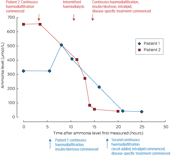
These measures should be initiated under the guidance of a metabolic physician, and in an intensive care unit where agitation or coma can be managed. Ammonia levels can be rapidly reduced by dialysis; its removal is dependent on flow rates, making intermittent haemodialysis the most effective method of clearance, as shown in Box 2. For this reason, we advocate intermittent dialysis rather than continuous venovenous haemodiafiltration for early ammonia control in the emergency setting.

While severe neurological impairment at the start of treatment is of great concern, this should not in itself be a reason to withhold treatment, as good neurological recovery is possible. This is illustrated by the case report of a middle-aged patient who recovered, despite decorticate posturing when therapy was initiated.5

We advocate early assessment of ammonia levels in patients with an unexplained altered conscious state, or when their cognitive disturbance seems disproportionate to any concurrent systemic illness. Many of the necessary elements of care can be initiated in non-tertiary intensive care units. Initiation of treatment at the hospital of presentation is essential, as this is a medical emergency; neurological outcome and survival are critically dependent on the timing of intervention. If recognised early and treated appropriately, the prognosis for neurological recovery is good.

Lessons for practice

  • Urea cycle disorders may first present in adulthood, unmasked by triggers such as systemic illness, increased protein load, surgery or corticosteroids.

  • Assessing ammonia levels is a simple but critical test in patients with unexplained impaired consciousness.

  • A session of intermittent haemodialysis is highly effective for rapid ammonia control, and superior to continuous haemodiafiltration for rapid correction.

  • Emergency treatment of hyperammonaemia should be undertaken early to prevent devastating neurological injury.

Box 1 –
Case histories of the two patients


Patient 1: 24 years old, male

Medical history

  • Obstructive sleep apnoea; no notable family history; high-functioning individual
  • Likely precipitant: intraoperative dexamethasone (8 mg) during nasal septoplasty

Progress

  • Vagueness and lethargy 48 h after operation, progressing over 24 h to incoherence
  • Intubated 12 h later for severe agitation
  • GCS declined to 5–6 over next 48 h; ammonia level, 334 μmol/L (RR, < 50 μmol/L); disease-specific treatment started
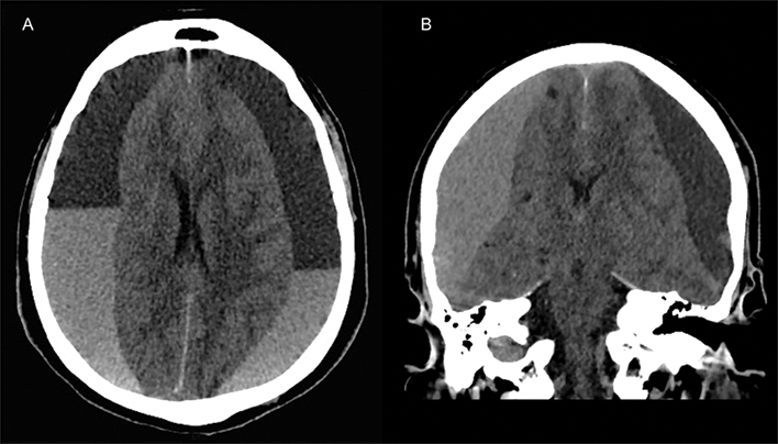
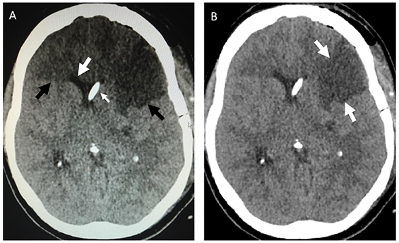
  • Raised intracranial pressures 6 h later (dilated pupils with cerebral oedema on CT brain), leading to decompressive craniectomy complicated by frontal haematoma, requiring evacuation
  • Prolonged intensive care unit and hospital stay

Outcome

  • Persistent minimally conscious state (at 22 months)
  • Discharged to nursing facility
  • Biochemical analysis of plasma and urine consistent with OTC deficiency (elevated urine orotic acid; plasma glutamine level high; plasma ornithine, citrulline and arginine levels low)
  • Genetic testing confirmed OTC gene mutation associated with OTC deficiency

Patient 2: 39 years old, male

Medical history

  • Chronic knee pain; no notable family history; high-functioning individual
  • Likely precipitant: cortisone injection into knee for knee pain

Progress

  • Headache, nausea, epigastric pain, blurred vision and incoordination 48 h after injection
  • Progressed over next 48 h to confusion, slowed speech
  • Progressive decline in GCS, requiring intubation
  • Seizure activity
  • Repeat CT brain showed cerebral oedema
  • Ammonia level: 652 μmol/L (RR, < 50 μmol/L); disease-specific treatment started; intracranial pressure monitor inserted; lack of control of intracranial hypertension; decision to palliate

Outcome

  • Proceeded to brain death and organ donation (except for liver donation: contraindicated)
  • Biochemical findings consistent with OTC deficiency (profound elevation of urine orotic acid, plasma glutamine level high, arginine level low)
  • Genetic testing confirmed OTC gene mutation associated with OTC deficiency

GCS = Glasgow coma score; RR = reference range; CT = computed tomography; OTC = ornithine transcarbamylase.

Box 2 –
Ammonia levels in our two patients, and their response to treatment*


* Note the rapid rate of decline of serum ammonia levels in Patient 2 achieved after initiating intermittent haemodialysis (about 10 hours after first measurement).