×

Stroke in a young man with untreated HIV infection and neurosyphilis

Clinical record

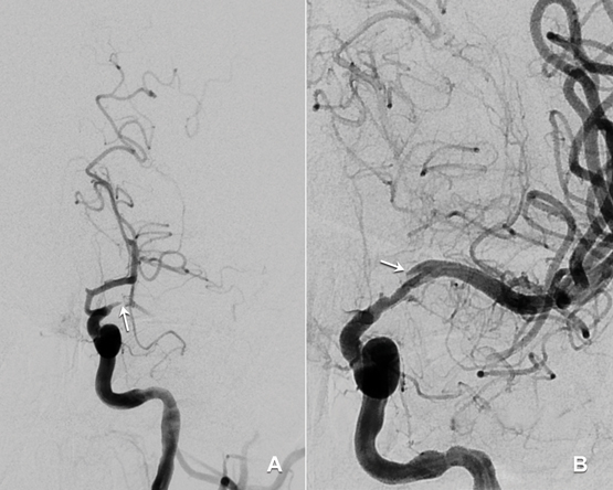
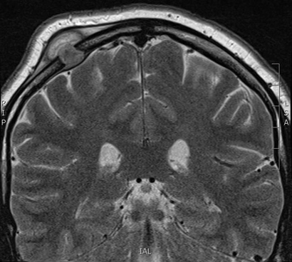
A 33-year-old man presented to an emergency department with acute dysphasia and a dense right hemiparesis. His National Institute Health Stroke Scale score was 12, indicating a moderate severity stroke (score range 0–42, with increasing values indicating increasing severity). His computed tomography (CT) brain scan was normal. A CT angiogram showed a filling defect in the left intracranial internal carotid artery. Intravenous thrombolysis was commenced 2.5 hours after stroke onset and completed during urgent transit to our hospital for endovascular thrombectomy. Combined stent retrieval and suction thrombectomy of the left internal carotid occlusion restored flow 4.5 hours after stroke onset. A small dissection in the left intracranial internal carotid artery was the source of the thrombotic occlusion (Figure). A magnetic resonance imaging scan of the brain showed small scattered infarctions in the left middle cerebral arterial territory.

The patient was later found to have a human immunodeficiency virus (HIV) infection that had been diagnosed 5 years earlier but for which he had not sought or received treatment. There was no history of screening for syphilis. He had a remote and brief history of recreational use of methamphetamines and cocaine (more than 12 months previously). He had no other vascular risk factors (non-smoker, normal fasting lipid and blood glucose levels, negative autoimmune serology). His CD4 cell count was 220 × 106 cells/L (reference interval [RI], ≥ 360 × 106 cells/L) and serum quantitative HIV RNA testing revealed 77 400 copies/mL. Hepatitis serology results were negative. Syphilis serology results were positive: reactive rapid plasmin reagin (RPR) with a titre of 1:256; reactive Treponema pallidum particle agglutination (TPPA) assay; and positive syphilis antibody enzyme immunoassay. His cerebrospinal fluid (CSF) protein level was 1.17 g (RI, < 0.45 g/L) and his white cell count was elevated at 62 μ/L (RI, < 5 μ/L), predominantly due to monocytosis (84%). CSF syphilis serology was positive, with reactive results from the venereal disease research laboratory, TPPA and fluorescent treponemal absorption antibody tests, confirming neurosyphilis. There were no other clinical or radiological features of tertiary syphilis. CSF polymerase chain reaction test results were negative for other pathogens including varicella-zoster virus, John Cunningham virus and tuberculosis. Cryptococcal antigen test results were negative. Other stroke investigations, including transoesophageal echocardiogram, returned negative results.

A 15-day course of intravenous benzylpenicillin (1.8 g, 4-hourly) with prednisone cover (three doses of 20 mg twice daily to prevent Jarisch–Herxheimer reaction) was completed as treatment for neurosyphilis. He received counselling and was commenced on antiretroviral therapy including abacavir–dolutegravir–lamivudine. Contact tracing was performed. The 3-month outcome was excellent, with only minor persistent dysphasic speech errors and a modified Rankin scale score of 1 (range 0–6, with increasing values indicating worse deficit and 6 for death). Progress serum RPR titres were significantly reduced (1:64), indicating a serological treatment response. A recent progress CD4 cell count was 630 × 106/L and quantitative HIV RNA testing revealed < 20 copies/mL.

Studies indicate that HIV infection increases the risk of ischaemic stroke, particularly in young patients (≤ 45 years) with low CD4 cell counts (< 350 × 106 cells/L).1,2 It is important for clinicians to recognise the various mechanisms by which HIV infection predisposes to stroke. These include a direct HIV-induced vasculopathy, and an indirect opportunistic co-infection-related arteritis with organisms such as tuberculosis, syphilis and varicella-zoster virus.2 HIV vasculopathy has been reported in the extracranial and intracranial cerebral circulations and may cause aneurysmal fusiform or saccular dilatation of vessels or a non-aneurysmal vasculopathy manifest by stenosis, occlusion or vasculitis.24 Additional factors contributing to stroke risk in HIV include a more frequent smoking history, coagulopathy, increased homocysteine levels and metabolic syndromes associated with antiretroviral therapies, which may result in accelerated atherosclerosis.1,3 Descriptions of intracranial arterial dissection in patients with HIV infection are limited to rare case reports.5 We postulate that HIV and syphilis co-infection in our patient may have caused a vasculopathy-associated intracranial arterial dissection.

The role and safety of intravenous thrombolysis in patients with HIV infection is not established.2,4 Thrombolysis could be theoretically harmful in patients with HIV vasculopathy or co-infection-related arteritis owing to a potential increased bleeding risk from abnormal vessel wall integrity.4 Despite this, intravenous thrombolysis has been used successfully in patients with untreated HIV infection to treat myocardial infarction and, in our patient, to treat acute ischaemic stroke without adverse outcomes.2,4 Clinicians should be aware that endovascular thrombectomy of proximal anterior cerebral circulation clots after intravenous thrombolysis is now evidence-based treatment for acute ischaemic stroke.6 Our case illustrates the “drip, ship and retrieve” concept of acute stroke management; with intravenous thrombolysis (“drip”) commenced at an initial hospital and completed while the patient was transferred (“shipped”) to another hospital for endovascular thrombectomy (clot “retrieval”). At present, only a limited number of stroke centres provide an endovascular thrombectomy service. Reorganisation of existing systems is required to allow rapid access to endovascular thrombectomy for all appropriate patients in Australia.6

This case presents an important reminder that HIV infection is a risk factor for stroke and that HIV testing should be performed in all young stroke patients. A lumbar puncture is recommended for diagnosis or exclusion of co-existing infections including tuberculosis, syphilis and varicella-zoster, which are all associated with vasculopathy in patients with HIV infection.

Lessons from practice

  • HIV infection is an important risk factor for stroke and HIV testing should be performed in all young stroke patients.

  • Patients with HIV infection and stroke should have a lumbar puncture to examine for co-existing opportunistic infections.

  • A diagnosis of neurosyphilis requires a cerebrospinal fluid (CSF) cell count and protein measurement and serological testing on serum and CSF.

  • There is evidence that the “drip, ship and retrieve” management approach to managing acute ischaemic stroke is effective. However, in patients with known HIV infection, acute stroke should be managed on a case-by-case basis.

Figure


Digital subtraction angiography: A: Pre-clot retrieval showing left internal carotid occlusion (arrow). B: Post-clot retrieval showing dissection (arrow) and restoration of flow.

Ankles jerk — yes, but how?

Eliciting an ankle jerk is not a trivial task. It requires the skills of an experienced bell-ringer, an expert golfer and a mechanical engineer. The experience of the examiner and the positioning of the patient are two factors contributing to the accuracy of reported findings.1,2 Accurate findings can provide invaluable clinical information.

When an ankle jerk is elicited, the response can be seen, felt and heard. When the response is present, the tendon produces an undamped oscillation heard as a “boing”, and when it is absent, the damped oscillation is heard as a dull “thud”.

Percussion on the Achilles tendon activates muscle spindles — specialised sensory receptors within the muscle that respond to stretch (lengthening) and vibration. Action potentials move centripetally via large diameter, myelinated, fast conducting fibres, and trigger responses in the spinal cord from α motor neurones that mediate muscle contraction. The magnitude and rapidity of the reflex response is modulated by both autonomic sympathetic tone and descending (upper motor neurone) inhibition.3

Reflex activation is faster and stronger when sympathetic nervous system tone is high. Patients who are anxious, thyrotoxic or withdrawing from alcohol will have stronger, brisker responses. Similarly, if descending inhibition from upper motor neurones is reduced by, for example, stroke, myelopathy or Jendrassik (reinforcement) manoeuvre, there will be stronger, brisker responses.

An intact ankle jerk response confirms normal function in the large diameter, myelinated, sensory fibres, in spinal cord integration at the L5-S1 level, and in the α motor neurones projecting to skeletal muscle. Symmetrical reduction or loss of ankle jerks occurs early in the course of large fibre sensory neuropathy (eg, in diabetes). Asymmetrical loss of the ankle jerk is a common sign of radiculopathy but may also occur with rarer conditions such as plexopathy. Symmetrical exaggeration of ankle jerks may simply reflect increased sympathetic nervous system activity. Whether symmetrically brisk responses are pathological or not depends on the “company they keep”. If the exaggerated ankle jerks are accompanied by clonus, spasticity, spread of reflexes, or an extensor plantar response, they will be judged to be pathologically exaggerated. Asymmetrical exaggeration of one ankle jerk indicates the presence of an upper motor neurone abnormality (either ipsilateral or contralateral, depending on how rostral the lesion is).

Absent ankle jerks are often due to faulty technique. If the tendon is too slack, it will not vibrate when struck, and if it is too taut, the response will be diminished or inevident. If the head of the reflex hammer strikes adjacent bone or connective tissue as well as the tendon, any response may be attenuated or lost. This is the bell-ringing skill. If the head of the hammer is pushed against the tendon, rather than allowed to swing freely against it and bounce off unimpeded, the response may be decreased or absent. This is the skill of the experienced golfer. Patient positioning can affect tendon length, sympathetic tone and the magnitude of descending inhibition. This is the mechanical engineering skill.

Unfortunately, most medical students have only a hazy notion of the required skill and are often taught the patient positioning that is least likely to offer early success. There are at least five different positions that can be used to elicit ankle jerks, each with advantages and disadvantages. The positions are illustrated, and their relative merits described, in Figures 1 – 5. Additional dynamic factors are discussed in the video at mja.com.au.

Figure 1 –
Patient kneeling backwards in a chair


In this position, the lower segment muscles are relaxed and the tendon is near optimal stretch. However, the examiner needs to be aware that gripping the back of the chair is a default Jendrassik manoeuvre, exaggerating the response. This is desirable if the ankle jerk is thought to be absent. This method is reported to have the lowest rate of false-negative results.

Figure 2 –
Patient with legs dangling over the edge of the bed


In this position, the examiner must gently position the ankle in the mid-position without the patient “helping” by contracting the ankle dorsiflexors and preventing the reflex plantar flexion.

Figure 3 –
Patient seated in a chair with the feet beneath the knees


This position almost automatically produces lower limb relaxation and optimal tendon stretch. However, it requires an examiner to “grovel” a little in front of the patient.

Figure 4 –
Patient supine in bed with legs relaxed


The technique illustrated here requires the examiner to strike his or her hand while it rests on the sole of the foot near the metatarsal heads. Patients will often become tense when first touched, and the examiner has to avoid excessive dorsiflexion at the ankle with this technique. It is said to be a more reproducible technique for general physicians, but it might also produce more false-negative results.

Figure 5 –
Patient supine with hip abducted and externally rotated, knee flexed and ankle dorsiflexed


This traditional positioning is easily the most problematic. Patients for whom this position is uncomfortable, unseemly or simply unfamiliar will experience excess muscle tension. In addition, if the patient is unable to hold the position unaided, the examiner is tempted to stabilise the limb by strongly dorsiflexing the foot at the ankle, thereby lengthening the tendon excessively and reducing the chance of obtaining any response. The effect is complete if the examiner, failing to elicit a response, exhorts the patient to relax. Feeling implicitly blamed, the patient inevitably becomes more tense.

A patient with sarcoidosis and a cryptococcal infection of the skull

A 29-year-old man of European ancestry, with a history of sarcoidosis treated with low-dose steroids, presented with a progressive swelling of the skull which had appeared 3 weeks earlier.

Physical examination revealed a 2.5 cm soft swelling of the right parietal part of the skull. A contrast-enhanced magnetic resonance imaging scan of the head showed a solitary lesion of the right parietal bone measuring 9 mm × 17 mm.

After neurosurgical resection, pus cultures grew Cryptococcus neoformans.

In the absence of disseminated cryptococcosis, the patient was treated successfully with oral fluconazole. Current literature suggests sarcoidosis as a risk factor for cryptococcosis, independent of the use of immunosuppressive agents.1,2

The role of neurosurgery in the treatment of chronic pain

Neurosurgical training should formally incorporate chronic pain management, and future generations will need to direct the development of rational surgical intervention

Until the early 1980s, neurosurgical intervention for intractable pain consisted almost exclusively of targeted neuroablative procedures aimed at disrupting nociceptive pathways at some point between peripheral nerve and cortex.

Used predominantly in the treatment of malignant pain in the trunk, pelvis and lower limbs, the most successful of these — dorsal rhizotomy, spinothalamic cordotomy and myelotomy — were considered to demonstrate, invariably in non-randomised case series, good to excellent results in selected patients with nociceptive cancer pain. Pain relief would usually be maintained through a survival period of 9–12 months but there were risks of operative mortality, post-lesion dysaesthesia, and impaired motor, sensory and sphincter function. Generally confined to use in cancer patients with a life expectancy of less than 1 year and with more widespread use of opiates, hospice care and the development of intrathecal drug delivery, these operations became almost redundant.

Recent advances in image-guided percutaneous cordotomy, a new understanding of the pain pathways within the dorsal columns, and the introduction of minimally invasive punctate myelotomy have led to some resurgence both of cordotomy and myelotomy in treating cancer pain patients.1,2 One ablative procedure yet to be unequivocally surpassed by newer techniques is that of dorsal root entry zone lesioning.3,4 Thermal lesions target the nociceptive fibres of the lateral part of the dorsal rootlets and medial aspect of Lissauer’s tract. Used in its proper context — for intractable upper limb pain following brachial plexus avulsion — it has demonstrated up to 80% long term excellent pain relief with few operative complications.

With the exception of microvascular decompression and ganglionic procedures for trigeminal neuralgia, almost all neurosurgical procedures for chronic pain have, over the past 30 years, moved firmly towards reversible, low risk neuromodulation techniques (electrical stimulation of some part of the nervous system or the use of intrathecal drug delivery to control pain or modify physiological function).

Medial thalamotomy and anterior cingulectomy — used to modify transmission and affective aspects of pain perception (via lateral and medial pain pathways respectively) have been supplanted by the use of deep brain stimulation. Used mostly for treatment of medically and surgically intractable trigeminal neuralgia and other facial pains, cluster headache, post-stroke pain and various painful deafferentation states, convincing data regarding long term effectiveness of deep brain stimulation in large numbers of chronic pain patients remain limited. Evidence for the benefits of motor cortex stimulation is even less compelling.5

Intrathecal infusion of baclofen for spasticity and mini-dose opiates for chronic nociceptive malignant pain consistently produce effective, durable results in appropriate cases. The potential for serious management morbidity coupled with the paucity of useful and safe intrathecal medications direct that extreme caution be exercised in applying this therapy to patients with neuropathic, non-malignant, and generalised pain syndromes. Neurosurgeons deal largely with complications such as catheter tip granuloma, infection, and catheter displacement or extrusion.

Pioneered in 1967 by the neurosurgeon C Norman Shealy using an intradural, radiofrequency-controlled system to relieve intractable malignant pain of the pelvis and lower limbs,6 spinal cord stimulation has evolved massively in terms of technology, technique, safety, and understanding of its uses and limitations. In Australia, percutaneous, epidural electrodes are now implanted predominantly by pain medicine specialists. Neurosurgical input is required for insertion of plate electrodes in cases where epidural access is limited, and for accessing difficult sites such as the cervico-medullary junction.

Advantages of spinal cord stimulation include its low risk, reversibility and straightforward procedural techniques. On this basis, some have advocated earlier neuromodulation so as to avoid or delay major or repeat surgeries such as in the treatment of failed back surgery syndrome, and of pure low back pain with poorly defined pain generators. The downside is the now widespread, often ill-considered and repeated application of an expensive mode of treatment to unsuitable patients and pathologies. Unfortunately, this trend seems matched by a substantial increase in equally ill-considered, minimally invasive, instrumented spinal fixation for back and neck pain. Both forms of surgery as applied to chronic spinal pain are in urgent need of rationalisation.

Evidence-based recommendations for patient selection in spinal cord stimulation in Australia and New Zealand were published in 2011.7 These have remained consistent with all literature reviews to date. The most valid indication is in cases of failed back surgery syndrome. Generalised acceptance of neuromodulation has been limited partly by medical territorialism but also by scepticism generated by the lack of high level evidence for efficacy and of cost benefit. It was not until 20058 and 20089 that level 2 evidence was established in these domains. The ability to provide level 1 evidence has been hampered until very recently by a lack of placebo controls owing to stimulation-induced paraesthesiae, while the very existence of an organic basis to some pain syndromes (eg, complex regional pain syndrome type 1 and non-traumatic occipital neuralgia) that may be treated by neural stimulation has been questioned.

Trials of stimulation using exteriorised leads (time-limited by infection risk) are sometimes too short to allow adequate assessment regarding permanent implantation. There is little commercial incentive to correct this problem. Careful patient selection is paramount, yet in the United States, where trial to permanent implantation may be office based, reported trial to permanent implantation rates vary between 20% and almost 100%. Research and development worldwide are heavily sponsored by industry, which gives rise to inevitable concerns of investigator bias and unseemly haste in publishing case series. All those entering or already working in this field are recommended to read the sobering chapter by Coffey in Surgical management of pain.10

Spinal cord stimulation aims at a spinal level to suppress wide dynamic range neuronal activity in the dorsal horns and suprasegmentally via the dorsal column nuclei to modulate activity in the medial thalamus and cingulate gyrus. It has traditionally entailed tonic stimulation with a frequency of around 40–60 Hz. Recent pragmatic randomised controlled trials suggest that high frequency stimulation at 10 kHz and burst (phasic) stimulation offer superiority in the treatment of chronic back and neuropathic leg pains.11 Each of these provides paraesthesia-free analgesia and has the potential finally to allow sham stimulation. Closed-loop feedback systems promise to even out stimulation efficiency while Wi-Fi and magnetic resonance imaging compatible devices are becoming well established.

Dorsal root ganglion stimulation may hold the key to treating pains less well treated by spinal cord stimulation, including groin pain, foot pain, post-herpetic neuralgia, persistent post-surgical pain (herniorrhaphy, mastectomy, thoracotomy), and in complex regional pain syndrome. Despite multiple positive case series, there is only weak evidence for occipital nerve stimulation benefitting a variety of chronic headaches, although it does appear useful in treating post-craniotomy neuropathic head pain.

Unless results from very necessary, very carefully conducted prospective, randomised, placebo-controlled trials dictate otherwise, neuromodulation is likely to continue as the predominant form of interventional treatment for intractable neuropathic pain for the foreseeable future. If so, incorporation of all these developments within a single system would maximise flexibility and efficacy. Advances in Wi-Fi and miniaturisation technology should simplify implantation beyond current imagination. Improved functional imaging may lead to anatomically discrete electrical or drug micro-implantation or even to highly focussed neuroablative procedures.

Nationally, the quality of intractable pain management remains erratic. Neurosurgeons are well qualified to play a leading research and clinical role in optimising both benign and cancer pain treatment. However, at present, fewer than ten practising neurosurgeons in Australia and New Zealand have a major subspecialist interest in pain surgery. Broader exposure to pain management in neurosurgical and spinal surgical training would be of substantial benefit to recruitment and in the treatment of acute (postoperative) and chronic pain states.

Surgical management of low back pain

Spinal surgery for chronic low back pain is controversial, and the disproportionate number of fusions in private hospitals is unexplained

In developed countries, low back pain is the most common presenting symptom in primary care practice, with a lifetime prevalence of 80% for episodes of back pain.1 It affects adolescent sportsmen, pregnant women, hospital nurses and middle-aged labourers, and it peaks in the elderly.1 Back pain disrupts the rhythm of daily life, affecting work, recreation, social life and family income. The level of disability has a significant cost to the community.

There is an encyclopaedic range of medical and alternative treatments for low back pain. Most have, at best, limited benefits.2 Disappointed patients resort to nerve blocks and ablative rhizotomies which also have limited evidence for long-term benefits.3

Armed with mobile phones, consumed with social media, and with rapid access to online information, our patients present with high expectations of modern technology, including surgery. With outstanding results from hip and knee arthroplasties, they expect similar results from spinal surgery. Not uncommonly, the patient attends the surgical consultation with an expectation that the problem scan be fixed.

Aetiology

At the initial consultation, the astute family physician will consider a wide range of diagnoses in the patient with the recent onset of back pain. Renal stones, aortic aneurysm, gastric and pancreatic conditions, malignant metastases, discitis and ankylosing spondylitis will all need to be considered before accepting the diagnosis of non-specific axial low back pain.

Clinical examination in the absence of a radiculopathy is likely to show non-specific signs and is unlikely to identify any specific injury. The supporting muscles, interspinous ligaments, lateral facet joints, vertebrae, discs and sacroiliac joints may all contribute to chronic low back pain, but their individual contribution can be difficult to elucidate clinically.4

Today, chronic pain is understood in terms of a biopsychosocial concept, although this can be difficult to explain to a patient. The surgeon John Loeser has untangled this concept with the Loeser rings.5 He likens the pain injury to the inner core of an onion, with additional layers surrounding it which enhance the experience of pain, including childhood issues, masked depression, substance abuse, pain behaviour and entitlements to secondary gain. For the benefit of the patient and the reputation of the surgeon, these aspects of chronic pain need to be carefully explored before considering spinal surgery.

In the absence of a diagnosis, magnetic resonance imaging of the lumbar spine is required to exclude congenital or advanced pathological changes in patients with low back pain. This tends to open Pandora’s Box. Imaging will identify degenerative changes from the third decade onwards, including disc dehydration (the black disc), disc narrowing, lateral facet joint arthropathy and bone spurs. However, there is “very little correlation between imaging findings of disc herniation and the clinical course. Imaging findings of structural change of osteoarthritis do not correlate with pain production”.6 The patient with chronic low back pain, having seen the report of the radiologist and suggestions for further pain interventions, then has renewed expectations of successful treatment.

For patients with non-specific axial back pain, clinical examination and radiological imaging are unreliable guides to surgical or other pain interventions. Because of this dilemma, and faced with anxious patients with high expectations from modern surgical technology, there has been a growing enthusiasm for surgical fusions.

Natural history

The spinal surgeon needs to be conscious of the natural history of low back pain. It is common in adolescence (18–50%), causing disability in up to 9%.1 There is the lifetime prevalence of 80%.1,7 There is a point prevalence of 37% in the adult population and, although 33% may make a recovery, up to 70% will still complain of pain 12 months later.7 Back pain can be transient, recurrent and chronic. Psychological, social and environmental issues compound the patient’s burden of pain.5

Surgery

Spinal fusion surgery for lumbar non-specific low back pain is controversial, particularly because the origin of the pain is undetermined4 and imaging of the spine is unhelpful.6 The procedure dates back to 1889 but it is in the past 20 years that there has been a great escalation internationally in its use, with an increase of 267% over 11 years in the United States alone.8 There has also been a disproportionate increase in the rate of spinal fusions in private hospitals compared with public hospitals, with a 10.8-fold increase in private hospitals. In addition, there is a range of techniques for spinal fusion, including anterior lumbar interbody fusions, posterior lumbar interbody fusions, 360° fusions (anterior and posterior approaches) in one or two stages, and anterior disc arthroplasties. Each of these procedures has different technical complications, and there remains little evidence of better outcomes for one over another.4,9

Results

A Cochrane review of surgical fusions for back pain in 1999 concluded that there were no published randomised controlled trials which established effectiveness of fusions for chronic pain.10 In 2004, a review again concluded that there was insufficient evidence for effectiveness of surgery for a firm conclusion to be drawn.11 A further Cochrane review in 2005 reported “variable clinical outcomes ranging between 16% and 95%”.9 There was no evident difference, over a period of 2 years, between artificial disc replacement and the less expensive fusion technique. It also found that the techniques using intradiscal thermal coagulation and spinal spacers had lost the support of surgeons and have since been discarded, and that there was insufficient evidence to support spinal fusion for degenerative disc disease, whether for back pain or in conjunction with spinal decompression.9

More recently, an Australian study on trends in spinal surgery has noted a significant increase in the rate of fusions, over a 10-year period, of 175%. The rate had increased from 8.4 per 100 000 to 23.1 per 100 000, and 69.9% were instrumented.12

The 2005 review by Gibson and Waddell was critical of the outcomes measured, preferring “patient-centred outcomes rather than an assessment of the short-term surgical outcomes.”9 This was to emphasise that reports needed to focus on the patient’s perception of pain relief and the patient’s return to the previous level of daily activities and employment. It was noted that “The limited evidence of the long-term effects of either surgical decompression or fusion remains a matter of concern given the magnitude of the clinical problem, the numbers and the cost of surgical procedures being performed”.9

A later study of a cohort of patients receiving workers’ compensation in New South Wales concluded that the outcomes were so poor that spinal fusions were not recommended for this group.12 In addition, independent reviews noted that the incidence of persistent post-operative pain syndrome was as high as 40% and that there was a 50% success rate, at best, from the first operation, 30% from the second and 15% from the third.7

Conclusions

The current high incidence of chronic low back pain in developed countries has little to do with biomedical explanations and is best understood in terms of a biopsychosocial framework, including work dissatisfaction, secondary gain and a cultural bias toward symptom relief. In Western medicine, current approaches to this problem, including the overuse of expensive diagnostic imaging, have failed and have in fact exacerbated the problem, because of the misunderstanding of the aetiology of the condition. The well documented ageing of our population and our increasingly sedentary lives are enhancing the epidemic of chronic low back pain. Our patients are presenting with high expectations of modern medicine and, in many cases, there are additional entitlements to monetary gains from workplace injury and third-party incidents. Multiple procedures have been carried out for spinal fusions, particularly in the past 20 years, but the surgery remains controversial with respect to aetiology and indication. There has been a lack of patient-oriented surgical outcomes, and there is a lack of outcomes for most things that we do for chronic low back pain.

In conclusion, there is a growing tendency for the astute spinal surgeon to have all patients assessed independently and, at times, for them to attend an interdisciplinary pain program to clarify issues of psychological origin that might complicate recovery. While the spinal fusion procedure remains controversial, it would be valuable for spinal surgeons to undertake a national audit of patient-centred outcomes for the procedure, similar to the excellent audit carried out for hip and knee arthroplasties by the Australian orthopaedic surgeons.

A case of subacute sclerosing panencephalitis in a 23-year-old recent immigrant to Australia

A 23-year-old woman presented with a generalised tonic–clonic seizure on a background of 9 months of progressive neurological decline (characterised by involuntary jerks, monocular visual disturbance and reduced speech) resulting in falls, impaired ability to perform activities of daily living and urinary incontinence. Examination showed right-sided myoclonus, bilateral parkinsonism, primitive reflexes present, akinetic mutism and retinal scarring apparent on fundoscopy.

Two years previously, she had migrated to Australia from the Philippines; her family reported that her neurodevelopment was normal and that she had received routine childhood vaccinations.

The results of computed tomography and angiography of the brain were normal. Magnetic resonance imaging of the brain showed cerebral volume loss and extensive white matter changes with ill-defined subcortical T2 hyperintensities. An electroencephalogram showed non-specific focal epilepsy disorder involving frontal regions. Results of extensive investigations for autoimmune, hereditary and infective causes were unremarkable with the exception of cerebrospinal fluid (CSF) analysis, which revealed unmatched oligoclonal IgG. Enzyme immunoassay of CSF and serum (with corrected optical density values of 2.24 and 4.21, respectively) gave a strongly positive result for measles IgG. Intrathecal measles antibody production was confirmed by the concurrent absence of varicella-zoster virus IgG in the CSF, despite it being detected in the serum.

A raised CSF:serum ratio of measles antibodies, oligoclonal IgG in CSF and clinical features of progressive mental deterioration with myoclonus fulfilled Dyken’s diagnostic criteria of probable subacute sclerosing panencephalitis (SSPE), outlined in the Box. Supportive measures were undertaken and isoprinosine treatment for SSPE was initiated. The patient’s neurological condition stabilised but there was no improvement in her condition.

SSPE is a fatal, progressive neurodegenerative disease caused by persistent infection with an altered measles virus. Although rare, SSPE should be considered in the differential diagnosis of subacute neurological deterioration and myoclonus, especially in incompletely vaccinated patients. SSPE is rare following measles infection, with an incidence of 4–22 cases per million measles cases.1 SSPE is rarer in adults, who account for 1–12.7% of cases.2 There is no association with the attenuated measles vaccine and SSPE; no vaccine strains have ever been isolated from tissue specimens of patients with SSPE.3 Vaccination unfortunately does not confer 100% protection against measles infection (and hence against developing SSPE, which may occur after subclinical measles infection). A single dose of MMR vaccine is 95% effective, and two doses are 99% effective for measles protection.

This case is important as it highlights a terrible consequence of a vaccine-preventable disease. Achieving whole-population vaccination is instrumental in preventing measles infections through the development of herd immunity. However, 96–99% of a population are required to be vaccinated to prevent sustained measles transmission. In Australia in 2012, only 91.9% of children aged 5 years had received two doses of measles vaccine.4 Lower immunisation rates have been observed in certain areas, such as parts of the New South Wales north coast. In Australia, there were 154 confirmed measles cases reported in 2013 and 335 cases in 2014.5 Our case highlights one of the possible consequences if measles vaccination rates are not improved.

Box –
Dyken’s criteria for diagnosis of subacute sclerosing panencephalitis1

Criterion

Description


1. Characteristic clinical features

Progressive, subacute mental deterioration with typical signs like myoclonus

2. Electroencephalogram

Periodic, stereotyped high-voltage discharges

3. Cerebrospinal fluid (CSF)

Raised γ-globulin level or oligoclonal pattern

4. Measles antibodies

Raised titre in serum (≥ 1 : 256) and/or CSF (≥ 1 : 4) with a CSF : serum ratio < 1 : 200

5. Brain biopsy or autopsy

Showing typical histopathology and/or culturing altered measles virus and/or detection of measles RNA by polymerase chain reaction


Definitive diagnosis: criterion number 5, plus 3 other criteria. Probable diagnosis: 3 or more of the 5 criteria.

A moving account of resilience and love

Bleed: surviving cerebral catastrophe: a tale of the mind, love and modern medicine. Bill Williams. Wild Man Press, 2015 (333 pp, $29.95). ISBN 9780646937649.

As a doctor, if you have ever watched a loved one in hospital — a parent, child or spouse — being carried along the medical diagnostic and treatment pathway, you will immediately recognise the feelings described in Bill Williams’s latest book. Fear, impotence, a desire to protect, but also not to offend or obstruct those whose role you can relate to. Where do you find the balance on the patient–doctor divide?

This compelling and emotionally intelligent story traces the weeks that follow after Bill’s wife Gisela suffers a subarachnoid haemorrhage, signalled by a thunderclap headache while they are camping in a riverbed in the western desert. He documents the roller-coaster of emotions — starting with the dash for medical help, and as the illness and management unfold, the hope and the grief, the repeated investigations, the surgery, the complications — and he carries the reader along with him. He chronicles the compassion and expertise of the carers, and wonders at the evolution of knowledge over the centuries that results in our current medical wizardry. And yet how little we still understand about the mysteries of the brain and what makes us who we are.

Much of the story is also about family and friends who provide love and support, and who are just as essential for the healing process. An intensely personal story, it is also a testament of love to his wife, and a story of her life and their lives together. Moving and engaging, the narrative is flowing and fast paced, with occasional diversions into medical history, neuroanatomy, and life and culture in the western desert. Written with medical insight but also with a novelist’s skill and sensitivity, it is a perceptive, honest and articulate story of struggling with human suffering and loss, and of the importance of love.

Male neurosurgeons highest ATO earners, GPs in top 50

With an average income of almost 577,000, male neurosurgeons have topped the list of the highest income status of Australians released by the Australian Tax Office.

Other medical specialities round out the top five for men, with Ophthalmologist, Cardiologist, Plastic and reconstructive surgeon and Gynaecologist/Obstetrician being the most lucrative jobs.

Among women, it was a slightly different story. The job with the highest income wasn’t in medicine at all, but was the role of a Judge. Neurosurgeon, plastic and reconstructive surgeon and Vascular surgeon came in at two, three and five respectively however future trader was number four in the list.

The statistics found that male specialists earned roughly twice as much as their female counterparts, however it didn’t distinguish between full and part time.

With an average income of $184,639, male general practitioners came in at 48th on the list while female GPs were 40th with an average income of $129,834.

According to the ATO, the statistics don’t include some of Australia’s top earners due to privacy protection or because their career title was unavailable.

Those with the lowest incomes, under $20,000 per year were fruit and nut pickers, deer farmer and fast food cook.

Read the full list at the ATO’s website.

Top 50 average incomes for Men and Women

  1. Neurosurgeon $577,674 Judge – law $355,844
  2. Ophthalmologist $552,947 Neurosurgeon $323,682
  3. Cardiologist $453,253 Plastic and reconstructive surgeon $281,608
  4. Plastic and reconstructive surgeon $448,530 Futures trader $281,600
  5. Gynaecologist; Obstetrician $446,507 Vascular surgeon $271,529
  6. Otorhinolaryngologist  $445,939 Gynaecologist; Obstetrician $264,628
  7. Orthopaedic surgeon $439,629 Gastroenterologist $260,925
  8. Urologist $433,792 Magistrate $260,161
  9. Vascular surgeon $417,524 Anaesthetist $243,582
  10. Gastroenterologist  $415,192 Ophthalmologist  $217,242
  11. Diagnostic and interventional radiologist  $386,003 Cardiologist $215,920
  12. Dermatologist  $383,880  Urologist $213,094
  13. Judge – law  $381,323 Surgeon – general  $210,796
  14. Anaesthetist  $370,492 Medical oncologist $208,612 
  15. Cardiothoracic surgeon  $358,043 Specialist physicians – other  $207,599
  16. Surgeon – general  $ 357,996 Specialist physician – general medicine $207,225
  17. Specialist physicians – other  $344,860 Otorhinolaryngologist $200,136
  18. Radiation oncologist $336,994 Dermatologist $195,030
  19. Medical oncologist   $322,178 Diagnostic and interventional radiologist  $180,695
  20. Securities and finance dealer $320,452 Cardiothoracic surgeon  $175,500
  21. Thoracic medicine specialist  $315,444 Paediatric surgeon  $175,314
  22. Specialist physician – general medicine $315,114 Endocrinologist $174,542
  23. Intensive care specialist $308,033 Member of parliament  $173,331
  24. Renal medicine specialist $298,681  Rheumatologist $169,409
  25. Neurologist   $298,543 Intensive care specialist $169,369
  26. Financial investment manager $288,790 Emergency medicine specialist $165,786
  27. Investment broker  $286,530 Orthopaedic surgeon $159,479
  28. Paediatric surgeon  $282,508 Neurologist $155,217
  29. Clinical haematologist $271,738 Renal medicine specialist $155,133
  30. Futures trader  $264,830 Psychiatrist $152,437
  31. Endocrinologist  $258,972 Clinical haematologist $147,970
  32. Cricketer  $257,527 Paediatrician $147,347
  33. Rheumatologist $256,933 Securities and finance dealer $145,208
  34. Dental specialist $253,442 Dental specialist $140,505
  35. Magistrate $246,737 Actuary $136,819
  36. Equities analyst; Investment dealer $245,826 Radiation oncologist $135,678
  37. Paediatrician $239,405 Financial investment manager $134,481
  38. Stock exchange dealer Stockbroker $238,192  Petroleum engineer $133,315
  39. Psychiatrist  $234,557  Mining production manager $133,061
  40. Emergency medicine specialist $232,595  General medical practitioner $129,834
  41. Member of parliament $232,093   Thoracic medicine specialist $127,645
  42. Pathologist $224,378 Stockbroker $124,433
  43. Company secretary – corporate governance $218,432 Paving plant operator $123,281
  44. State governor  $212,652 Mining engineer $119,564
  45. Actuary  $196,144 Tribunal member $119,219
  46. Doctor – medical practitioner – other; Occupational medicine specialist; Public health physician; Sports physician $187,468 Doctor – medical practitioner – other; Occupational medicine specialist; Public health physician; Sports physician  $118,310
  47. Petroleum engineer $185,808  Geophysicist $117,575
  48. General medical practitioner $184,639  Chief executive officer; Executive director; Managing director; Public servant – secretary or deputy secretary $116,855
  49. Chief executive officer; Executive director; Managing director; Public servant – secretary or deputy secretary  $181,849  Engineering manager $116,732
  50. Mining production manager  $179,439 Metallurgist $110,359

Latest news:

First national dementia clinical guidelines released

The first national clinical guidelines for dementia have been released, in a move welcomed by experts.

The guidelines were outlined in a Medical Journal of Australia article after being developed by the National Health and Medical Research Council (NHMRC) Partnership Centre for Dealing with Cognitive and Related Functional Decline in Older People.

The dementia clinical guidelines are a collaboration of 23 authors from 17 institutions around the country. They are an adaptation of the UK’s National Institute for Health and Care Excellence recommendations.

Related: Dementia challenge for general practice model

The guidelines focus on timely diagnosis as well as living well with dementia and delaying functional decline. It also looks into training and supporting staff and carers in providing optimal care and non-pharmacological treatment options.

There are 109 recommendations in the guidelines. Key recommendations include:

  • Getting a timely diagnosis but exploring symptoms when they’re first raised.
  • Finding a systematic approach to diagnosis. This includes patient and informant history taking, cognitive assessment, medication review, blood tests and computed tomography or magnetic resonance imaging to exclude other cerebral pathologies.
  • A clinical cognitive assessment which should include examination with a screening tool with established reliability and validity.
  • Reviewing people with mild cognitive impairment after 6-18 months.
  • At diagnosis of dementia and regular intervals afterwards, there should be assessment for medical comorbidities and key psychiatric features associated with dementia, including depression, to ensure optimal management of coexisting conditions.
  • Comprehensive  role-appropriate  dementia-specific  training  for  health  and  aged  care professionals.
  • Emphasising promoting and maintaining independence. This can be through activities of daily living, continuing exercise and supporting patients to continue maintaining activities that are of interest to them.
  • Understand the person and symptoms via a comprehensive assessment and analysis of the behaviour, antecedent, behaviour description and consequence.
  • Those with mild to moderate behavioural and psychological symptoms of dementia shouldn’t usually be prescribed antipsychotic medications. This is because it leads to increased risk of cerebrovascular adverse events and death.
  • Those with advanced dementia should have a palliative care approach and involve a palliative care service if indicated.
  • Family and carers should be involved in the planning and decision making of the care and management of people with dementia.
  • Programs including dementia education should be provided to carers.

Related: MJA Podcast: Advances and challenges in dementia care, with Dr Terence Chong and Prof Nicola Lautenschlager

Read the summary of the guidelines Medical Journal of Australia.

Latest news:

Clinical practice guidelines for dementia in Australia

Dementia is a National Health Priority Area in Australia. As our population ages, the number of people with dementia will increase.1 People with dementia have deficits in one or more of the areas of memory, communication, attention, thinking and judgement.2

The quality of clinical practice in dementia care in Australia is variable. The availability of high-quality services to support workforce training, diagnosis and ongoing care, advance care planning and support for families to provide care is inconsistent.

Clinical practice guidelines can improve uptake of research findings by identifying, synthesising and disseminating evidence to clinicians.3 Most importantly, adherence to clinical practice guidelines can improve the quality and consistency of care.4

The National Health and Medical Research Council (NHMRC) Partnership Centre for Dealing with Cognitive and Related Functional Decline in Older People was established in 2013 with funding support from the NHMRC, HammondCare, Alzheimer’s Australia, Brightwater Care Group and Helping Hand Aged Care. One of the activities of the Partnership Centre was to develop Australian clinical practice guidelines for dementia. The guidelines were adapted from existing guidelines5 using ADAPTE methodology6 to reflect the Australian context and the latest evidence. A multidisciplinary guideline committee, which included consumers, was appointed to refine the scope of the guidelines and form recommendations based on systematic reviews of the evidence.

The purpose of the guidelines is to provide recommendations for an agreed standard of practice for the diagnosis and management of people with dementia in Australia. The guidelines address care of people with dementia in community, residential care and hospital settings and are relevant to medical practitioners, nurses, aged care workers and allied health professionals. They are also useful for researchers, educators, policy makers and decision makers.

The full guidelines can be accessed via the Australian Clinical Practice Guidelines portal (https://www.clinicalguidelines.gov.au).

Main recommendations

The guidelines provide 109 recommendations, categorised as evidence-based recommendations (formulated after a systematic review of the evidence), consensus-based recommendations (formed where a systematic review has failed to identify sufficient studies to inform a recommendation) and practice points (based on expert opinion). Key recommendations prioritised by the committee for implementation are presented in the Box.

Changes in management

Delays between the onset of symptoms and diagnosis of dementia are widely acknowledged.7 There is currently a lack of information regarding the benefits and harms of population screening for cognitive impairment.8 The guidelines focus on timely diagnosis by recommending that symptoms are explored when first raised by the person experiencing the symptoms and/or their carer or family and are not dismissed as “just a part of ageing”. People with a possible diagnosis of dementia should be referred to a service or specialist in dementia diagnosis (eg, a memory clinic, neurologist, geriatrician or psychiatrist).9

The guidelines recommend a systematic approach to diagnosing dementia; this includes patient and informant history taking, cognitive assessment, medication review, blood tests and computed tomography or magnetic resonance imaging to exclude other cerebral pathologies. The use of single-photon emission computed tomography is not recommended.10 More recent diagnostic techniques using biomarkers (including the use of positron emission tomography) are not recommended for routine use.11

Clinical cognitive assessment should include examination with a screening tool with established reliability and validity. A number of tools are recommended in the guidelines including the Mini-Mental State Examination. The Kimberley Indigenous Cognitive Assessment tool for remote living Aboriginal and Torres Strait Islander populations and the Rowland Universal Dementia Assessment Scale for people from non-English speaking backgrounds are recommended for use where illiteracy, language or cultural considerations deem their use appropriate.

The committee recommended review of people with mild cognitive impairment after 6–18 months. This recommendation was formulated based on an existing systematic review which found that, in a clinic setting, the annual conversion rate of mild cognitive impairment to Alzheimer disease was close to 10%.12

At the time of diagnosis of dementia, and at regular intervals subsequently, assessment should be made for medical comorbidities and key psychiatric features associated with dementia, including depression, to ensure optimal management of coexisting conditions.

The guidelines recommend comprehensive role-appropriate dementia-specific training for health and aged care professionals. Such training can improve the quality of life for the person with dementia13 and reduce restraint use14,15 by teaching staff how to understand a person with dementia and to read body language and behaviour as signs of communication and respond appropriately. The evidence supports training models that focus on understanding symptoms and behaviours and providing person-centred care.16

The guidelines recommend a greater emphasis on promoting and maintaining independence through activities of daily living, continuing exercise and supporting the person to pursue activities that are meaningful and of interest to them. Adequate nourishment and hydration through maintaining a healthy, balanced diet should be encouraged and supported. People with dementia should have their weight monitored and nutritional status assessed regularly. Oral health is important17 and, on diagnosis, the medical practitioner should recommend that the person with dementia (or their carer[s] or family) make an appointment to see a dentist to conduct an assessment and formulate a long-term treatment plan.

Acetylcholinesterase inhibitors and memantine are routinely prescribed for people with mild to moderate Alzheimer disease in order to delay functional decline, and the guidelines support their use.18 Based on recent evidence, the guidelines also state that any one of the three acetylcholinesterase inhibitors (donepezil, galantamine or rivastigmine) could also be considered for people with dementia with Lewy bodies, Parkinson disease dementia, vascular dementia or mixed dementia.1922 The combination of an acetylcholinesterase inhibitor and memantine could be considered for managing the symptoms of functional decline for people with moderate to severe Alzheimer disease.23 Clinicians should be aware that not all of these indications are reimbursed under the Pharmaceutical Benefits Scheme and that acetylcholinesterase inhibitors are associated with a number of side effects including (but not limited to) nausea, vomiting, diarrhoea, dizziness, increased urinary frequency, falls, muscle cramps, weight loss, anorexia, headache and insomnia.24 Acetylcholinesterase inhibitors should not be prescribed for people with mild cognitive impairment.25

If people with dementia cannot express their needs through communication, they may communicate through their actions and behaviour. The guidelines recommend the need to understand the person and symptoms via a comprehensive assessment and analysis of the behaviour (eg, antecedent [triggers], behaviour description and consequence [ABC approach]). The objective measurement of behavioural and psychological symptoms of dementia should be undertaken using tools to monitor the type and patterns of behaviours. The provision of care that is consistent with the ten principles of dignity in care26 and non-pharmacological interventions should be implemented before considering use of medications. Non-pharmacological interventions should ideally involve engagement in activities that are enjoyable for the person with dementia and individualised support. Working with the carer and family to build skills in managing symptoms, communicating effectively and problem solving have been shown to be effective in reducing symptoms.27,28

A number of pharmacological treatments are recommended to complement non-pharmacological approaches when the person with dementia is severely distressed or there is an immediate risk of harm. Analgesics are recommended when pain is suspected.29 A trial of selective serotonin reuptake inhibitors is recommended for agitation; the strongest evidence is for citalopram.30 The role of antidepressants in the treatment of depression in people with dementia is uncertain. Larger trials conducted in people with dementia have not shown benefit (in group data) for antidepressants for treatment of depression per se.31 Nevertheless, the committee considered that those with a pre-existing history of major depression (before developing dementia) who develop a comorbid major depression should be treated in the usual way.

People with Alzheimer disease, vascular dementia or mixed dementias with mild to moderate behavioural and psychological symptoms of dementia should not usually be prescribed antipsychotic medications, owing to the increased risk of cerebrovascular adverse events and death.32 For people with severe symptoms who are distressed or causing distress to others, treatment with an antipsychotic may be offered following a full discussion with the person with dementia and/or their carer(s) or family about the possible benefits and harms. Treatment should be reviewed every 4–12 weeks, considering the need for antipsychotics and possible cessation of medication.

Care for people with advanced dementia should be based on a palliative approach and involve a palliative care service if indicated. Treatment and care should be provided as per the person’s advance care plan.

Carers and families should be included in the planning, decision making and care and management of people with dementia. Carers are often not provided with enough support or adequate training to effectively provide care.33 There is evidence that tailored multifaceted programs involving both the carer and the person with dementia can improve quality of life for both.34 Carers should have access to programs that include education regarding dementia; information regarding relevant services such as respite; information about support organisations such as Alzheimer’s Australia; individualised care management strategies to overcome specific problems; training in providing care and communicating most effectively with the person with dementia; and support regarding coping strategies to maintain their own wellbeing, including stress management.

Box –
Key recommendations identified by the committee as priorities for implementation*

  • Health and aged care professionals should provide person-centred care, by identifying and responding to the individual needs and preferences of the person with dementia, their carer(s) and family. The 10 principles of dignity in care (http://www.dignityincare.org.uk) should be used as the standard by which care is delivered and evaluated.
  • People with a possible diagnosis of dementia should be offered referral to memory assessment specialists or services for a comprehensive assessment.
  • The medical practitioner should be honest and respectful and use a gradual and individualised approach when communicating the diagnosis to the person with dementia and their carer(s) and family.
  • Health system planners should ensure that people with dementia have access to a care coordinator who can work with them and their carers and families from the time of diagnosis.*
  • Health and aged care organisations should ensure that all staff working with people with dementia receive dementia-care training (attitude, knowledge and skill development) that is consistent with their roles and responsibilities. Training should reflect programs that have been shown to optimise care for people with dementia.*
  • Training programs should be comprehensive and have a strong focus on communicating effectively with the person with dementia and his or her carer(s) and family and recognising, preventing and managing behavioural and psychological symptoms of dementia. Staff should be trained in the principles of person-centred care and how these principles are applied in practice.
  • People with dementia living in the community should be offered occupational therapy interventions which should include: environmental assessment and modification to aid independent functioning; prescription of assistive technology; and tailored intervention to promote independence in activities of daily living.*
  • People with dementia who develop behavioural and psychological symptoms should be offered a comprehensive assessment at an early opportunity by a professional skilled in symptom assessment and management. This should involve their carer(s) and families as appropriate and include; analysis of the behaviours, assessment of physical and mental health, pain or discomfort, side effects of medication, the influence of religious and spiritual beliefs and cultural norms, physical environmental and interpersonal factors, an assessment of carer(s) health and communication style, understanding the behaviour as a form of communication.*
  • People with Alzheimer’s disease, vascular dementia or mixed dementias with mild-to-moderate behavioural and psychological symptoms of dementia should not usually be prescribed antipsychotic medications because of the increased risk of cerebrovascular adverse events and death.
  • The person with dementia, their carer(s) and family should be offered respite appropriate to their needs.
  • Carers and families should have access to programs designed to provide support and optimise their ability to provide care for the person with dementia. Programs should be individualised, multifaceted and delivered over multiple sessions.*

* Note that some recommendations have been shortened in this publication.