×

Postcards

Winner — Practitioner category

She started in the medical wards,
With the dishevelled old gents,
Their missing dentures and distant adventures.
Hour by hour punctuated with shower by shower,
And dutifully dosing dopamine, Duodart, Diabex …
Or the old fellas might fade.

Call bells are calling …

Then to the bubbling, boiling, batty and breathless.
Resuscitation replaced restoration,
With routine catastrophes in apostrophes.
Chaos curtained, cubicle by cubicle,
Frantically she fetches fluids, fentanyl, frusemide …
To keep the misfortunate alive.

The sirens are stalling …

“I’m going on a holiday”
Swapping cases for suitcases.
Zips sweating and pulling to grasp sweaters, pullovers, high-heels,
Dressing for thrills in footwear that kills,
Eagerly ordering edamame, escargot, eel …
To keep the glasses clinking.

And the cameras clicking …

Pale like a china doll — it’s winter?
Fragile and flagging — jet lagging?
Losing weight — that’s great!
The red spatter — that’s no matter?
Sirens sing.
She shifts outfits.
Up the lifts.

Now she calls the bell
For the old gent next door,
He’s ranting about the war …
And lost his dentures somewhere on the floor.
While she updates her last post,
With selfies from abroad.

Death in offshore detention: predictable and preventable

Asylum seeker Hamid Khazaei suddenly became ill last year from a leg infection which escalated to septicaemia. He needed rapid evacuation from Manus Island to receive appropriate specialist medical care. This was delayed by almost 30 hours, by which time he was gravely ill. He died a few days later.

The course of events leading up to Khazaei’s death, while harrowing, came as no surprise. Health professionals have long warned that conditions in offshore detention centres are inhumane, degrading and pose life-threatening risks to asylum seekers and refugees.

Monday’s ABC Four Corners program highlighted a disturbing litany of delays, poor judgement, ill-informed decisions and above all, the serious consequences of a detention system that neglects warnings and fails to heed clinical advice.

Offshore detention centres cannot provide quality health care. Their remote location prevents rapid and accessible specialist level care. Many health professionals have expressed concern about the standard of local health care provided and the absence of transparent, independent oversight.

Doctors’ responsibilities

Few issues have united the health profession as strongly as the dissatisfaction with our country’s response to people fleeing persecution. Australia’s asylum seeker policies prevent health professionals from treating all our patients with clinical excellence, dignity and respect.

The Royal Australasian College of Physicians advised the government in 2015 that:

asylum seekers seeking protection from Australia should not be transferred to, detained or resettled in regional processing countries.

Over the past year, many professional and religious organisations, public protests and social media campaigns have called on the government to “end detention” and expedite the processing of refugee claims in the community.

Under the Australian Border Force Act, health professionals, including those who spoke on the Four Corners program, can be jailed for speaking in public about conditions in immigration detention. This includes unmet health care needs, children suffering abuse, or even severe errors of judgement that could be regarded as criminal or negligent in Australia.

The options for health care professionals are limited: silently accept, speak out and be labelled “whistle-blowers”, or boycott, which presents its own health risks to patients.

The Border Force Act compels health professionals to be complicit with an inadequate and harmful system. It should be repealed without delay.

Harms of detention

Immigration detention is detrimental to the health of people of all ages, both in the short and long term.

A recent review of Australian government mental health screening data reveals that around half of the 215 adult detainees who completed the Harvard Trauma Questionnaire in 2014 had symptoms of post-traumatic stress disorder. On clinician-rated measures, one-third of adults and children had symptoms severe enough to require referral for specialist mental health assessment.

People detained for more than two years have particularly poor health, both mental and physical. Health problems in adults increase with the duration of detention, and include mental health and musculoskeletal problems, dental issues and lacerations.

The most extreme risk is to children. A review of the evidence over the last 15 years in Australia shows immigration detention has harmful health, developmental and psychological consequences for children. It also has negative impacts on parenting. Typical symptoms among children include developmental regression, nightmares, abnormal sleep and toileting, behavioural disturbance, self-harm, depression and anxiety.

Children who have been detained in immigration detention have higher rates of psychiatric disorders than children with similar pre-migration traumatic events who have not been detained.

There is a clear link between the duration of detention and developmental and psychological symptoms. However even brief periods in detention can affect children’s functioning.

Policy response

While we welcome the government’s recent decision to release children and their families from detention facilities in Australia, major issues are unresolved. Nearly 150 children and their families remain on Nauru.

At the end of February, 1,753 people were in mainland immigration detention facilities, and a further 1,379 in offshore facilities.

Those who are released into the community cannot be assured of permanent protection, but face years of uncertainty on temporary visas. These uncertain conditions are harmful to health, with those offered temporary protection experiencing worse health and mental health outcomes than those with permanent protection. This includes an increase in anxiety, depression and post-traumatic stress disorder.

It is not possible to provide high quality and timely health care in offshore detention. And health providers cannot address health issues inherently caused by detention offshore, such as self-harm, suicide or treatment delays caused by inadequate local health care systems. They also face the challenge of bureaucratic barriers over-riding their clinical decisions.

If quality health care in offshore detention cannot be provided, offshore detention simply needs to cease.

Hamid Khazaei’s friend told Four Corners that he “wanted to be a person that Australia would be proud of” and his death should prompt an independent high-level inquest.

We predict that in ten or 20 years there will inevitably be an inquiry to investigate the serious harms inflicted by immigration detention more broadly. We will listen to the stories people will tell and ask ourselves why “good people stood by”.

The Conversation

Karen Zwi, Paediatrician and Associate Professor, UNSW Australia and Nicholas Talley, Pro Vice-Chancellor, Global Research and Laureate Professor of Medicine, University of Newcastle

This article was originally published on The Conversation. Read the original article.

Other doctorportal blogs

Note: Since this article was published in The Conversation, the Papua New Guinea Prime Minister Peter O’Neill has said the Manus Island regional processing centre will be closed following a ruling from the country’s Supreme Court. The Australian Government has been asked to make alternative arrangements for the asylum seekers.

A moving account of resilience and love

Bleed: surviving cerebral catastrophe: a tale of the mind, love and modern medicine. Bill Williams. Wild Man Press, 2015 (333 pp, $29.95). ISBN 9780646937649.

As a doctor, if you have ever watched a loved one in hospital — a parent, child or spouse — being carried along the medical diagnostic and treatment pathway, you will immediately recognise the feelings described in Bill Williams’s latest book. Fear, impotence, a desire to protect, but also not to offend or obstruct those whose role you can relate to. Where do you find the balance on the patient–doctor divide?

This compelling and emotionally intelligent story traces the weeks that follow after Bill’s wife Gisela suffers a subarachnoid haemorrhage, signalled by a thunderclap headache while they are camping in a riverbed in the western desert. He documents the roller-coaster of emotions — starting with the dash for medical help, and as the illness and management unfold, the hope and the grief, the repeated investigations, the surgery, the complications — and he carries the reader along with him. He chronicles the compassion and expertise of the carers, and wonders at the evolution of knowledge over the centuries that results in our current medical wizardry. And yet how little we still understand about the mysteries of the brain and what makes us who we are.

Much of the story is also about family and friends who provide love and support, and who are just as essential for the healing process. An intensely personal story, it is also a testament of love to his wife, and a story of her life and their lives together. Moving and engaging, the narrative is flowing and fast paced, with occasional diversions into medical history, neuroanatomy, and life and culture in the western desert. Written with medical insight but also with a novelist’s skill and sensitivity, it is a perceptive, honest and articulate story of struggling with human suffering and loss, and of the importance of love.

English as a second language and outcomes of patients presenting with acute coronary syndromes: results from the CONCORDANCE registry

Australia is one of the most culturally and linguistically diverse nations in the world, its population of 24 million including an estimated 6 million people who were born overseas.1 Australian research has shown a higher prevalence of cardiovascular risk factors among southern European, Middle Eastern and Indian immigrants.2,3 The lack of a mutually comprehensible and usable written and spoken language is a major barrier to effective communication between health care providers and patients. This potentially affects the provision of primary and secondary disease preventive care, which, in turn, may affect patient outcomes. Immigrants from non-English-speaking backgrounds have a higher incidence of admissions for acute myocardial infarction, and remain in hospital longer than their English-speaking peers.4 Communication and language barriers have also been shown to affect the provision of quality care for South Asian immigrants.5

The Cooperative National Registry of Acute Coronary Care, Guideline Adherence and Clinical Events (CONCORDANCE) is an Australian observational registry that describes the management and outcomes of patients presenting to hospitals with acute coronary syndromes (ACS) from geographically diverse regions of Australia.6 Information about patients’ demographic characteristics, including whether the patient reported English as their second language, their presenting characteristics, past medical history, in-hospital management, and outcomes at 6 months and 2 years are collected. In our study, we examined the effect of having English as the second spoken language on the treatment and outcomes for patients presenting with an ACS.

Methods

The CONCORDANCE registry currently includes data from 41 hospitals representative of regional and acute care facilities, with a range of clinical and treatment characteristics and procedural capabilities. Enrolment processes and inclusion and exclusion criteria for the registry have been described previously.6 In brief, every month each site enrols the first ten patients who present to hospital with symptoms of an ACS together with significant electrocardiographic changes, elevated cardiac enzyme levels, or newly documented coronary heart disease.

Data collection and measures

The data collected included details about pre-hospital assessment and management, and the admission diagnosis; demographic characteristics; medical history; in-hospital investigations and management, including the timing of invasive therapy; medication dosage and timing; and in-hospital events. Clinical events are defined in the Appendix. Patients were followed up to assess quality of life measures, clinical events, and medication compliance 6 months and 2 years after discharge.

Definition of English as a second language

Demographic information collected included data pertaining to Indigenous status, country of birth, and language (English as first language [EFL] v English as second language [ESL] v unknown/undocumented). Ethnic background was classified as documented in the medical record, according to participant self-report or determined when discussing the study with the participant. The focus of our study was an analysis of demographic differences, use of evidence-based therapies, and cardiovascular outcomes, according to whether the subject reported using English as their first or second language.

Statistical analysis

Patients were dichotomised according to their English language status (ESL v EFL). Data for continuous variables were summarised as means and standard deviations (SDs), and for dichotomous variables as numbers of people and percentages.

Data were analysed in SAS 9.3 (SAS Institute). Baseline data were compared in univariate analyses (χ2 tests, t tests and Wilcoxon rank sum tests). Stepwise logistic regression was used to model the predictors associated with mortality. To account for the clustering effect of hospital as a factor, generalised estimating equation correction with an exchangeable working correlation matrix was applied. Similarly, a robust variance estimator was applied to the Cox regression analysis used to model overall mortality (from admission to 6-month follow-up). The proportional hazards assumption was visually checked by plotting the estimated survivor functions, and the ratio of hazards was found to be constant across time. Estimates of adjusted survival curves from the proportional hazard model were generated using the mean of covariates method.

The following factors were included in the Cox regression model: age, sex, diagnosis, Global Registry of Acute Coronary Events (GRACE) risk score, prior percutaneous coronary intervention (PCI), prior coronary bypass grafting (CABG), in-hospital PCI, in-hospital CABG, diabetes, prior myocardial infarction, congestive heart failure, renal failure, and prior stroke.

Candidate variables for the stepwise logistic regression models included those statistically significant at α = 0.1 in univariate comparisons. GRACE risk score was included in the model regardless of the statistical significance in the univariate comparison. Generalised linear modelling was performed using a forward selection process with entry and exclusion criteria set at α = 0.1.

Missing data

For 106 patients there was no registry information about language; these patients were excluded from the models that included language as a variable. In addition, about 2% of responses for other variables were missing; patients with missing data have been omitted from analyses where these variables were included in the model.

Ethics approval

The study protocol conforms with the ethics guidelines of the 1975 Declaration of Helsinki as reflected by a priori approval in the guidelines of the institutions’ human research ethics committees. Concord Hospital was the lead site for the New South Wales sites (approval reference, HREC 08/CRGH/180). Two authors (BD and DB) had full access to the data and assume full responsibility for the integrity of the data and the accuracy of the analysis. The lead author (CJ) affirms that this article is an accurate and transparent account of the study.

Results

Enrolment in the CONCORDANCE registry commenced in February 2009. In this article we present data on the first 6304 patients enrolled at 41 sites, of whom 27.7% presented with ST elevation myocardial infarction (STEMI), 47.3% with non-STEMI (NSTEMI), and 25% with unstable angina. The data used in this analysis include patients registered to June 2014.

Nearly three-quarters of the patients (4578, or 73%) were from Australia or New Zealand; 287 were from Asia (Chinese, 37; Southern Asian, 117; other Asian, 133), 498 from the United Kingdom or North America, 578 from Europe, 45 from Africa, and 318 were categorised as “other”. A total of 418 patients (6.6%) identified themselves as Aboriginal or Torres Strait Islander.

One thousand and five patients (15.9%) reported English as their second language. These patients were younger than those in the EFL group, and the prevalence of cardiovascular risk factors was higher (Box 1). The incidence of comorbidities was also higher in the ESL group, but there were no significant differences between the ESL and EFL groups in median GRACE risk score. ESL patients were more likely to be admitted to a metropolitan than to a rural hospital (70.4% v 61.1%; P < 0.001), but there were no differences in median symptom onset to admission time (2.4 h v 2.6 h; P = 0.44), median door-to-needle time (47 min v 42 min; P = 0.59), or median door-to-balloon time (118 min v 113 min; P = 0.40).

Rates of coronary angiography and PCI were lower in the ESL group, while rates of CABG were similar. ESL patients were also less likely to be referred for outpatient cardiac rehabilitation (Box 2).

The median length of hospital stay was greater for ESL patients (5 days v 4 days; P < 0.001). Antiplatelet agents such as ticagrelor and prasugrel were used less by ESL patients, but the use of other evidence-based therapies was similar in both groups (Box 3).

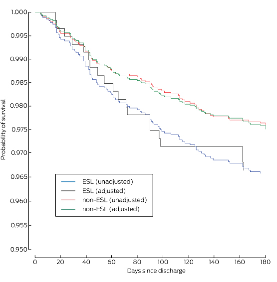
There was a higher incidence of heart failure, renal failure, stroke, recurrent myocardial infarction, major bleeding, and in-hospital mortality in the ESL group (Box 4). Independent predictors of in-hospital mortality included presentation in cardiogenic shock, cardiac arrest in hospital, in-hospital renal or cardiac failure, and having English as a second language (Box 5). At the 6-month follow-up, all-cause mortality was also higher in the ESL group (13.8% v 8.3%; P = 0.0001) (Box 6). ESL, age, in-hospital renal failure, and recurrent ischaemia were independent predictors of 6-month mortality (Box 7). The Cox regression model of survival at 6 months is outlined in Box 8.

Discussion

We found that mortality among patients presenting with an ACS who report English as their second language was higher than among those who report English as their first language. Other studies of Australian populations have not reported such a difference,7,8 but analyses of administrative data have compared populations according to standardised mortality rates, and did not specifically assess language as a marker of risk.

The incidence of diabetes mellitus, hypertension, dyslipidaemia and smoking was higher in our ESL group than among than those who used English as their first language. A systematic review2 found a higher incidence of smoking among male immigrants from the Middle East, the UK and Ireland, western and southern Europe, China, and Vietnam than among Australians of the same age. Another UK study suggested that people of South Asian origin had a higher incidence of diabetes mellitus, hypertension, dyslipidaemia and obesity than comparable Europeans,9 although the authors concluded that the measured metabolic risk factors did not entirely explain the overall differences in the incidence of coronary heart disease. Similarly, a study of Asian Indians living in Australia found a higher incidence of hypertension, diabetes mellitus and obesity than the national average.3 The proportion of patients from South Asia in our study was less than 2%, too small for us to identify this particular group as being at increased risk.

Several American studies have found longer delays to treatment for myocardial infarction for Hispanics than for non-Hispanic whites.1012 The median door-to-needle and door-to-balloon times were slightly longer for our ESL patients, but the differences were not statistically significant.

Although patients with ESL were more likely to be admitted to a metropolitan rather than a rural hospital, we identified a reduced rate of cardiac catheterisation in ESL patients presenting with an ACS and a much lower rate of PCI than for those with English as their first language. Similar findings have been reported for Hispanic migrants in the United States13 and migrants of South Asian background in the UK.14 This is thought to reflect not a physician bias in recommending patients for revascularisation,14 but rather patient treatment preferences15 and perhaps their lack of language skills.16,17 We did not collect data on why revascularisation was not performed, but ESL patients may have had more diffuse disease, not amenable to revascularisation. Lack of English comprehension can affect access to health education that promotes health maintenance (primary prevention), which may mean patients present later, at a more advanced stage of disease.18

An Australian study4 that examined hospital discharge data for the period between 1993–94 and 1997–98 compared outcomes using an English-speaking background (ESB) v non-English-speaking background (NESB) dichotomy, based on the patient’s country of birth rather than their actual capacity to speak English. It was found that NESB patients were more likely to be admitted for acute myocardial infarction than ESB patients, possibly because of a delay in diagnosis. This study found a longer average length of hospital stay for NESB than for ESB patients, which the authors attributed to complications associated with later presentation. We also found a slightly longer length of stay in our ESL group; this may reflect differences in hospital events, but may also reflect delays in organising professional interpreters for obtaining an accurate history and facilitating informed consent for procedures.

Deficiencies in intercultural communication may play a role in the adverse outcomes for patients from culturally and linguistically diverse backgrounds.19,20 A review of the literature on cultural differences in communication suggested that doctors behave less effectively when interacting with patients from ethnic minorities, and patients themselves are also less verbally expressive and seem to be less assertive and effective during medical encounters.4

Fewer patients in our ESL group were referred for cardiac rehabilitation after discharge than patients with English as their first language. Two systematic reviews of secondary prevention programs of randomised controlled trials have shown that either supervised exercise alone21,22 or risk factor education or counselling without an exercise component22 reduced mortality. It is therefore possible that reduced attendance at cardiac rehabilitation in the ESL group also contributed to poorer outcomes. Hospitals should extend the reach of cardiac rehabilitation services by offering multilingual opportunities for patients of different backgrounds.

Second-line antiplatelet agents, such as ticagrelor and prasugrel, were used less in our ESL group. This may be partially explained by fewer PCIs in this cohort and a higher incidence of major bleeding, but clinicians may also have been concerned that they could not accurately exclude contraindications of their use in ESL patients. Prasugrel23 and ticagrelor24 reduce major adverse ischaemic events in patients presenting with an ACS more effectively than clopidogrel, and the less frequent use of these agents in our ESL patients may have contributed to their poorer outcomes.

Limitations

The patients themselves identified whether English was their first or second language, and we have no data on their fluency or length of time in Australia, and this may affect patient care and understanding. Further, we did not collect data on the use of interpreters with our ESL patients. A professionally trained medical interpreter can provide a higher degree of accuracy and confidentiality than family members or bilingual staff members; using non-professional interpreters can be expedient, but has the disadvantage of loss of productivity and additional stress on the staff involved, together with the risk of inadequate or incomplete translation. It is therefore unknown whether the use of interpreter services was adequate, and whether improving their availability would enhance clinical outcomes.

While we attempted to adjust for baseline differences, there may have been unmeasured variables, such as socio-economic status and educational level, that may have confounded the impact of English proficiency on outcomes. Finally, we did not rigorously collect data on compliance with medications and physician follow-up, which may also have affected long term outcomes.25

Conclusion

In this large registry study of patients presenting with an ACS, in-hospital and 6-month mortality was greater for patients who reported English as their second language. While this may be explained by a higher prevalence of cardiovascular risk factors leading to more advanced disease, problems of culturally specific communication may also play important roles, and should be the subject of further research.

Box 1 –
Baseline demographics of the 6304 patients at 41 sites enrolled in the CONCORDANCE database

English as second language

English as first language

Difference (95% CI)

P


Number

1005

5299

Age [SD], years

63.3 [14.9]

65.3 [13.5]

–2.06 (–3.05 to –1.07)

< 0.001

Sex (men)

691 (68.8%)

3699 (69.8%)

–1% (–4% to 2%)

0.507

Diabetes

403 (40.1%)

1351 (25.5%)

15% (11% to 18%)

< 0.001

Hypertension

669 (66.6%)

3287 (62.0%)

5% (1% to 8%)

0.005

Dyslipidaemia

628 (62.5%)

2970 (56.0%)

6% (3% to 10%)

< 0.001

Current smoker

310 (30.8%)

1429 (27.0%)

4% (1% to 7%)

0.011

Median GRACE risk score [IQR]

124 [100.8–147.6]

125 [104.0–147.6]

–1.00 (–3.30 to 1.30)

0.400

Prior cerebrovascular accident

73 (7.3%)

430 (8.1%)

–1% (–3% to 1%)

0.396

Prior myocardial infarction

368 (36.6%)

1609 (30.4%)

6% (3% to 10%)

< 0.001

Prior heart failure

134 (13.3%)

488 (9.2%)

4% (2% to 6%)

< 0.001

Prior coronary artery bypass grafting

131 (13.0%)

681 (12.9%)

0% (–2% to 3%)

0.840

Prior percutaneous coronary intervention

201 (20.0%)

1125 (21.2%)

–1% (–4% to 1%)

0.380

Chronic renal failure

185 (18.4%)

411 (7.8%)

11% (8% to 13%)

< 0.001


GRACE = Global Registry of Acute Coronary Events.

Box 2 –
In-hospital interventions in 6304 patients at 41 sites enrolled in the CONCORDANCE database

English as second language

English as first language

Difference (95% CI)

P


Number

1005

5299

Cardiac catheter

714 (71.0%)

4003 (75.5%)

–5% (–8% to –2%)

0.002

Percutaneous coronary intervention

346 (34.4%)

2240 (42.3%)

–8% (–11% to –5%)

< 0.001

Coronary artery bypass grafting

89 (8.9%)

411 (7.8%)

1% (–1% to 3%)

0.234

Rehabilitation referral

495 (49.3%)

3120 (58.9%)

–9% (–13% to –6%)

< 0.001


Box 3 –
Discharge medications for 6304 patients at 41 sites enrolled in the CONCORDANCE database

English as second language

English as first language

Difference (95% CI)

P


Number

1005

5299

Aspirin

790 (85.9%)

4276 (85.0%)

1% (–2% to 3%)

0.917

Clopidogrel

497 (54.0%)

2596 (51.6%)

2% (–1% to 6%)

0.341

Ticagrelor or prasugrel

79 (8.6%)

627 (12.5%)

–4% (–6% to –2%)

< 0.001

β-Blockers

719 (78.1%)

3881 (77.1%)

1% (–2% to 4%)

0.733

Angiotensin-converting enzyme inhibitor or angiotensin receptor blocker

704 (76.5%)

3799 (75.5%)

–1% (–4% to 2%)

0.740

Statin

815 (88.6%)

4508 (89.6%)

–1% (–3% to 1%)

0.548


Box 4 –
In-hospital events for 6304 patients at 41 sites enrolled in the CONCORDANCE database

English as second language

English as first language

Difference (95% CI)

P


Number

1005

5299

Left ventricular failure

153 (15.2%)

444 (8.4%)

7% (5% to 9%)

< 0.001

Renal failure

103 (10.2%)

268 (5.1%)

5% (3% to 7%)

< 0.001

Myocardial infarction

41 (4.1%)

144 (2.7%)

1% (0 to 3%)

0.021

Stroke

12 (1.2%)

25 (0.5%)

1% (0 to 1%)

0.007

Major bleed

106 (10.5%)

441 (8.3%)

2% (0 to 4%)

0.020

Death

71 (7.1%)

200 (3.8%)

3% (2% to 5%)

< 0.001


Box 5 –
Logistic regression analysis of in-hospital mortality

Characteristic

Odds ratio

95% CI

P


English as first language

0.56

0.33–0.94

0.029

Cardiac arrest in hospital

16.95

10.53–27.78

< 0.001

Cardiogenic shock

7.04

4.46–11.11

< 0.001

GRACE score

1.04

1.03–1.05

< 0.001

Renal failure

5.08

3.29–7.81

< 0.001

Congestive failure

2.08

1.26–3.44

0.005


GRACE = Global Registry of Acute Coronary Events.

Box 6 –
Kaplan–Meier estimates of survival at 6 months for 6304 patients at 41 sites enrolled in the CONCORDANCE database*


ESL = English spoken as second language. * Adjusted for age, diagnosis, sex, previous stroke, chronic renal failure, chronic heart failure, diabetes, prior myocardial infarction, prior coronary artery bypass grafting and prior percutaneous coronary intervention.

Box 7 –
Logistic regression analysis of mortality to 6 months after discharge for 6304 patients at 41 sites enrolled in the CONCORDANCE database

Characteristic

Odds ratio

95% CI

P


English as first language

0.55

0.31–0.97

0.040

Age

1.09

1.07–1.11

< 0.001

Renal failure

4.67

2.88–7.58

< 0.001

Ischaemia

2.54

1.54–4.22

< 0.001


Box 8 –
Cox regression model of survival at 6 months for 6304 patients at 41 sites enrolled in the CONCORDANCE database

Characteristic

Hazard ratio

95% CI

P


Age

1.08

1.06–1.10

< 0.001

Diagnosis

3.96

3.22–4.70

< 0.001

Sex

0.86

0.42–1.30

0.502

Prior stroke/transient ischaemic attack

0.95

0.35–1.55

0.868

Chronic renal failure

0.43

0.05–0.90

< 0.001

Congestive heart failure

0.36

0.12–0.84

< 0.001

Diabetes

0.90

0.46–1.34

0.642

Prior myocardial infarction

0.84

0.36–1.33

0.489

Prior coronary artery bypass grafting

1.03

0.52–1.53

0.922

Prior percutaneous coronary intervention

1.08

0.57–1.59

0.764

Percutaneous coronary intervention

1.36

0.85–1.87

0.239

Coronary artery bypass grafting

8.00

6.01–9.98

0.040


Teledermatology and clinical photography: safeguarding patient privacy and mitigating medico-legal risk

Teledermatology is an innovative model of health care that has the potential to deliver significant benefits for both patients and medical practitioners. These include increased access to specialist services and reductions in travel times, waiting times and costs for patients, and reduced professional isolation and improved access to professional education for doctors.

“Store-and-forward” is a popular form of teledermatology. It involves capturing a still clinical image that is forwarded to a specialist, who later responds with an opinion on diagnosis and management. Teledermatology at the Princess Alexandra Hospital is a successful example of this model of care.1,2

As dermatology is a visually oriented specialty, using digital images for diagnosis is a natural fit. Numerous studies have already shown the diagnostic accuracy and reliability of store-and-forward teledermatology. More rapid diagnosis and initiation of treatment coupled with improved patient outcomes have also been demonstrated.1,3,4

The growth of store-and-forward services coincides with the increasing use of email, faster Internet speeds, the development of electronic health records and the advent of smartphones. Importantly, smartphones provide an accessible mode of capturing and transmitting patient images.

To date, at least two studies have surveyed the use of clinical photography in two separate Australian tertiary hospitals.5,6 Both showed that the use of personal smart phones for capturing clinical images was widespread. However, both studies also revealed inadequate privacy practices, including inconsistencies in the consent process, inappropriate disclosure of images to third parties and poor security practices when personal devices were used.

Mitigating the risks

In Australia, medical indemnity insurers have identified privacy as an emerging medico-legal risk for the profession.7 Breaches of privacy may result in legal action against and reputational damage for individuals and institutions.8,9 Individuals also risk being the subject of a complaint to the Australian Health Practitioner Regulation Agency (AHPRA), a health complaints entity, or an internal hospital investigation.7

In 2014, the Australian Medical Association (AMA) in conjunction with the Medical Indemnity Industry Association of Australia (MIIAA) released a guide for medical students and doctors for the use of clinical imaging and personal mobile devices.7 Its main recommendations include:

  • ensuring the patient understands the reasons for taking the image, how it will be used, and with whom it will be shared;

  • obtaining informed consent before taking a clinical image;

  • documenting the consent process in the patient record;

  • having controls on mobile devices to prevent unauthorised access; and

  • deleting clinical images from mobile devices after saving them to patients’ health records.7

These recommendations highlight key privacy practices for practitioners. However, the AMA and MIIAA advise that the guide should always be read in conjunction with any relevant legislation, and hospital policies and contracts related to clinical images and the use of personal mobile devices.7

Mitigating the risk of breaches of patients’ privacy by health practitioners begins with an awareness of their privacy obligations under the law, together with the privacy protocols of their employer organisations and professional indemnity insurers. Apart from explaining to the patients why an image is necessary, how it will be used, and who will see it when obtaining consent, the law also requires that health practitioners take reasonable steps to protect patient information (including images) from loss, disclosure, unauthorised access or misuse.8,9 Consider the following case scenario:

An elderly man presents to the emergency department of a regional base hospital with a new onset rash. The attending resident is not confident to make a diagnosis, but knows of a teledermatology service provided by a tertiary hospital in the nearest capital city. She asks the patient if she can “take some photos of the rash”. The patient agrees. She takes several photos of the patient with her personal smartphone and emails them, from her personal email address, to the on-call dermatology registrar at the tertiary hospital, requesting an opinion. The dermatology registrar reviews the images together with the patient’s history, and replies 2 hours later with a diagnosis of discoid eczema and a recommendation for management. Meanwhile, the dermatology registrar decides to use the patient images for a teaching session for the interns at the tertiary hospital.

Two weeks later the resident’s mobile phone is stolen. The phone had no security features enabled. The thief finds the patient’s images stored on the phone and uploads them to a public website. The thief also peruses the resident’s emails and finds details of the patient, and these are also shared publicly.

In the first instance, the resident should have made it clear to the patient that the purpose of taking the photo was to email the image to a specialist at another hospital to obtain an expert opinion on diagnosis and management. Further, notwithstanding the absence of a direct relationship with the patient, the teledermatology provider does not have consent to use the images for teaching, as the treating doctor did not obtain consent to use the images for this purpose.

Consent may be obtained orally or in writing, although practitioners should follow their institution’s guidelines. When consent is obtained orally, the consent process should be documented in the patient notes. Written consent must still be accompanied by a proper verbal explanation of the procedure.9

After the theft of her mobile phone in this case, the resident would be liable for a breach of privacy and confidentiality. She may also be in breach of the privacy and confidentiality obligations of her employment contract, as well as the conditions of her professional indemnity insurance. As a consequence, the resident could face disciplinary action from her employer or the Medical Board. The hospital may also be vicariously liable for the breach.9 Serious and repeated privacy breaches can attract substantial civil penalties for individuals and organisations under privacy legislation.

Technological precautions

The case above illustrates how easily a simple photograph can result in a costly legal dispute, not to mention untold harm to the patient. This situation could have been avoided if a number of simple precautions had been adopted.

An unlocked phone with patient photographs stored on it is a breach of privacy waiting to happen. Best practice dictates that patient images should be deleted from personal devices as soon as they are added to patients’ health records.7

As best practice may be overlooked during busy periods, security controls on personal smart phone devices, such as passcode locks, should be enabled to prevent unauthorised access. Installing remote locking or data wiping software to personal devices is recommended, as it will allow practitioners to delete data from their devices in the event of theft or loss.

Device settings should be adjusted so that clinical images on devices are not auto-uploaded to social media or back-up sites.7 Once a photo makes its way into the public domain via social media, it is very difficult to limit further sharing of that image.

The process of transmitting a patient image during the teledermatology consultation introduces an additional risk of a breach of privacy. Images should be transmitted via secure methods. Transmitting images through personal email or text messages is not considered secure by some sources, as such methods are typically not encrypted or password protected.10 Despite these concerns, the practice of emailing images is common in many hospitals. Practitioners should be familiar with the policies and systems of their institution or health service in relation to transmitting clinical images.

If a practitioner is required to send images by email or a text message to a colleague or specialist, they should send a test message first to ensure they have the correct email address or phone number for the intended recipient.7,10 Sending patient information or images to incorrect email addresses or phone numbers constitutes an automatic breach of privacy. Once sent, a practitioner has limited control over what the recipient does with that information. Ideally, one should encrypt or password-protect any images before transmitting them, although some may see this extra precaution as an inconvenience.

Like images, email or text messages that were part of the teledermatology consultation should be deleted from personal email accounts and devices after they have been uploaded to the patient record, as these may contain sensitive patient information that is still accessible if a device is lost or stolen. This includes emails saved in the “sent” folder. Automatic forwarding of emails to another email account should be disabled.

Practitioners should also consider the security of other devices that they use for sending emails. Computers and tablets with email management systems that allow email to be accessed on the desktop without logging in are a potential privacy risk. They should avoid sending patient images from an email address that is connected to an email management system. Practitioners should also implement automatic log off on computers and laptops at home and at work that are used to capture and store patient images, and change computer passwords regularly.

Where videoconferencing is being used, the room should be adequately sound-proofed and access should be restricted to those permitted according to the patient’s consent. Any videorecording of the consultation needs to be securely stored.

When consent for using an image for teaching or research purposes is appropriately obtained, best practice dictates that the image should be de-identified where possible, and must comply with relevant research or ethical guidelines.7 Identifying features such as birthmarks, tattoos, metadata, or even the condition itself, if it is rare, should be removed for such purposes.

Practitioner liability

Health care practitioners are personally responsible for patient information they choose to capture and transmit on their personal devices. Practitioners can limit their legal liability in the event of a breach of privacy by demonstrating that reasonable measures were taken to protect patient information. If a privacy breach does occur, clinicians should adopt an open disclosure approach, whereby the breach is notified and acted on. In these circumstances, practitioners should seek advice from hospital management and their medical defence organisations. Once appropriate legal advice has been sought, it may be necessary to inform the patient of the breach, and to explain and apologise.

Conclusions

It remains to be seen how health institutions and policy makers will tackle the issue of patient privacy in the new era of smart phones and teledermatology services. Fear of legal action should not preclude doctors from embracing novel approaches to health care that benefit patients and doctors. Rather, practitioners should take a few sensible precautions to reduce the likelihood of sensitive patient information falling into the hands of malicious third parties.

Before taking that next clinical photograph, individual practitioners should take a moment to review their own privacy practices and make any necessary adjustments. By doing so, practitioners can avoid the financial and emotional cost of a potential lawsuit in the future.

Female doctors in Australia are hitting glass ceilings – why?

Over the past 30 years, there have been some great achievements in gender equity. The number of women enrolled in professional degrees, such as law and medicine, rose from less than 25% in the 1970s to more than 50% in 2015. Australia has introduced a number of equal opportunity policies in health care and in 2000 achieved gender parity in medical schools.

Today, women are typically the dominant group within medical schools and yet remain under-represented in formal leadership positions and particular speciality areas. Although today there is greater female participation in medical roles, it still appears that women are hitting the glass ceiling.

Similar sorts of trends in gender participation are found in other countries such as the UK, Canada and the US. Given these broader trends, we could infer that these patterns are the result of “natural” processes related to the relative merits of the sexes.

Yet studies in Sweden show remarkably similar preferences for speciality areas across male and female medical students. Like Australia, these preferences have not typically translated into representation across the health workforce. This suggests there are forces in place that mean women do not go into their preferred roles.

Women in leadership

Despite the significant representation of women within the medical workforce, today fewer than 12.5% of hospitals with more than 1000 employees have a female chief executive. 28% of medical schools have female deans. 33% of state and federal chief medical officers or chief health officers are female.

In 1986, fewer than 16% of specialists were women. This rose to 34% in 2011.

While this is a substantial increase, women are also woefully underrepresented in these figures. There are also distinct gender patterns across specialist roles. Women’s participation is skewed towards pathology (58%), paediatrics (53%), obstetrics and gynaecology (49%) and underrepresented in orthopaedic surgery (6%), vascular surgery (11%) and cardiothoracic surgery (12%).

A popular explanation for these patterns is that there is a lag phenomenon at play. Once current women advance through their career, figures will self-correct and result in more gender balance in the system.

A more pessimistic view (and one we would subscribe to) is that women are being channelled into particular areas of the profession that are lower status and attract lower pay, while more high-profile roles remain in the hands of men.

This is not to say that male doctors are (all) actively working to keep women excluded from these roles. There are a range of reasons for the barriers around perceptions of capability, capacity and credibility.

Capability, capacity, credibility

The evidence suggests that women are as (and possibly more in some cases) intellectually capable of the high-profile roles that they are poorly represented in.

Our research found that some women may lack self-confidence or doubt their ability to undertake certain roles. What this means is that women may be less willing to self-promote or to put themselves forward for positions traditionally held by men.

Women are more likely than men to have caring responsibilities, which can have implications for perceived capacity. Juggling leadership or the long hours associated with some speciality areas with motherhood can be a challenge.

Many of the areas where women are poorly represented in offer limited options for flexible ways of working and cultivating work-life balance. Some speciality areas have additional years of training which, again, can make them difficult to access for some women.

Perceived credibility is a further barrier, with women not being taken seriously as leaders or surgeons – roles typically associated with males. Sociology has a long tradition of scholarship arguing that organisations and professions are highly gendered and valorise masculine values.

Where work environments are heavily gendered, they can be alienating for some women. Some speciality areas are traditionally considered to be highly male (think surgery) in a way that paediatrics or palliative care may be less so.

These barriers are not simply externally imposed on women by men and may be internalised in women through the broader culture and values of organisations. Internalised beliefs about the traits and qualities required for particular roles can dissuade some women from actively seeking out these roles, unless they received mentoring and support from others.

At an interpersonal level, unconscious biases, sexist micro-aggressions, and a “club culture” contribute to a hostile environment for women within some health-care settings. At a structural level, conservative social norms and male-dominated career pathways can make it difficult for women to balance the pressures and demands of maternity leave, child-rearing, care-giving and running a household with leadership roles.

What is clear is that with such a broad range of barriers, there will be no easy or quick solutions. If we are to successfully smash these glass ceilings then solutions will need to be structural as well as cultural. Women will be unable to overcome these issues alone and solutions will need to be both multifaceted and supported through a broad base.

The Conversation

Helen Dickinson, Associate Professor, Public Governance, University of Melbourne and Marie Bismark, Senior Research Fellow, Public Health Law, University of Melbourne

This article was originally published on The Conversation. Read the original article.

Other doctorportal blogs

Season’s Greetings from doctorportal

Here at doctorportal, we’d like to wish you a Merry Christmas and thank you for all your support this year.

Since our launch in May, we have gradually been building doctorportal to be a useful tool for Australian medical professionals.

We have an array of  news sources including 2 Minute Medicine and AAP, so you can find out the health news your patients are reading. You also have an opportunity to write for us in our blog section, giving you a chance to have your voice heard.

We have grown our events page, feature the latest Australian medical conferences. Please contact us if you’d like your event to be covered.

Many of you have searched for your personal listing in the ‘Find the Doctor‘ tool, a national doctor search service to find colleagues for referrals.

Our CPD tracker has been particularly popular, giving you the opportunity to earn CPD points and track all your learning in one place.

Most recently we have launched our Discussions platform, a place where you can debate the clinical aspects of the MBS review with your colleagues.

It’s been a big year at doctorportal and we will soon be taking a few weeks off, however we are looking forward to coming back in 2016 with more exciting launches and products.

We wish you all a wonderful, restful and safe Christmas and a Happy New Year!

The Value of Audio Devices in the Endoscopy Room (VADER) study: a randomised controlled trial

Music has been used in medical therapy and healing since Ancient Greek times.1 Its use in the operating theatre has been longstanding, with reports of its potential therapeutic effects dating back to the 1940s.2 Music therapy before and during an operation has been shown to reduce anaesthetic requirement and patient anxiety,3 and improve overall satisfaction.4 There is also evidence that proceduralist-selected music improves surgical skill acquisition.5

In gastrointestinal endoscopy, music has shown benefits for patients;614 however, there are no data on the effects of music on improving the performance of endoscopists and on whether a specific style of music would lead to optimal performance. This is particularly relevant in an era where high volumes of endoscopic procedures are performed in an often stressful clinical environment, and where procedural outcomes, such as adenoma detection rate (ADR) in colonoscopy, are reported as validated benchmarks of quality endoscopy.

The appropriate style of music to use and avoid in the operating theatre has been previously proposed,15 but music from epic movies was absent from this list. The soundtracks from such movies often contain uplifting musical tracks associated with glory, success and large-scale victory. Given we are avid fans of the Star Wars movies (Lucasfilm Ltd), and with the imminent release of the latest instalment, Star Wars: The Force Awakens, we felt it would be important and timely to evaluate the effect of music from the Star Wars soundtrack in colonoscopy. Therefore, we hypothesised that Star Wars music (SWM) would be superior to endoscopist-selected popular music (PM) when measuring quality outcomes in colonoscopy.

Materials and methods

In a hospital far, far away (Melbourne, Australia), patients were recruited from endoscopy lists from a single quaternary-centre gastroenterology unit between June and August 2015. All human subjects undergoing colonoscopy for any indication were included in the study. Ewoks and Wookies were excluded from the study owing to their inability to provide informed consent, but fortunately none were identified on the colonoscopy lists. The Austin Health Human Research Ethics Committee approved the project. The Jedi Council could not be contacted despite numerous attempts — clouded their contact details were.

Bowel preparation was performed using three sachets of sodium picosulfate for healthy patients and a volume-reduced regimen for patients with substantial comorbidities. A split-dose bowel preparation was used for afternoon procedures. All colonoscopies were performed under anaesthetist-administered monitored sedation with intravenous propofol. An Olympus 190-Series colonoscope (Olympus Medical Systems) was used in all cases. ProVation MD (ProVation Medical) was used for procedure documentation and coding.

Randomisation was performed using a coin toss by the endoscopist before colonoscopy to determine which music would be played: heads for SWM; tails for endoscopist-selected PM. SWM included the soundtrack from Star Wars: Episode III Revenge of the Sith, of which “Battle of the Heroes” (composed by John Williams) was the most commonly played track. The music was played through a personal computer in the endoscopy suite at a volume of between about 40 and 70 decibels, depending on the tolerance of the other endoscopy staff, and began before commencement of the colonoscopy. Patients were blinded to the music type being used.

Five endoscopists participated in the study: one consultant with 10 years’ experience (Jedi Master) and four specialist registrars with 1–2 years’ experience (Padawans). An independent member of the endoscopy team kept timings of procedures, and endoscopists were blinded to these timings. Timing commenced when the scope entered the anus (outer rim) and continued until the scope was withdrawn from the colon. The Padawans were supervised by a Jedi Master endoscopist for each procedure and advised to use the Force, and not force, during the colonoscopy.

Patient demographics and the presence of diverticular disease were recorded. The quality of bowel preparation was assessed by the endoscopist using qualitative descriptors (“excellent”, “good”, “fair” or “poor”). Primary end points were the procedure time, polyp detection rate (PDR) and adenoma detection rate (ADR). The PDR was defined as the number of colonoscopies in which one or more polyps were removed, divided by the total number of colonoscopies performed. The ADR was defined as the number of colonoscopies in which one or more adenomas were removed, divided by the total number of colonoscopies. A secondary end point was the recently proposed quality measure, adenomas per colonoscopy (APC), defined as the total number of adenomas detected divided by the number of colonoscopies.16

Data were analysed using the statistical software GraphPad Prism, version 6 (GraphPad Software, Inc). Non-parametric data comparisons between the two groups used Mann–Whitney tests to determine differences in procedure time and PDR. P ≤ 0.05 was used as the statistical significance level.

Results

Box 1 details the patient characteristics and colonoscopy outcomes. One hundred and three colonoscopies were included in the analysis: 58 in the SWM group and 45 in the PM group. A similar number of colonoscopies were performed by the Jedi Master in both groups (P = 0.45). Mean ages were 64 and 66 years (P = 0.13); 62% and 51% of patients in the SWM and PM groups, respectively, were men (P = 0.055). The most common indications for colonoscopy were positive faecal occult blood test, anaemia for investigation, and a personal history of polyps. Colonoscopies for bowel cancer screening and in patients with a history of polyps were evenly distributed between the two groups (P = 0.18).

Bowel preparation was assessed as good or excellent in 57% of the SWM group compared with 69% in the PM group (P < 0.01). Caecal intubation rates were high in both groups (98% and 97%, SWM and PM groups, respectively). The procedure duration was similar in both groups, with total procedure times of 20 minutes and 22 minutes and withdrawal times of 14 minutes in both groups (P = 0.87) in the SWM and PM groups, respectively.

The PDR was significantly higher in the SWM group than in the PM group (60% v 35%; P = 0.006). Similarly, the ADR was significantly higher in the SWM group than the PM group (48% v 27%; P = 0.01). The APC in the SWM group was 84% compared with 35% in the PM group (P = 0.01).

Discussion

Colonoscopy is a key intervention to prevent bowel cancer.17 With an increasing uptake of bowel cancer screening, demand on colonoscopy services is high. Of note, individuals from the baby boomer generation, many of whom were fans of the original Star Wars trilogy, are approaching the appropriate age to undergo bowel cancer screening. It is therefore essential that strategies to enhance quality outcomes in colonoscopy are evaluated. Environmental considerations are important, and music has been shown to improve a proceduralist’s skills. Our prospective, single-centre randomised controlled trial study supports the positive effects of music in procedures, and demonstrated a higher PDR and ADR with SWM compared with PM during colonoscopy.

Higher PDRs and ADRs were achieved in the SWM group compared with the PM group, despite a similar procedure time and a lower rate of optimal bowel preparation. It would be expected that a poorer quality of bowel preparation would lead to a lower PDR and ADR and prolonged procedure duration. Poor preparation leads to missed lesions. Missed lesions lead to interval cancers. Interval cancers may have serious consequences for the affected patients. SWM may break this cycle. Our finding that the PDR and ADR were higher in the SWM group may relate to the heightened observation skills of the endoscopist in a conducive musical environment. Proceduralist-selected music, which was used in both groups of our study, has been found to induce neurohormonal and immune system changes that lead to improved procedural performance;18 however, this does not explain the differences observed between the groups. As Star Wars fans, we must suspect that the Force was also involved, enhancing our Force-sensitive abilities to detect polyps, despite an overall more hostile luminal environment. Akin to Luke Skywalker’s destruction of the Death Star in Star Wars: Episode IV A New Hope, despite being pursued by Darth Vader and TIE fighters, we trusted the Force to guide us through the murky colonic waters to locate and destroy polyps.

There are some limitations in our study. Despite being randomised, there was a non-significant trend towards a greater proportion of men in the SWM group compared with the PM group. Also, there were disproportionate numbers in the two groups. Both may be explained by flaws in randomisation; however, other key parameters known to influence PDR and ADR, such as being referred for colonoscopy after receiving a positive faecal occult blood test result from participation in the National Bowel Cancer Screening Program, were distributed evenly. Differences by sex between the groups were unlikely to account for the almost twofold increase in ADR in the SWM group compared with the PM group. Given that the Star Wars fan base is predominantly male and there were more males randomised to the SWM group, it is again plausible that the Force was involved — influence the coin toss it may have.

Without concealment allocation, it could be argued that the higher PDRs and ADRs in the SWM group were due to performance bias. However, the ADRs in the PM and SWM groups were at the expected level for the study population,1922 so it is unlikely that deliberate underperformance in the PM group led to the observed difference in PDRs and ADRs between the groups. Additionally, the attainment of at least a 90% caecal intubation rate and an ADR of 20% was achieved in both groups and is in keeping with the current recommended guidelines for quality colonoscopy.23

It is unclear from this study if the improved endoscopic outcomes are specific to SWM or can be generalised to other epic movie soundtracks. A future study could, for example, assess the impact of music from the Lord of the Rings on endoscopic parameters. Of course, other non-Star Wars soundtracks would not be able to harness the power of the Force. If the Force is responsible for the superior endoscopic outcomes demonstrated in this, the addition of other Star Wars paraphernalia to the endoscopy suite may be of additive value. The wearing of Star Wars costumes may become a standard of practice in the future (Box 2).

In conclusion, SWM improves PDR and ADR in colonoscopy despite poorer quality bowel preparation when compared with PM. We recommend the widespread use of SWM in endoscopy rooms to improve quality outcomes. May the Force be with you.

Box 1 –
Patient characteristics and colonoscopy outcomes

Variable

Popular music (n = 45)

Star Wars music (n = 58)

P


Sex, men

23

36

0.055

Mean age, years (range)

63.8 (22–88)

65.5 (23–93)

0.13

Indications

Anaemia

6

18

National bowel colon cancer screening

19

21

Follow-up

Polyp

5

5

Cancer

1

0

Constipation

1

4

Diarrhoea

2

3

Pain

3

2

Perirectal bleeding

3

0

Weight loss

0

1

Inflammatory bowel disease surveillance

2

2

Other

3

2

Preparation quality

Excellent

1

1

Good

30

32

Fair

12

20

Poor

2

5

Good or excellent quality (%)

31/45 (69%)

33/58 (57%)

< 0.01

Procedures carried out by:

Consultant (Jedi Master)

22

28

0.45

Registrar (Padawan)

23

30

Extent

Caecum

13

22

0.016

Terminal ileum

31

35

0.23

Insertion time, min

Mean (SD)

8.0 (7.6)

6.8 (5.8)

0.056

Median

6

6

Range

2–42

1–35

Withdrawal time, min

Mean (SD)

13.7 (8.6)

14.0 (8.0)

0.87

Median

12

12

Range

6–50

6–52

Polyp detection rate (%)

16/45 (36%)

35/58 (60%)

0.006

Second polyp detection rate (%)

7/16 (43%)

20/35 (57%)

0.19

Polyp retrieval rate (%)

15/16 (93%)

32/35 (91%)

Adenoma detection rate (%)

12/45 (27%)

28/58 (48%)

0.01

Adenoma per colonoscopy (%)

16/45 (35%)

49/58 (84%)

0.01

Total adenomas

16

49

Diverticulosis (%)

23/45 (51%)

16/58 (27%)

0.007


∗Data are the number of patients unless otherwise indicated. †There was one aborted procedure in each group due to poor bowel preparation.

Box 2 –
Artist’s impression of the future standard practice in endoscopy

Mondayitis

A little-known condition — and there is much work to be done

Mondayitis is a widely known and discussed condition in popular culture. Searching for “Mondayitis” on PubMed, however, will not return any positive results (as of Sunday 6 September 2015). We hope to address this gap in knowledge by proposing a number of possible hypotheses as to its cause, and so assist a keen researcher in directing his or her energies in the most fruitful direction for further exploration.

Definition

In the absence of a formal medical definition, one must be forgiven for resorting to “Doctor Google” for the popularly accepted meaning. It leads us to a number of definitions all of which emphasise that the symptoms, consisting of apathy and lethargy, are seen on Monday. We believe that this definition requires broadening as it unjustly excludes sufferers who do not work a regular Monday to Friday working week. Hence, we would like to propose a more generalised definition: a systemic illness with a non-specific constellation of symptoms including fatigue, lethargy or asthenia, dysthymia, irritability, light-headedness, photophobia, dry mouth, myalgia, and headache in the absence of another focal or systemic illness. These symptoms commonly manifest as an overwhelming desire to skip work or, if sufficiently mild to allow attendance at work, in a dishevelled appearance and feeling poorly. Symptoms typically occur on the first working day after a period of time off work, which may be as short as 2 days (“the weekend”) or longer (“the long weekend” or “a holiday”).

Pathophysiology

While there are no studies examining this topic, there would be a general agreement with Urban Dictionary’s assertion that “this feeling [of general distress] is usually enhanced after a large weekend”.1 While exploring the popular definition and threshold of “a large weekend” is beyond the scope of this article, we believe that it would not be unreasonable to assert that it involves a disruption to the usual sleep pattern, consumption of more food and beverages (alcoholic or otherwise) than usual and partaking in extra-occupational and extra-familial social engagements. Based on the above assumptions, we propose a number of possible pathophysiological processes causing Mondayitis.

1 Direct effect of alcohol

Mondayitis could be a version of hangover, a well recognised consequence of alcohol consumption. This may relate to toxic metabolite (mostly acetaldehyde) accumulation, hormonal changes, or dehydration and electrolyte imbalance from alcohol-induced diuresis. This topic is expounded in more detail in Penning and colleagues’ excellent review.2

2 Metabolic derangement, including accumulation of toxic metabolites

Penning’s review also covers the effects of metabolic derangement, particularly acidosis, in the setting of alcohol consumption resulting in hangover-like symptoms.2 In addition, many food items consumed over a “long weekend”, particularly those that are sweet, salty or fatty, may also have a role in creating and sustaining the metabolic derangement and causing the detrimental symptoms of Mondayitis.

3 Transient systemic inflammation

The symptoms of Mondayitis are also consistent with excess cytokine production and a generalised pro-inflammatory state. This state could be triggered, for example, by a viral infection acquired during social engagements in the periods leading up to Monday. Further epidemiological studies are needed to confirm this hypothesis, particularly since the causative virus may not yet have been identified.

4 Reduced sleep and the jet lag effect

Disruption to normal circadian rhythm is known to cause fatigue, reduced concentration, headache and malaise.3 This effect, when associated with the change in time zone after a long-haul flight, is known as jet lag and is widely accepted by the medical and the general community. It is not impossible that alterations to the normal sleep pattern imposed by extra socialisation and “having fun” during the “large weekend” disrupt the normal circadian rhythm, resulting in the same clinical picture as jet lag. If this was indeed the case, we believe that the term “Mondayitis” should be reserved exclusively for the constellation of such symptoms that occur without changing time zones by flying on a jet plane.

5 Withdrawal from an environmental agent

The symptoms of Mondayitis typically resolve by the afternoon of the first working day. This may be because the symptoms are due to substance withdrawal, occurring over 2–3 days, from an environmental agent only found at the workplace, or perhaps even the actual work itself. Admittedly, this seems unlikely and some readers may find this suggestion incredulous and even upsetting. But currently, there is nothing to preclude this possibility and only further research will exclude this hypothesis.

Conclusion

There are many possible causes of Mondayitis. Our suspicion is that Mondayitis has multiple contributing pathophysiological processes, with the exact manifestation determined by the genetic variations in each individual. What is clear, however, is that we know very little about this condition and that there is much work to be done. While Mondayitis may not be a life-threatening disorder, it is a common disease responsible for a significant amount of misery and, as professionals dedicated to alleviating human suffering, we cannot, and must not, continue to neglect this condition.

Americans shooting themselves in the foot: the epidemiology of podiatric self-inflicted gunshot wounds in the United States

The United States is home to about one third of all firearms worldwide, with 90 guns for every 100 American citizens.1 It is therefore perhaps not surprising that gunshot wounds (GSWs) are among the leading causes of injury in the US.2,3 The statistics indicate that 93% of the wounded are men, 56% are unemployed, and 56% tested positive for drugs or alcohol after the incident.46 As the incidence of GSWs is increasing, epidemiological studies that provide insight into their general nature and the circumstances in which they occur are useful for developing preventive education. Further, an understanding of terminal ballistics is important for determining the appropriate clinical management of GSWs.

The extent of injury inflicted by a GSW is determined by the energy of the primary projectile, its dissipation in the tissue, and the generation of secondary projectiles following osseous injury. The kinetic energy of a bullet before impact is equal to half its mass multiplied by its velocity squared; the energy of a projectile thus increases exponentially with its velocity. In order to maximise mass (and minimise energy loss caused by air resistance), bullets are often made with pointed or rounded tips from metals with a high specific gravity, such as lead. The energy transferred to the tissue after impact is the difference in kinetic energy of the bullet as it enters and leaves the tissue. This difference is dependent on the bullet’s diameter on impact and the density of the tissue. The more a bullet deforms or mushrooms on impact, the greater the amount of energy transferred to the tissue.7,8

High-velocity projectiles create large temporary cavities that fill with water vapour, causing tissue damage and wound contamination distal to the primary tract of the bullet. When a bullet collides with a dense object, such as bone, secondary missiles may be generated, the number of which increases with the velocity of the bullet.9 These secondary missiles have less predictable trajectories and often do more soft tissue damage than the primary projectile. The velocity of the bullet is thus a primary determinant of tissue damage.

The foot has a number of anatomical and biomechanical features that make it unique in terms of GSW injury and management. The function of the foot depends on its ability to painlessly and efficiently transfer the energy generated by the leg muscles into locomotion. Unlike long bone and other joint injuries, low-velocity GSWs to the foot often result in significant morbidity, and are managed in the same manner as high-velocity injuries. The ratio of bone to soft tissue in the foot is high, with a particularly large number of articular surfaces. More than 80% of GSWs to the foot result in osseous injury,10 and such fractures frequently generate secondary projectiles that damage the densely populated neurovascular structures. The resulting inflammation and haemorrhage within the restricted fascial compartments of the foot predispose to compartment syndrome and other complications.

Management includes antibiotic therapy, operative debridement, bone stabilisation, revascularisation and soft tissue coverage. Low-velocity GSWs to the foot have traditionally been treated with intravenous antibiotic therapy for 1–5 days,1114 followed by operative assessment of soft tissue contamination and irrigation. However, it is now generally accepted that both low- and high-velocity injuries require careful debridement of non-viable soft tissue and non-essential osseous fragments to prevent necrosis and wound infections.1320 As mentioned earlier, the vast majority of GSWs to the foot involve intra-articular osseous injury. Even low-velocity injuries may require both internal and external percutaneous fixation of fractures to achieve adequate alignment.21 High-velocity injuries are often allowed to heal by secondary intention, while others may require wound closure with myocutaneous flaps, skin grafting or, in extreme cases, amputation.

Until now there has been no large-scale epidemiological examination of the injury characteristics and circumstance of GSWs to the foot. Given the anatomical and biomechanical features of the foot, these GSWs are unique in their presentation, and, while they have been studied on a case-by-case basis,2224 the overarching trends of self-inflicted GSWs to the foot have not been investigated in a large sample. We undertook a large-scale epidemiological examination of Americans who had shot themselves in the foot.

Methods

Study sample

Using a stratified probability sample of all US hospitals with more than six beds that provide 24-hour accident and emergency services, the National Electronic Injury Surveillance System (NEISS) collected data for the period 1993–2010 as part of the Firearm Injury Surveillance Study. Based on the number of emergency department visits per year, hospitals were stratified as very large, large, medium or small. An additional stratum for children’s hospitals was also used. Between 1993 and 1996, 91 emergency departments were included in the sample. An additional 10 hospitals were added between 1997 and 1999, with two dropouts between 2000 and 2002; 99 hospitals were included in the sampling frame 2002–2010.

Data collection

NEISS, the primary data collection body for the Consumer Product Safety Commission, was responsible for data collection. Data on initial emergency department visits that resulted from non-fatal firearm-related injuries were extracted from the patients’ medical records.

Outcomes

The characteristics of the patients and the conditions in which each sustained self-inflicted GSWs to the foot were the primary outcomes.

Statistical analysis

All statistical analysis was conducted in Stata 12 (StataCorp). Participants were identified as either generic firearm victims or patients who had a self-inflicted GSW to the foot. χ2 tests were used to compare the categorical variables of groups; ie, sex, age group, marital status, illicit drug use, involvement in criminal activities, weapon used, location of incident, and diagnosis. Logistic regression was undertaken for sex and marital status (married v not married).

Results

Of the 69 111 reported firearm-related injuries, 667 (1.0%) were self-inflicted GSWs to the foot. Individuals who shot themselves in the foot were typically men (597, 89.6%) aged 15–34 years (345, 51.7%). Incidents generally occurred in the home (381, 51.1%), involved a handgun (208, 31.2%) or BB gun (228, 64.2%) while the individual was neither committing a crime nor under the influence of alcohol. Significant differences between individuals who shot themselves in the foot and those who had other firearm-related injuries with respect to sex (χ2 = 3.19, P = 0.048), age group (χ2 = 116.39, P < 0.0001), marital status (χ2 = 87.18, P < 0.0001), illicit drug use (χ2 = 24.49, P < 0.0001), involvement in criminal activities (χ2 = 330.79, P < 0.0001), weapon used (χ2 = 457.56, P < 0.0001), location of the incident (χ2 = 571.16, P < 0.0001) and the physician’s diagnosis (χ2 = 273.18, P < 0.0001) were noted (Box 1).

Logistic regression indicated that individuals who shot themselves in the foot were significantly more likely than individuals with other firearm-related injuries to be male (odds ratio [OR], 1.28; 95% CI, 1.0–1.7) and married (OR, 2.6; 95% CI, 2.1–3.4).

Incidences of shooting oneself in the foot were most common in October, November and December (Box 2).

Discussion

Contrary to popular belief, incidents of Americans shooting themselves in the foot are relatively rare; the characteristics of these incidents, however, are unique. When these auto-foot shooters were compared with individuals who had sustained other firearm-related injuries, significant differences were noted in the demographic characteristics of the victim/assailant, weapon of choice, the circumstances of the incident, and the nature of the injury itself.

There are several limitations that must be acknowledged in the interpretation of these data. The study included only individuals who presented to US emergency departments with non-fatal firearm-related injuries, so that our comparisons cannot be generalised to the broader population. The primary source of most data was the individual who had shot themselves in the foot; while data about the injury were provided by health care professionals, information about the incident itself may be subject to self-report biases. A social desirability bias may have caused under-reporting of self-inflicted wounds, as individuals who shoot themselves in the foot may not be entirely forthcoming about the nature and cause of their injuries.

Never-married men between the ages of 15 and 34 years were the most common perpetrators of self-inflicted GSWs to the foot. Due to the disproportionate number of males who possess firearms, it is to be expected that the prevalence of these injuries would be higher among men. Of interest, however, was the strength of the relationship between being married and shooting oneself in the foot when compared with the odds of other firearms-related injuries and non-married self-saboteurs. These results are consistent with anecdotal reports from disgruntled spouses and depictions of married men in the mainstream media, such as sitcoms and reality television programs. However, due to the nature of our data, evidence-based generalisations to the broader American population cannot conclusively be made.

Shooting oneself in the foot was extremely uncommon during the commission of a crime or while under the influence of drugs; only five individuals shot themselves in the foot while committing a crime. Given that inhibitions are reduced and cognitive capacity diminished by drug use, it is a somewhat counterintuitive finding that the association of illicit drug use with shooting one’s foot was not stronger. An investigation into the relationship between alcohol use and self-inflicted podiatric injuries is an area for future research, given the ease of access to alcohol and the prevalence of alcohol use in other firearm-related injuries.

There was a strong positive correlation between the month of the year and the number of self-inflicted GSWs to the foot; a disproportionate number of incidents occurred in October, November and December. It is notable that these trends were much stronger than for other firearm-related incidents, with a relatively constant number of these incidents throughout the year.

The epidemiology of firearm-related podiatric trauma has been neglected until now; to our knowledge, ours is the first large-scale epidemiological investigation of GSWs to the foot or of self-inflicted GSWs to the foot. Given the anatomical and biomechanical features of the foot, the nature of the wounds caused by GSWs is unique. A major epidemiological study is required to examine overarching trends in the circumstances and scenarios in which these events occur. Although it may not be possible to prevent Americans from shooting themselves in the foot, large-scale investigations of the nature of these incidents provides invaluable information for those at greatest risk. Particular caution must be taken during the festive season if one is to avoid being caught under the missing toe.

Box 1 –
Characteristics of self-inflicted gunshot wounds to the foot

Self-inflicted gunshot wounds to the foot


Other firearm-related injuries


P*

Number

Percentage

Number

Percentage


Number

667

1.0%

68 444

99.0%

Demographics

Sex (male)

597

89.6%

59 562

87.0%

0.048

Age

0–14 years

158

23.7%

7 691

11.2%

< 0.0001

15–34 years

345

51.7%

45 286

66.2%

35–54 years

134

20.1%

12 109

17.7%

≥ 55 years

30

4.5%

2 843

4.2%

Marital status

Married

103

24.0%

6 117

11.6%

< 0.0001

Never married

154

35.9%

26 826

51.0%

Divorced or separated

21

4.9%

1 262

2.4%

Other

8

1.6%

635

1.2%

Not stated

143

33.3%

17 760

33.8%

The incident

Drugs involved

11

1.7%

1 644

2.4%

< 0.0001

Crime involved

5

1.0%

11 680

17.1%

< 0.0001

Weapon

Handgun

208

31.2%

19 002

27.8%

< 0.0001

Rifle

80

12.0%

3 169

4.6%

Shotgun

60

9.0%

2 697

3.9%

BB gun

228

64.2%

10 094

14.8%

Unknown

91

13.6%

33 482

48.9%

Location

Home

381

57.1%

14 661

21.4%

< 0.0001

Farm

5

0.8%

109

0.2%

Apartment or condominium

1

0.2%

75

0.1%

Street or highway

12

1.8%

14 049

20.5%

Other public area

24

3.6%

7 500

11.0%

Mobile home

1

0.2%

112

0.2%

School

1

1.0%

276

0.4%

Recreational area

18

18.0%

1 140

1.7%

Unknown

224

33.6%

30 473

44.5%

The injury

Diagnosis

Amputation

2

0.3%

128

0.2%

< 0.0001

Contusion or abrasion

7

1.1%

3 936

5.8%

Foreign body

164

24.6%

7 289

10.7%

Fracture

75

11.2%

3 738

5.5%

Laceration

25

3.8%

7 818

11.4%

Puncture

265

39.7%

24 317

35.5%

Avulsion

1

0.2%

99

0.1%

Other

128

19.2%

16 680

24.4%


∗Self-inflicted wounds v other firearm-related wounds. †Number of injuries and percentage of all firearm-related injuries. All other percentages in the table are column percentages.

Box 2 –
Gunshot wounds in the United States, by month; expressed as a percentage of all foot- or non-foot-related incidents