×

Acute HIV infection presenting as erythema multiforme in a 45-year-old heterosexual man

Clinical record

A 45-year-old heterosexual man of European descent presented to our hospital with a 3-day history of fever, myalgia, headache and a macular papular rash. The rash originated on his left shoulder and anterior chest wall before extending to chest, back and abdomen. The rash also involved the palmar aspect of his hands and the plantar aspect of his feet.

He had a background history of dyslipidaemia, excessive alcohol consumption, gastro-oesophageal reflux disease, recurrent pancreatitis and gout. He had been taking statin medications, esomeprazole, allopurinol and creon for several years. There had been no new complementary medicines.

On arrival in hospital, his heart rate was 80 beats/min; respiratory rate, 16 breaths/min; blood pressure, 140/88 mmHg; and his temperature was 37.6°C. Cardiovascular, respiratory and abdominal examinations were otherwise unremarkable. Inguinal lymphadenopathy was noted on examination. A skin examination revealed an extensive macular papular rash affecting his face, trunk and limbs, with an erosion noted on his soft palate.

Investigations revealed low levels of haemoglobin (124 g/L; reference interval [RI], 135–175 g/L), white blood cells (2.4 × 109/L; RI, 4.0–11.0 × 109/L) and lymphocytes (0.62 × 109/L (1.50–3.50 × 109/L). His platelet count was normal (192 × 109/L; RI, 150–450 × 109/L), and his C-reactive protein level was slightly elevated (8.1 mg/L; RI, < 8.0 mg/L).

The patient reported having unprotected sex with one new female partner in the previous 3 months. On advice from the hospital’s infectious diseases team, molecular testing for measles and serological testing for syphilis and HIV were performed. The patient was discharged home after 24 hours of observation with investigations pending. The day after discharge, an HIV enzyme immunoassay (EIA) screen was reactive but western blot was negative. The patient was recalled for further HIV testing, which revealed an HIV viral load of 1 060 000 copies/mL.

The patient was reviewed in the hospital’s infectious diseases clinic 4 days later and was found to have a persisting generalised pruritic papular rash of urticarial appearance involving his trunk and proximal limbs, including his elbows and knees (Figure 1). Target lesions typical of erythema multiforme (EM) were noted on the plantar aspects of his feet, appearing red and blue centrally, with tense oedema surrounding the pale area and a well defined erythematous peripheral margin (Figure 2). A dermatologist’s opinion was sought, who agreed on a diagnosis of EM secondary to acute HIV infection.

The patient’s rash was treated with topical steroids and oral antihistamines and abated within 1 week. Follow-up serological testing revealed a rising HIV EIA titre and positive western blot.


Figure 1. Papular rash on the patient’s trunk and arm. Figure 2. Target lesions on the plantar aspects of the feet.

An acute, self-limiting hypersensitivity mucocutaneous reaction pattern, EM is frequently associated with viral infections, most often herpes simplex, and Mycoplasma pneumoniae infection. EM can also arise due to drug reactions, most commonly non-steroidal anti-inflammatory drugs, penicillins, sulfonamides, phenothiazines and anticonvulsants.1

The incidence of EM is unclear, but it is thought to affect less than 1% of the population, with a slight predominance among young women.1 EM is characterised by targetoid lesions distributed in peripheral acral regions, and macular, papular and urticarial patterns peripherally and on the extensor surfaces, and, less frequently, diffusely on the trunk. Oral and genital lesions are generally present. Typical target lesions are most frequently seen in acral locations, especially the dorsal and palmar surfaces of the hands and the dorsal and plantar surfaces of the feet.

Acute HIV infection generally occurs within 4–10 weeks from the time of HIV exposure, with 95% of patients seroconverting within 6 months.24 Acute HIV infection is associated with a maculopapular rash in up to 80% of cases, which presents 48–72 hours after the onset of fever and typically affects the upper trunk and neck. Acute HIV is also commonly associated with headache, lymphadenopathy and myalgia.5,6 Vesicular, pustular exanthematous and enanthematous patterns have also been described.7 However, EM in acute HIV infection is considered extremely rare and we are aware of only three reports.5,8,9

The pathogenesis of EM has largely been derived from studies of herpes simplex virus. The mechanism for mucocutaneous lesions is thought to begin with the release of viral DNA into the blood. DNA fragments are phagocytosed by mononuclear CD34+ cells, then transferred to keratinocytes. Expression of herpes simplex virus genes in the epidermis leads a herpes-specific CD4+ Th1 cell-mediated immune response directed against viral antigens and subsequent epidermal damage.10

If oral or genital erosions are noted, serological testing for syphilis should be performed after taking a detailed sexual history. Clinicians should also consider screening for secondary syphilis among patients presenting with a new rash of unknown cause.

Treatment of EM differs depending on the underlying cause and the severity. If drug aetiology (prescribed or non-prescribed) is considered, the suspected medications should be ceased. In cases where an underlying infection is suspected to be the cause, the pathogen should be identified and treated appropriately. A combination of topical steroids and oral antihistamines can provide symptomatic relief for cutaneous EM. Mucosal involvement may require oral anaesthetic and antiseptic solutions with topical corticosteroids. Ocular involvement should be managed in conjunction with an ophthalmologist.

This case highlights the importance of thorough sexual history-taking and clinical examination. Clinicians should suspect acute HIV infection in any patients who present with a new rash and viral prodrome. EM presenting without the typical new drug introduction, or the more frequently associated herpes simplex or M. pneumoniae infections, mandates us to ensure our clinical review includes a detailed sexual history-taking. When a new diagnosis of HIV is detected, the clinician should consider consulting an infectious diseases specialist for ongoing care.

Lessons from practice

  • HIV should be considered in all patients presenting with a new rash.
  • Acute HIV infection can present as febrile illness associated with any rash, including erythema multiforme.
  • A detailed sexual history should be obtained when a patient presents with a new generalised rash.

Facing the challenge of multidrug-resistant gram-negative bacilli in Australia

Antimicrobial resistance is a major challenge for current and future medical practice.1,2 Yet the magnitude of the problem we face and its solutions are not obvious. It is estimated that at least 2 million people acquire infections with bacteria that are resistant to standard therapy each year in the United States alone.3 The World Health Organization recently reported alarmingly high rates of bacterial resistance across all WHO regions.2 This is not just a problem in hospitalised patients; community-acquired infections are now increasingly likely to be caused by resistant bacteria.4

The emerging phenomenon of multidrug-resistant (MDR) gram-negative bacilli (GNB) is a pressing contemporary concern. This challenge has been compounded by the paucity of new antibiotics in late-stage development for MDR GNB. Without effective antibiotics, many health care interventions (such as intensive care, transplantation or orthopaedic surgery) would be excessively risky. In this article, we aim to describe the current gram-negative resistance landscape in Australia and the implications for clinical care more broadly.

Antimicrobial resistance in gram-negative bacilli

Standardised definitions for multidrug resistance in GNB have recently been promulgated (Appendix 1).5 Many bacterial species have been described with MDR or extensively drug-resistant phenotypes, but the greatest concern arises from this phenomenon occurring among Enterobacteriaceae. This family of bacteria includes common species such as Escherichia coli and Klebsiella pneumoniae. These bacteria are ubiquitous human gut commensals and frequent pathogens, causing the vast majority of gram-negative bacterial infections in community and health care settings. Other non-Enterobacteriaceae species such as Pseudomonas aeruginosa and Acinetobacter baumannii also have the propensity to develop multidrug resistance but tend to be problematic in defined patient groups (eg, cystic fibrosis) or clinical settings (eg, intensive care units).

The importance of β-lactams and β-lactamases

Beta-lactams are a broad group of antimicrobials, based on penicillin and its derivatives, including many essential antimicrobial classes: penicillins, cephalosporins, carbapenems and monobactams.

Beta-lactamases are bacterial self-defence enzymes capable of inactivating β-lactams.6 They remain the primary mechanism of antibiotic resistance in gram-negative bacteria, with more than 1000 unique β-lactamase enzymes described. Although β-lactamases have been present in nature for millions of years,7 their diversity and host range have flourished in response to antibiotic selection pressure in the contemporary world.

The genes encoding β-lactamases in MDR GNB often reside on plasmids (small mobile packages of DNA) associated with genes encoding resistance to other non- β-lactam antibiotics (co-resistance). Plasmids can be passed between different species of bacteria, leading to the rapid spread of multidrug resistance (Appendix 2).

The scientific terminology and classification systems used for β-lactamases can be confusing and are not always clinically useful. From a clinical perspective, β-lactamases associated with MDR GNB can be divided into two main groups: those that confer resistance to third-generation cephalosporins (3GCs; eg, ceftriaxone, cefotaxime), most commonly caused by extended-spectrum β-lactamases (ESBLs); and carbapenemases, which confer resistance to carbapenems (eg, meropenem) (Appendix 1). Some species carry intrinsic, chromosomally encoded, broad-spectrum β-lactamases (AmpC enzymes) that may be expressed at a high level, leading to resistance against 3GCs and other β-lactams; these are also increasingly seen to spread via plasmids.

Carbapenems are some of the most broad-spectrum antimicrobials we have available and are often viewed as the last-line antimicrobial for treatment of MDR GNB. However, carbapenem-resistant Enterobacteriaceae (CRE) have emerged as a major concern in recent years, largely driven by the widespread dissemination of carbapenemase genes.8 CRE almost invariably possess numerous other resistance mechanisms, drastically limiting treatment options.

Morbidity and mortality

High mortality rates have been reported for patients with bloodstream infections caused by antibiotic-resistant GNB,8 in excess of those caused by susceptible isolates in otherwise comparable patients.9 Rates of attributable mortality have ranged from 18.9% to 48.0% for CRE,10 but this may also reflect significant confounding comorbidity. Even for non-severe infections, morbidity and cost may be incurred by the need for parenteral therapy because of the lack of oral options.

Epidemiology

The epidemiology of MDR GNB varies greatly by organism, resistance mechanism and geographical location.2 For an Australian clinician, understanding a patient’s risk of harbouring MDR GNB may pose a challenge. The proportion of the Australian population born overseas has been increasing since the 1950s, as has travel between Australia and other countries in our region. Increasingly, the exposure of Australian patients to MDR bacteria may occur overseas. This is particularly because of hospitalisation overseas, although in some situations community acquisition of MDR bacteria may occur.

Risk factors for becoming infected or colonised with MDR GNB have been described (Appendix 3). Risk-predictive tools have been developed for MDR pathogens but remain to be well validated in Australian populations.11

Third-generation cephalosporin-resistant Enterobacteriaceae

Within Australia, the rates of resistance to 3GCs in E. coli and other Enterobacteriaceae continue to rise, although they remain low by global standards. In 2012, a national survey suggested that about one in 25 community E. coli isolates (4.2%) were 3GC resistant, although the rate varied by state.12 This is a considerable rise on previous data; a national survey in 2007 found that less than 2% of E. coli were 3GC resistant (coming from a mix of hospitals and community).13 Perhaps the most comprehensive surveillance data are reported from the European Union, where an exponential rise in the prevalence of 3GC-resistant Enterobacteriaceae has been seen in recent years.14 Such an outcome could be seen in Australia if timely action is not taken.

Risk factors for community-onset 3GC-resistant E. coli infection in Australia have been defined. Health care contact and antimicrobial use still constitute the greatest risks. New risk groups include recent travellers to countries with high rates of resistance, and birth on the Indian subcontinent, independent of travel.15 Nursing homes may also act as a reservoir for resistant infections.16 Globally, fluoroquinolone use is a strong risk factor for infection with ESBL producers, due to co-selection. While these are used sparingly in Australia, in instances where fluoroquinolone use is common (eg, prophylaxis for transrectal prostate biopsy), subsequent infection by ESBL producers is of considerable concern.16

From a regional perspective, our Asia–Pacific neighbours have some of the highest rates of 3GC-resistant E. coli in the world. Current publications suggest in excess of 25% of E. coli are 3GC resistant in many Asia–Pacific countries; in mainland China and the Indian subcontinent, the rate is over 50%. Alarmingly, the vast majority of these infections are community acquired.17

Carbapenem-resistant Enterobacteriaceae

CRE of various types have been identified in Australia for many years. After a sustained outbreak in the 1990s, CRE are now endemic in some intensive care units and other high-acuity settings (eg, burns units) on Australia’s east coast.18 Until recently, CRE were infrequently isolated from Australian patients unless they had exposure to these endemic settings. Dramatic changes in global epidemiology of CRE are changing this picture.

Although CRE infections remain infrequent in Australia by global standards, patients with overseas health care contact in countries of high incidence for CRE2 are increasingly identified with colonisation or infection on return. The type of overseas health contact has ranged from minor procedures to receipt of a commercial organ transplant.19 Consequences of CRE importations have included secondary transmission leading to deaths within Australian hospitals20 and secondary spread to family members of carriers.21

Clinical management

The key investigation for detecting MDR GNB remains adequate clinical samples for culture. Screening rectal swabs, although potentially limited by imperfect sensitivity, may be necessary to identify carriers in certain high-risk groups.22 For patients with suspected infection with MDR bacteria or prior risk factors, early communication between the laboratory, infection control services and infectious disease services is essential. The inherent delay in laboratory identification and susceptibility testing has proved an obstacle to providing real-time notification of bacterial resistance. Evolving technological advances in this area have the potential to reduce this lag time, although they remain some years from routine use.

Infection prevention and control

Two national Australian guidelines pertain to the prevention of infection with MDR GNB in health care settings. Guidelines published by the National Health and Medical Research Council cover all pathogens23 and the 2013 guidelines by the Australian Commission on Safety and Quality in Health Care pertain to CRE specifically.22 Several states have also published local guidelines or directives. Recommendations are summarised in Box 1.

In addition to standard precautions, typical interventions include microbiological screening to actively identify carriers of MDR GNB and the use of contact precautions (placement in a single room with gown and glove use for contact) for known or suspected carriers.23 In all settings, a key factor in reducing transmission and optimising treatment of these patients is clear communication within and between facilities and care teams.18

A guiding tenet of infection control is to ensure that a patient is never denied quality care as a result of harbouring a resistant pathogen. The potential negative effects of contact precautions on patient care and the burden on resources needs to be balanced against the consequences of transmission. In low-acuity settings the consequences of transmission may be less significant than in an acute-care hospital. Australian guidelines stress the importance of individualising policies for the health service involved.

Minimising the risk of MDR GNB becoming firmly established in Australian health care facilities will require a multifaceted approach. Essential key strategies include antimicrobial stewardship, hand hygiene adherence, attention to environmental decontamination, and enhanced local and national microbiological surveillance. Limitation of antibiotic exposure — especially to 3GCs, quinolones and carbapenems — has been shown to reduce rates of subsequent resistance.24 Broader measures across the community as a whole will also be required to help preserve the future of our current antimicrobials (Box 2).25

Therapy

On receipt of a culture result reporting MDR GNB, the first decision is whether antibiotic therapy is warranted. Typical specimens where treatment may not necessarily be warranted include urine from an asymptomatic patient with an indwelling urinary catheter and wound or respiratory tract specimens without convincing clinical signs to suggest infection rather than colonisation.

Significant cultures growing organisms that are ceftriaxone resistant (eg, ESBL producers) have historically been treated with carbapenems when intravenous therapy is required. However, there is increasing recognition that carbapenems should be not be used as workhorse antibiotic therapy. Beta-lactam–β-lactamase inhibitor combination drugs (eg, piperacillin–tazobactam) have generally been avoided for the treatment of serious infections caused by ESBL producers, despite in-vitro susceptibility. However, recent studies would suggest they may be safe and effective treatment for ESBL producers in many circumstances.26 In all cases, source control (adequate surgical debridement of the nidus of infection) should be performed if appropriate.27 Urinary tract infections with ceftriaxone-resistant organisms may be resistant to all orally available antibiotics, although amoxicillin–clavulanate, nitrofurantoin and fosfomycin may be useful if the organisms show susceptibility on testing. However, the application of minimum inhibitory concentration break points calibrated for systemic infections to urinary infections may explain why patients can respond to antibiotics despite apparent resistance in vitro.

Carbapenem-resistant organisms pose a greater therapeutic problem because they are typically resistant not just to carbapenems but also β-lactam–β-lactamase inhibitor combination drugs, fluoroquinolones and sometimes to all aminoglycosides. Much Australian research has been conducted into the optimal use of colistin.28 This older antibiotic (first used in the 1950s) may be the only active antibiotic against CRE, but potential nephrotoxicity has been a major concern. Increasingly, colistin is used in combination with other antibiotics. Tigecycline, amikacin and fosfomycin are rarely used antibiotics that may be given under specialist advice for CRE infections.

Conclusions

Discussion of antimicrobial resistance can often seem a lost cause. Yet there may be reasons for optimism. The US Food and Drug Administration has initiated revised strategies to facilitate new antibiotic development.29 There are several promising antimicrobial agents (eg, ceftolozane–tazobactam, ceftazidime–avibactam) against MDR GNB currently undergoing clinical trials,30 and further compounds in earlier stages of development. Antimicrobial stewardship is now an essential part of Australian hospital accreditation. The public is increasingly aware of the risks of health care-associated infection and the overuse of antibiotics. Although most health care providers understand these risks, applying this awareness to daily practice is a challenge that we all have to confront.

1 Strategies for managing infections caused by multidrug-resistant gram-negative bacilli


Third-generation cephalosporin-resistant Enterobacteriaceae

Carbapenem-resistant Enterobacteriaceae (CRE)


Treatment


Severe illness and/or requiring intravenous therapy (includes pyelonephritis)

Discuss with an infectious diseases physician or clinical microbiologist. Typically treated with a carbapenem (eg, meropenem). Occasionally aminoglycosides or fluoroquinolones are a suitable alternative if susceptible. Piperacillin–tazobactam may be effective but clinical experience is limited.

Highly specialised therapy required. Always discuss with an infectious diseases physician or clinical microbiologist. Often requires combination therapy.

Non-severe illness (eg, cystitis)

Fluoroquinolone or trimethoprim–sulfamethoxazole can be used if susceptible. For cystitis, amoxicillin–clavulanate or nitrofurantoin can be used if susceptible. Fosfomycin and pivmecillinam are commonly available overseas but not easily obtained in Australia. Agents that test resistant in vitro using minimum inhibitory concentration break points calibrated for systemic infection may still be effective in uncomplicated urinary infection. Even for non-severe infection outside of the urinary tract, intravenous therapy may be required owing to a lack of suitable oral therapies.

Always discuss with an infectious diseases physician or clinical microbiologist. Frequently no oral options are available for therapy. In non-severe and potentially self-limiting illnesses, occasional observation without treatment is appropriate.

Infection control22,23


Universal

Practices aimed at preventing patient infection including:

  • minimising the use of invasive devices (eg, urinary catheters, short-term intravenous catheters and long-term vascular access devices such as peripherally inserted central catheters or central venous catheters) and their rapid removal when not required
  • antimicrobial stewardship
  • hand hygiene
  • care with environmental cleaning
  • surveillance of infection rates and antimicrobial resistance patterns (these should be tailored to the clinical setting and resources available and may range from a simple audit to a broad-based integrated program)
  • education of health care workers, patients and the public about multidrug-resistant bacteria.

Acute care hospitals

Current National Health and Medical Research Council guidelines recommend use of contact precautions (although with consideration given to local circumstances). Data suggest a low risk of transmission of extended-spectrum β-lactamase Escherichia coli in an acute care setting. Practice among Australian hospitals varies widely from full contact precautions to use of only standard precautions.

Covered by Australian Commission on Safety and Quality in Health Care guidelines, which recommend active case finding with screening of high-risk patients and contact precautions for all patients harbouring CRE.

Nursing homes and subacute settings

Transmission may be higher in these settings owing to longer cohabitation and greater sharing of facilities. However, contact precaution use is very problematic due to staff levels, cost and impact on patients, hence it is used infrequently. Careful attention should be paid to the universal measures above.

Guidelines suggest risk-based stratification dependent on factors such as patient continence and clinical site of CRE. Use of contact precautions in high-risk patients is recommended.

Outpatient and clinic settings

Risk of transmission likely low; no precautions recommended.

No specific recommendations from guidelines. Gloves for examination and cleaning of examination bed recommended by some authorities overseas.


2 Long-term strategies to reduce the burden of antibiotic resistance*

  • Establish a central database of national antimicrobial use
  • Restrict agricultural use of antibiotic classes used in human medicine
  • Prevent nosocomial infections using a systematic implementation plan
  • Strong advocacy and implementation of antimicrobial stewardship
  • Improve microbiological diagnostics, including rapid testing
  • Reduce legislative barriers to drug development
  • Facilitate public–private partnerships to help bring new drugs to market

* Adapted from Bartlett and colleagues.25

Antibiotic prescribing in residential aged care facilities — health care providers’ perspectives

To the Editor: I was interested to gain some insight into the drivers of antibiotic prescribing in Australian residential aged care facilities (RACFs).1 I was surprised, though, that Lim and colleagues did not include residents or their families in consultations, opting to focus solely on the perspectives of health care providers.

It is apparent that consumers have a key role to play in antimicrobial stewardship initiatives;2 expectations of consumers are often identified by clinicians as a contributing factor in inappropriate antimicrobial prescribing in primary care. It should not have come as a surprise that the impact of patients and their families emerged as a recurring theme in responses from general practitioners and nurses in the study (see Box 4), with the discussion citing pressure from family as having a “significant influence”.1 It has been shown, however, that it is not only patients’ views, but clinicians’ perceptions of these views, that influence prescribing decisions.35

Recommendations made in the discussion to overcome the impact of patient or family pressures are at risk of being ineffective in the absence of consumer consultation. Recognition of these limitations, and comment on opportunities for further research, were notably absent in this article. I implore the authors to consider consumers a central component of any further work in this area. RACFs should not be exempt from the move towards patient-centred care and shared decision making, both of which are very much entwined in workplace culture.

The rise of targeted HIV oral rapid testing in Australia

General awareness of HIV in Australia has declined since the mid 1980s when the “Grim Reaper” campaign depicted the threat of AIDS to everyday Australians. This is due largely to antiretroviral therapy, which has improved quality of life and reduced mortality among people with AIDS; but the incidence of HIV in Australia has steadily increased since the nadir of 724 infections in 1999, to a 15-year high of 1236 cases in 2013.1 This increase has happened despite 90% uptake of antiretroviral therapy among patients with HIV,1 more of them attaining undetectable viral load (58% in 2004 compared with 88% in 2013)1 and ongoing prevention efforts (safer sex promotion, testing campaigns, post-exposure prophylaxis).

Men who have sex with men (MSM) continue to account for most diagnoses (85%) of newly acquired HIV infection (ie, acquired in the past 12 months).1 A multidimensional approach — including promotion of safer behaviour, increased HIV testing, early diagnosis, antiretroviral therapy and biomedical prevention — is called for. Australia’s Seventh national HIV strategy 2014–2017 aims to reduce the national incidence of HIV infection by 50% by 2015.2

In New South Wales, data from the first three quarters of 2014 showed 263 new HIV diagnoses: 211 (80%) were in MSM, 37 (14%) were heterosexually acquired, five (2%) came from injecting drugs and nine (3%) were from unknown sources of exposure.3 The proportion of new infections among MSM increased from 71% in 2009 to 84% in 2011, thereafter plateauing at about 80% per year.3 Despite this increase, the overall incidence of HIV infections in NSW has remained relatively stable at 5.3 per 100 000 population between 2004 and 2013.1

The NSW HIV strategy 2012–2015 aims to reduce HIV transmission among MSM by 60% by 2015 and 80% by 2020.4 Increasing the amount of testing is a cornerstone of this strategy; but individuals may elect not to test. Reasons for this include perceiving themselves to be at low risk, fear of a positive result, stress associated with waiting for a result, general reluctance to attend health services, reluctance to re-attend health services to receive results (positive or negative) and fear of venepuncture or generalised discomfort with needles.5,6 The stigma of having HIV itself is another reason.7

Such barriers to being tested may be overcome by the use of HIV rapid tests that are reliable (sensitive and specific), efficient (render results quickly) and acceptable to the individuals being tested. However, it is important to remember that rapid tests are screening tests and do not replace diagnostic serological tests.

At the time of writing, the Alere Determine HIV 1/2 Ag/Ab Combo test (Alere Pty Ltd), requiring a finger-prick blood sample, was the only HIV rapid test approved by Australia’s Therapeutic Goods Administration (TGA). The manufacturer lists the sensitivity and specificity of this test as 100% and 99.66%, respectively.8

The OraQuick Advance Rapid HIV-1/2 Antibody Test (ORT) (OraSure Technologies Inc), requiring an oral fluid sample, is currently being considered by the TGA for approval. The ORT was approved by the United States Food and Drug Administration (FDA) for clinical use in 2004 and for home testing in 2012. Serological testing is recommended to confirm reactive results of this test. The manufacturer lists the sensitivity and specificity of the ORT as 99.3% and 99.8%, respectively.9

Here, we report the first large-scale Australian study assessing the performance of the ORT compared with standard serological testing (enzyme immunoassay; EIA) in the clinical setting, and its acceptability to individuals at high risk of HIV infection.

Methods

Between 1 January and 31 December 2013, we conducted a cross-sectional study in eight NSW sites with high MSM caseloads (five public HIV or sexual health services, two general practices and one community clinic). Eligible participants were aged 18 years or older, consented to testing with one ORT plus a confirmatory fourth-generation EIA (either an Elecsys HIV Combi PT [Roche Diagnostics] or Architect HIV Ag/Ab Combo [Abbott Diagnostics] assay, depending on the local laboratory).

Confirmatory tests

Reactive ORT results were confirmed with the Serodia HIV 1/2 particle agglutination assay (Fujirebio Inc), Genscreen HIV 1/2 Antibody EIA (Bio-Rad Laboratories), Genscreen HIV-1 Ag Assay immunoassay (for p24 antigen; Bio-Rad Laboratories) and the HIV Blot 2.2 Western Blot Assay (MP Diagnostics). Test kits were funded by the South Eastern Sydney Local Health District HIV and Related Programs Unit. Quality assurance was performed externally by the National Reference Laboratory (Point of Care Test External Quality Assessment Scheme Pilot 2013), and internally with manufacturer-supplied control solutions.

Testing protocol

Before being tested, participants were surveyed for risk history, demographics and their “usual” frequency of HIV testing. Site investigators or trained staff conducted one ORT as per the manufacturer’s instructions. The two-step process involved sweeping the test swab across participants’ upper and lower gum lines and submerging it in a developer solution vial; this yielded an HIV-1/2 antibody result in 20 minutes (minimum).

Each ORT was interpreted by two staff members and results were reported as reactive, non-reactive or invalid (Box 1). A second ORT was performed for invalid tests (invalid in this context is not synonymous with “indeterminate”, as in evolving western blots). After the test, participants with non-reactive results were surveyed about how stressful they found the testing process and whether they would have an ORT test in the future or recommend this test to their peers. They were not asked whether having to pay for the test would affect their desire to be tested again, recommend the test or be tested more frequently. Participants with reactive test results were not surveyed and received immediate counselling and expedited medical review. All ORT results were confirmed by the study site laboratory using the available EIA, with results available in 1 to 5 days. Data were analysed using SPSS version 20 (SPSS Inc).

Ethics approval

The South Eastern Sydney Local Health District Human Research Ethics Committee approved this study (Study 12/139).

Results

Overall, 1074 participants were tested; 83.5% (874/1046, 28 null responses) were MSM, and 90.3% were aged less than 50 years (969/1073, 1 null response). The self-reported frequency of HIV testing for study participants is shown in Box 2.

Box 3 shows that, overall, there were 11 true-positive results (10 MSM and one heterosexual man — all had been tested before), two false non-reactive results (early infections — non-reactive on ORT, negative result on EIA HIV-1/2 antibody test, positive for p24 antigen, indeterminate on western blot) and one false-positive result (due to reader error). The sensitivity and specificity of the test were 84.6% and 99.8%, respectively. Results did not differ by age, sexual orientation, number of sexual partners, or testing frequency.

Not all participants answered all questions about ORT acceptability. Of those who completed the survey:

  • 74.0% (730/987) found the ORT less stressful than standard venous sampling (EIA);
  • those who usually had tests less frequently (at intervals of greater than 3 months) deemed the ORT less stressful than those who had more frequent (quarterly) tests (77.5% v 64.8%; χ2 = 16.217; df = 1; P < 0.001);
  • 99.2% (998/1006) would have another ORT;
  • 99.4% (994/1000) would recommend the ORT to peers; and
  • 69.1% (720/1042) felt TGA approval would encourage them to test more.

Discussion

We consider our study population as broadly representative of urban MSM and individuals at high risk of acquiring HIV infection in Australia. The ORT is a potential alternative HIV rapid test to the Alere Determine HIV 1/2 Ag/Ab Combo test. As stated earlier, both tests have high (and similar) sensitivity and specificity under clinical trial conditions.8,9 However, in actual clinical use, they perform less well. In our study, ORT sensitivity and specificity were 84.6% and 99.8%, respectively. This compares to a clinical trial of 3190 Alere Determine tests and parallel serological tests in MSM who were offered one or more tests in Sydney, in which the sensitivity and specificity were 87.2% (94.4% for antibody, 0% for antigen, contributing 33% of false-positive results) and 99.4%, respectively.11 Overall, both tests perform similarly under clinical trial conditions and in clinical use, and both are portable and yield results in about 20 minutes.

The main advantage of the ORT is the simple two-step process, making it ideal for home self-testing. In contrast, the Alere Determine test requires a blood spot by finger-prick, an exact aliquot to be pipetted onto the test strip and a buffer solution to be applied — a process that is less ideal for home self-testing.

The ORT does, however, have a potential limitation in that its sensitivity may be reduced in two contexts.

Reader error: In a study submitted to the US FDA Blood Products Advisory Committee before its approval of the home-testing kit in 2012, 5499 untrained individuals collected their own samples and interpreted their test results at home as if they had purchased the kit over the counter,12 revealing a test sensitivity of 92% (compared with the ORT sensitivity of 99.3% listed by the manufacturer9) while the specificity was 99.8% (the same as listed for the ORT by the manufacturer9). The low sensitivity shown in that study may cause confusion and requires explanation. A sensitivity analysis of 114 participants was conducted, of whom 106 returned true-positive results (ie, ORT reactive, positive confirmatory serological test result), but eight incorrectly reported non-reactive results when the serological test result was positive. Thus, the sensitivity was 92.0% (106/114; 95% CI, 86.6%–96.9%).12 As reader error can lower ORT sensitivity, it is important to confirm negative results with serological tests, especially if seroconversion or early infection is suspected. Note that the sensitivity of the ORT in our study was 84.6% because of the small number of true-positive results confirmed by EIA, not because of reader error. Reader error may be reduced by providing training by experienced staff at the point of sale or point of testing.

Early infection: The ORT only detects HIV-1/2 antibody (which takes 2–4 weeks or more to develop), so early infections (eg, a positive result on an HIV p24 antigen test) may be missed. Such cases are viraemic and highly infectious, and identifying these is crucial to enable consideration of antiretroviral therapy and to prevent HIV disease progression and transmission.

Referring again to the study submitted to the US FDA Blood Products Advisory Committee, one of the eight participants reporting a non-reactive result was seroconverting.12 Similarly, in our study, we identified two asymptomatic MSM as having early infections. Together, the studies highlight the decreased sensitivity during early infection and the need to confirm non-reactive ORT results with serological tests — which underpins our decision to confirm all ORT results (reactive and non-reactive) with EIA.

For the general population at low risk of HIV infection, using serological tests to confirm reactive ORT results only (as recommended by the US FDA) is practical and appropriate. However, for those at high risk, like MSM, we suggest that clinicians consider confirmatory serological tests on a case-by-case basis. A careful history to identify recent exposure (eg, unprotected anal intercourse), symptoms of seroconversion (eg, flu-like illness, rash) and consideration of the “window period” (ie, time for HIV antibody to develop since the last potential exposure) will help inform this decision. In public NSW sexual health services, serological tests are routinely performed for syphilis and hepatitis A, B and C, so requesting a confirmatory HIV antibody test is a simple matter.

The current recommendation for MSM is to have four HIV tests per year.10 Seventy per cent of the participants in our study did not follow this recommendation, and only 20% did. Ten per cent had never been tested for HIV. These findings suggest that more work is required to encourage MSM and individuals with numerous sexual partners or who engage in other high-risk behaviour to test quarterly for HIV.

Overall, participants were satisfied with the ORT, finding it less stressful than standard venous sampling. They would have future ORTs, recommend the test to peers and test more frequently if the TGA approved the ORT. Participants who usually had tests less frequently found the ORT less stressful than those who had quarterly tests. This was not because those who had more frequent tests reported more unsafe sex compared with those who had less frequent tests. It may simply be that those who had more frequent tests were more anxious and motivated to test often and to know their HIV serostatus.

Conclusions

With the incidence of HIV in Australia at its highest level since 1992, HIV rapid tests such as the ORT can play a key role in early diagnosis, initiation of antiretroviral therapy and preventing transmission, particularly among MSM and individuals at high risk. This will deliver clear individual and public health benefits.

We found that ORT specificity is very high for established infection but that its sensitivity is lower in early HIV infection. Thus, we suggest confirmatory serological tests for MSM and individuals at high risk or those with symptoms of seroconversion.

The ORT is a highly acceptable HIV screening test; our participants reported that they would recommend it to peers and, importantly, test more frequently if the test was approved by the TGA. Compared with the TGA-approved Alere Determine test, the ORT has similar sensitivity and specificity under both clinical trial conditions and in clinical use. However, the ORT has practical advantages that make it suitable for self-testing at home. As is the case in the US, we believe the ORT is an appropriate alternative rapid test to tackle Australia’s HIV epidemic.

1 Standard results of the OraQuick Advance Rapid HIV-1/2 Antibody Test


C◄ indicates the control line and T◄ indicates the HIV-1/2 antibody test line. The control line must be present for the test to be valid. Lines at both C◄ and T◄ indicate a reactive test; lines may vary in intensity but must fall in the centre of each ◄. Reactive test results should be confirmed with a standard HIV test such as enzyme immunoassay. Tests with invalid results should always be repeated.
Reproduced with permission from OraSure Technologies Inc and Integrated Sciences Pty Ltd.

2 Self-reported frequency of HIV testing among 1074 study participants


* As per HIV testing guidelines.10

3 Results of the OraQuick Advance Rapid HIV-1/2 Antibody Test for the 1074 study participants


Result


Site

Total no. of tests

True-positive

False-positive

True-negative

False-negative


Public HIV or sexual health service (n = 5)

871

9

1

859

2

General practice (n = 2)

129

2

0

127

0

Community clinic (n = 1)

74

0

0

74

0

Total

1074

11

1

1060

2

Domestically acquired hepatitis E successfully treated with ribavirin in an Australian liver transplant recipient

We describe a rare case of domestically acquired hepatitis E in Australia and the first in an Australian liver transplant recipient. The infection was successfully treated with ribavirin.

Clinical record

A 48-year-old Australian man of European ancestry received his third liver transplant in February 2013 for hepatic failure precipitated by ischaemic cholangiopathy and secondary biliary cirrhosis. His first liver transplant was performed 10 years earlier for complications of cirrhosis arising from autoimmune hepatitis – primary sclerosing cholangitis overlap syndrome, but required retransplantation after 3 months due to hepatic vein thrombosis and hepatic infarction. The second liver transplant was complicated by hepatic artery thrombosis, resulting in ischaemic cholangiopathy.

For his third transplant, from 13 days before to 13 days after transplantation, the patient received blood products from 22 individual donors. His initial immunosuppressive regimen comprised cyclosporin 150 mg twice daily, prednisolone 20 mg once daily and mycophenolate mofetil (MMF) 500 mg twice daily. Subsequently, moderate renal impairment (creatinine, 170 µmol/L; reference interval [RI], 60–110 µmol/L) and cytopaenias prompted gradual cyclosporin and MMF dose reductions. Blood products were not required, and results of liver function tests (LFTs) remained normal.

Ten weeks after transplantation, an elevation in LFT levels occurred (alanine aminotransferase [ALT], 131 U/L [RI, < 40 U/L]; aspartate aminotransferase [AST], 53 U/L [RI, < 45 U/L]; γ-glutamyltransferase [GGT], 76 U/L [RI, < 60 U/L]). Abdominal ultrasound was unremarkable, and the raised LFT results gradually settled without adjustment of the immunosuppressive regimen.

Twenty-two weeks after the patient’s transplantation, he developed significant transaminitis (ALT, 338 U/L; AST, 215 U/L; GGT, 183 U/L; alkaline phosphatase, 126 U/L [RI, 35–135 U/L]; total bilirubin, 13 µmol/L [RI, < 20 µmol/L]) (Box 1).

The patient adhered to his immunosuppressive regimen and had not commenced other medications. Physical examination was unremarkable and abdominal ultrasound did not show biliary or hepatic vascular abnormalities. A liver biopsy was performed and reported as consistent with moderately active acute rejection. This prompted methylprednisolone therapy (500 mg once daily for 3 days) followed by prednisolone (50 mg once daily) and replacement of cyclosporin with tacrolimus (3 mg twice daily). MMF was continued unchanged. A second liver biopsy was performed 1 week later due to a further rise in the ALT level (456 U/L), which demonstrated non-specific hepatitis without definite features of rejection or an autoimmune aetiology (Box 2). Subsequent comparison of these biopsies by a specialist histopathologist confirmed acute hepatitis in both samples, without significant features of rejection on either biopsy.

Investigation for infectious causes excluded hepatitis A, hepatitis B, hepatitis C, Epstein–Barr virus, cytomegalovirus, and human herpesvirus 6. Anti-hepatitis E IgG antibody (HEV ELISA, MP Biomedicals Asia Pacific) was not detected, but an in-house hepatitis E virus (HEV) reverse transcription polymerase chain reaction (RT-PCR) assay (Appendix) detected HEV RNA in the patient’s blood. HEV RNA was also detected at the Victorian Infectious Diseases Reference Laboratory (VIDRL), and nucleotide sequencing demonstrated genotype 3 HEV. Paraffin-embedded liver tissue from the biopsy collected 22 weeks after transplantation was HEV RNA positive, as were blood samples collected 22 and 23 weeks after transplantation. Retrospective testing of the patient’s and liver donor’s blood at the time of transplantation was negative for anti-HEV IgG antibody and HEV RNA. A biopsy of the donor liver, collected at the time of transplantation, was also HEV RNA negative.

Stored blood from all donors of the blood products were tested for anti-HEV IgG antibody by two assays (HEV IgG ELISA, Genelabs Diagnostics; HEV-IgG ELISA, Beijing Wantai), anti-HEV IgM antibody (HEV-IgM ELISA, Beijing Wantai) and by in-house (VIDRL) and commercial (RealStar HEV RT-PCR 1.0, Altona Diagnostics) HEV RT-PCR. All 22 donors’ samples were negative for anti-HEV IgM antibody and HEV RT-PCR. Three donors were found to have detectable anti-HEV IgG antibody levels. Two of these donors were found to have detectable IgG anti-HEV antibody levels in both assays; one was born in South Africa but had not left Australia in 6 years, and the second had travelled frequently to India in the past 5 years, most recently 8 months before donation. The third donor had never travelled outside of Australia, but had worked in a piggery. Repeat samples collected 9 months after donation from these donors and 18 of the 19 seronegative donors produced the same serological results.

The patient was born in Australia, had not recently travelled overseas, and worked in a city office. He had no contact with overseas travellers, and had not visited rural areas, farms or had any livestock exposure. He consumed pork regularly, which was sourced from local supermarkets.

Despite reducing the patient’s immunosuppression, the hepatitis continued to worsen over the next 5 weeks (ALT, 669 U/L) and HEV RNA remained detectable (Box 1). Ribavirin at a dose adjusted for his renal impairment (200 mg once daily) was commenced with an immediate improvement of his liver function. HEV RNA was last detected 17 days after commencement, and HEV RT-PCR was negative after 24 days. Pegylated interferon alfa was not used due to pre-existing cytopaenia and the risk of precipitating acute rejection. The patient received a 12-week course of ribavirin and remained HEV RNA negative 15 weeks after cessation. The patient had not developed anti-HEV IgG antibody 7 months after onset of hepatitis.

Discussion

HEV is a non-enveloped RNA virus identified in 1980 as the cause of “epidemic, non-A, non-B hepatitis”, a waterborne illness similar to hepatitis A.1 After an incubation period of 2–9 weeks2 the illness is usually self-limiting, but can progress to severe disease, particularly during advanced pregnancy, and among very young children and those with pre-existing chronic liver disease.3

There are four human HEV genotypes. Genotypes 1 and 2 cause large outbreaks in Asia, Africa and Central America via contaminated water. Genotypes 3 and 4 are predominantly swine viruses causing sporadic zoonotic disease in Europe, the United States and Eastern Asia.4

Acute hepatitis E is most reliably diagnosed either serologically by IgG anti-HEV antibody seroconversion,1,3 or by detection of HEV RNA in blood or faeces. HEV RNA is detectable in blood samples from up to 2 weeks before and 1 week after the onset of jaundice, and in stool it is detectable for up to 3 weeks after the onset of jaundice.

In developed countries, genotype 3 HEV is mostly transmitted by the consumption of undercooked pork or raw offal,3 and occasionally from animal contact or blood transfusion.1,4 The seroprevalence in Europe and the US is lower than for hepatitis A, but higher than for hepatitis B and hepatitis C.1,3 However, the incidence of acute hepatitis E in developed countries is unknown, with only five US cases of domestically acquired acute hepatitis E reported from 1997 to 2006.3

The source for HEV infection in our patient was unknown, but the donor liver, blood products, or contaminated food or water could have been responsible. Solid organ donors are not routinely screened for HEV infection in Australia, although this is recommended where HEV is endemic.4 Donor liver transmission of HEV, presenting 5 months after transplantation from an anti-HEV antibody negative but HEV RNA positive donor has occurred,5 but in our case both donor blood and liver tested negative for anti-HEV IgG antibody and HEV RNA. The blood donor who had travelled to India is a possible source as this donor’s anti-HEV IgG antibody sample-to-cut-off ratio was high in both IgG assays, which has been correlated with a recent illness compatible with hepatitis E and overseas travel.2 A contaminated food source cannot be excluded, as anti-HEV seropositive pigs have been found in Australian piggeries6 and, although processed, up to 80% of the ham, bacon and smallgoods sold in Australia is made from imported pig meat, mostly from the US, Canada and the European Union.7

Chronic hepatitis E can develop in solid organ transplant recipients, patients receiving cancer chemotherapy, and people with HIV. About two-thirds of solid organ transplant recipients infected with HEV develop chronic disease,8 which can be severe and cause significant inflammation and fibrosis.1 In our patient, the undetectable anti-HEV IgG antibody is likely a reflection of his immunosuppression.9

Management options for hepatitis E in solid organ transplant recipients include reducing immune suppression, pegylated interferon alfa or ribavirin therapy, or a combination of these. Reduction of immunosuppression alone can clear HEV in a minority of solid organ transplant recipients and pegylated interferon alfa has been used effectively to treat chronic hepatitis E after transplantation, but may precipitate donor organ rejection.4,8 Although not approved for this use in Australia, ribavirin for at least 3 months has been shown to produce sustained virological responses in at least two-thirds of patients with chronic hepatitis E,8 and is recommended as first-line treatment in solid organ transplant recipients who do not clear the virus despite reducing the immunosuppression.4

Domestically acquired hepatitis E has been reported rarely in Australia since the mid 1990s.1012 To our knowledge, this is the first case of an Australian organ transplant recipient with hepatitis E successfully managed with antiviral therapy. Domestically acquired cases in Australia may be missed due to infrequent HEV serological testing in the absence of a travel history and the relative unavailability of HEV RNA testing. Hepatitis E should be considered in patients with unexplained hepatitis, and solid organ transplant recipients or those with compromised immune systems with hepatitis should be tested for HEV RNA because anti-HEV antibody tests may be negative in these patients.

1 Timeline of laboratory test results and treatment


HEV = hepatitis E virus. PCR = polymerase chain reaction. RI = reference interval, < 40 U/L.

2 Liver biopsy findings 5 months after transplant


A: Mild portal tract inflammation with interface and lobular hepatitis (haematoxylin and eosin stain [H & E], x20). B: Lobular disarray with spotty necrosis (H & E, x40). C: Portal tract expansion by early fibrosis with extension into the lobule (Masson trichrome, x10).

Chronic Q fever prosthetic valve endocarditis — an important cause of prosthetic valve dysfunction in Australia

Clinical record

An 86-year-old man, a retired structural engineer, was referred to our tertiary centre with a 3-week history of New York Heart Association Class III–IV heart failure symptoms. Past medical history included bioprosthetic aortic valve (23 mm Perimount) implanted 10 years previously for severe aortic stenosis due to age-related degeneration, ischaemic heart disease (coronary artery bypass grafting in 2003), chronic kidney disease stage 2, dyslipidaemia, and previous smoking. He had been closely followed by his local cardiologist with slowly progressive dysfunction of the aortic valve replacement, thought to represent prosthetic valve degeneration, which was asymptomatic and managed conservatively. Importantly, there were no symptoms or clinical suspicion of chronic endocarditis. A sudden clinical deterioration, with clinical signs of left ventricular failure, prompted a referral to hospital.

On examination, the patient was haemodynamically stable and afebrile. There were no peripheral stigmata of infective endocarditis. His jugular venous pressure was raised at 5 cm, and he had lower limb pitting oedema. Bibasilar crackles were noted on chest auscultation, and there were murmurs consistent with mixed prosthetic valve stenosis and regurgitation. The patient’s abdomen was soft, with no evidence of hepatosplenomegaly.

Laboratory investigations showed a white blood cell count of 10.2 × 109/L (reference interval [RI], 3.5–11.0 × 109/L), a haemoglobin level of 120 g/L (RI, 120–180 g/L) and a platelet count of 149 × 109/L (RI, 140–400 × 109/L). Chest x-ray showed cardiomegaly.

Transthoracic echocardiography showed severe prosthetic valve aortic regurgitation and thickening of the prosthetic valve leaflets with moderate aortic stenosis, with a maximum gradient of 96 mmHg and a mean gradient of 51 mmHg. Transoesophageal echocardiography confirmed severe transvalvular aortic regurgitation and thickened prosthetic valve leaflets, but no defined vegetations were present (Box 1).

Three sets of blood samples were cultured, the results of which were all negative. Cardiac catheterisation showed native triple vessel coronary disease, a patent left internal mammary arterial graft and mildly diseased but patent venous grafts.

Based on the patient’s presenting signs and echocardiography findings, it was concluded that repeat aortic valve replacement was indicated. His case was discussed at the multidisciplinary heart valve team meeting to decide between valve-in-valve transcutaneous aortic valve implantation (TAVI) and redo sternotomy and surgical aortic valve replacement. The consensus was for repeat cardiac surgery with a redo bioprosthetic aortic valve.

The patient’s condition was stabilised medically, and he underwent a redo sternotomy and transverse aortotomy. Macroscopic assessment of the explanted bioprosthetic aortic valve showed significant structural deterioration, with calcified central nodularity on all three leaflets surrounded by erythema, and destruction of leaflet tissue (Box 2). A 23 mm Perimount valve (Edwards Lifesciences) was secured in place. He developed atrial fibrillation after surgery, which was managed medically. The patient’s recovery was otherwise uncomplicated, and he was discharged home 8 days after surgery.

Pathological examination of the specimen showed marked nodular fibrosis and calcification. Preliminary microscopy and cultures of operative specimens revealed no microorganisms. However, transmission electron microscopy of tissue sections confirmed intracellular organisms, in keeping with Coxiella burnetii chronic active endocarditis (Box 3 and Box 4).

The patient and his general practitioner and cardiologist were immediately informed, and the patient was commenced on 18 months’ doxycycline and hydroxychloroquine therapy, under advice from the infectious diseases team.

Q fever serological investigations subsequently showed antiphase I IgG titre of 3200 (normal titre, < 800), which represented chronic Q fever. Further detailed history-taking ruled out any known Q fever infection in the past, contact with farm animals or consumption of unpasteurised milk. However, he lives in a regional area of Australia, which is endemic for C. burnetii.

The bacterium C. burnetii is a small, obligate intracellular gram-negative organism that was first described by Derrick in Australia in 1937.1 Q fever is a zoonosis transmitted from its primary reservoirs, usually farm animals, to humans mainly via inhalation but also by consuming unpasteurised dairy products.2 C. burnetii infection can affect the liver, spleen, skin, lungs, kidneys and central nervous system. Cardiac manifestations include culture-negative endocarditis, myocarditis and pericarditis.2 Presenting symptoms are usually non-specific and protean, making the diagnosis, particularly of chronic Q fever, challenging.

Q fever endocarditis (QFE) is one of the main causes of culture-negative endocarditis.3 Degenerated and damaged native aortic or mitral valves are most commonly targeted by C. burnetii.2 Prosthetic valves and prior valve surgery also predispose to QFE.4

The diagnosis of QFE is usually based on serological investigations, bacterial cultures and polymerase chain reaction (PCR) testing. Echocardiography may detect valvular vegetation in only 30% of cases.5

QFE is a potentially fatal disease if not diagnosed and treated in time.5 It has been recommended that patients with prosthetic valves but without infective endocarditis who are successfully treated for acute Q fever should have serological follow-up every 4 months for 2 years.6 In the setting of QFE, due to risk of relapse even after successful treatment, it is recommended that patients should have serological follow-up for at least 5 years.7

This case provides a unique learning point that QFE of a prosthetic valve (QFE-PV) can present as silently progressive, asymptomatic prosthetic valve dysfunction. In the past 5 years, 13 patients with progressive bioprosthetic valve dysfunction were found to have infective endocarditis at redo surgery at the Prince Charles Hospital in Brisbane, Queensland; two of these cases were due to QFE-PV. Clinicians should have a low threshold for investigation of QFE-PV in patients with prosthetic heart valves living in a region endemic for Q fever.

A second issue is the performance of Q fever serological investigations before consideration for TAVI. If TAVI were to occur in the context of unsuspected C. burnetii infection, the infection is likely to go undetected and result in early degeneration of the prosthesis from recurrent endocarditis.

In conclusion, QFE-PV is an unsuspected cause of prosthetic valve dysfunction, but may be present in a proportion of patients requiring redo surgery. QFE-PV should be suspected in patients with degenerative heart valve replacements living in C. burnetii endemic countries such as Australia. Increased physician awareness of this condition, and early performance of C. burnetii serological investigations for any patient with early prosthetic valve dysfunction (regurgitation or stenosis), is recommended.

Lessons from practice

  • Q fever endocarditis of a prosthetic valve can result in premature prosthetic valve dysfunction among patients with cardiac valve replacements.
  • Clinicians should have a low threshold to exclude Q fever endocarditis of a prosthetic valve in a patient with prosthetic valve dysfunction who lives in a Q fever endemic region.
  • After successful treatment of acute Q fever endocarditis, regular follow-up is required to detect and treat chronic Q fever.

1 Transoesophageal echocardiograph of the aortic valve prosthesis in the short axis orientation


Thickening of the leaflets is visible on the left, and a central zone of severe aortic regurgitation is visible on the right (arrow).

2 Macroscopic surgical specimen of the excised bioprosthetic valve


Nodular thickening of the leaflets and destruction of the leaflet tissue are visible on the specimen.

3 Giemsa stain showing macrophages with Giemsa-positive cytoplasmic granules


Light microscopy, × 400 magnification.

4 Electron microsgraph showing granules consistent with intracellular Coxiella burnetii

Antimicrobial stewardship resources and activities for children in tertiary hospitals in Australasia: a comprehensive survey

Antimicrobial resistance is a serious threat to global health13 and there are few new antimicrobials in the pipeline, particularly for gram-negative bacteria.4 By ensuring rational prescribing, antimicrobial stewardship (AMS) plays a critical role in reducing the development of microbial resistance. Since the 2011 call for action by the World Health Organization,3 AMS activities have been increasing worldwide.

While guidelines exist for implementing hospital AMS programs,5,6 there is little information about what is being done in paediatric hospitals.7 We aimed to identify current AMS resources and activities for children in hospitals throughout Australasia, to identify gaps in services, to identify gaps in services.

Methods

Tertiary paediatric hospitals (children’s hospitals and combined adult and paediatric hospitals that offer tertiary paediatric care) were surveyed in every state and territory of Australia and the North and South Islands of New Zealand.

The survey was adapted with permission from a Victorian AMS survey that was developed in 2012 by the Victorian Department of Health and Melbourne Health’s AMS Research Group.8 Questions were adapted for the paediatric context and administered via a web-based survey by the Australian and New Zealand Paediatric Infectious Diseases Group – Australasian Stewardship of Antimicrobials in Paediatrics (ANZPID-ASAP) group. In June 2013, the survey was sent to a paediatric infectious diseases (ID) physician, paediatrician or pharmacist responsible for AMS at each hospital.

Data collected included: hospital demographics (paediatric and adult bed numbers, paediatric specialist services); types of AMS resources (a paediatric AMS program [a multidisciplinary program within the structure and governance of the hospital responsible for oversight of and strategies to improve antimicrobial prescribing], antimicrobial prescribing guidelines, ID personnel specifically funded for AMS, AMS pharmacist, other personnel); and AMS activities (education, approval for restricted antimicrobials, audit of antimicrobial use, monitoring of antimicrobial resistance, selective susceptibility reporting and point-of-care interventions). Unpaired t tests were used to compare means and SEMs.

Ethics approval was not required as no patient data were included.

Results

The survey was completed by 14 hospitals: seven children’s hospitals, six hospitals that had a large majority of adults, and one hospital that had a majority of children plus a maternity unit. Paediatric bed numbers ranged from 40 to 300. At 12 hospitals, the survey was completed by paediatric ID physicians; at one it was completed by a paediatrician on the hospital AMS team, and at one it was completed by an AMS pharmacist.

Personnel resources

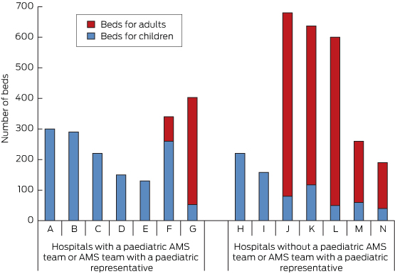
Nine hospitals had AMS programs (in two there was no paediatric representation), and one further AMS program was in development. Five hospitals had a dedicated paediatric AMS team, and only one of the hospitals with a large majority of adults had a paediatric representative on the hospital AMS team (Box 1).

Hospitals with a paediatric AMS team or AMS team with a paediatric representative had a higher number of paediatric beds than those without (mean, 200 [SD, 92] v mean, 104 [SD, 66]; = 0.04; 95% CI, 5–191). They also had a higher number of paediatric specialist services (mean, 8 [SD, 1.4] v mean, 4 [SD, 2.4]; P = 0.009; 95% CI, 1.0–5.6).

However, having paediatric specialist services did not necessarily mean a hospital had an AMS program with paediatric representation; for example, seven of the 12 hospitals with a neonatal unit did not have one. Throughout the rest of the survey, paediatric AMS activities were generally associated with having a dedicated paediatric AMS team.

While 11 hospitals had funding for an AMS pharmacist, only eight had a paediatric component (Box 2), and only four of these had committed ongoing funding for a permanent paediatric AMS pharmacist. Only two hospitals had any funding for a paediatric ID physician for AMS — both were part-time and one was about to cease (Box 2). Thus in total there was a single hospital out of 14 tertiary paediatric hospitals across Australia and New Zealand that had an ongoing part-time (half a day per week) paediatric ID position dedicated to AMS, amounting to 0.1 equivalent full-time (EFT).

Guidelines

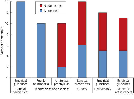
All hospitals had empirical antimicrobial prescribing guidelines and all of those with a haematology and oncology service had guidelines for febrile neutropenia. However, fewer than half of the hospitals had guidelines for antifungal prophylaxis, surgical prophylaxis, neonatology or paediatric intensive care (Box 3).

Review of antimicrobial prescribing

Some point-of-care interventions relating to antimicrobial prescribing were carried out in 12 hospitals, including empirical choice, dose optimisation, de-escalation based on microbiology test results and intravenous to oral switch. At four hospitals, the interventions were used for most antimicrobial prescriptions; for three they were only used in units that have high prescribing rates (eg, haematology and oncology); and for five they were only used in consultation with an ID physician or for restricted antibiotics. For the four hospitals where point-of-care interventions were used for most antimicrobial prescriptions, feedback was provided by the AMS team; for the remainder it was provided by the ID team.

Only two hospitals had automatic stop orders, and these were for restricted antimicrobials.

All hospitals had restricted antimicrobials but only four hospitals had an electronic approval system. At 13 hospitals, approval for restricted drugs was done by ID physicians.

Monitoring

Seven hospitals monitored antimicrobial usage by unit use (eg, number of vials), cost or both. Auditing methods varied widely but were mainly ad hoc: 10 hospitals had done at least one hospital-wide audit including assessment of appropriateness and one did occasional ward audits, but three hospitals had not done any audits. Results of audits were mostly fed back in an untargeted way through grand rounds or clinical quality meetings; only five hospitals provided feedback at departmental meetings and two provided the results to the relevant head of department or consultant. Of the 11 hospitals that did any audits, two did not feed the results back at all.

All hospital microbiology laboratories selectively reported antimicrobial susceptibility results (“cascade reporting”). Only five, however, monitored and regularly reported hospital-wide sensitivity patterns, and a further three reported on selected bacteria only.

Education

There was a paucity of education about AMS in general: no hospitals had education for senior medical staff, eight had education for junior medical staff, five for pharmacists, one for nurses, and four had no education for any staff. One hospital covered AMS at hospital orientation.

Barriers to antimicrobial stewardship

In the opinions of paediatric ID physicians and pharmacists, there was a wide range of reasons for slow implementation of AMS. The commonest perceived barriers to successful AMS were lack of education (11 hospitals), lack of dedicated pharmacy staff (eight) and lack of dedicated medical staff (seven). Other perceived barriers included lack of willingness to change, lack of leadership by executive and senior clinicians, lack of enforcement and transient junior staff.

Discussion

This is the first comprehensive survey of AMS resources for hospitalised children as measured by the inclusion of every children’s hospital in Australia and New Zealand. The survey responses are likely to be highly reliable as they were completed by an ID physician, AMS paediatrician or AMS pharmacist at each hospital — the most qualified local staff.

The results represent large hospitals with paediatric patients. There are many other hospitals with paediatric patients across both countries and, as these would predominantly have fewer children, paediatric AMS resources are likely to be even more scarce.

To our knowledge, only one other study of paediatric AMS programs in hospitals has been conducted. This was a larger study that was done in the United States, but it had only a 60% response rate and did not include all children’s hospitals.7

Personnel resources

In our study, most hospitals had an AMS program, either in existence or in development, which compares favourably with the 33% reported from the US study.7 This may reflect the fact that AMS has been a requirement of national hospital accreditation in Australia since 1 January 2013.

Hospitals included in our study that had a majority of children and those that offered more specialist services were more likely to have a paediatric AMS team or representative. While this is not surprising, it raises the question of how hospitals with fewer children might manage.

One possibility is to link in with the hospital’s adult AMS services. However, having a high number of adult patients did not necessarily equate to having an adult AMS team (data not shown), and differences between children and adults (in terms of antimicrobial use, measurement of antimicrobial use and patient outcomes) would make it difficult to use combined resources.

An alternative model is to have a network of paediatric AMS services between hospitals to share resources. The ANZPID-ASAP group is one such network of paediatric ID physicians and AMS pharmacists with representation from every state and territory in Australia and from New Zealand. Resources such as AMS guidelines, surveys and data on antimicrobial use can and have been shared among the group.

Members of the ANZPID-ASAP group also provide information and consultations via email for practical antimicrobial questions. This model could be replicated at a local level. It is less straightforward to share personnel resources such as AMS pharmacists, although this may be possible in a geographically smaller area such as a city that has several hospitals.

Paediatric representation on adult AMS networks for the acquisition and dissemination of information is also important. One such network is the Australian Commission on Safety and Quality in Health Care Antimicrobial Stewardship Jurisdictional Network.

The total of 0.1 EFT ongoing paediatric ID physician dedicated to AMS in the whole of Australasia is concerning. Even the proportion of hospitals with any paediatric AMS pharmacist time was only just over half. In the US study, almost half of AMS programs had greater than 0.25 EFT of paediatric ID physician time, but up to 40% had none and 40% had no paediatric AMS pharmacist.7

Guidelines

It is reassuring that every hospital had empirical guidelines for children attending the emergency department, as emergency departments have a high turnover of junior medical staff and having guidelines to direct empirical antimicrobial decision making for children is important.9

Conversely, the lack of guidelines in specialist areas is concerning. This may reflect lack of evidence or resources, or complacency in some instances. In some areas (eg, antifungal prophylaxis for haematology and oncology patients), there is little specific evidence in children.10

Lack of evidence, however, should not be an impediment to consensus guidelines. In other areas (eg, surgical prophylaxis), adult guidelines can be used. Guidelines should always be adapted to the specific population, taking into account age, disease burden and resistance patterns of local organisms. Guidelines help to standardise prescribing and represent an area for improvement.

Review of antimicrobial prescribing

The hospitals that were able to do most point-of-care interventions were those where the AMS team was heavily involved (data not shown), indicating that widespread AMS activities are dependent on time and resources.

The low rate of automatic stop orders is a potential area for intervention. AMS programs in hospitals are likely to have a greater impact on strategies for stopping antimicrobials than on those for starting antimicrobials. By having AMS systems and processes built into everyday workflow for patient care (eg, guidelines, protocols, routine data collection on antibiotic use), the need for individual patients to have consultations with ID, microbiology or AMS teams decreases.

While all hospitals had a list of restricted drugs, a minority had an electronic approval system. Phone approval systems are easier to circumvent intentionally or unintentionally, so the drugs that are most important to preserve may be missed. It has been suggested that prospective audit of and feedback on the use of selected drugs would be more effective than restriction and approval.6 However, audits are time consuming and require dedicated AMS personnel.

Monitoring

Only half of hospitals included in our survey routinely monitored antimicrobial use and fewer than half monitored hospital-wide organism resistance patterns (antibiograms). These represent two of the few measurable outcomes for paediatric patients.11 Half of the hospitals monitored unit use and/or cost — similar to the 55% in the US study.7

Because dosing for children is weight-based, unit use and cost do not provide a useful snapshot of antimicrobial use, although they can be used to monitor trends. One innovative strategy that has been proposed is adapting methods used for adults — for example, using average weight bands for different paediatric age groups12 — although such strategies have not been validated.

In the absence of electronic systems that enable the number of days of antimicrobial use to be recorded, audits in the form of antimicrobial point prevalence surveys remain the mainstay of paediatric monitoring. In our study, 10 hospitals had done at least one hospital-wide audit; of those 10, eight were involved in an international point prevalence survey during 2012, undertaken in Australia by the ANZPID-ASAP group.13,14 To be useful, such surveys (which are labour intensive) need to be repeated.

Hospital antibiograms are helpful for drafting guidelines that account for local resistance patterns, and for detecting significant changes over time, to inform empirical antimicrobial choices.15 It is possible that hospitals will eventually move away from antibiograms, and instead adopt strategies such as mathematical modelling and likelihood of inadequate therapy,16 but antibiograms are the best choice at present.

Education

Lack of AMS education for senior medical staff in the hospitals we surveyed likely reflects a lack of hospital-based education programs for this group in general. Since senior medical staff are crucial in the support of AMS principles, this area needs to be addressed. While junior medical staff received the most education, this was often under the umbrella of other teaching that included antibiotics (eg, management of urinary tract infections) rather than specific teaching about AMS. Other clinical staff (pharmacists and nurses) received even less education, failing to support the notion of AMS as multidisciplinary. In the US study, 64% of AMS programs had education as a specific component.7 For all but one hospital in our study, AMS was not considered important enough to be included in hospital orientation.

Barriers to antimicrobial stewardship

While the US study identified perceived loss of prescribing autonomy and lack of hospital administrative awareness about the importance of AMS as barriers, lack of resources ranked most important (70% of respondents).7 Two recent Australian adult AMS surveys found that lack of education, lack of pharmacy resources and lack of ID and microbiology resources were in the top three perceived barriers to AMS.8,17

In our study, the main barriers to effective AMS that were identified by the paediatric ID physicians and AMS pharmacists of Australia and New Zealand who completed the survey were lack of education and lack of personnel (dedicated pharmacy and medical staff). The latter may reflect the difficulty in showing cost benefits in paediatric health care compared with adult health care: for example, incidence of Clostridium difficile infection in paediatrics is not a useful outcome measure. Other measures, such as reduced length of hospital stay and intensive care unit stay may be more useful in children,15 but these reductions cannot be realised without resources.

Lack of education and lack of personnel are the two main areas that should be targeted in efforts to keep hospital executives informed about best practice in AMS. They should be seen as the focus of quality improvement activities in the care of children in hospital.

Conclusions

Australasian children’s hospitals have implemented some AMS activities, such as audits of antimicrobial use and monitoring of antimicrobial resistance, but most lack human resources. There was consensus among the staff who completed our survey that lack of education and personnel are major barriers to effective AMS. These must be addressed to improve antimicrobial use in hospitalised children.

Members of the Australian and New Zealand Paediatric Infectious Diseases Group – Australasian Stewardship of Antimicrobials in Paediatrics (ANZPID-ASAP) group

Penelope Bryant (Chair) (Royal Children’s Hospital, Melbourne), David Andresen (Children’s Hospital at Westmead, Sydney), Minyon Avent (Mater Children’s Hospital, Brisbane), Sean Beggs (Royal Hobart Hospital, Hobart), Chris Blyth (Princess Margaret Hospital for Children, Perth), Asha Bowen (Menzies School of Health Research, Darwin), Celia Cooper (Women’s and Children’s Hospital, Adelaide), Nigel Curtis (Royal Children’s Hospital, Melbourne), Julia Clark (Royal Children’s Hospital, Brisbane), Andrew Daley (Royal Children’s Hospital, Melbourne), Jacky Dobson (Canberra Hospital, Canberra), Gabrielle Haeusler (Monash Children’s Hospital, Melbourne), David Isaacs (Children’s Hospital at Westmead, Sydney), Brendan McMullan (Sydney Children’s Hospital, Sydney), Pam Palasanthiran (Sydney Children’s Hospital, Sydney), Tom Snelling (Princess Margaret Hospital for Children, Perth), Mike Starr (Royal Children’s Hospital, Melbourne), Lesley Voss (Starship Children’s Health, Auckland).

1 Paediatric and adult bed numbers at hospitals with and without a paediatric antimicrobial stewardship (AMS) team or AMS team with a paediatric representative in June 2013

2 Numbers of hospitals with antimicrobial stewardship (AMS) pharmacists and/or AMS infectious diseases physicians in June 2013 (n = 14)


Number


AMS pharmacists


Paediatric full-time

2

Paediatric part-time

6

Adult only

1

Paediatric position not filled, no ongoing funding

2

None

3

AMS infectious diseases physicians


Paediatric full-time

0

Paediatric part-time

1

Adult only

2

Paediatric position not filled, no ongoing funding

1

None

10


3 Numbers of hospitals with and without antimicrobial prescribing guidelines for different services (n = 14)


* General paediatrics includes children presenting to the emergency departments.

Infliximab therapy in two cases of severe neurotuberculosis paradoxical reaction

Clinical record

Patient 1

A 60-year-old HIV-negative woman presented with a week’s history of fever, vomiting and confusion, followed by progressive personality change. On admission, she was noted to have urinary retention, left oculomotor nerve palsy and an upgoing right plantar response. A magnetic resonance image (MRI) of the brain showed leptomeningeal enhancement with gyral swelling and subtle cortical T2 signal hyperintensity in the right frontal lobe, suggesting meningoencephalitis. Cerebrospinal fluid (CSF) cultures grew fully susceptible Mycobacterium tuberculosis. Antituberculous therapy was started with isoniazid, rifampicin, ethambutol and pyrazinamide, plus dexamethasone. CSF cultures tested negative by Week 1. Over the next month, she had ongoing fevers and fluctuating conscious state. High CSF pressures necessitated ventriculoperitoneal (VP) shunting. An MRI 3 months into therapy showed numerous granulomas, microabscesses and infarcts. Her condition failed to improve with a further course of dexamethasone, and an MRI at 5 months showed increasing size and number of granulomas, with worsening oedema and midline shift (Figure 1, A). She was given a trial of three doses of infliximab 10 mg/kg, 1 month apart, resulting in marked improvement in neurological status and radiological findings (Figure 1, B). She regained movement of her limbs, opened her eyes spontaneously and was able to articulate a few words. After completing 2 months of four-drug therapy, she received isoniazid and rifampicin for 10 months, with ongoing improvement. She was left with mild cognitive deficit and required some assistance with activities of daily living.

Patient 2

A 32-year-old HIV-negative woman presented with delirium and back pain. A chest radiograph suggested miliary tuberculosis. A computed tomography brain scan was unremarkable. Results of CSF molecular testing were positive for M. tuberculosis complex, and cultures from CSF, blood and a laryngeal swab grew fully susceptible M. tuberculosis. Isoniazid, rifampicin, ethambutol, pyrazinamide and prednisolone 50 mg were commenced. One month into therapy, she developed headache in the context of weaning from prednisolone. An MRI showed multiple rim-enhancing nodules in the CSF spaces, with leptomeningeal enhancement and enhancing lesions in the right cerebellum and hemipons. CSF cultures tested negative.

The prednisolone dose was increased to 60 mg, with little response, then converted to dexamethasone 12 mg/day. One month later, while steroid tapering, she developed diplopia. An MRI showed worsening tuberculomas with increasing oedema (Figure 2, A). Dexamethasone was reinitiated at 12 mg/day. Three months into therapy, she developed obstructive hydrocephalus requiring VP shunting. Over the following weeks, she developed peripheral visual field loss. After 4 months of tuberculosis therapy, a trial of infliximab 5 mg/kg was initiated. The steroid dose was tapered over the next week without worsening of symptoms, and she was discharged. One month later, an MRI showed moderate improvement (Figure 2, B). Two further doses of infliximab were given over the subsequent 6 weeks, with complete resolution of visual symptoms. She completed 2 months of four-drug therapy, followed by 10 months of isoniazid and rifampicin. The course was complicated by a seizure at Month 8, necessitating antiepileptic therapy, but she made an otherwise full neurological recovery.

A paradoxical reaction (PR) in tuberculosis (TB) is the worsening of disease after starting TB therapy, usually despite microbiological response. It may represent an inflammatory response to the release of antigen from dying bacilli.1 Such disease exacerbation has also been observed in people with HIV when antiretrovirals are started,2 and in individuals with TB when tumour necrosis factor alpha (TNF-α) antagonists are discontinued.3 A PR may manifest with new pulmonary lesions or lymphadenopathy3 and can be life-threatening, especially in patients with neurotuberculosis.1 Management involves high-dose corticosteroids, but in intractable cases success has been reported with TNF-α blockade.1,35

Our two patients had severe neurotuberculosis PRs unresponsive to dexamethasone, which abated after administration of the anti-TNF-α antibody infliximab. Before these two cases, there was only one report of therapeutic use of infliximab for TB PR in an individual without prior history of TNF-α antagonist use.1 Our two cases add weight to this approach being safe and effective in patients with steroid-refractory TB PR. The previously reported patient had steroid-refractory neurotuberculosis that did not respond to a trial of cyclophosphamide. Radiological and neurological parameters improved only after infliximab was given.1 In that case and ours, cultures tested negative soon after antituberculous therapy was started, suggesting that ongoing disease was due to an immunologically mediated PR rather than inadequate microbiological control.

These cases highlight the potentially devastating effects of central nervous system (CNS) TB, which, despite contemporary therapeutic approaches, still results in permanent disability or death in half of those treated.6 Much of this morbidity can be attributed to the inflammatory response. A key inflammatory cytokine is TNF-α, which plays an integral role in granuloma formation to contain TB infection. However, in mouse models of neurotuberculosis, TNF-α has been shown to increase blood–brain barrier permeability, resulting in increased CSF leukocytosis and CNS inflammation.7

Attenuation of the inflammatory response with routine administration of corticosteroids in patients with neurotuberculosis has been shown to reduce mortality.6 However, in cases of PR, outcomes are often poor despite steroids. There is growing evidence that medications with anti-TNF-α activity may have a role in controlling this inflammatory response, without compromising microbiological response.1,35

Thalidomide, a potent TNF-α inhibitor, was administered to two patients with steroid-refractory neurotuberculosis, with apparent improvement.8 While also showing promise in rabbit models and a small pilot study, it was poorly tolerated and failed to show clinical benefit when used as adjunctive therapy for childhood TB meningitis in a randomised trial.9

There are accumulating data on the role of the anti-TNF-α monoclonal antibodies infliximab and adalimumab and the soluble TNF-α receptor etanercept. They have potent anti-inflammatory properties and are well tolerated, but have been associated with increased risk of TB in those taking them for autoimmune conditions.10 TB developing in patients receiving TNF-α antagonists is more likely to be extrapulmonary or disseminated,11 and early reports suggested that it was more refractory to treatment.12 However, as experience with these agents grew, it became apparent that the poor response could be a PR to the TNF-α antagonist withdrawal. As the immunosuppressive effect of the TNF-α antagonist wanes, the recovering immune system can generate an intense inflammatory reaction against mycobacterial antigens. Two patients with steroid-refractory disease were successfully treated with reintroduction of the offending TNF-α antagonist.4,5

Our report supports an additional role for TNF-α inhibition in severe PRs in immunocompetent individuals. Given that TNF-α antagonists appear to be safe in TB PR, further studies of their role in management are warranted.

Lessons from practice

  • Central nervous system tuberculosis remains a potentially devastating disease that, despite contemporary therapeutic approaches, still results in permanent disability or death in half of those treated.
  • A paradoxical reaction is an inflammatory reaction that can cause disease progression and complications after initiation of antituberculous therapy.
  • High-dose corticosteroids are recommended but if these are ineffective, there is mounting evidence for the use of tumour necrosis factor alpha antagonists such as infliximab.

Are we doing enough to promote the effective use of mosquito repellents?

Health authorities need to review recommendations on how to choose and use mosquito repellents

Mosquito-borne pathogens remain a threat to public health in Australia. The activity of dengue and chikungunya viruses has increased across South-East Asia and the Pacific region in recent years, and the number of travellers returning to Australia infected with mosquito-borne pathogens has steadily grown.1 Annual notifications of endemic mosquito-borne disease resulting from infection with Ross River or Barmah Forest viruses persist at around 5000 cases a year in Australia, and local transmission of dengue viruses remains a threat in Far North Queensland.2

Notwithstanding the risk to human health, the nuisance biting of local mosquito species is also a problem. With the threat of exotic mosquito introduction, particularly of Aedes albopictus (Asian tiger mosquito), which could become established in major metropolitan regions,3 management of public health risks associated with mosquito populations will continue to be of concern.

While broad-scale mosquito control can reduce the risk of mosquito-borne disease in certain circumstances,4 local authorities rarely have the financial or operational capacity to implement and maintain an effective program. New “technological fixes”, such as the use of insect-specific intracellular bacteria to prevent mosquitoes transmitting dengue viruses,5 may assist in reducing disease risk, but these approaches are typically specific to mosquito species or pathogens.

Personal protection strategies, particularly the use of topical insect repellents, are therefore likely to remain the most widely encouraged strategy to reduce mosquito-borne disease risk.

What do health authorities recommend?

Recommendations on personal protection measures to avoid mosquito bites are provided in fact sheets and accompany seasonal warnings issued by a range of health authorities, including federal and state health departments and local governments. These recommendations usually include:

  • use of insect repellents or insecticides (eg, topical repellents, mosquito coils)
  • source reduction (eg, minimising opportunities for mosquitoes around dwellings by emptying or covering water-holding containers)
  • behavioural practices (eg, avoiding mosquito habitats or times of the day when mosquitoes are most active)
  • physical barriers (eg, bed nets, long-sleeved shirts).6

With regard to insect repellents, generally only products containing either N,N-diethyl-3-methylbenzamide (commonly known as DEET) or 2-(2-hydroxyethyl)-1-piperidinecarboxylic acid 1-methylpropyl ester (commonly known as picaridin) are recommended. Published laboratory and field-based studies have shown DEET and picaridin to be the most effective mosquito repellents.7 These two products have also been shown to pose no substantial adverse health effects to users and are considered safe,8 although they are generally not recommended for children under 3 months of age.6 There should be no hesitation in recommending either of these products for use by older children or adults to protect against mosquito bites.

However, the advice provided rarely reflects the wide range of commercially available mosquito repellent formulations, how the concentrations of active ingredients in these products should be interpreted, or how they should be used.

How can we compare repellents?

All products marketed as mosquito repellents must be approved by the Australian Pesticides and Veterinary Medicines Authority (APVMA). There are currently more than 100 registered repellent formulations (including creams, gels, lotions, sprays, patches and wristbands). However, repellent formulation is generally far less important than the active ingredients contained therein. While most registered products contain DEET or picaridin, there are also a few formulations that contain botanical-based active ingredients.

In assessing repellents, both repellency (the percentage of mosquitoes landing on treated skin compared with untreated controls) and protection time (the period of time after application of a repellent during which no mosquito landings occur) are typically reported. Preventing, not just reducing, mosquito bites is critical in minimising mosquito-borne disease transmission.9 An understanding of how protection times differ between repellent formulations not only provides a guide to the most effective products but can also guide better communication of recommended use patterns, such as reapplication times.

Formulations containing DEET or picaridin can vary in the concentration of active ingredients. There is often a misconception that “stronger” formulations provide better protection by preventing more bites. However, the concentration of active ingredient determines how long a repellent provides protection, not the degree of protection that is provided. For example, “high-dose” and “low-dose” DEET-based repellents will provide comparable protection against biting mosquitoes up to about 2 hours, but the protection provided by the high-dose repellent will persist beyond the time at which the low-dose repellent fails.10

Repellents containing botanical extracts are often perceived as “safer” or “natural” alternatives to DEET or picaridin. The most common such active ingredients include Melaleuca and Eucalyptus oils. However, studies have shown that these repellents provide much shorter periods of protection against biting insects.11 By way of comparison, botanical-based repellent formulations would need to be reapplied three to four times more frequently than a 20% DEET- or picaridin-based repellent to provide comparable protection times.10 Botanical repellents are rarely recommended but given the interest in them in the community, an emphasis on their high required rate of reapplication may be needed.

A product often perceived to be a botanical repellent is p-menthane-3,8-diol (PMD). In Australia, PMD is registered with the APVMA as “Extract of lemon eucalyptus, being acid modified oil of lemon eucalyptus (Corymbia citriodora)”. It is not an essential oil but rather a by-product of the hydrodistillation process. Laboratory and field tests have demonstrated that PMD provides effective protection against mosquito bites,12 but it is rarely included in lists of recommended repellents. This may be due to the relatively limited distribution of PMD products locally.

Do we know how to effectively use repellents?

Very rarely is advice provided on how repellents should be applied. This communication gap may contribute to deficiencies in the protection provided by even the most effective repellent formulations. Repellents, regardless of their active ingredient, should be applied as a thin, even coating on all exposed areas of skin. Spraying on clothes or a dab “here and there” will not provide complete protection. Reapplication after swimming and exercise is typically required.

There is a paucity of attitudinal and behavioural research into the use of topical insect repellents. Although instructions to “apply to exposed skin” are commonly included on repellent formulation labels, perhaps stronger advice should also accompany recommendations from local health authorities.

It is also important to consider other registered products among commercial mosquito repellents. Wristbands and patches purporting to provide protection against mosquitoes are registered, but studies have shown these “spatial” repellents to be ineffective.13 There is also an emerging range of cosmetic products combining mosquito repellents with sunscreens and skin moisturisers, which may require specific recommendations on use.14

Finally, different recommendations for repellent use will be required in regions where day-biting mosquitoes are present and pose a risk of dengue virus transmission (eg, Far North Queensland) compared with those needed elsewhere, where predominantly evening-biting mosquitoes are present.15

Topical mosquito repellents will remain a key component of recommended personal protection strategies against mosquitoes. When developing recommendations, it is important to consider not only the published scientific literature but also the current range of commercially available formulations, as this is likely to fuel concerns and drive enquiries from the public.

Ideally, nationally consistent guidelines would help local and state government health authorities develop strategies to promote the effective use of mosquito repellents to reduce nuisance biting and mosquito-borne disease risk.

[Comment] Rising wealth, improving health? Adolescents and inequality

The linkage between social inequalities and health disparities is well established.1 Less well described are the connections between social inequalities during childhood and adolescence and adult health status.2 Additionally, little is known about changes in social inequalities during early life, and virtually nothing is known about how these changes might affect social gradients in non-communicable diseases (NCDs) later in life. In The Lancet, Frank Elgar and colleagues3 break new ground by examining secular trends in adolescents in socioeconomic inequalities and health disparities in factors contributing to NCDs.