×

Anti-vax dodge dismissed by Commonwealth

The Federal Government has confirmed that a form being circulated by anti-vaccination campaigners attempting to circumvent new ‘No Jab, No Pay’ laws has no legal standing, backing AMA advice that doctors are under no obligation to sign it.

Social Services Minister Christian Porter has written to AMA President Brian Owler confirming that medical practitioners were under no obligation to sign the form, which asks doctors to acknowledge the ‘involuntary consent’ of a parent to the vaccination of their children, and which is deemed to be ineffective in any case.

“I am able to advise you that under the No Jab, No Pay Act, immunisation providers are not obligated to sign such declarations,” Mr Porter wrote. “This statutory declaration is not relevant evidence for the purposes of family assistance payments, [so that] even if such a form were signed by a doctor…it would not in any circumstances make the relevant parent eligible for payments that would otherwise be suspended.”

The form has been circulated by anti-vaccination campaigners following Federal Government welfare changes aimed at denying certain welfare payments to parents who refuse to vaccinate their child.

Under the No Jab, No Pay laws, from 1 January this year parents of children whose vaccination is not up-to-date are no longer eligible for the Family Tax Benefit Part A end-of-year supplement, or for Child Care Benefit and Child Care Rebate payments. The only exemption will be for children who cannot be vaccinated for medical reasons.

The new laws were introduced amid mounting concern that vaccination rates in some areas were slipping to dangerously low levels, increasing the risk of a sustained outbreak of potentially deadly diseases such as measles.

The Australian Childhood Immunisation Register shows there has been a sharp increase in the proportion of parents registering a conscientious objection to the vaccination of their child, from just 0.23 per cent in late 1999 to 1.77 per cent by the end of 2014.

In all, around a fifth of all young children who are not fully immunised are that way because of the conscientious objection of their parents.

The form being circulated by anti-vaccination groups, headed “Acknowledgement of involuntary consent to vaccination”, is intended to circumvent the No Jab, No Pay laws and allow conscientious objectors to receive Government benefits without allowing the vaccination of their children.

But Mr Porter said the aim of the new laws was to boost immunisation rates “by providing a level of encouragement and incentive for families to more thoroughly inform themselves about the importance of immunising their children”.

The Minster said the Government recognised the right of parents to decide not to vaccinate their children, but the new laws meant there would be consequences.

“An individual is not prohibited in any way from maintaining their vaccination objection; it is simply the case they will not receive some of their family assistance,” he said. “This is a relatively small financial cost, particularly when compared to the cost that the spread of crippling, debilitating and deadly diseases has on our health system and community.”

“It is the Government’s view that when an individual decides not to vaccinate their child, they are putting their child and the community at risk of infectious diseases.”

Last month, the AMA’s senior legal adviser John Alati advised that, where there was no medical reason for vaccination exemption, the doctor’s job was to outline the relevant facts about immunisation and to provide vaccination where consent was given. Where it was withheld, “the doctor should not perform the procedure as it might constitute trespass to the person”.

His advice was backed by Mr Porter, who said that “the appropriate path for a doctor or medical profession who may be requested to sign [the form being circulated by anti-vaccination campaigners] is simply to vaccinate where there is consent, and decline where consent is absent”.

Adrian Rollins

A case of bilateral endogenous bacterial endophthalmitis from Streptococcus pneumoniae bacteraemia

Clinical record

A 55-year-old woman was admitted to an orthopaedic unit of a metropolitan hospital in Australia with right shoulder septic arthritis. She had been experiencing 3 days of right shoulder pain, fevers, rigors and delirium. These occurred in the context of a right rotator cuff repair 3 months previously. Her medical history included hypertension, hypercholesterolemia and an L4-S1 spinal fusion for lumbar spine degeneration. Her medications included hydrochlorothiazide, olmesartan and atorvastatin.

On examination, she was febrile with a temperature of 38.9°C, with a painful, erythematous and swollen right shoulder with movement limited by pain. Her chest and abdominal examinations were normal. Her relevant admission blood test results were white cell count, 30 × 109/L (reference interval [RI], 4–11 × 109); neutrophil count, 21.9 × 10⁹ (RI, 2–8 × 10⁹); and C-reactive protein level, 438 mg/L (RI, 0–5 mg/L). Platelet, haemoglobin, uric acid and blood sugar levels were all normal. Her shoulder and chest x-rays were unremarkable. Several sets of blood cultures as well as a shoulder aspirate showed gram-positive cocci and she was empirically commenced on intravenous (IV) flucloxacillin 2 g four times a day and vancomycin 1.5 g twice daily.

Shortly after admission, she revealed that she had been experiencing 3 days of blurred vision and pain in her left eye. Further history revealed that aside from mild myopia, her vision was previously normal and she had never undergone any ophthalmic procedures. Her right eye was asymptomatic and appeared slightly injected but otherwise normal on initial assessment (Figure, A). However, her left sclera was severely injected, and the pupil was fixed and mildly dilated with corneal and anterior chamber haze and a circumferential hypopyon (Figure, B). Visual acuity (VA) was 6/12 in her right eye, but only hand movements were appreciated in her left eye. Fundoscopy was not possible on the left eye owing to the severity of the corneal haze; however, in the right eye, fundoscopy showed a vitreous haze.

An urgent assessment by an ophthalmologist found that she had bilateral endogenous bacterial endophthalmitis. Despite appearing mostly unremarkable on external inspection, the right eye was deemed to be in the early stages of infection owing to the presence of vitreous haze on fundoscopy. After aqueous humour samples were taken, she was given bilateral intravitreal antibiotic injections of 1 mg vancomycin and 2 mg ceftazidime. This was followed up with intravitreal injections of 1 mg vancomycin every second day for three further doses. The cultures from her shoulder, blood and eyes all grew Streptococcus pneumoniae. Her antibiotics were changed to IV benzylpenicillin and vancomycin; IV azithromycin was also added, as evidence has shown that it has a survival benefit in S. pneumoniae infections.1 Further investigations performed to exclude immunosuppression and locate the source of the septicaemia included HIV, tuberculosis and hepatitis serology; glycated haemoglobin testing; vasculitic screening; a transthoracic echocardiogram; computed tomography of the chest, abdomen and pelvis to look for malignancy or abscesses; and a magnetic resonance imaging scan of her lumbosacral spine to exclude infection of the implants from her spinal fusion. The results of these tests were unremarkable and no cause was found to explain the S. pneumoniae septicaemia.

Twelve days after admission, she was transferred to another centre with a vitreoretinal unit, where she underwent a left vitrectomy to debride the posterior chamber of her eye. Unfortunately, the vision in her left eye only made a small recovery and, 6 months later, the VA was limited to counting fingers. Fortunately, VA in her right eye remained at its baseline and she has been managing well in the community.

Bacterial endophthalmitis is an infection of the eye involving the aqueous and/or vitreous humour. It is an ophthalmic emergency and requires urgent treatment to prevent permanent blindness. The vast majority of cases are due to exogenous inoculation of microbes into the eye via trauma, surgery, intravitreal injections or an invasive corneal infection. Rarely, in 2–6% of cases, it can occur from haematogenous spread of organisms to the eye, which is referred to as endogenous or metastatic endophthalmitis. Endogenous spread is associated with immunosuppressive predisposing factors such as diabetes or malignancy in 90% of cases.2

Patients usually experience 12–24 hours of eye pain and decreasing vision. They are rarely systemically unwell. However, patients with the endogenous variant are usually unwell from the underlying bacteraemia. On examination, there is often conjunctival injection or oedema of the cornea. VA will be decreased. Slit-lamp examination of the anterior chamber may show cells and flare (small floating particles and a generalised haziness, respectively, in the anterior chamber representing inflammation). As in this patient, it may be also be associated with a hypopyon. Hypopyon is pus in the anterior chamber of the eye which will collect inferiorly and form a horizontal layer in an erect patient (Figure, B shows a circumferential hypopyon in our patient as she was supine at the time she was photographed; the Box highlights the difference between hypopyon in erect and supine patients). On fundoscopy, there is usually poor visualisation of the retina secondary to vitreous haze.2,3 The diagnosis can be confirmed via aqueous or vitreous culture, but it should be noted that history and examination are the main diagnostic tools, as a negative culture cannot reliably exclude the condition.4

Bacterial endophthalmitis is usually exogenous and postoperative. It is most commonly caused by coagulase-negative staphylococci (the most common normal flora of the ocular surface).5 The organisms for the endogenous form depend on the underlying infection. A major study showed only 30% of patients with streptococcal endophthalmitis had a final VA of 6/30 or better, which is worse than for Staphylococcus aureus, gram-negative bacteria (50% for both) and coagulase-negative staphylococci (80%).6 Regardless of the organism involved, the strongest indicator of visual prognosis is VA at presentation.5

Treatment of endophthalmitis requires prompt referral to an ophthalmology team. Treatment of bacterial endophthalmitis involves intravitreal antibiotic injections as the intravenous route does not deliver sufficient concentrations to the relatively avascular posterior chamber of the eye. Empirical therapy is usually vancomycin with ceftazidime or amikacin. This is often combined with vitrectomy, which debrides the vitreous, reducing the bacterial load. Patients should also be treated with systemic antibiotics for sufficient duration to clear the underlying infection.7

Bilateral endogenous bacterial endophthalmitis is very rare, with one study estimating that only around 12% of patients with endophthalmitis had both eyes infected.2 A rare bilateral case like ours, where one eye is severely infected and the other is in the early stages of infection, serves to demonstrate the importance of early diagnosis, as once vision has been lost it is far less likely to return. Further, our case is unusual in that no immunosuppression was found in a relatively young patient.

Lessons from practice

  • Endogenous bacterial endophthalmitis can rapidly cause blindness and should be considered in all bacteraemic patients with decreased visual acuity or ocular pain.

  • Risk factors include diabetes, malignancy and immunosuppression.

  • Early diagnosis is important as visual acuity on diagnosis is the strongest indicator of final visual prognosis.

  • Early ophthalmology involvement and intravitreal antibiotic injections are essential, as intravenous therapy alone will not deliver adequate concentrations to the relatively avascular vitreous.

Figure


The patient’s right eye (A) showing mild scleral injection but appearing otherwise unremarkable on initial assessment, and left eye (B) showing corneal haze, circumferential hypopyon and injected left sclera.

Box –
Difference between hypopyon in erect and supine patients

Repeat exposure to active tuberculosis and risk of re-infection

Clinical record

A 20-year-old woman of Indo-Fijian background who had lived in Australia for 10 years presented to the emergency department with a 3-day history of pain in the lower back and right hip. She had been coughing for 3 months and reported fevers and weight loss of 6 kg over the same period. Her medical history included treatment for latent (dormant) tuberculosis infection (LTBI) at 13 years of age with an unproblematic 6-month course of isoniazid 300 mg daily at a hospital chest clinic when her father, who lived in the same household, had sputum smear-positive pulmonary tuberculosis (TB). She had had a positive tuberculin skin test result of 15 mm before commencing treatment for LTBI, and had been given the BCG vaccine as a baby. Chest x-rays at the beginning and end of her treatment for LTBI had been normal. Her older sister, who also lived in the same household, was diagnosed with sputum smear-positive miliary TB 16 months after the father was diagnosed with TB (and 10 months after our patient had completed treatment for LTBI). Although our patient lived in the same household as her sister at that time, she did not receive a repeat course of preventive TB treatment. Six years after the sister was diagnosed with TB, our patient developed the symptoms described above.

When our patient presented at the emergency department, she had a heart rate of 120 beats/min, was normotensive, and was febrile at 38.3°C. A chest x-ray showed bilateral pulmonary nodular opacities (Box 1). Magnetic resonance imaging of the lumbar spine 2 days later revealed signs of early osteomyelitis with subchondral bony oedema and contrast enhancement of the right sacroiliac joint (Box 2). Mycobacterium tuberculosis DNA was detected in two sputum samples by polymerase chain reaction. We diagnosed our patient with pulmonary and osseous TB and commenced anti-TB treatment: isoniazid, rifampicin, pyrazinamide, ethambutol and vitamin B6 backup.

Two weeks later, M. tuberculosis complex was isolated from a sputum culture and was found to be fully sensitive to first-line (standard) anti-TB drugs. The patient’s overall condition improved rapidly on treatment and the opacities seen on her chest x-ray cleared. Two weeks into treatment, however, the patient developed nausea and vomiting, and blood tests showed an increase in liver transaminase levels, supporting the clinical suspicion of drug-induced hepatitis. After all anti-TB medications were withdrawn, the patient felt better within 2 days and her liver transaminase levels were normal 10 days later. The anti-TB drugs were re-introduced sequentially and additively in escalating doses following international recommendations for the order of re-introduction,1 starting with ethambutol 800 mg daily and an increasing dose of rifampicin (up to 600 mg daily) over 4 days, adding isoniazid over the next 5 days (up to 300 mg daily) and finally adding pyrazinamide (up to a dose of 1500 mg daily) over 3 days. Two days after re-introduction of full dose pyrazinamide (and thus on full TB treatment), the patient experienced nausea, her liver transaminase levels increased again, and we suspected pyrazinamide as the cause of drug-induced hepatitis. After all anti-TB medications were again withdrawn and her liver transaminase levels again normalised, all anti-TB drugs excluding pyrazinamide were restarted at full dose. However, 16 days later, the patient again developed hepatitis. Subsequent treatment with rifampicin, moxifloxacin, ethambutol and pyrazinamide, but without isoniazid, was tolerated well by the patient without laboratory evidence of hepatitis.

Genotyping of the M. tuberculosis organisms by the 24-loci mycobacterial interspersed repetitive units variable number tandem repeat (MIRU-VNTR) method2 showed that the patient, her father and her sister had indistinguishable genotypes, suggesting that all had been infected with the same organism.

Contacts of patients who have active TB are routinely screened in countries with a low incidence of TB such as Australia. If they have evidence of LTBI, but not active TB, they are usually offered a course of preventive TB treatment with isoniazid daily for 6–9 months. Isoniazid is estimated to be up to 90% effective in eradicating LTBI,3 but it does not offer protection from subsequent re-infection.

This case emphasises the importance of repeating a course of preventive TB treatment with each significant new exposure. It is highly likely that our patient was re-infected with TB through contact with her sister (who was sputum smear-positive and likely to have been infected by the father), but we cannot exclude with certainty that active TB developed as a consequence of failed LTBI treatment and re-activation of TB after the first exposure. In both scenarios, the patient’s M. tuberculosis organism would be the same as the father’s. It is also possible that all three family members (or at least the father and the sister) were infected at the same time. Treatment of LTBI and full anti-TB treatment do not offer protection from subsequent re-infection with M. tuberculosis and individuals do not develop immunity to TB after exposure.4 Thus, treatment of LTBI is not a priority in countries with a high incidence of TB, ongoing transmission and a high risk of re-infection.5

In patients with previous evidence of LTBI, repeat testing with a tuberculin skin test or blood test (interferon gamma release assay) is not helpful to assess the risk of re-infection, as these tests often remain positive even after a full course of LTBI treatment.6,7 In people with repeat contact with active TB, the decision to give a course of preventive TB treatment should be informed by an assessment of the risk that transmission occurred (taking into account the infectiousness of the index case with active TB, and the duration and proximity of contact) and factors that affect the risk of TB re-activation (eg, immunosuppression or use of biological agents such as infliximab) in the person under consideration for preventive TB treatment.

Our patient developed isoniazid-induced hepatitis when she was on full TB treatment, while she previously had no problems with preventive isoniazid monotherapy. Patients with low-acetylator status of N-acetyltransferase 2 have a significantly increased risk of developing drug-induced hepatitis on anti-TB drugs (our patient was not tested for this).8 The concurrent exposure to drugs that induce cytochrome P450 enzymes (including rifampicin, which is routinely prescribed for TB) could have increased the risk of isoniazid-induced hepatotoxicity.9 This needs to be kept in mind when patients who tolerated isoniazid preventive therapy well develop hepatitis on full anti-TB treatment.

Lessons from practice

  • It is possible for a person to be re-infected with tuberculosis (TB), even after a completed course of preventive TB treatment.

  • A person with repeat significant exposure to active TB should be considered for repeat preventive TB treatment.

  • Isoniazid could be the cause of drug-induced hepatitis in a person on multi-drug therapy for active TB, independently of whether isoniazid was previously tolerated well by the same person as monotherapy for TB prevention.

Box 1 –
Chest x-ray showing bilateral nodular opacities of the lungs

Box 2 –
Magnetic resonance image of the sacroiliac joints


Note the subchondral bony oedema and contrast enhancement of the right sacroiliac joint (arrow).

Influenza vaccine effectiveness in general practice and in hospital patients in Victoria, 2011–2013

The 9th edition of the Immunisation Handbook sponsored by the National Health and Medical Research Council maintained that influenza vaccines were 70%–90% effective in preventing influenza when the match between vaccine strains and circulating strains was good.1 Even when published in 2008, this was probably a generous assessment of the evidence. The 10th edition, published in 2013, maintained that influenza vaccines were 59% effective in preventing influenza in healthy adults and at least as effective in children, although in some years there was no evidence of any benefit.2

Although not explicitly stated in the handbooks, these estimates referred to efficacy in protecting against influenza infections managed in the community, the majority of which are relatively mild. While protection against the mild disease seen in primary care might be modest, it is nevertheless possible that the protection provided against more serious disease, including confirmed influenza infections requiring admission to hospital, might be greater.

In Victoria, two surveillance schemes make it possible to investigate whether there was any major difference in vaccine effectiveness estimates in community and hospital patients. The Victorian Sentinel Practice Influenza Network (VicSPIN) is a group of sentinel general practitioners in Melbourne and regional Victoria, operating since 1997, that has provided estimates of influenza vaccine effectiveness for protection against laboratory-confirmed influenza since 2003.3 The Influenza Complications and Alert Network (FluCAN) is a national hospital-based sentinel surveillance scheme that has provided estimates of influenza vaccine effectiveness since 2010.4 About 40% of patients registered by this scheme were reported by Victorian hospitals.

We compared influenza vaccine effectiveness estimates for 3 years in Victoria, basing our analysis on data from these two sentinel surveillance systems. Each scheme has published separate vaccine effectiveness estimates for the three study years.510

Methods

We reviewed data for the influenza seasons of 2011–2013 from the general practice and hospital-based schemes. Vaccine effectiveness was estimated by comparing the vaccination status of influenza cases (patients with laboratory-confirmed influenza) with that of non-cases (patients for whom influenza test results were negative). The use of test-negative controls is an established variation of the case–control study design.11

VicSPIN uses a community-based, test-negative design. Sentinel GPs were located in metropolitan Melbourne, Geelong and regional Victoria, and patients were recruited by sentinel GPs when they presented with symptoms consistent with influenza infection. At presentation and before their case status was known, patients were swabbed at the discretion of the GP. Patients with positive influenza test results were defined as cases, and those with negative results as non-cases or controls. Vaccine effectiveness was calculated as 1 − odds ratio (OR), and expressed as a percentage, where the OR compared the odds of vaccination for cases with the odds for controls. Logistic regression was used to adjust estimates for age group (0–17 years, 18–64 years, ≥ 65 years), comorbidity (yes v no), and time within influenza season (number of weeks from peak). Estimates were restricted to patients vaccinated at least 14 days before the onset of symptoms, accepted as the time needed to produce protective antibodies, and to those presenting within 7 days of symptom onset, as data from shedding studies suggest that influenza virus detection declines after 7 days.12 On the assumption that vaccine effectiveness would not be detectable when influenza virus was not circulating, we also restricted our analyses to patients who presented during the influenza season, as defined by positive case ascertainment and sentinel surveillance of influenza-like illness.13 Year was added as a covariate to the regression analysis for the combined 3-year estimate. Although the number of sentinel practitioners participating in the scheme in different years varied slightly, the approach to surveillance remained constant. Vaccination status was determined by patient or GP report, with vaccine date requested as a proxy for a register record. All samples were tested by polymerase chain reaction (PCR) assays at the Victorian Infectious Diseases Reference Laboratory, a designated National Influenza Centre of the World Health Organization. The assay detected influenza A(H3N2), influenza A(H1N1), influenza B and influenza C viruses. Two patients with influenza C were excluded from the analysis.

FluCAN is a sentinel surveillance system that receives data from 17 Australian hospitals. It provides data on the number of cases admitted with severe influenza A or B confirmed by PCR nucleic acid assays in the reporting hospitals’ laboratories. We based our analysis on the four Victorian hospitals reporting to FluCAN (the Alfred Hospital, Monash Medical Centre, University Hospital Geelong, the Royal Melbourne Hospital). Only adults (over 18 years of age) were included in the study. The vaccination status of cases was compared with that of controls (1:1); each selected control was the next patient after each case who presented with an acute respiratory infection and a negative influenza test result. Vaccination was defined as the patient having received the inactivated influenza vaccine at least 14 days before presentation, and the patient’s status was based on patient report and medical record. Vaccine effectiveness was estimated in the same way as for the community patients, but adjusted for potential confounders using conditional logistic regression to account for the matched design. Binary covariates included in the model were: being over 65 years of age, chronic comorbidities, Indigenous Australian status, and pregnancy. Because the control group was frequency-matched by the date of admission using the incidence density control selection strategy, we did not adjust or stratify estimates for time. We conditioned the analysis on the basis of hospital site. For the pooled analysis of all three seasons, the analysis accounted for year by adjusting standard errors with the Huber–White robust sandwich estimator.

During the years included in our study, FluCAN did not collect control data for people aged 0–17 years. We estimated vaccine effectiveness for all ages from the VicSPIN data, but, to improve the comparability of results, we also calculated vaccine effectiveness for the VicSPIN data after excluding the 0–17-year-old age group. All vaccines used in Australia during the study period were trivalent inactivated vaccines. We did not collect information on vaccine manufacturer, and assumed that all vaccines performed equivalently.

Ethics approval for FluCAN data collection and reporting was obtained from the Human Research Ethics Committees of all participating hospitals and the Australian National University. VicSPIN data were collected, analysed and reported under the legislative authorisation of the Victorian Public Health and Wellbeing Act 2008 and the Public Health and Wellbeing Regulations 2009, and therefore did not require formal Human Research Ethics Committee approval.

Results

In the VicSPIN surveillance system, before exclusion of patients vaccinated within 14 days of symptom onset or presenting outside the influenza season, 1680 patients for whom vaccine status by age group was known were available for the three seasons 2011–2013. Their number varied from 354 in 2013 to more than 600 in each of the two earlier years (Box 1). Eighty-five per cent of swabbed patients were from Melbourne or Geelong, the locations of the Victorian sentinel hospitals. Most patients consulting a sentinel GP were aged between 18 and 64 years. Older people were under-represented, but more than 70% were vaccinated each year (Box 1). For the 3 years combined, 5% of patients aged 0–17 years reported a comorbidity, compared with 16% of those aged 18–64 years and 48% of those aged 65 years or more. Comorbidity status was not recorded for 12% of patients.

In the FluCAN surveillance system, 1289 patients were enrolled in Victorian hospitals for whom vaccine effectiveness estimates could be made for the three seasons 2011–2013. The number of participants varied from 271 in 2011 to 737 in 2012. The majority of patients admitted to hospital were at least 65 years old. For the 3 years combined, 76% of adults aged 18–64 years had a comorbidity, compared with 91% of those aged 65 years or more. The estimated vaccine coverage varied during the 3 years between 76% and 82% in those aged at least 65 years, and between 38% and 44% in adults aged 18–64 years.

Information on Indigenous status was not recorded in the VicSPIN data. Fifteen Indigenous patients (nine influenza-positive) were recorded in the FluCAN data. VicSPIN included six pregnant patients, while FluCAN recorded 26 (including 19 who were influenza-positive).

Estimates of protection afforded by influenza vaccines were similar in both schemes. On the basis of the VicSPIN data, vaccine effectiveness against influenza for all age groups managed in general practice varied between 37% in 2011 (when the confidence interval included zero) and 61% in 2013 (Box 2). Vaccine effectiveness estimates changed by no more than four percentage points when the 0–17 years age group was omitted from analysis of the VicSPIN dataset. The pooled estimate for the 3 years was 50% (95% CI, 26%–66%). When the youngest age group was excluded in order to improve comparability with the data for hospitalised patients, the pooled vaccine effectiveness was 51% (95% CI, 27%–67%) (Box 2).

The estimates based on the data from the Victorian sentinel hospitals reporting to FluCAN varied between 35% in 2012 and 52% in 2013, with a pooled estimate of 39% (95% CI, 28%–47%). The crude and adjusted VicSPIN estimates were very similar to those of FluCAN, apart from in 2012. Point estimates were highest in both settings for 2013, and confidence intervals for each estimate included zero in 2011. The point estimates were higher in the general practice than the hospital setting in two of the three years when a significant protection could be demonstrated, but the confidence intervals for the two schemes overlapped in each year. The difference between the pooled vaccine effectiveness in general practice and hospital settings was 12% (P = 0.23).

Discussion

We found that the estimated protection provided by inactivated influenza virus vaccines, after adjustment for important confounders, was slightly higher in general practice than in hospital-based studies, but with overlapping confidence intervals. All FluCAN and most VicSPIN patients were recruited from metropolitan Melbourne and Geelong; very ill patients from regional Victoria can be transferred to any of the FluCAN hospitals.

Our estimates are similar to those found by other studies of similar design that have used PCR-confirmed influenza as the outcome,14 and suggest that older vaccine effectiveness estimates based on serology data15 or non-specific endpoints16 may have overestimated protection.

By targeting populations at risk of severe outcomes, the national immunisation program has assumed that the vaccine protects against severe outcomes associated with laboratory-confirmed influenza, such as hospitalisation, as well as influenza infections managed in the community. Our data support this view, while the small differences in the point estimates of vaccine effectiveness in community and hospital patients suggest that the influenza vaccine prevents hospital admission by preventing symptomatic infection rather than by attenuating the severity of illness. The difference in vaccine effectiveness may reflect the population at risk of hospitalisation, which includes people more likely to be elderly and to have comorbidities, characteristics that may be associated with impaired vaccine-induced immunity.17

A limitation of this study was the potential for selection bias, given that clinicians (GPs or hospital doctors) had discretion as to which patients were swabbed. However, we have shown there was no association between swabbing and vaccine status in VicSPIN patients during 2011–2014. In an unpublished study of 3649 patients with influenza-like illness who presented to VicSPIN GPs during the influenza seasons of 2011–2014, 2224 samples (64%) were submitted for testing. In the crude analysis, age, sex and year were associated with testing, but vaccination status was not. After adjustment, none of the variables were statistically associated with testing (Lisa McCallum, epidemiologist, Hunter New England Health; personal communication, 20 October 2015). We have not explored this association in the FluCAN dataset.

The selection of patients for inclusion in both VicSPIN and FluCAN distinguish them from surveillance schemes reporting the same outcomes, such as those in the United States18 and New Zealand,19 although the vaccine effectiveness estimates were broadly similar.

Another limitation of our study was that the two surveillance systems collected different covariate data, limiting combined data reporting. Details about covariates have been reported elsewhere.510 Influenza subtyping was incomplete in the FluCAN data, but the match between circulating and vaccine strains in the VicSPIN data has been previously explored.20 In all 3 years of our study, circulating influenza A(H1N1) and influenza B strains were matched to the vaccine strains. The influenza A(H3N2) subtype was matched in 2011, but was partially mismatched in the following two years.

A further substantial limitation of this study was that it was underpowered to detect small differences, if they existed, in the two clinical settings.

Vaccine status was incompletely reported in the FluCAN system, but ascertainment has improved in recent seasons. In particular, previous sensitivity analyses using multiple imputation have found similar estimates for vaccine effectiveness when comparing collected and imputed missing data; this suggests that missing data are unlikely to significantly bias estimates of vaccine effectiveness.8 A date of vaccination was provided for at least 85% of VicSPIN patients during this period.

Despite these shortcomings, published results from the VicSPIN studies are consistent with estimates of protection based on meta-analyses of community trial data.21,22 No reviews of efficacy in preventing hospital admission have been published because there have been no trials examining this outcome. However, in schemes that recruit patients from the same defined population in the same year, such as those conducted in Navarra (Spain) and Auckland (New Zealand), vaccine effectiveness estimates have been reported to be similar for hospital and community patients. For instance, vaccine effectiveness in Navarra during 2010–2011 was 75% (95% CI, 61%–84%) in preventing outpatient influenza cases, and 60% (95% CI, 37%–75%) in preventing influenza-associated hospitalisations.23 In Auckland, interim estimates of vaccine effectiveness against laboratory-confirmed influenza for 2014 were 67% (95% CI, 48%–79%) for presentation to a sentinel GP and 54% (95% CI, 19%–74%) for hospitalisation.24 While point estimates of protection in this and in two other studies where patients in community and hospital settings were recruited from the same population were higher for community than for hospital patients, the comparisons are illustrative rather than exhaustive; further, the confidence intervals for vaccine effectiveness estimates overlapped.

A randomised controlled trial of vaccine efficacy in averting hospital admission for laboratory-confirmed influenza might help to resolve the question. To account for annual variation in influenza circulation and vaccine effectiveness, however, this would require a study including tens of thousands patients conducted over more than one influenza season. As well as being extremely expensive, such a trial would not be ethical in view of current recommendations that all people aged 65 years and over, the age group most commonly admitted to hospital for influenza infection, receive the influenza vaccine.4,810 This emphasises the importance of observational studies in this context.

The Australian and international surveillance systems provide continuous data that support the effectiveness of the national influenza immunisation program. While the magnitude of benefit may not be as great as earlier studies had suggested, and while variation from year to year is acknowledged, influenza vaccination remains an important intervention for protecting vulnerable patients, as shown by our pooled analyses; this is especially true in the FluCAN setting, where a large majority of patients reported a comorbidity. Further evidence that protection against confirmed influenza infection managed in the community is similar to protection against hospitalisation will require additional studies in which patients in both clinical settings are drawn from exactly the same population.

Box 1 –
Vaccination status by age group and case/non-case status for hospitalised and community patients in two sentinel surveillance schemes, Victoria, 2011–2013

Year

Age group

FluCAN*vaccinated patients/all patients (%)


VicSPINvaccinated patients/all patients (%)


Influenza-positive

Influenza-negative

Influenza-positive

Influenza-negative


2011

0–17 years

2/78 (3%)

4/123 (3%)

18–64 years

19/60 (32%)

51/116 (40%)

9/96 (9%)

58/320 (18%)

≥ 65 years

23/34 (68%)

49/61 (80%)

5/6 (83%)

13/19 (68%)

All

42/94 (45%)

100/177 (56%)

16/180 (9%)

75/462 (16%)

2012

0–17 years

2/79 (3%)

6/93 (6%)

18–64 years

45/141 (32%)

103/274 (38%)

33/171 (19%)

85/289 (29%)

≥ 65 years

103/153 (67%)

129/169 (76%)

15/18 (83%)

27/34 (79%)

All

148/294 (50%)

232/443 (52%)

50/268 (19%)

118/416 (28%)

2013

0–17 years

1/17 (6%)

4/50 (8%)

18–64 years

27/106 (25%)

36/84 (43%)

10/59 (17%)

65/199 (33%)

≥ 65 years

26/37 (70%)

44/54 (82%)

0/3 (0%)

21/26 (81%)

All

53/143 (37%)

80/138 (60%)

11/79 (14%)

90/275 (33%)


FluCAN = Influenza Complications Alert Network; VicSPIN = Victorian Sentinel Practice Influenza Network. * Victorian hospital data only. † Excluded because no controls were available for this age group in these years.

Box 2 –
Vaccine effectiveness against influenza, as indicated by hospital admission or presentation to a general practitioner in Victorian sentinel surveillance systems, 2011–2013

Year

Effectiveness against hospital admission with laboratory-confirmed influenza to a Victorian sentinel hospital (FluCAN)


Effectiveness against presentation with laboratory-confirmed influenza to a sentinel general practitioner (VicSPIN)


Crude (95% CI)

Adjusted (95% CI)

Crude (95% CI)

Adjusted (95% CI)

Adjusted (95% CI)*


2011

39% (−1% to 64%)

40% (−6% to 66%)

50% (11%–72%)

37% (−34% to 70%)

35% (−44% to 71%)

2012

18% (−11% to 40%)

35% (8%–54%)

42% (16%–60%)

52% (19%–71%)

53% (19%–72%)

2013

57% (31%–73%)

52% (19%–71%)

67% (34%–83%)

61% (1%–85%)

65% (5%–87%)

Pooled: 2011–2013

34% (9%–52%)

39% (28%–47%)

47% (31%–60%)

50% (26%–66%)

51% (27%–67%)


* Excludes patients aged 0–17 years.

Severe ulcerative herpes zoster

A 78-year-old man presented to a regional emergency department with a severe progressive rash, on a background history of chronic lymphocytic leukaemia, dementia and malnourishment.

The rash was multidermatomal, with patchy areas of ulceration, crusting, excoriation and necrosis (Figure, A and B). Active bleeding, seborrhoeic discharge and occasional vesicles were also noted, extending to the left pelvis. Subsequently, the patient developed concurrent Pseudomonas aeruginosa cellulitis and bacteraemia.

Punch biopsies were non-specific with dermal necrosis, excoriation and possible lichenoid reactivity. However, swabs revealed varicella-zoster virus. The patient was successfully treated with intravenous piperacillin–tazobactam, intravenous acyclovir, normal saline (0.9% sodium chloride) washes, and 50% liquid paraffin with 50% white soft paraffin cream (Figure, C and D). Multifactorial immunodeficiency was deemed to be the aetiology.

Figure

Figure

How severe was the 2015 influenza season in Australia?

The answer depends on how much and which data one consults

Media coverage of the 2015 influenza season in Australia depicted a severe influenza season, focusing on the record number of laboratory-confirmed influenza cases notified to health authorities.1,2 We used sentinel surveillance data and a weekly online symptoms survey to compare the magnitude of the 2015 influenza season in Australia with recent seasons.

Data were provided by the Australian Sentinel Practices Research Network (ASPREN), the Victorian Sentinel Practice Influenza Network (VicSPIN) and the Sentinel Practitioners Network of Western Australia (SPNWA). Together, these networks conduct Australia-wide surveillance of influenza-like illness (ILI; defined as cough, fever and fatigue). Participating general practitioners submit weekly reports on the number of patients presenting with ILI and the total number of consultations. Influenza polymerase chain reaction (PCR) test results of nose and throat swabs from ILI patients collated by ASPREN (a 20% systematic sample) and VicSPIN (selected at the discretion of the GP) were also available. Data were also provided by Flutracking, a national, weekly online survey of volunteers that can estimate the proportion of participants with ILI (defined as cough and fever, and/or the absence from normal duties). Notifications data are publicly available (http://www9.health.gov.au/cda/source/cda-index.cfm).

As shown in the Box, peak ILI rates in ASPREN, VicSPIN and Flutracking data during 2015 were not notably higher than during the preceding 5 years, and were similar to those of 2012. However, the percentage of positive PCR test results was lower in 2015 than in 2012. The peak ILI rate for SPNWA in 2015 was similar to the low peak of 2013. In contrast, cases of laboratory-confirmed influenza notified to the National Notifiable Diseases Surveillance System (NNDSS) increased by an average of 67% annually between 2010 and 2014, with a record 93 303 cases notified to 31 October 2015.

This discrepancy suggests that the increase in notified cases of influenza may be attributable to increased testing, rather than to increased morbidity. Medicare has reimbursed the costs of PCR testing since 2005, and public funding enabled the purchase of new PCR testing equipment after the 2009 influenza H1N1 pandemic; both developments facilitated increased routine influenza testing.3 A similar increase in notified pertussis cases was observed during 2008–2011.4 These data highlight the need for reporting negative laboratory results to calculate the proportion of positive influenza laboratory tests, and account for the effects of changing testing practices.5

Sentinel syndrome and laboratory surveillance data suggest that the magnitude of the 2015 influenza season was slightly higher than but nevertheless comparable with that of other post-2009 pandemic years. Increased media coverage that sensationalised the 2015 season, on the basis of notification data alone, was misleading. Interpretation of a season’s magnitude should draw on the multiple systems available that include data about community ILI, general practices, and emergency departments and hospitals. Selective use of data to motivate vaccination and other health-protecting behaviours in the population can mislead our health care professionals and compromise the community’s confidence in public health messages.

Box –
Weekly influenza-like illness consultations per 1000 consultations by sentinel general practice networks (ASPREN, SPNWA, VicSpin)*; proportion of Flutracking participants reporting fever and cough; and notified cases of laboratory-confirmed influenza in Australia (January 2010 – October 2015)


ASPREN = Australian Sentinel Practices Research Network; ILI = influenza-like illness; SPNWA = Sentinel Practitioners Network of Western Australia; VicSPIN = Victorian Sentinel Practice Influenza Network. * For ASPREN and VicSPIN: overall proportions of positive swabs collected during the influenza season (May–October).

News briefs

Harvey named to Friends of Science in Medicine board

Associate Professor Ken Harvey, from Monash University’s School of Public Health and Preventive Medicine, has been appointed to the executive of Friends of Science in Medicine (FSM). He has been an influential member of the Commonwealth Pharmaceutical Health and Rational Use of Medicines Committee and most recently served on the Federal Government’s Natural Therapies Review Committee, which found no evidence for the effectiveness of any of the 18 common taxpayer-supported alternative treatments reviewed. Dr Harvey was a member of the expert group that drafted the World Health Organization’s Ethical Criteria for Medicinal Drug Promotion. FSM was established in 2011 and is supported by almost 1200 leading Australian scientists and clinicians. “No one has done more to protect consumers from the unethical marketing of prescription and ‘alternative’ medicines in our country,” said FSM president, Professor John Dwyer, AO.

Anatomy bestseller from 1613 published online

Columbia University in New York has digitised the 1661 translation of an anatomy “flapbook”, first published in 1613, and which remained a bestseller for 150 years. Catoptrum Microcosmicum, originally in Latin, “explains the human body, using movable flaps to take people down through successive layers”, reports Gizmodo. “The first layer was the person delicately draped in a way that preserved their modesty. The layer of drapery came off first. The book features a female figure and a male figure, both shown from the front and the back. Each figure is drawn with one foot standing on a skull.” Also featured is a pregnant female torso, which Gizmodo described as “the creepiest experience imaginable” and includes a “crotch-demon”. Available online at https://archive.org/details/ldpd_11497246_000.

Chromium in the spotlight

Gizmodo reports that University of New South Wales and University of Sydney researchers have found that popular chromium supplements are partially converted into a carcinogenic form when they enter cells. The National Health and Medical Research Council recommends 25–35 micrograms of chromium daily as the adequate adult intake. A maximum of 200 micrograms per day is considered safe by the US National Academy of Sciences. Over-the-counter supplement tablets, available in Australia and most commonly used for weight management, body building and type 2 diabetes, have been found to contain up to 500 micrograms each. The research, originally published in the chemistry journal Angewandte Chemie, was conducted on animal fat cells, which were x-rayed to allow scientists to observe the behaviour of chromium in the cell. The researchers say more study is needed to conclusively say whether the supplements significantly alter cancer risk.

Zika joins list of mosquito-borne nasties

A rare mosquito-borne virus called Zika is spreading from its African home through Asia and the Americas, with the United States Centers for Disease Control issuing its first travel advisory for the disease, for travellers through Puerto Rico, Wired reports. “In Brazil, the number of infants born with shrunken, malformed brains has gone up by a factor of 10 since Zika entered the country, and scientists there are trying to establish a causal link to the virus.” Closely related to dengue fever and yellow fever, Zika is hard to detect because “the classic test for Zika — checking a person’s blood for antibodies that bind to the Zika virus — spikes a false positive when it sees antibodies for those other two diseases”. Complicating the issue is that Zika also appears to be spread through sexual contact.

Malaria detection using breath biomarkers

Although there were almost 200 million cases of malaria in 2013, resulting in over half a million deaths, this lethal infection is in retreat.1 Better control through prevention with insecticide-treated nets and more effective drugs (including artemisinin, for the discovery of which the 2015 Nobel Prize in Physiology or Medicine was awarded) mean that the ambition of elimination is again on the agenda. Sensitive diagnosis of malaria is becoming increasingly important, particularly for low-level and asymptomatic cases. Currently, most diagnoses of malaria use microscopy, which is not sufficiently sensitive to enable elimination and is dependent on highly trained operators with good equipment.

In a collaboration between the CSIRO and QIMR Berghofer Medical Research Institute, we tested the breath of volunteers who had been given a controlled Plasmodium falciparum malaria infection in a clinical trial of new drug treatments. The levels of four sulfur compounds in the breath of the volunteers consistently rose and fell with the life cycle of the parasite and cleared with the recovery process.2

It was particularly exciting that elevation of the levels of the sulfur compounds could be detected at the earliest stages of infection, when blood-smear microscopy is unable to detect the malaria infection. This finding suggests that the biomarkers could be exploited in a simple diagnostic test with better sensitivity than blood smears or rapid immunodiagnostic tests.3 In addition to its potentially great sensitivity, the non-invasive nature of breath sampling could be advantageous for on-the-spot diagnosis in resource-limited settings.

The CSIRO and QIMR Berghofer teams are expanding their collaboration to investigate whether the same changes in breath composition are reliably detected in patients in malaria-endemic areas. If so, this will open the way to developing a robust portable sensing device, specific for the four sulfur compounds, that could be used in the field to test for malaria infection.

[Perspectives] Transforming health in New York City

Like all of the USA, and now most countries, New York City (NYC) at the start of this century was experiencing a steady rise in obesity and diabetes that portended reversal of hard-earned gains in life expectancy. Smoking rates had decreased from their highest levels, but had plateaued. The burden of non-communicable diseases (NCDs) was concentrated in low-income and minority areas. Even the smallest steps to address these challenges at the national level were systematically blocked by powerful industries representing junk food, soda, and tobacco.

SEPSIS KILLS: early intervention saves lives

The increasing incidence of sepsis is well recognised, and is generally attributed to the growing prevalence of chronic conditions in ageing populations.13 In New South Wales, the number of patients with a diagnosis of sepsis in the Admitted Patient Data Collection (APDC) has increased, and sepsis was involved in 17.5% of in-hospital deaths in 2009, compared with a mortality of 1.5% for all hospital separations (unpublished data).

The clinical presentation of sepsis may be subtle; fever is not always present.4,5 In NSW, failure to recognise and respond to sepsis has been regularly reported. In 2009, 167 incidents were highlighted in a clinical focus report published by the Clinical Excellence Commission (CEC).6 A Quality Systems Assessment in 2011, completed by over 1500 respondents across the NSW hospital system, reported that 34% of clinical units did not have guidelines or protocols for managing sepsis.7

This article reports on the SEPSIS KILLS program of the CEC, which aims to promote the skills and knowledge needed for recognising and managing patients with sepsis in NSW hospital emergency departments.

Methods

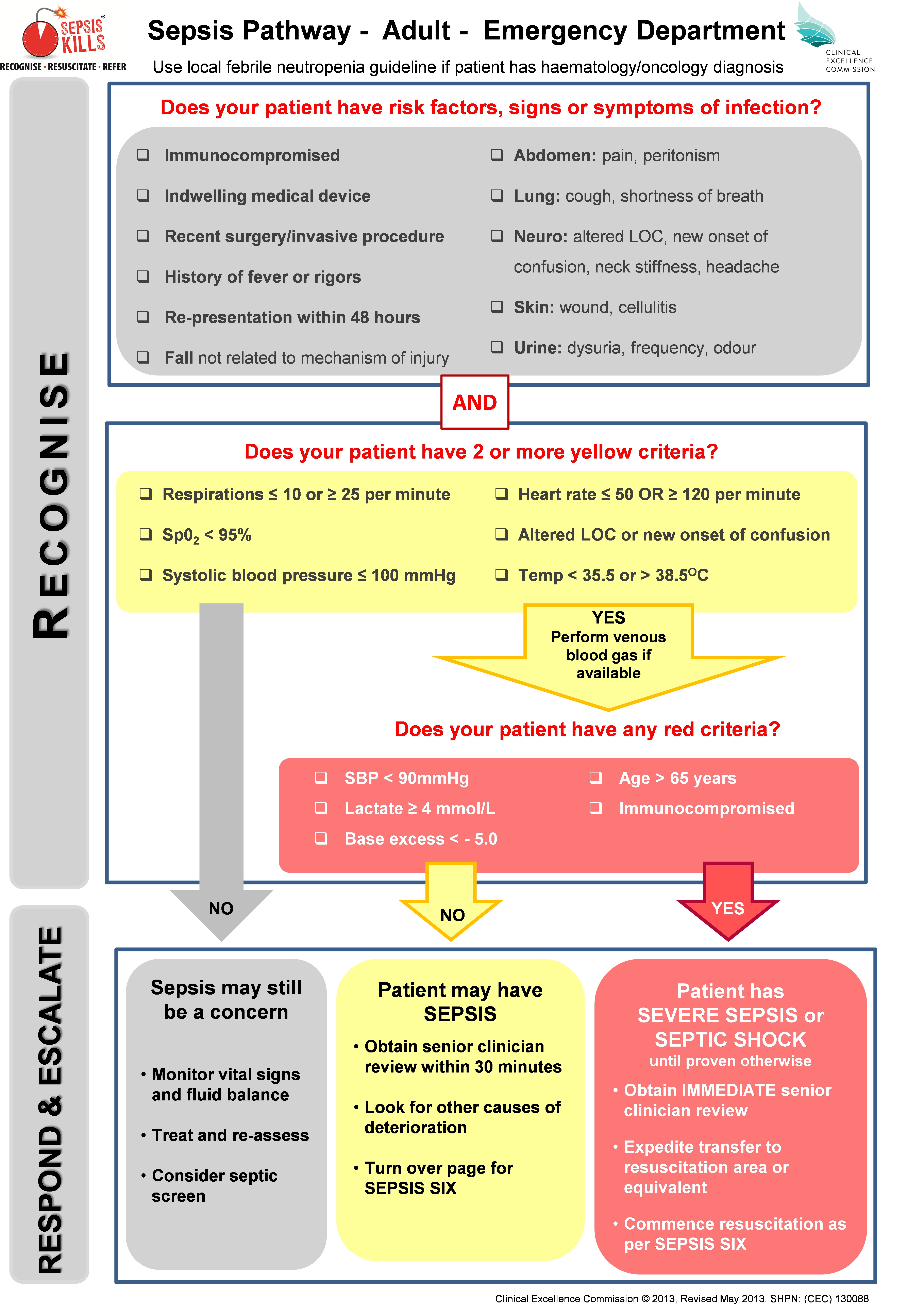
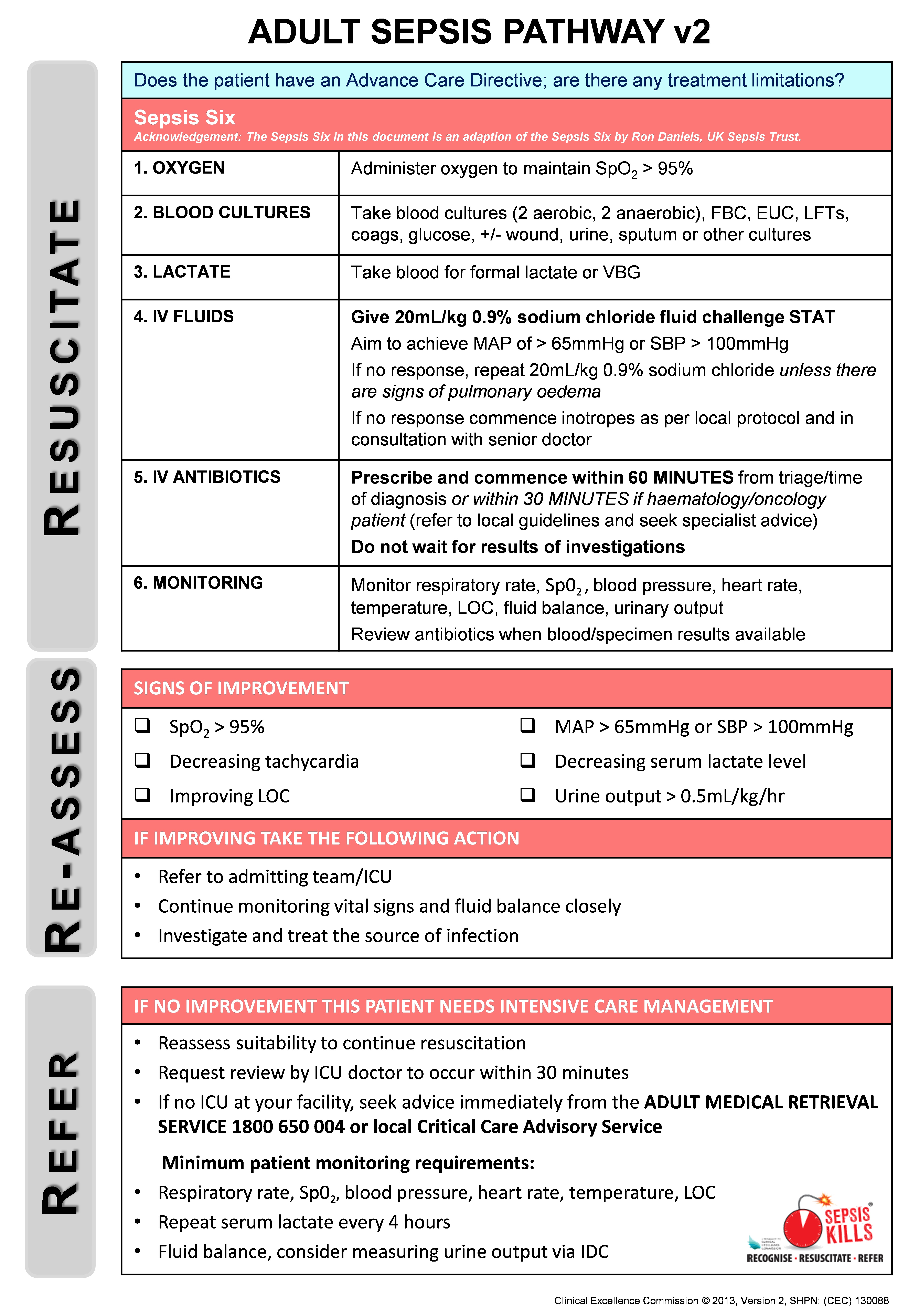
The focus of the program is to RECOGNISE risk factors, signs and symptoms of sepsis; RESUSCITATE with rapid intravenous fluids and antibiotics; and REFER to senior clinicians and teams. Standardised sepsis tools were developed in consultation with NSW emergency physicians, and included adult and paediatric pathways that built on the NSW deteriorating patient system, Between the Flags (BTF).8 The vital signs assessed in the sepsis pathways were consistent with BTF, and varied marginally from accepted systemic inflammatory response criteria (Box 1).

The SEPSIS KILLS pathways promote bundles of care, with the emphasis on early intervention. The adult bundle includes taking blood cultures, measuring serum lactate levels, administering intravenous antibiotics within an hour of triage and recognition, and administering a fluid bolus of 20 mL/kg, followed by another bolus of 20 mL/kg (if necessary) and inotropic drugs if the patient’s condition is not fluid-responsive. If no improvement is observed, senior medical review and admission to intensive care or retrieval to a major centre should be considered (Box 2). The paediatric bundle emphasises the importance of early senior clinical review and decision making. In addition, an empiric antibiotic guideline was developed with advice from expert infectious disease physicians. Emphasis was placed on the first dose of antibiotics, thereby allowing time for further assessment and diagnosis. Because of wide variations in practice, the guideline also contained details on how antibiotics could be administered most expeditiously.

The program was implemented in 2011 with a top-down, bottom-up approach, with strong leadership from medical and nursing clinical leads, and supported by the local health district Clinical Governance Units. Participation was not mandatory, and no extra resources were provided to participating emergency departments who implemented the program. The CEC team supported clinicians by holding a preliminary launch, monthly CEC–hospital teleconferences, executive reports, newsletters, site visits and workshops. A range of online resources was provided, including a Sepsis Toolkit (implementation guide) and various education tools.

Emergency departments were encouraged to collect prospective data on paediatric and adult patients with a provisional diagnosis of sepsis who had received intravenous antibiotics. An online sepsis database (from August 2011) facilitated collection of a minimum dataset for each patient that included their date of birth, triage time and date, triage category, clinical observations (including systolic blood pressure [SBP] and serum lactate levels), time and date of initial intravenous antibiotic treatment and of commencement of the second litre of intravenous fluid, the presumptive source of sepsis, and the disposition of patients following emergency department treatment. Data were collected either prospectively or by retrospective chart review. The database allowed emergency departments to monitor time to antibiotics and fluids in real time, and to compare this with the corresponding local health district and NSW data.

Ethics approval was obtained from the NSW Sepsis Register, which was developed as a public health and disease register under s98 of the Public Health Act 2011. The Sepsis Register is maintained by the CEC.

Data analysis

Analysis of process measures (time to antibiotic, time to intravenous fluid) was based on data from the SEPSIS KILLS database. A total of 13 567 SEPSIS KILLS records were submitted for linkage to the APDC to assess associations between in-hospital mortality and sepsis severity and patient disposition. Patients were classified by emergency department staff according to the Australasian Triage Scale (ATS).9 The cases were further classified as being severe or uncomplicated sepsis according to the serum lactate levels and presenting SBP of the patient.

To assess the population-level impact of the SEPSIS KILLS program, we analysed health outcomes (in-hospital mortality, hours in intensive care, length of stay) for paediatric and adult patients separated from NSW hospitals with ICD-10-AM (International Classification of Diseases, 10th revision, Australian modification) discharge diagnosis codes consistent with sepsis10 recorded in the Admitted Patient, Emergency Department Attendance and Deaths Register. This register was accessed through the NSW Ministry of Health Secure Analytics for Population Health Research and Intelligence (SAPHaRI) system. Linkage was undertaken by the Centre for Health Record Linkage (CHeReL). Only patients admitted to public hospitals with emergency departments were included in the analysis. Trend analysis was performed for the run-in period, August 2009 – December 2011, and for the two following years, 2012 and 2013. Outcomes by sepsis severity could not be analysed at the population level because of the lack of consensus about using ICD-10-AM codes to differentiate between severe and uncomplicated sepsis.

Descriptive and inferential analyses included the calculation of frequencies, odds ratios (ORs) and 95% confidence intervals, and χ2 tests for trends. Trends over time for process and outcome measures were assessed in regression models. Logistic regression was used to analyse in-hospital deaths, while linear regression models were used for time in intensive care and lengths of stay. Models were adjusted as appropriate for covariates (age, year, triage category and severity of sepsis). Statistical significance was defined as P < 0.05.

Results

The SEPSIS KILLS program was implemented as individual emergency departments became ready during 2011. Both retrospective and prospective data were collected by 97 hospitals to 31 December 2013 and entered into the sepsis database. Data were submitted by 13 tertiary, 19 metropolitan and 65 rural hospitals. Because of the low number of paediatric patients, analysis was restricted to data for adult patients.

The provisional sources of sepsis included the lungs (5216 patients, 40.5%), urinary tract (2998, 23.2%), abdomen (1077, 8.4%), skin or soft tissue (975, 7.6%), musculoskeletal system (98, 0.8%), central nervous system (96, 0.7%), vascular device (82, 0.6%), and other systems (973, 7.6%). For 1238 patients (9.6%) the source was unidentified, for 133 (1.0%) no source was recorded.

There were age data in 12 879 records. There was a significant reduction in the mean age of patients between 2009 and 2013, from 67.3 years in 2009–2011 to 64.8 years in 2013 (P < 0.001 for trend; Box 3).

Data for the process indicators from the CEC sepsis database are summarised in Box 3. The proportion of patients who were categorised at triage as ATS 1 (“see immediately”) rose from 2.3% in 2009–2011 to 4.2% in 2013. Those categorised as ATS 2 (“see within 10 minutes”) increased from 40.7% in 2009–2011 to 60.7% in 2013 (P < 0.001). There were small reductions in the proportions of patients classed as ATS 3, 4 or 5.

The proportion of patients who received antibiotics within 60 minutes of triage or recognition increased from 29.3% in 2009–2011 to 52.2% in 2013 (linear trend test, P < 0.001). Similarly, the number of patients who started a second litre of intravenous fluid within one hour rose from 10.6% to 27.5% (linear trend test, P < 0.001).

The analysis of population-based APDC data, which included all separations with emergency department involvement from public hospitals in NSW between January 2009 and December 2013, is presented in Box 4. There were 15 801 sepsis hospital separations during the run-in period of 2009–2011, with a mortality of 19.3%. This rate declined to 17.2% in 2012 and 14.1% in 2013. There was a significant linear decrease over time (P < 0.0001); the OR for death (compared with the run-in period) was 0.87 (95% CI, 0.80–0.94) in 2012, and 0.69 (95% CI, 0.63–0.74) in 2013. Significant linear declines were also observed for time in intensive care and length of stay (for each trend: P < 0.0001).

Linkage of the APDC and sepsis databases showed that the mortality rate for the 1616 patients with severe sepsis (serum lactate ≥ 4 mmol/L or SBP < 90 mmHg) was 19.7%; these patients were significantly more likely to die than patients with uncomplicated sepsis (serum lactate < 4 mmol/L and SBP ≥ 90 mmHg) (OR, 3.7; 95% CI, 3.2–4.4; P < 0.0001). The mortality rate for the 893 patients with hyperlactataemia (a lactate level of 4 mmol/L or more; reference interval, 0.5–2.0 mmol/L) was 24.9%, while that for 637 patients presenting with cryptic shock — hyperlactataemia together with normotension (SBP ≥ 90 mmHg) — was 21.2%. There was no change in mortality for either group over time. For 734 patients who presented with SBP < 90 mmHg and lactate levels < 4 mmol/L, mortality was 13.5%, which declined significantly across the study period (2009–2011, 16.5%; 2012, 16.0%; 2013, 9.8; P = 0.03). The overall mortality rate for uncomplicated sepsis patients increased significantly over time: 3.7% (21/567) in 2009–2011, 6.2% (145/2336) in 2012, and 6.7% (145/2164) in 2013 (P = 0.02).

The mortality rate for the 268 ATS 1 patients was 28.8%. The risk of death for patients over 65 years of age was 3.3 times higher (95% CI, 2.6–4.1) than for patients under 65 years of age (P = 0.001).

The mortality rate for 543 severe sepsis patients admitted to intensive care did not change significantly over time — 23.4% (2009–2011), 19.5% (2012) and 16.0% (2013) (P = 0.145) — nor did the proportion of the 1073 patients with severe sepsis who were admitted to the ward and died — 21.4% (2009–2011), 21.5% (2012) and 18.4% (2013) (P = 0.263). In contrast, the risk of death for 4225 patients with uncomplicated sepsis admitted to the ward increased significantly: 3.2% (15/466) during 2009–2011, 6.0% (115/1914) during 2012, and 6.2% (115/1845) during 2013 (P = 0.047).

Discussion

SEPSIS KILLS is a quality improvement program that aims to reduce preventable harm to patients with sepsis by recognising the condition early and managing it promptly. It is based on the principle that early recognition and aggressive management with antibiotics and fluids will improve outcomes.1113 It is consistent with the 3-hour component of the resuscitation bundle outlined in the international guidelines of the Surviving Sepsis Campaign.3

The program was not planned as a before-and-after project, but was independently implemented by individual emergency departments during 2011. More than 80% of NSW emergency departments (175 of 220) used the sepsis pathways, and 97 emergency departments submitted over 13 000 records. The resulting increase in the number of patients for whom antibiotics were initiated within 60 minutes of recognition and the increased likelihood of the second litre of fluids being started in the first hour indicate that the program was successful. Greater urgency is also apparent from the marked increase in the number of patients classified at triage as ATS 2. We cannot, however, explain the significant age difference between the patients seen in 2012 and 2013.

Reviewing the population-based APDC hospitalisations with an ICD-10-AM code for sepsis showed that there was a steady reduction in mortality over time. Contrary to what we expected, the survival benefit in our patients appears to have been greatest for those with evidence of haemodynamic instability (SBP < 90 mmHg) but normal lactate levels.

The mortality rate of 15%–20% for patients admitted to intensive care with severe sepsis (one-third of the overall sample) is consistent with the overall mortality rate in Australian and New Zealand intensive care units.14,15 In 2013, the crude mortality rate for the patients admitted to wards was higher than that for the intensive care group. We believe the relatively high proportion of ward patients may be the result of an underappreciation of the potential mortality of sepsis, of the significance of elevated lactate levels, and of the time course of the septic process, as well as of failure to recognise cryptic shock16 and the obvious and practical problem of intensive care unit bed availability. We did not assess how many had end-of-life treatment limitations in place.

Managing large numbers of patients with sepsis on the wards has been described elsewhere.17,18 These patients have not been well studied, although a number of studies have identified deficiencies in care.1921 The significant increase in mortality among patients with uncomplicated sepsis admitted to the ward causes concerns that some of our ward patients may have qualified for intensive therapy. An increase in mortality in less severe sepsis has also been documented by other authors.22

The major challenge was managing the prescribing of antibiotics. Despite general acceptance of expert guidelines for prescribing antibiotics, differences in their prescription and administration were observed. This evidence–practice gap is well recognised,23 and the empiric antibiotic guideline was developed to promote appropriate antibiotic prescribing practices and optimal outcomes.24 The empiric guideline was consistent with the principles of antimicrobial stewardship, and, while each site was allowed to modify it according to local opinion and antibiotic resistance patterns, changes were infrequent. Particular anxieties were expressed about prescribing and administering gentamicin. The administration component of the guideline was developed to promote the most expeditious method of administration rather than favouring the slow infusion that had become normal practice. Despite the emphasis on the first dose and timely review, antibiotics were often continued long after they should have been reviewed, following consideration of the results of pathology investigations.

Other challenges beyond our control included educating a high turnover workforce in emergency departments, as well as medical engagement, particularly in rural facilities where governance is difficult and there is no doctor on site, or locum medical staff are more common. There were wide variations in the methods of blood culture collection, and a standardised guideline for blood cultures was subsequently added to the Sepsis Toolkit.

Limitations

This work is subject to the limitations of any quality improvement project at multiple sites. The prolonged run-in period was not ideal. Our approach was not to measure compliance with the care bundle, as undertaken elsewhere, but to use time as a measure for promoting behavioural change among emergency department clinicians. Assessing patient outcomes was the major difficulty. The voluntary nature of data collection resulted in its inconsistent submission, and the lack of strict diagnostic criteria for sepsis resulted in patients with conditions from across the inflammatory condition–sepsis spectrum being included in the SEPSIS KILLS database. Resource limitations also meant that some sites implemented the pathways but did not submit data.

Reviewing the outcomes of patients with an ICD-10-AM code for sepsis in the population-based administrative APDC is an accepted approach. This, however, entails the risk of reviewing the outcomes of a different group of patients, a group for whom the final diagnosis might not be directly related to sepsis or its severity. This is a potential problem when comparing the final diagnosis in the APDC database with the provisional diagnosis in the SEPSIS KILLS data.

Finally, the improved outcomes described in our article may be the result of the SEPSIS KILLS program, but may also be related to other initiatives for improving quality of care.

Implications for clinicians, researchers and policy makers

The observation that patients with severe sepsis are being managed on the wards highlights the need for a shift in the focus of both practice improvement and research from intensive care to ward management. It also raises the problem of sepsis and the deteriorating patient. We informally estimated that around 30% of patients who required a Rapid Response call had sepsis, but this may be an underestimate, with rates possibly as high as 50%–60%.25 Finally, our work confirms the need for continued research into risk stratification tools for sepsis in the emergency department. In the meantime, all patients with lactate levels of 4 mmol/L or greater require intensive care unit review and admission.

The SEPSIS KILLS program promotes early recognition and management of sepsis during the first few hours in NSW emergency departments. By focusing on the principle of “Recognise, Resuscitate, Refer” it is possible to reduce the time before antibiotics are administered and fluid resuscitation initiated. This program could be applied in other jurisdictions and its integration with antimicrobial stewardship requirements should be considered.

Box 1 –
The SEPSIS KILLS pathway for adult patients in hospital emergency departments, page 1

Box 2 –
The SEPSIS KILLS pathway for adult patients in hospital emergency departments, page 2

Box 3 –
Characteristics, and process and outcome indicators of sepsis-related hospital separations before and after the launch of the SEPSIS KILLS program

Characteristics

Run-in period


SEPSIS KILLS program in operation


P for trend

Aug 2009 – Dec 2011

2012

2013


Number of separations

1585

5396

5905

Mean age ± SEM, years

67.3 ± 0.5

67.6 ± 0.3

64.8 ± 0.3

< 0.001

Triage category*

< 0.001

1

37 (2.3%)

176 (3.3%)

242 (4.2%)

2

463 (40.7%)

2765 (51.5%)

3532 (60.7%)

3

683 (43.2%)

2034 (37.9%)

1767 (30.4%)

4

207 (13.1%)

378 (7.0%)

267 (4.6%)

5

10 (0.6%)

16 (0.3%)

12 (0.2%)

Missing data

5

22

83

Antibiotic received within 60 min

464 (29.3%)

2165 (40.2%)

3083 (52.2%)

< 0.001

Second litre of intravenous fluid within 60 min

135 of 1272 patients (10.6%)

521 of 3631 patients (14.3%)

991 of 3609 patients (27.5%)

< 0.001


SEM = standard error of the mean.

Box 4 –
Hospital outcomes prior to and after the launch of the SEPSIS KILLS program, NSW, January 2009 to December 2013

Outcomes

Descriptive statistics

Odds ratio (95% CI)

P for trend


Deaths, numbers (percentage)

< 0.0001

2009–2011

3053/15 801 (19.3%)

1

2012

979/5683 (17.2%)

0.87 (0.80–0.94)

2013

870/6167 (14.1%)

0.69 (0.63–0.74)

Mean time in intensive care (SEM), hours

< 0.0001

2009–2011

32.7 (1.0)

2012

26.6 (1.4)

2013

25.8 (1.3)

Mean length of stay (SEM), days

< 0.0001

2009–2011

13.5 (0.1)

2012

12.2 (0.2)

2013

11.5 (0.2)


SEM = standard error of the mean. Source: Admitted Patient Data Collection, NSW Ministry of Health Secure Analytics for Population Health Research and Intelligence (SAPHaRI). Data extracted on 1 June 2015.