×

It’s official: Zika causes birth defects

The United States’ Centers for Disease Control and Prevention has declared that the Zika virus is a cause of microcephaly and other severe foetal brain defects, confirming long-held suspicions about the infection’s link to serious neurological disorders.

As the US gears up for outbreaks of the potentially deadly virus, the CDC has reported that an accumulation of evidence proves Zika can cause birth defects and pregnant women living in or travelling to areas of where it is prevalent should strictly follow steps to avoid mosquito bites and prevent sexual transmission of the virus.

“This study marks a turning point,” CDC Director Dr Tom Frieden said. “It is now clear that the virus causes microcephaly. We’ve now confirmed what mounting evidence has suggested, affirming our early guidance to pregnant women and their partners to take steps to avoid Zika infection.”

The CDC report, published in the New England Journal of Medicine, said its conclusion was not based on any one discovery but rather an accumulation of evidence from a number of recently published studies and a careful evaluation using established scientific criteria.

The CDC announcement came as the Australasian Society for Infectious Diseases reminded GPs to be on heightened alert for tropical diseases in patients with febrile illnesses – particularly those who have recently travelled overseas.

Society President Professor Cheryl Jones said serious tropical diseases including Zika, multi-drug resistant malaria and dengue were endemic in many overseas destinations popular with Australians, including Thailand, Vietnam, Myanmar, Laos and Cambodia, and there was also a local outbreak of dengue in northern Queensland.

“There has never been a more critical time for Australian health professionals to get up to speed with developments in tropical medicine,” Professor Jones said. “With malaria resistance growing and no antiviral treatment available for dengue, Zika and other mosquito-borne viruses, it is imperative that Australian doctors are able to identify these diseases and refer patients swiftly.”

Her warning came as a senior US public health official, Dr Anne Schuchat, told a White House briefing that the virus “seems to be a bit scarier than we initially thought”.

Dr Schuchat, who is a deputy director of the US Centers for Disease Control and Prevention, said that initially it was thought the species of mosquito primarily associated with carrying the disease was only present in about 12 states, but that had now been revised up to 30 states.

Authorities are particularly concerned about the US territory of Puerto Rico, where they fear there may be hundreds of thousands of infections, but the speed of the disease’s spread has them concerned it may soon appear in continental US as temperatures rise.

“While we absolutely hope we don’t see widespread local transmission in the continental US, we need the states to be ready for that,” Dr Schuchat said.

While the Zika virus has been documented in 61 countries since 2007, the World Health Organization said its transmission has really taken off since it was first detected in Brazil in May last year, and it is now confirmed in 33 countries in Central and South America, as well as 17 countries and territories in the Western Pacific, including New Zealand (one case of sexual transmission), Fiji, Samoa, Tonga, American Samoa, Micronesia and the Marshall Islands.

Its appearance has been linked to a big jump in cases of microcephaly, Guillian-Barre syndrome (GBS) and other birth defects and neurological disorders, and the WHO said that there was now “a strong scientific consensus” that the virus was the cause.

In Brazil, there were 6776 cases of microcephaly or central nervous system malformation (including 208 deaths) reported between October last year and the end of March. Before this, an average of just 163 cases of microcephaly were reported in the country each year.

The WHO reported 13 countries or territories where there has been an increased incidence of GBS linked to the Zika virus. French Polynesia experienced its first-ever Zika outbreak in late 2013, during which 42 patients were admitted to hospital with GBS – a 20-fold increase compared with the previous four years. All 42 cases were confirmed for Zika virus infection.

Similar increases in the incidence of GBS cases have been recorded in other countries where there is Zika transmission, including Brazil, Colombia, El Salvador, Venezuela, Suriname and the Dominican Republic.

Scientists have also detected potential links between the infection and other neurological disorders. In Guadeloupe, a 15-year-old girl infected with Zika developed acute myelitis, while an elderly man with the virus developed meningoencephalitis. Meanwhile, Brazilian scientists believe Zika is associated with an autoimmune syndrome, acute disseminated encephalomyelitis.

Scientists worldwide are working to develop a vaccine for the virus, and an official with the US National Institute of Allergy and Infectious Diseases said initial clinical trials of a vaccine might begin as soon as September.

Meanwhile, research on other aspects of Zika, including its link with neurological disorders, sexual transmission and ways to control the mosquitos that spread the disease is being coordinated internationally.

So far, the only confirmed cases of Zika in Australia have involved people who were infected while travelling overseas, and authorities are advising any women who are pregnant or seeking to get pregnant to defer travelling to any country where there is ongoing transmission of the virus.

Adrian Rollins

Get back to the BEACH, Govt told

AMA President Professor Brian Owler has urged the Federal Government to reverse its decision to axe funding for one of the most extensive and sustained studies of general practice in the world, arguing the move is “completely at odds” with its stated primary care focus.

In a decision that has shocked and dismayed medical practitioners and researchers, the long-running Bettering the Evaluation and Care of Health (BEACH) program, which began tracking the activities of Australian GPs in 1998, is being wound up after the Federal Department of Health announced it would not be renewing funding for the research after the current contract expires on 30 June.

Professor Owler has written to Health Minister Sussan Ley urging her to reconsider the move, which he said was particularly ill-considered given major changes planned for primary care.

“Research into general practice and primary care attracts very little funding support in comparison to other parts of the health system,” the AMA President said. “The reality is that we need more of this type of research, not less.”

The Government’s decision to axe its funding for BEACH has come less than two week after Ms Ley unveiled the Health Care Homes initiative to give GPs a central role in improving the care of patients with chronic and complex disease. Simultaneously, the Government is trialling its My Health Record e-health record system and is persisting with a four-year freeze on Medicare rebates.

Professor Owler said the Commonwealth had contributed just $4.6 million of the $26 million that had been used to fund the BEACH program over the years.

“This is a very small investment that has delivered significant policy outcomes and, with all the changes planned for general practice and primary care, I think there is a very strong case to extend funding for the program,” he said.

The wealth of data on general practice that the program had collected had proven invaluable in driving evidence-based policy development, Professor Owler said, and warned that there was “no credible source of information and analysis that is capable of filling the gap that will be left when the program ceases”.

The program’s director, Professor Helena Britt of Sydney University’s Family Medicine Research Centre, said the Government’s decision to cease its contribution had come at a time when the program was already facing a funding crunch caused by a downturn in contributions from other sources including non-government organisations and pharmaceutical companies.

“BEACH has always struggled to gain sufficient funds each year,” Professor Britt said. “However, this notification comes when we also have a large shortfall in funding coming from other organisations…due to the closure of many government instrumentalities and authorities, and the heavy squeeze on pharmaceutical companies’ profits resulting from changes to the PBS.

“We therefore have no choice but to close the BEACH program.”

Professor Britt said she had been inundated with inquiries and messages of support from individuals and groups around the country and internationally.

Professor Britt said the BEACH data, which is drawn from an annual sample of GPs providing detailed information on everything from the hours they work to the diseases and other conditions they treat, was a unique resource, and the program’s closure would “leave Australia with no valid reliable and independent source of data about activities in general practice”.

“BEACH has been the only continuous national study of general practice in the world which relies on random samples of GPs, links management actions to the exact problem being managed, and provides extensive measurement of prevalence of diseases, multi-morbidity and adverse medication events,” a statement issued by the Family Medicine Research Centre said.

The data from the latest BEACH survey, which began in April last year and closed at the end of March this year, is being collated and Professor Britt said she hoped to issue a report on the results, possibly in mid-June.

Asked about the possibility of funding coming from other sources, Professor Britt said it was “early days”.

One of the biggest concerns is what will happen to the rich store of data accumulated through the program’s 18 years of operation, during which time more than 11,000 GPs have been surveyed.

Professor Britt said the data was used by a huge range of researchers and organisations, and her group was looking at ways to ensure people would continue to have access to it.

“We would be happy to find a place with a senior analyst who could take request to analyse the data for specific purposes,” she said. “We would like to be able to keep that access up there for at least a little while.”

Adrian Rollins

US gears up for ‘scary’ Zika

The United States is gearing up for outbreaks of the potentially deadly Zika virus amid concerns the mosquito-borne infection can also be sexually transmitted and may cause neurological disorders in adults as well as children.

As Australian health authorities monitor the appearance of the disease, particularly in areas of the country where mosquito vectors are present, a senior US public health official, Dr Anne Schuchat, told a White House briefing that the virus “seems to be a bit scarier than we initially thought”, and health authorities are ramping up efforts to research the disease and raise public awareness of the threat.

Dr Schuchat, who is a deputy director of the US Centers for Disease Control and Prevention, said that initially it was thought the species of mosquito primarily associated with carrying the disease was only present in about 12 states, but that had now been revised up to 30 states.

Authorities are particularly concerned about the US territory of Puerto Rico, where they fear there may be hundreds of thousands of infections, but the speed of the disease’s spread has them concerned it may soon appear in continental US as temperatures rise.

“While we absolutely hope we don’t see widespread local transmission in the continental US, we need the states to be ready for that,” Dr Schuchat said.

Related: MJA – First report of Zika virus infection in a returned traveller from the Solomon Islands

While the Zika virus has been documented in 61 countries since 2007, the World Health Organization said its transmission has really taken off since it was first detected in Brazil in May last year, and it is now confirmed in 33 countries in Central and South America, as well as 17 countries and territories in the Western Pacific, including New Zealand (one case of sexual transmission), Fiji, Samoa, Tonga, American Samoa, Micronesia and the Marshall Islands.

Its appearance has been linked to a big jump in cases of microcephaly, Guillian-Barre syndrome (GBS) and other birth defects and neurological disorders, and the WHO said that there was now “a strong scientific consensus” that the virus was the cause.

In Brazil, there were 6776 cases of microcephaly or central nervous system malformation (including 208 deaths) reported between October last year and the end of March. Before this, an average of just 163 cases of microcephaly were reported in the country each year.

The WHO reported 13 countries or territories where there has been an increased incidence of GBS linked to the Zika virus. French Polynesia experienced its first-ever Zika outbreak in late 2013, during which 42 patients were admitted to hospital with GBS – a 20-fold increase compared with the previous four years. All 42 cases were confirmed for Zika virus infection.

Similar increases in the incidence of GBS cases have been recorded in other countries where there is Zika transmission, including Brazil, Colombia, El Salvador, Venezuela, Suriname and the Dominican Republic.

Related: Zika app, registry for pregnant women

Scientists have also detected potential links between the infection and other neurological disorders. In Guadeloupe, a 15-year-old girl infected with Zika developed acute myelitis, while an elderly man with the virus developed meningoencephalitis. Meanwhile, Brazilian scientists believe Zika is associated with an autoimmune syndrome, acute disseminated encephalomyelitis.

Scientists worldwide are working to develop a vaccine for the virus, and an official with the US National Institute of Allergy and Infectious Diseases said initial clinical trials of a vaccine might begin as soon as September.

Meanwhile, research on other aspects of Zika, including its link with neurological disorders, sexual transmission and ways to control the mosquitos that spread the disease is being coordinated internationally.

So far, the only confirmed cases of Zika in Australia have involved people who were infected while travelling overseas, and authorities are advising any women who are pregnant or seeking to get pregnant to defer travelling to any country where there is ongoing transmission of the virus.

Adrian Rollins

Latest news:

BEACH washed up

Attempts to gauge the effect of big changes to chronic disease management and primary care being planned by the Federal Government have been dealt a blow by revelations one of the most extensive and sustained studies of general practice in the world is facing shutdown.

The long-running Bettering the Evaluation and Care of Health (BEACH) program, which began tracking the activities of Australian GPs in 1998, is being wound up after the Federal Department of Health announced it would not be renewing funding for the research after the current contract expires on 30 June.

The program’s director, Professor Helena Britt of Sydney University’s Family Medicine Research Centre, said the Department’s decision had come at a time when the program was already facing a funding crunch caused by a downturn in contributions from other sources including non-government organisations and pharmaceutical companies.

“BEACH has always struggled to gain sufficient funds each year,” Professor Britt said. “However, this notification comes when we also have a large shortfall in funding coming from other organisations…due to the closure of many government instrumentalities and authorities, and the heavy squeeze on pharmaceutical companies’ profits resulting from changes to the PBS.

“We therefore have no choice but to close the BEACH program.”

The announcement has been met with shock and dismay by medical practitioners and researchers. Professor Britt said she had been inundated with inquiries and messages of support from individuals and groups around the country and internationally.

BEACH’s shutdown comes at a particularly uncertain time for general practice as the Government moves to implement its Health Care Homes model of chronic care while simultaneously trialling its My Health Record e-health record and persisting with a four-year freeze on Medicare rebates.

Professor Britt said the BEACH data, which is drawn from an annual sample of GPs providing detailed information on everything from the hours they work to the diseases and other conditions they treat, was a unique resource, and the program’s closure would “leave Australia with no valid reliable and independent source of data about activities in general practice”.

“BEACH has been the only continuous national study of general practice in the world which relies on random samples of GPs, links management actions to the exact problem being managed, and provides extensive measurement of prevalence of diseases, multi-morbidity and adverse medication events,” a statement issued by the Family Medicine Research Centre said.

The data from the latest BEACH survey, which began in April last year and closed at the end of March this year, is being collated and Professor Britt said she hoped to issue a report on the results, possibly in mid-June.

Asked about the possibility of funding coming from other sources, Professor Britt said it was “early days”.

One of the biggest concerns is what will happen to the rich store of data accumulated through the program’s 18 years of operation, during which time more than 11,000 GPs have been surveyed.

Professor Britt said the data was used by a huge range of researchers and organisations, and her group was looking at ways to ensure people would continue to have access to it.

“We would be happy to find a place with a senior analyst who could take request to analyse the data for specific purposes,” she said. “We would like to be able to keep that access up there for at least a little while.”

Adrian Rollins

Old but not forgotten: Antibiotic allergies in General Medicine (the AGM Study)

The prevalence of antibiotic allergy labels (AAL) has been estimated to be 10–20%.1,2 AALs have been shown to have a significant impact on the use of antimicrobial drugs, including their appropriateness, and on microbiological outcomes for patients.3,4 Many reported antibiotic allergies are, in fact, drug intolerances or side effects, or non-recent “unknown” reactions of questionable clinical significance. Incorrect classification of patient AALs is exacerbated by variations in clinicians’ knowledge about antibiotic allergies and the recording of allergies in electronic medical records.57 The prevalence of AALs in particular subgroups, such as the elderly, remains unknown; the same applies to the accuracy of AAL descriptions and their impact on antimicrobial stewardship. While models of antibiotic allergy care have been proposed8,9 and protocols for oral re-challenge in patients with “low risk allergies” successfully employed,10 the feasibility of a risk-stratified direct oral re-challenge approach remains ill defined. In this multicentre, cross-sectional study of general medical inpatients, we assessed the prevalence of AALs, their impact on prescribing practices, the accuracy of their recording, and the feasibility of an oral antibiotic re-challenge study.

Methods

Study design, setting and population

Austin Health and Alfred Health are tertiary referral centres located in north-eastern and central Melbourne respectively. This was a multicentre, cross-sectional study of general medical inpatients admitted between 18 May 2015 and 5 June 2015; those admitted to an intensive care unit (ICU), emergency unit or short stay unit were excluded from analysis.

At 08:00 (Monday to Friday) during the study period, a list of all general medical inpatients was generated. Baseline demographics, comorbidities (age-adjusted Charlson comorbidity index11), infection diagnoses, and inpatient antibiotic medications (name, route, frequency) were recorded. Patients with an AAL were identified from drug charts, medical admission notes, or electronic medical records (EMRs). A patient questionnaire was administered to clarify AAL history (Appendix), followed by correlation of the responses with allergy descriptions in the patient’s drug chart, EMR and medical admission record. To maintain consistency, this questionnaire was administered by pharmacy and medical staff trained at each site. Patients with a history of dementia or delirium who were unable to provide informed consent were excluded only from the patient questionnaire component of the study. A hypothetical oral antibiotic re-challenge in a supervised setting was offered to patients with a low risk allergy phenotype (Appendix).

Definitions

An AAL was defined as any reported antibiotic allergy or adverse drug reaction (ADR) recorded in the allergy section of the EMR, drug chart, or medical admission note. AALs were classified as either type A or type B ADRs according to previously published definitions (Box 1):12,13

  • type A: non-immune-mediated ADR consistent with a known drug side effect (eg, gastrointestinal upset);

  • type B: immune-mediated reactions consistent with an IgE-mediated (eg, angioedema, anaphylaxis, or urticaria = type B-I) or a T cell-mediated response (type B-IV):

    • Type B-IV: delayed benign maculopapular exanthema (MPE);

    • Type B-IV* (life-threatening in nature): severe cutaneous adverse reactions (SCAR),14 erythema multiforme (EM), fixed drug eruption (FDE), serum sickness, and antibiotic-induced haemolytic anaemia.

Study investigators JAT and AKA categorised AALs; if consensus could not be reached, a third investigator (LG) was recruited to adjudicate.

An AAL was defined as a “low risk phenotype” if it was consistent with a non-immune-mediated reaction (type A), delayed benign MPE without mucosal involvement that had occurred more than 10 years earlier (type B-IV), or an unknown reaction that had occurred more than 10 years earlier. Unknown reactions in patients who could not recall when the reaction had occurred were also classified as low risk phenotypes. All low risk phenotypes were ADRs that did not require hospitalisation. A “moderate risk phenotype” included an MPE or unknown reaction that had occurred within the past 10 years. A “high risk phenotype” was defined as any ADR reflecting an immediate reaction (type B-I) or non-MPE delayed hypersensitivity (type B-IV*).

AAL mismatch was defined as non-concordance between a patient’s self-reported description of an antibiotic ADR in the questionnaire and the recorded description in any of the medical record platforms (drug charts, medical admission notes, EMR). Infection diagnosis was classified according to Centers for Disease Control/National Healthcare Safety definitions.15

Statistical analysis

Statistical analyses were performed in Stata 12.0 (StataCorp). Variables of interest in the AAL and no antibiotic allergy label (NAAL) groups were compared. Categorical variables were compared in χ2 tests, and continuous variables with the Wilcoxon rank sum test. P < 0.05 (two-sided) was deemed statistically significant.

Ethics approval

The human research ethics committees of both Austin (LNR/15/Austin/93) and Alfred Health (project 184/15) approved the study.

Results

Antibiotic allergy label description and classification

The baseline patient demographics for the AAL and NAAL groups are shown in Box 2. Of the 453 patients initially identified, 107 (24%) had an AAL. A total of 160 individual AALs were recorded: 27 were type A (17%), 26 were type B-I (16%), 45 were type B-IV (28%), and 62 were of unknown type (39%) (Box 3). Sixteen of the type B-IV reactions (35%) were consistent with more severe phenotypes (type B-IV*). When the time frame criterion (more than 10 years v 10 years or less since the index reaction) was applied to phenotype definitions, this translated to 63% low risk (101 of 160), 4% moderate risk (7 of 160), and 32% high risk (52 of 160) phenotypes. The antibiotics implicated in AALs and their ADR classifications are summarised in Box 3; 34% of reactions were to simple penicillins, 13% to sulfonamide antimicrobials, and 11% to cephalosporins. Three AAL patients (2.8%) were referred to an allergy specialist for assessment (one with type A, two with type B-I reactions). No recorded AALs were associated with admission to an ICU, while eight either ended or occurred during the index hospital admission (two type A, five type B-I, and one type B-IV).

Antibiotic use

Ceftriaxone was prescribed more frequently for patients with AALs (29 of 89 [32%]) than for those in the NAAL group (74 of 368 [20%]; P = 0.02); flucloxacillin was prescribed less frequently (0 v 21 of 368 [5.7%]; P = 0.02). The rate of prescription of other restricted antibiotics, including carbapenems, monobactams, quinolones, glycopeptides and lincosamides, was low in both groups (Box 4).

Antibiotic cross-reactivity

Seventy patients had a documented reaction to a penicillin (a total of 72 penicillin AALs: 55 to penicillin V or G, eight to aminopenicillins, nine to anti-staphylococcal penicillins), including two patients with two separate penicillin allergy labels to members of different β-lactam classes. Of these, 23 (32.9%) were prescribed and tolerated cephalosporins (Box 5). Of the 55 patients with a penicillin V/G AAL, β-lactam antibiotics were prescribed for 19 patients (34%); one patient received aminopenicillins (1.8%), four first generation cephalosporins (7%), two second generation cephalosporins (3.6%), and 12 received third generation cephalosporins (21.8%). Conversely, 18 patients had documented ADRs to cephalosporins, with a total of 19 AALs (14 to first generation, one to second generation, two to third generation cephalosporins, and two to cephalosporins of unknown generation). Of these, five patients (27.8%) were again prescribed cephalosporins without any reaction, and a further five (27.8%) tolerated any penicillin (Box 5).

Eight patients with AALs (7%) were administered an antibiotic from the same antibiotic class. No adverse events were noted in any of the patients inadvertently re-challenged. Eighty-six AAL patients (77%) reported a history of taking any antibiotic after their index ADR event. Thirteen patients (12%) believed they had previously received an antibiotic to which they were considered allergic, 62 had not (58%), and 32 were unsure (30%).

Recording of AALs

Almost all AALs (156 of 160 [98%]) were documented in medication charts, but only 115 (72%) were documented in admission notes and 81 (51%) in the EMR. Twenty-five per cent of patients had an AAL mismatch. No patients received the exact antibiotic recorded in the AAL.

Hypothetical oral antibiotic re-challenge

Fifty-eight AAL patients (54%) were willing to undergo a hypothetical oral antibiotic re-challenge in a supervised environment, of whom 28 (48%) had a low risk phenotype, seven a moderate risk phenotype (12%), and 23 a high risk phenotype (40%). If patients had received and tolerated an antibiotic to which they were previously considered allergic, they were more likely to accept a hypothetical re-challenge than those who had not (9 of 12 [75%] v 3 of 12 [25%]; P = 0.04).

Discussion

The major users of antibiotics in community and hospital settings remain our expanding geriatric population.16 An accumulation of AALs, reflecting both genuine allergies (immune-mediated) and drug side effects or intolerances, follows years of antibiotic prescribing. This is reflected in the high AAL prevalence (24%) in our cohort of older Australian general medical inpatients, notably higher than the national average (18%) and closer to that reported for immune-compromised patients (20–23%).4,17

To understand the high prevalence of AALs and the predominance of low risk phenotypes in our study group requires an understanding of “penicillin past”, as many AALs are confounded by the impurity of early penicillin formulations and later penicillin contamination of cephalosporin products.18,19 Re-examining non-recent AALs of general medical inpatients is therefore potentially both a high yield and a low risk task, considering the low pre-test probability of a persistent genuine penicillin allergy.2022 While the definition of a low risk allergy phenotype is hypothetical, it is based upon findings that indicate the loss of allergy reactivity over time,20,21,23 the low rate of adverse responses to challenges in patients with mild delayed hypersensitivities,20,22,23 and the safety of oral challenge in patients with similar phenotypes.24

The high rate of type A, non-severe MPE and of non-recent unknown reactions in our patients (74% of all AALs; 63% low risk phenotypes) provides a large sample size to explore further, while the higher use of antibiotics that are the target of antimicrobial stewardship programs (eg, ceftriaxone) in AAL patients provides an impetus for change. The increased use of restricted antibiotics (eg, ceftriaxone and fluoroquinolones) and the reduced use of simple penicillins (eg, flucloxacillin) in patients with an AAL were marked. The effects of AALs on antibiotic prescribing have been described in large hospital cohorts and in specialist subgroups (eg, cancer patients).3,4 Associations between AALs and inferior patient outcomes, higher hospital costs and microbiological resistance have also been recently noted.24,8,17,25 Re-examining AALs in older patients from an antimicrobial stewardship viewpoint is therefore essential, particularly in an era when multidrug-resistant (MDR) organisms are being isolated more frequently in Australia.26 The fact that third generation cephalosporins and fluoroquinolones are associated with MDR organisms and with Clostridium difficile infection generation further supports the need for re-examining AALs, especially in those with easily resolved non-genuine allergies.2730

The high rate of potential patient acceptance of an oral re-challenge (54%), especially by those with low risk phenotypes (48%), suggests that this should be explored in prospective studies. The idea of an antibiotic allergy re-challenge of low risk phenotypes is a practical extension of the work by Blumenthal and colleagues,24 who found a sevenfold increase in β-lactam uptake and a low rate of adverse reactions. Another group found that oral re-challenge was safe in children with a history of delayed allergy.23 These are both important advances; while skin-prick allergy testing is sensitive for immediate penicillin hypersensitivity, skin testing (delayed intradermal and patch) lacks sensitivity for delayed hypersensitivities.8,22,31 Incident-free accidental re-challenge with the culprit antibiotic or a drug from a similar class had occurred in some of our patients, adding further support for exploring this approach. A structured oral re-challenge strategy is attractive, as skin-prick testing is potentially expensive and inaccessible for most people.8

Analysing the high rate of AAL mismatch may be a more pragmatic low-cost approach, as not only were AAL labels absent from a number of medical records, the EMR AAL often differed from patients’ reports. Incorrect and absent AALs in other centres have been raised as a concern from a drug safety viewpoint.6,7,10 Education programs aimed at improving clinicians’ (pharmacy and medical) understanding of allergy pathogenesis could also assist antibiotic prescribing in the presence of AALs.5,10 Interrogation of the patient and their relatives about allergy history and examination of blood investigations at the time of the ADR for evidence of end organ dysfunction or eosinophilia may also provide greater accuracy in phenotyping and severity assessment. Many accumulated childhood allergies reflect the infectious syndrome that resulted in the implicated antibiotic being prescribed, rather than an immunologically mediated drug hypersensitivity.21,23 Referral to allergy specialists at the time of drug hypersensitivity may also reduce over-labelling.

That a clinician questionnaire about antibiotic prescribing attitudes was not administered is a limitation of this study, as was the inability to obtain AAL information from all patients (eg, because of dementia or delirium) or to further clarify “unknown” reactions. Some AAL descriptions are also likely to be affected by recall bias; however, this reflects real world attitudes and prescribing in the presence of AALs. While the prevalence of AALs in younger patients is probably lower than found in this study, the distribution of genuine, non-genuine and low risk allergies may well be the same. In a group of paediatric patients with an AAL for β-lactam antibiotics following non-immediate mild cutaneous reactions without systemic symptoms, none experienced severe reactions after undergoing oral re-challenge.23

Conclusion

AALs were highly prevalent in our older inpatients, with a significant proportion involving non-genuine allergies (eg, drug side effects) and low risk phenotypes. Most patients were willing to undergo a supervised oral re-challenge if their allergy was deemed low risk. AALs were sometimes associated with inadvertent class re-challenges, facilitated by poor allergy documentation, without ill effect. AALs were also associated with increased prescribing of ceftriaxone and fluoroquinolone, antibiotics commonly restricted by antimicrobial stewardship programs. These findings inform a mandate to assess AALs in the interests of appropriate antibiotic use and drug safety. Prospective studies incorporating AALs into antimicrobial stewardship and clinical practice are required.

Box 1 –
Classification of reported antibiotic allergy labels into adverse drug reaction groups12,13


EM=erythema multiforme; FDE=fixed drug eruption; MPE=maculopapular exanthema; SCAR=severe cutaneous adverse reactions (includes Stevens–Johnson syndrome, toxic epidermal necrolysis, drug rash with eosinophilia and systemic symptoms, and acute generalised exathematous pustulosis). *These adverse reactions are classified as type B-IV* in this study, denoting their potentially life-threatening nature.

Box 2 –
Baseline demographics for patients with and without antibiotic allergy labels

Characteristic

Patients with an antibiotic allergy label

Patients with no antibiotic allergy label

P


Number

107

346

Median age [IQR], years

82 [74–87]

80 [71–88]

0.32

Sex, men*

38 (36%)

194 (56%)

< 0.001

Immunosuppressed

25 (23%)

29 (8%)

< 0.001

Median age-adjusted Charlson Comorbidity Index score [IQR]

6 [4–7]

6 [4–7]

0.17

Ethnicity

0.38

European

106 (99%)

334 (97%)

African

0

2 (1%)

Asian

1 (1%)

10 (3%)

Infection diagnosis

50 (47%)

140 (41%)

0.25

Infections (205 patients)

56

151

0.002

Cardiovascular system

0

2 (1%)

Central nervous system

1 (2%)

3 (2%)

Gastrointestinal

9 (16%)

9 (6%)

Eyes, ears, nose and throat

0

3 (2%)

Upper respiratory tract

7 (13%)

30 (20%)

Lower respiratory tract (including pneumonia)

12 (21%)

54 (36%)

Skin and soft tissue

7 (13%)

14 (9%)

Urinary system

11 (20%)

21 (14%)

Pyrexia (no source)

3 (5%)

4 (3%)

Sepsis (unspecified)

5 (9%)

8 (5%)

Other

0

2 (1%)

Received antibiotics

45 (42%)

162 (46%)

0.43


* There were a total of 232 men and 221 women in the study.

Box 3 –
Spectrum of implicated antibiotics linked with reported antibiotic allergy labels according to adverse drug reaction classification

Implicated antibiotics

Antibiotic allergy labels: adverse drug reactions


Type A

Type B


Unknown

Total

Type B-I

Type B-IV

Type B-IV*


All antibiotics

27 (17%)

26 (16%)

29 (18%)

16 (10%)

62 (39%)

160

Simple penicillins*

7 (26%)

14 (54%)

16 (55%)

4 (25%)

14 (23%)

55 (34%)

Aminopenicillins

1 (4%)

2 (8%)

2 (7%)

1 (6%)

2 (3%)

8 (5%)

Anti-staphylococcal penicillins

0

0

1 (3%)

5 (31%)

3 (5%)

9 (6%)

Cephalosporins

3 (11%)

1 (4%)

1 (3%)

2 (13%)

11 (18%)

18 (11%)

Carbapenems§

0

0

0

0

1 (2%)

1 (0.6%)

Monobactam

0

0

0

0

0

0

Fluoroquinolones

2 (7%)

0

2 (7%)

0

3 (5%)

7 (4%)

Glycopeptides

0

0

1 (3%)

1 (6%)

1 (2%)

3 (2%)

Lincosamides

0

0

1 (3%)

0

2 (3%)

3 (2%)

Tetracyclines

4 (15%)

1 (4%)

0

1 (6%)

5 (8%)

11 (7%)

Macrolides

1 (4%)

2 (8%)

1 (3%)

1 (6%)

6 (10%)

11 (7%)

Aminoglycosides

0

0

1 (3%)

0

0

1 (0.6%)

Sulfonamides

4 (15%)

4 (15%)

3 (10%)

1 (6%)

9 (15%)

21 (13%)

Others

5 (19%)

2 (8%)

0

0

5 (8%)

12 (8%)


All percentages are column percentages, except for the “all antibiotics” row. * Benzylpenicillin, phenoxymethylpenicillin, benzathine penicillin. † Amoxicillin, amoxicillin–clavulanate, ampicillin. ‡ Flucloxacillin, dicloxacillin, piperacillin–tazobactam, ticarcillin–clavulanate. § Meropenem, imipenem, ertapenem. ¶ Trimethoprim–sulfamethoxazole, sulfadiazine.

Box 4 –
Antibiotic use in patients with and without an antibiotic allergy label

Antibiotic class prescribed

Antibiotic prescriptions


P

Antibiotic allergy label group

No antibiotic allergy label group


Total number of patients

89

368

β-Lactam penicillins

14 (16%)

120 (35%)

0.02

Simple penicillins*

4 (5%)

32 (9%)

0.27

Aminopenicillins

8 (9%)

52 (14%)

0.22

Anti-staphylococcal penicillins

2 (2%)

36 (10%)

0.02

Carbapenems§

2 (2%)

5 (1%)

0.63

Cephalosporins (first/second generation)

8 (9%)

20 (5%)

0.22

Cephalosporins (third or later generation)

29 (33%)

82 (22%)

0.05

Monobactam

0

0

NA

Fluoroquinolones

5 (6%)

6 (2%)

0.04

Glycopeptides

3 (3%)

12 (3%)

1

Tetracyclines

6 (7%)

46 (13%)

0.14

Lincosamides

0

0

NA

Others

26 (29%)

109 (30%)

1


NA = not applicable. * Benzylpenicillin, phenoxymethylpenicillin, benzathine penicillin. † Amoxicillin, amoxicillin–clavulanate, ampicillin. ‡ Flucloxacillin, dicloxacillin, piperacillin–tazobactam, ticarcillin–clavulanate. § Meropenem, imipenem, ertapenem. Some patients received more than one antibiotic.

Box 5 –
Antibiotic use in patients with penicillin and cephalosporin antibiotic allergy labels


Patients with documented allergy to penicillins* (n = 70)

Antibiotics prescribed:

Any antibiotics

28 (40%)

More than one class of antibiotic

31 (44%)

Culprit group penicillins

1 (1.4%)

Non-culprit group penicillins

2 (2.9%)

First generation cephalosporins

4 (5.7%)

Second generation cephalosporins

2 (2.9%)

Third generation cephalosporins

17 (24%)

Carbapenems

2 (2.9%)

Fluoroquinolones

4 (5.7%)

Glycopeptides

2 (2.9%)

Aminoglycosides

2 (2.9%)

Lincosamides

0

Patients with documented allergy to cephalosporins (n = 18)

Antibiotics prescribed:

Any antibiotics

10 (56%)

More than one class of antibiotic

7 (39%)

Culprit generation cephalosporins

1 (5.6%)

Non-culprit generation cephalosporins

3 (17%)

Other

1 (5.6%)

Any penicillins*

5 (28%)

Carbapenems

1 (5.6%)

Fluoroquinolones

1 (5.6%)

Glycopeptides

1 (5.6%)

Aminoglycosides

1 (5.6%)

Lincosamides

0


* Penicillins (benzylpenicillin, phenoxymethylpenicillin, benzathine penicillin); aminopenicillins (amoxicillin, amoxicillin–clavulanate, ampicillin), and anti-staphylococcal penicillins (flucloxacillin, dicloxacillin, ticarcillin–clavulanate and piperacillin–tazobactam). † Prescription of culprit group penicillin: received any penicillin from the same group as that to which the patient is allergic. This patient had a documented allergy to an unknown generation of cephalosporin, and received ceftriaxone.

[Editorial] Beat diabetes: an urgent call for global action

The theme of this year’s World Health Day on April 7—Beat diabetes—adds to a 2011 UN initiative to stem the rise in prevalence of diabetes by 2025, as well as to reduce premature deaths from non-communicable diseases, part of Sustainable Development Goal 3. In today’s Lancet, the NCD Risk-Factor Collaboration (NCD-RisC) report that in 2014, an estimated 422 million people worldwide were living with diabetes—roughly a four-fold increase over the past 35 years. The NCD-RisC pooled data from 751 studies that measured either fasting plasma glucose or haemoglobin A1c to determine global and regional trends in diabetes prevalence.

Cord blood vitamin D and the risk of acute lower respiratory infection in Indigenous infants in the Northern Territory

One fifth of Indigenous infants born in the Northern Territory are hospitalised with an acute lower respiratory infection (ALRI) during their first year of life.1 Several international studies have reported an inverse relationship between cord blood vitamin D levels and infant respiratory infections.24 As exposure to sunshine is the most important influence on vitamin D status, there has been little consideration of the relationship between vitamin D status and disease in the tropical north of Australia.

Vitamin D is produced in the skin after exposure to sunlight. Subsequent hydroxylation in the liver yields the dominant circulating vitamin D metabolite, 25(OH)D3. The discovery that vitamin D receptors are widely distributed throughout human tissues and that several cell types, including those of the immune system, can synthesise the active vitamin D metabolite (1,25(OH)2D) from 25(OH)D3 has prompted renewed interest in the role of vitamin D. Vitamin D is required for innate (antimicrobial peptide production) and adaptive (favours response by Th2 effector T cells) immune responses.5 These may be particularly important in the respiratory tract of the developing infant, and perhaps relevant to the relationship between cord blood vitamin D levels and the risk of respiratory infection.24

Circulating 25(OH)D3 and the less common 25(OH)D2 are together referred to as 25(OH)D. In the United States, vitamin D deficiency is defined as a serum 25(OH)D level under 50 nmol/L, and vitamin D insufficiency as levels of 50–75 nmol/L.6 In Australia, 25(OH)D levels of 50 nmol or more are considered sufficient, although higher levels are regarded as optimal.7

Neonates and breastfed infants rely almost exclusively on maternal vitamin D.8,9 According to national population surveys, the prevalence of low 25(OH)D levels (< 50 nmol/L) in women during pregnancy varies from 10% among women in south-east Queensland10 to more than 80% in dark-skinned and/or veiled women in Melbourne, Victoria.11 In Far North Queensland, a small study of pregnant women at mid-gestation (93 non-Indigenous and 23 Indigenous women) found that only eight (7%) had 25(OH)D values under 75 nmol/L.12 While little is known about the vitamin D status of pregnant Indigenous women and children, dark skin is a risk factor for low vitamin D levels,7 and our recently published data indicate that about 40% of hospitalised Indigenous infants in the NT (median age, 7 months) had 25(OH)D3 levels below 75 nmol/L.13

The aims of our study were to describe the natural history of vitamin D status from the third trimester of pregnancy to infancy (age 7 months), and to determine whether low vitamin D levels at birth (cord blood 25(OH)D3) were associated with an increased risk of ALRI hospitalisation during the first year of life.

Methods

Participants and study design

From our randomised controlled trial of maternal pneumococcal vaccination (PneuMum; ClinicalTrials.gov NCT00714064), we established a cohort of 109 Indigenous mother–infant pairs from the Top End of the Northern Territory, in regions serviced exclusively by Royal Darwin Hospital. Participants were recruited from 2006 to 2011, and followed over several visits from the third trimester of pregnancy until the infant was 7 months old. Within this cohort, blood was available from 33 mothers during the third trimester of pregnancy (< 36 weeks), from 106 mothers at birth, from 84 cord specimens (< 72 hours after birth), and from 37 infants at age 7 months. Vitamin D levels were measured in each of these blood samples to assess temporal trends in vitamin D status during the birth period, and to establish the exposure of interest (cord blood vitamin D status) before ascertaining the primary outcome, ALRI hospitalisation before 12 months of age.

Vitamin D measurements

Serum 25(OH)D3 and 25(OH)D2 levels were measured using isotope dilution–liquid chromatography–tandem mass spectrometry (ID-LC-MS/MS), as described previously.13,14 Low, medium and high commercial controls (UTAK Laboratories) were used to monitor assay precision. Sample identity was concealed during testing. As 25(OH)D2 levels were undetectable or negligible in all specimens, we defined 25(OH)D3 levels below 75 nmol/L as vitamin D insufficiency,6 and below 50 nmol/L as vitamin D deficiency.7

ALRI hospitalisations

Infant ALRI hospitalisations during the first 12 months of life were identified by International Classification of Diseases, 10th revision, Australian modification (ICD-10-AM) codes recorded during admission to Royal Darwin Hospital (J09–J22, A37–A37.9).15 Hospital and study data were linked via the Hospital Registration Number, common to each dataset and unique to each infant. Diagnoses made during the birth admission (ICD-10-AM, Z37.0–Z39.2) and related admissions within 7 days of birth were excluded from the analysis.

Analysis

Vitamin D levels are reported for all available blood samples at each time point. Participant characteristics were assessed according to cord blood vitamin D status categories (< 50 nmol/L, 50–74 nmol/L, ≥ 75 nmol/L) to assess potential confounders of the exposure. The Fisher exact test (proportional data) and Kruskal–Wallis test (continuous data) were used to assess differences between groups. The primary analysis was a comparison of cord blood 25(OH)D3 levels in infants who were subsequently hospitalised with an ALRI with those of infants who were not. Student t tests were used to compare the normally distributed vitamin D data; P < 0.05 (two-tailed) was defined as statistically significant. With 84 cord blood samples and assuming that 20% of infants would be hospitalised with an ALRI1 and that mean cord blood 25(OH)D3 levels for healthy infants ranged between 50 and 75 nmol/L (standard deviation, 25 nmol/L), our analysis had 80% power to detect a difference in 25(OH)D3 levels (between those of infants who were hospitalised for ALRI and of those who were not) of 20 nmol/L.

Ethics approval

The study was approved by the Human Research Ethics Committee of the NT Department of Health and by the Menzies School of Health Research (HREC 05/52, HREC-2012-1882). Written consent was obtained for access to each child’s medical records and the analysis of their blood samples.

Results

Participant characteristics

In general, participant characteristics were similar across the cord blood 25(OH)D3 categories, except that remote dwelling was associated with lower cord blood 25(OH)D3 levels (Box 1). The median maternal age at recruitment was 24 years, and almost half (43%) reported smoking during pregnancy. Uptake of the influenza vaccine during pregnancy was low (14%). Most infants (91%) had received three doses of the pneumococcal conjugate vaccine (PCV; 7-valent or 10-valent plus Haemophilus influenzae protein D) by 12 months of age.

Vitamin D levels

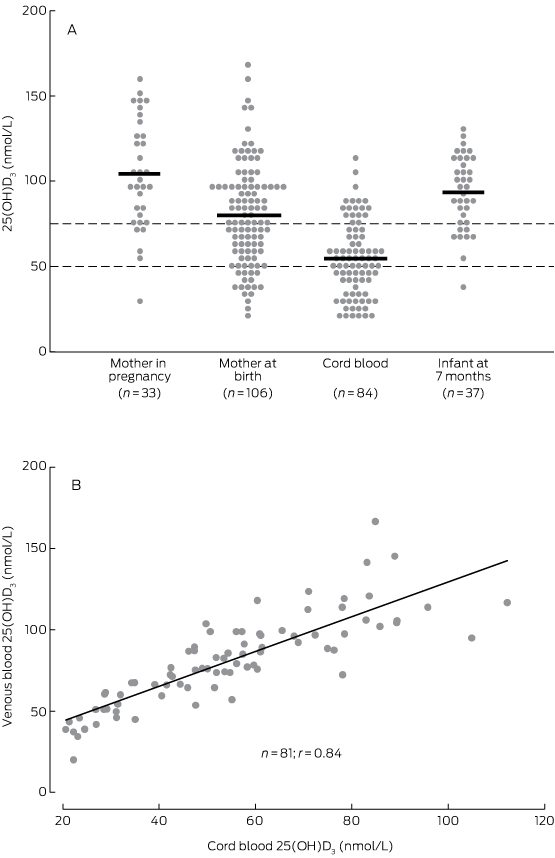
As assessed in maternal venous blood, the prevalence of vitamin D insufficiency was 21% (7 of 33) during the third trimester (median gestation time, 32 weeks; range, 28–36 weeks) and 45% (48 of 106) at birth (median gestation time, 39 weeks; range, 34–41 weeks) (Box 2). In cord blood (median gestation time, 39 weeks; range, 36–41 weeks), the prevalence of vitamin D insufficiency was 80% (67 of 84); 44% (37 of 84) had 25(OH)D3 levels below 50 nmol/L, and 10% (8 of 84) below 25 nmol/L. The prevalence of vitamin D insufficiency among infants at the 7 month visit (median age, 7.1 months; range, 6.6–8.1 months) was 22% (8 of 37).

Considering all samples (unmatched), the relative difference in mean 25(OH)D3 levels between maternal venous blood during the third trimester and at birth was 23% (104 nmol/L v 80 nmol/L) and between maternal venous and cord blood levels at birth 33% (80 nmol/L v 54 nmol/L) (Box 2; Box 3A). Overall, there was a 48% relative difference in 25(OH)D3 levels between mothers’ levels during the third trimester and those of cord blood. This trend in relative difference was similar in matched samples (data not shown). At birth, the 25(OH)D3 concentrations of the 81 matched maternal venous and cord blood samples exhibited a linear correlation (r = 0.84; P < 0.001; Box 3B).

Vitamin D levels in urban and remote participants

The mean 25(OH)D3 concentration was lower in remote than in urban participants during pregnancy, at birth, and at infant age 7 months (Box 4). The relative difference in 25(OH)D3 concentration between maternal blood in the third trimester and cord blood in remote participants was 57% (87 nmol/L v 37 nmol/L), compared with 46% in urban participants (108 nmol/L v 58 nmol/L). The cord blood 25(OH)D3 concentrations of all 14 remote infants were below 75 nmol/L; 86% (12 of 14) were under 50 nmol/L and 14% (2 of 14) were under 25 nmol/L.

Vitamin D and ALRI hospitalisation

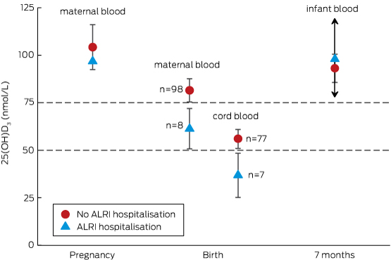
Of the 84 infants for whom cord blood samples were available, seven (8%) were hospitalised with an ALRI during their first 12 months of life; the median age at initial admission was 5.3 months (range, 1.9–7.5 months). In our primary analysis (Box 5), the mean cord blood 25(OH)D3 concentration in these seven infants was 37 nmol/L (95% CI, 25–48 nmol/L), compared with 56 nmol/L (95% CI, 51–61 nmol/L) for the 77 infants not hospitalised for an ALRI (P = 0.025). Mean 25(OH)D3 levels in maternal venous blood at birth were similarly lower in the mothers of infants subsequently hospitalised with an ALRI than in the mothers of those not hospitalised for an ALRI (Box 5).

ALRI among urban and remote participants

The proportion of remotely dwelling infants who were hospitalised with an ALRI (4 of 14, 29%) was higher than for urban infants (3 of 70, 4%; P = 0.013). The low number of ALRI hospitalisations was insufficient for a model including remote dwelling as a confounding factor.

Discussion

This is the first study to longitudinally assess vitamin D levels in pregnant Indigenous mothers and their infants. We found that the mean 25(OH)D3 level in cord blood was about half (48%) that of maternal blood during the third trimester of pregnancy (about 7 weeks earlier), a difference due equally to a decline in maternal levels late in pregnancy and to a gradient across the placenta. We also found that the 25(OH)D3 concentration was less than 75 nmol/L in 80% of cord blood samples, and that the mean cord blood 25(OH)D3 concentration was lower in infants who were subsequently hospitalised with an ALRI than in those who were not (37 nmol/L v 56 nmol/L; P = 0.025). This comparison of cord blood vitamin D levels according to ALRI hospitalisation outcome should be interpreted with caution, however, given the small number of ALRI hospitalisations (seven) and an inability to adequately investigate potential confounders, such as remote dwelling. This characteristic was associated with both lower cord blood vitamin D levels and a higher proportion of ALRI hospitalisations, and may have independently caused both low vitamin D levels and increased risk of ALRI hospitalisation. Further, this study did not measure specific factors known to influence vitamin D status, such as skin pigmentation, time spent outdoors, and diet. Despite the limitations of this study, our findings warrant further investigation.

Physiological changes in vitamin D metabolism occur during pregnancy to support the increased calcium demands of the fetus, but the specific mechanisms are not fully understood. Levels of vitamin D-binding protein and the active vitamin D metabolite, 1,25(OH)2D, increase steadily during pregnancy, while concentrations of 25(OH)D3 generally remain stable.16,17 In our study, maternal 25(OH)D3 concentrations fell both late in pregnancy (by 23%) and across the placenta (by 33%). The observed difference in 25(OH)D3 concentrations between venous (maternal) and cord blood at birth is consistent with other studies;18 however, few have specifically characterised 25(OH)D3 levels late in pregnancy. In 2003, a small study of 20 Hungarian women found no difference in mean maternal 25(OH)D3 levels between 22–24 weeks’ gestation and birth,19 while a study of 14 healthy French women found a 20% decline in mean 25(OH)D3 levels between 36 weeks’ gestation (46.8 nmol/L) and birth (37.4 nmol/L).20 As there is little seasonal variation in 25(OH)D3 levels in the tropical NT,13 the drop in late pregnancy may reflect natural progression, perhaps related to maternal–fetal immune tolerance or neonatal immune development,21 increased calcium demands of the growing fetus,22 or the emergence of risk factors, such as increased body mass index or more time spent indoors.7

The cord blood vitamin D data in our study suggest that 80% of infants were born with vitamin D insufficiency (< 75 nmol/L), 44% with mild deficiency (< 50 nmol/L), and 10% with moderate deficiency (< 25 nmol/L). The significance of these definitions for the neonate, however, is unclear, and more work is needed to define vitamin D reference ranges in cord blood. Vitamin D status was generally normal by infant age 7 months, the next sampling point.

Concordant with the trends in our data, a recent trial of maternal-plus-infant vitamin D supplementation for the prevention of deficiency among New Zealand infants8 showed that mean 25(OH)D levels in the placebo group were higher in mothers at 36 weeks’ gestation (50 nmol/L) than in cord blood (33 nmol/L), and that infant levels steadily increased through ages 2 (50 nmol/L), 4 (75 nmol/L) and 6 months (78 nmol/L). As the authors did not report maternal 25(OH)D levels at birth, it was not possible to determine whether the drop between 36 weeks’ gestation and cord blood was the result of a maternal decline or of the placental differential. Compliant daily maternal (from 27 weeks’ gestation to birth) and infant supplementation (from birth to age 6 months) at low (1000 and 400 IU/day respectively) and high doses (2000 and 800 IU/day respectively) increased mean cord blood 25(OH)D3 levels to 60 nmol/L and 65 nmol/L respectively (v 33 nmol/L in placebo-treated participants); mean infant 25(OH)D3 levels of 75 nmol/L or more were maintained at each of the 2, 4 and 6 months sampling points.

Among the participants who contributed a cord blood sample to our study, there were fewer infant ALRI hospitalisations in the first 12 months of life (8%) than predicted by historical NT data (22%).1 As ALRI hospitalisation rates are highest among remote and Central Australian infants,1 this difference was probably caused by the over-representation of Top End and urban infants in our cohort (Top End, 100%; urban, 83%) compared with the NT-wide historical data (Top End, 71%, urban, 39%). Nevertheless, cord blood 25(OH)D3 concentrations were lower in infants who were subsequently hospitalised with an ALRI than in those who were not. We could not reliably adjust our analysis to account for remote dwelling as a confounder because of the low number of ALRI hospitalisations, but our unadjusted findings are consistent with those of several other studies. In the Netherlands, cord blood 25(OH)D concentration was lower in infants who developed a respiratory syncytial virus-associated ALRI during their first 12 months than in controls (65 nmol/L v 84 nmol/L; P = 0.009);3 in New Zealand, lower cord blood 25(OH)D concentration was associated with an increased risk of any respiratory infection by 3 months of age (odds ratios, 1.00 for ≥ 75 nmol/L; 1.39 for 25–74 nmol/L; 2.16 for < 25 nmol/L).2 In a Korean study, 90% of 525 cord blood samples tested had 25(OH)D levels below 75 nmol/L, and reduced cord blood 25(OH)D concentration was strongly associated with increased risk of acute nasopharyngitis during the first 6 months of life.4 In their recent randomised controlled trial of vitamin D supplementation, Grant and colleagues23 audited health care visits by children (to age 18 months) as a secondary outcome; infants in the high dose group (87%; 66 of 76) but not the low dose supplementation group (95%; 76 of 80) had significantly fewer health care presentations for acute respiratory infections than infants in the placebo group (99%; 79 of 80).

Although not all studies support an inverse association between vitamin D levels and ALRI risk, a supplementation strategy beginning in the third trimester of pregnancy may be useful in preventing both vitamin D insufficiency and subsequent acute respiratory infections in Indigenous neonates in the NT. Acute respiratory infections are endemic in remote Indigenous communities because of factors such as overcrowding and exposure to tobacco smoke.24 However, it is less obvious why remote participants in our study had lower 25(OH)D3 levels during pregnancy and at birth, or why the relative difference in mean concentrations in maternal blood at 30–36 weeks’ gestation and in cord blood was greater than in their urban counterparts (remote, –57% v urban, –48%). Similar vitamin D levels in urban and remote infants at age 7 months suggest that the negative influence of remoteness on vitamin D levels was confined to the mothers. As the climate and time spent outdoors are likely to be similar for urban and remote participants, risk factors other than exposure to sunlight require further investigation. Interestingly, vitamin D has also been found to be a negative acute phase reactant that is depleted after an inflammatory insult.25 Lower vitamin D levels seen among remote participants may therefore be the result, rather than a cause, of their high burden of infection.

Conclusions

While only one in five Indigenous mothers had 25(OH)D3 levels below 75 nmol/L midway through the third trimester of their pregnancy, four in five cord bloods tested had lower levels as the result of declining 25(OH)D3 levels late in pregnancy and differences in levels across the placenta. The significance of low cord blood 25(OH)D3 concentrations is unclear, but the seven infants hospitalised with an ALRI during their first 12 months of life had significantly lower levels than those not hospitalised with an ALRI. Our findings, in conjunction with emerging international data, support the need for further longitudinal studies and for randomised controlled trials of vitamin D supplementation for the prevention of infant ALRI.

Box 1 –
Participant characteristics, by cord blood 25(OH)D3 status

Total

Cord blood 25(OH)D3 levels (nmol/L)


P

< 50

50–74

≥ 75


Number

84

37

30

17

Maternal characteristics

Median maternal age, years (range)

24 (17–37)

25 (17–37)

23 (17–33)

26 (17–33)

0.197

Household occupancy, people (range)

4 (1–11)

5 (2–11)

4 (1–11)

4 (3–10)

0.117

Remote community residence

14 (17%)

12 (32%)

2 (7%)

0

0.003

Smoker

36 (43%)

19 (51%)

8 (27%)

9 (53%)

0.086

Influenza vaccine during pregnancy

12 (14%)

6 (16%)

5 (17%)

1 (6%)

0.613

Infant characteristics: at birth

Boys

47 (56%)

20 (54%)

17 (57%)

10 (59%)

0.960

Low birth weight (< 2500 g)

2 (2%)

1 (3%)

0

1 (6%)

0.491

Premature (< 37 weeks)

1 (1%)

1 (3%)

0

0

1.000

Special or intensive care admission

9 (11%)

4 (11%)

4 (13%)

4 (6%)

0.901

Infant characteristics: after the birth

Exclusively breastfed

1 month after the birth

48 (47%)

18 (49%)

18 (60%)

12 (71%)

0.290

2 months after the birth

34 (40%)

14 (38%)

12 (40%)

8 (47%)

0.807

7 months after the birth

31 (37%)

16 (43%)

9 (30%)

6 (35%)

0.536

Mother smoking*

1 month after the birth

35 (46%)

19 (61%)

7 (25%)

9 (53%)

0.016

2 months after the birth

31 (45%)

15 (56%)

9 (32%)

7 (50%)

0.196

7 months after the birth

38 (55%)

17 (57%)

13 (50%)

8 (62%)

0.856

Vaccination

2 doses of PCV by 7 months

57 (68%)

25 (68%)

22 (73%)

10 (59%)

0.595

3 doses of PCV by 12 months

68 (91%)

31 (89%)

24 (96%)

13 (87%)

0.591

23vPPV during pregnancy

24 (29%)

8 (22%)

9 (30%)

7 (41%)

0.322

23vPPV at birth

29 (35%)

11 (30%)

14 (47%)

4 (24%)

0.221


23vPPV = 23-valent pneumococcal polysaccharide vaccine; PCV = pneumococcal conjugate vaccine (the 7-valent pneumococcal conjugate vaccine [7vPCV] or the 10-valent pneumococcal Haemophilus influenzae protein D conjugate vaccine [10vPHID-CV]). All figures are numbers of individuals and column percentages unless otherwise indicated. Data were compared across categories using the Fisher exact test for proportional data and the Kruskal–Wallis test for continuous data. * Smoking data were unavailable for eight mothers at 1 month and for 15 mothers at 2 and 7 months post partum.

Box 2 –
Serum vitamin D (25(OH)D3) levels measured during pregnancy, at birth, and in the infant at age 7 months

Visit

Blood sample

n

Median age (range)

25(OH)D3 levels (nmol/L)


Mean (95% CI)

Relative difference*

< 50

50–74

≥ 75


Pregnancy

Maternal

33

32 weeks (28–36 weeks)

104 (93–115)

Base

1 (3%)

6 (18%)

26 (79%)

Birth

Maternal

106

39 weeks (34–41 weeks)

80 (74–86)

−23%

18 (17%)

30 (28%)

58 (55%)

Birth

Cord

84

39 weeks (36–41 weeks)

54 (50–59)

−48%

37 (44%)

30 (36%)

17 (20%)

7 months

Infant

37

7.1 months (6.6–8.1 months)

93 (86–101)

−10%

1 (3%)

7 (19%)

29 (78%)


* Compared with maternal vitamin D blood concentration during pregnancy. † Gestational age. ‡ Infant age.

Box 3 –
(A) Maternal and infant vitamin D levels.* (B) Correlation between 84 matched maternal venous and cord blood vitamin D measurements at birth


* Dashed lines indicate reference 25(OH)D3 values for vitamin D deficiency (< 50 nmol/L) and insufficiency (< 75 nmol/L).

Box 4 –
Vitamin D levels (nmol/L) for urban and remote dwelling participants

Visit

Blood sample

Urban


Remote


n

Median age (range)

Mean 25(OH)D3 levels (95% CI)

Relative difference* (%)

n

Median age (range)

Mean 25(OH)D3 levels (95% CI)

Relative difference* (%)


Pregnancy

Maternal

26

33 weeks (30–36)

108 (95–122)

Base

7

32 weeks (28–35)

87 (68–107)

Base

Birth

Maternal

85

39 weeks (35–41)

86 (79–92)

−23%

21

39 weeks (34–41)

57 (49–66)

−34%

Birth

Cord

70

39 weeks (36–41)

58 (53–63)

−46%

14

39 weeks (37–41)

37 (30–43)

−57%

7 months

Infant

33

7.1 months (6.8–8.1)

94 (86–101)

−13%

4

7.1 months (6.6–8.1)

90 (56–124)

+3%


* Compared with maternal vitamin D blood concentration during pregnancy. † Gestational age. ‡ Infant age.

Box 5 –
Mean vitamin D levels during pregnancy, at birth, and in the infant at age 7 months, according to infant hospitalisation with an acute lower respiratory infection (ALRI) during the first 12 months of life


Only one maternal vitamin D measurement during the third trimester of pregnancy was associated with an infant ALRI hospitalisation, so that there is no confidence interval for the open triangle. Only two infants with vitamin D measurements at 7 months were hospitalised with an ALRI, so that the upper and lower confidence boundaries around the open triangle are very wide (exceeding the graph scale), as indicated by the arrows.

Resistance sans frontières: containing antimicrobial resistance nationally and globally

Coordinated action on several fronts is required

Antimicrobial resistance is everywhere, and everywhere invisible. Bacteria, which comprise the bulk of microscopic life, have lived on planet Earth for 3.4 billion years, giving them a huge amount of time to diversify, to establish themselves in almost all terrestrial and aquatic niches, and to develop advanced survival skills. Antimicrobial resistance is one of these skills. The agility with which bacteria acquire resistance to antimicrobial drugs is a perfect demonstration of those skills and of Darwin’s “survival of the fittest”.

Antimicrobials developed for therapeutic use are a very recent addition to the range of toxins in the bacterial environment. The introduction of sulfonamides in the 1930s was followed in the early 1940s by the development of penicillin, the first “miracle drug”, capable of killing bacteria causing infection in host tissues while causing no harm to the host.1 Resistance to both drug types emerged quite rapidly after their introduction into medical practice.2 Resistance has since developed, sooner or later, to all other classes of antimicrobials that have made their way into human and veterinary medicine, and into other fields of human activity.

Wherever antimicrobials are used, bacteria will be exposed and ultimately acquire resistance, by mutation or, more commonly, by acquiring resistance genes from other bacteria or the environment. The same is true for antiseptic agents, which are, in reality, antimicrobials that can only be safely administered topically. Although we are familiar with their use in humans, antimicrobials are currently used in a variety of other settings for the treatment and control of bacterial infections: in food-producing animals, companion animals, performance animals; in aquaculture, apiculture, and agriculture. We are extending the reach of antimicrobials by including antiseptics in home cleaning and personal hygiene products.

The alarm bells about resistance have been ringing for some time, but it has taken more than 20 years and several false starts before minds have responded collectively and focused on controlling resistance nationally and internationally. Antimicrobial resistance is now a major item on the agendas of the World Health Assembly3 and the World Organisation for Animal Health (OIE),4 and has been revived as a major work focus for both the World Health Organization and the OIE. Many developed and some developing countries have generated strategies and action plans in recent years; Australia did so in 2015, when it released Australia’s First National Antimicrobial Resistance Strategy.5 Although not the first attempt in this country to address the problem of resistance,6,7 it was the first to fully embrace the idea that resistance has no borders, ensuring that all aspects of antimicrobial use and resistance were considered. Both the Department of Health, and the Department of Agriculture and Water Resources drove the development of the Strategy.

What does the Strategy hope to achieve? It incorporates seven objectives, each with a strong motivation to cut through and achieve the changes needed to make a difference.

Increase awareness and understanding: There is ample evidence that most in our community have a poor understanding of what antibiotics can and cannot do, and what resistance is. At least half believe that antibiotics will help with the common cold, and many also believe that antimicrobial resistance means that they personally become resistant to antibiotics. NPS MedicineWise has been running advertising and other programs in response to this problem,8 but the impact has yet to be fully felt. Awareness and understanding are also sometimes lacking among prescribers. Although almost all doctors and veterinarians prescribe antimicrobials as part of their daily practice, few are aware of rational prescribing principles and their benefits. Doctors and vets need to improve their own awareness and understanding, as well as that of their clients, of the negative effects of using antimicrobials inappropriately.

Implement effective antimicrobial stewardship: Antimicrobial stewardship is the coordination of activities to ensure and promote rational prescribing in a defined context; for example, in hospitals, in the community, or in veterinary practice. Having a stewardship program is now part of hospital accreditation requirements, thanks to the efforts over many years of the Australian Commission on Safety and Quality in Health Care (ACSQHC).9 There is an obvious need to extend stewardship into residential aged care, general practice, small animal and equine practice, and food animal practice. The establishment last year of a Centre for Research Excellence, the National Centre for Antimicrobial Stewardship, will lay the groundwork for the development of stewardship programs in all these areas.

Develop national surveillance: Without surveillance data it is impossible to know which control strategies are effective, or how effective they are. Although Australia has for some decades had several antimicrobial use and resistance surveillance programs in human medicine, their work has lacked coordination and correlation. The ACSQHC has received funding for the development of a national coordinated use and resistance surveillance system for human health, due for completion by June 2016. This project will coordinate all existing programs, enhance them as needed, and fill important gaps, including through regular and timely reporting and trend analysis of antimicrobial dispensing data from the Pharmaceutical Benefits Scheme, and linking data on antimicrobial resistance from laboratory information systems around the country. On the veterinary and agriculture side, there have been a number of small pilot programs. Funding was recently found for a project in pig production, and there is interest in extending this initiative to the poultry sector. However, more needs to be done to establish a national surveillance program in the non-human sector that is integrated with surveillance in the human community.

Improve infection prevention and control: Control of antimicrobial use is essential, but by itself is insufficient to control the spread of antimicrobial resistance. Controlling the spread of bacterial diseases (eg, with vaccines) is a very effective way of reducing the need for antimicrobials. Infection prevention and control systems are essential components of resistance containment. Australia has national infection control guidelines for human health,10 and infection control systems are a mandatory requirement of hospital accreditation. In the non-human sectors, infection control is a key part of animal husbandry in intensive food animal industries. In veterinary practice, guidelines on infection prevention and control are available, including information on personal protection for vets and staff;11 however, more could be done to prevent the spread of infection between animals.

Agree on a national research agenda: Containing antimicrobial resistance is not currently an explicit research priority in Australia. The National Health and Medical Research Council, the Australian Research Council and other funding bodies support research in antimicrobial resistance, but only on a competitive funding basis (ie, in competition with all other types of research). While some excellent studies have been supported by these organisations, there is no strategic or targeted approach which ensures that the most important research questions are prioritised, such as new drug discovery and development, rapid diagnostics, and the identification of optimal education, community and professional strategies.

Strengthen international collaboration: Australia may be an island, but we are certainly not protected from exposure to new resistances. We have a long history of effective control of the introduction of exotic infectious diseases, but have not yet recognised that the same objective should apply to exotic resistances. The recent introduction in Victoria of an exotic resistance to last-line antibiotics (carbapenem resistance), with subsequent spread in the human population, highlights the fact that this aspect of resistance crossing borders cannot be neglected.12 Developing partnerships with countries across the world will assist Australia to learn from international best practice, avoid duplication of effort, contribute to public health outcomes in our region, and provide early warning of emerging threats.

Establish and support clear governance: None of these objectives can work without a clear, forward-looking and stable governance structure. As a federation, our national strategy requires the cooperation and coordination of the activities of nine governments and, more importantly, of numerous ministries and agencies. This is where the national Antimicrobial Resistance Prevention and Containment (AMRPC) Steering Group, reporting to the federal ministers for Health and Agriculture, supported by the Australian Strategic and Technical Advisory Group, and working in collaboration with the Australian Health Protection Principal Committee, is forging the way forward. A coordinated approach is essential. The efforts of these groups will align with international efforts and contribute to the global control of antimicrobial resistance.

All prescribers and users of antimicrobials have a responsibility to preserve their long term effectiveness and to protect the health of their nation’s citizens, animals and ecosystems. With the ever increasing global movements of people, animals and goods, all nations must work together to protect each other. Resistant bugs don’t respect borders.

New recommendations for Hepatitis C treatment

New recommendations have been released for the management of hepatitis C virus (HCV) infection in a consensus statement.

The statement was drawn up by Gastroenterological Society of Australia, the Australasian Society of Infectious Diseases, the Australasian Hepatology Association, the Australasian Society for HIV, Viral Hepatitis and Sexual Health Medicine, Hepatitis Australia and the Royal Australian College of General Practitioners.

A summary, published in the Medical Journal of Australia, says that the recommendations for Hepatitis C treatment were drawn up in the wake of the new direct-acting antiviral therapies that were added to the Pharmaceutical Benefits Scheme earlier this month.

Related: 5 things you need to know about the new Hepatitis C medicines on the PBS

“The introduction of DAA therapies for HCV that are highly effective and well tolerated is a major medical advance,” said Professor Alexander Thompson, director of gastroenterology at St Vincent’s Hospital in Melbourne.

“All Australians living with HCV should now be considered for antiviral therapy.”

Recommendations in the consensus statement include:

  • All individuals with a risk factor for HCV infection should be tested.
  • Annual HCV serological testing is recommended for seronegative individuals with risk factors for HCV transmission.
  • People with confirmed HCV infection should be tested for HCV genotype (Gt).
  • All concomitant medications should be reviewed before starting treatment, using the University of Liverpool’s Hepatitis Drug Interactions website.
  • The use of any DAA regimen during pregnancy is not recommended.
  • People who are not cured by a first-line interferon-free treatment regimen should be referred to a specialist centre.
  • All people with decompensated liver disease, extra-hepatic manifestations of HCV, HCV–HIV or HCV–HBV co-infection, renal impairment or acute HCV infection, as well as people who have had a liver transplant should be referred for management by a specialist who is experienced in the relevant areas.
  • All people living with HCV infection should have a liver fibrosis assessment before treatment to evaluate for the presence of cirrhosis.
  • People with no cirrhosis can be treated by general practitioners working in consultation with specialists.

Read the full recommendations on the Gastroenterological Society of Australia’s website.

Latest news:

[Correspondence] Tuberculosis control

Vikram Patel and colleagues1 discussed most of the health issues pertaining to tuberculosis control in India, but possibly ignored very important issues of tuberculosis control, which contribute to international health because tuberculosis is an aerosol-mediated transmissible disease. India has the highest estimated incidence of tuberculosis (2 200 000) in the Global Tuberculosis Report 20152 compared with its neighbouring countries China (930 000), Bangladesh (360 000), and Pakistan (500 000). In 2014, 250 000 deaths from tuberculosis (including HIV-positive tuberculosis) were officially classified as deaths caused by HIV/AIDS in the International Classification of Diseases.