×

Ebola outbreak in West Africa: considerations for strengthening Australia’s international health emergency response

It is time for a common vision and strategy for deploying Australian expertise to international public health emergencies

An effective response to health emergencies such as the Ebola virus disease outbreak in West Africa relies on global capacity to rapidly surge the supply of skilled workers, particularly when they are limited in affected countries and increasingly depleted during the emergency. Before the Ebola outbreak, health professionals in West Africa were already scarce; for example, in Liberia the doctor-to-population ratio was 1:70 000, compared with 1:300 in Australia.1,2 In addition to clinicians, an effective response to a large outbreak of Ebola virus disease in resource-limited settings requires international technical support across a range of public health and other disciplines, including infection prevention and control, epidemiology, laboratory diagnostics, communication, mental health, anthropology, social mobilisation, logistics, security and coordination.

Early in the Ebola virus disease outbreak in West Africa, many international non-government organisations (NGOs) and several governments established treatment centres and sent public health professionals to provide clinical care and augment control efforts. Timely public health interventions in Ebola-affected rural communities achieved crucial reductions (about 94%) in Ebola transmission.3 The Centers for Disease Control and Prevention (CDC) in the United States is a case study in how governments can deploy significant public health staff to countries affected by health emergencies. At time of writing, the CDC had effected 2206 staff deployments since July 2014 to support the public health response in Ebola-affected countries across a wide range of areas including surveillance, contact tracing, database management, laboratory testing, logistics, communication and health education.4 Most CDC public health staff were deployed into roles with a low risk of Ebola virus infection (ie, non-patient care roles) and none have become infected. In assessing the CDC’s exemplary response, it is important to note its pre-existing Global Health Strategy that clearly articulates the CDC’s vision, rationale, role, strategy, funding, partnerships, staff and areas of expertise for working in international public health, including in health emergencies such as Ebola virus disease.5

In contrast, nearly 6 months into the outbreak — when almost 5000 deaths had already been recorded — the Australian Government was being criticised by public health experts for its lack of substantive assistance to Ebola-affected countries.6 The Public Health Association of Australia called on the government to help strengthen the medical and public health capacity in the region by deploying an Australian Medical Assistance Team (AUSMAT), and by supporting Australians who wanted to volunteer their services through the World Health Organization or international NGOs by encouraging their employers to provide special leave and continuation of entitlements.6 Ultimately, the Australian Government declined to deploy AUSMAT resources, stating it would not consider sending people to Ebola-affected countries until it could get assurances from developed countries closer to West Africa that Australians would be able to be evacuated for treatment in the event they became infected.7 Instead, the Australian Government chose to fund a private contractor to staff a single treatment centre built by British army engineers in Sierra Leone. To date, anecdotal reports suggest no public health professionals have been deployed to Ebola-affected countries by the Australian Government, although the risk of infection is low.

The current and all previous Australian governments have not clearly articulated a vision for providing public health support during an international health emergency. AUSMATs are multidisciplinary health teams of doctors, nurses, paramedics, firefighters (logisticians) and allied health staff such as environmental health workers, radiographers and pharmacists8 who provide timely acute medical relief immediately after disasters in Australia and overseas. Staff of state and territory governments can be members of AUSMATs, and these agencies are reimbursed by the federal government for the salaries of staff who deploy through this mechanism. AUSMATs have a relatively small number of public health professionals on a roster largely drawn from staff of state and territory health authorities, but this list includes only a limited number with relevant outbreak response skills. While AUSMATs have a proven track record in providing emergency medical care in post-disaster settings, they are not currently designed to support the public health response required for a large outbreak.

The decision not to use AUSMAT assets and the lack of federal support for other Australian health professionals who wanted to volunteer to help contain the Ebola outbreak transferred the pressure to institute human resource policies (ie, special leave and continuation of entitlements) to state and territory health authorities. Without the type of financial arrangements that the AUSMATs afford state and territory health authorities, and with a general lack of jurisdiction-funded strategies for their staff engaging in emergency responses overseas, the environment for staff deploying independently was not always supportive.

Despite the challenges, many public health professionals from state and territory health authorities and academic institutions in Australia deployed as volunteers for NGOs or United Nations agencies, including about 15% of the Australian National University Master of Applied Epidemiology (MAE) program’s alumni since 1991 (Martyn Kirk, MAE Program Director, Australian National University, personal communication). Still, only one current Australian MAE participant was deployed, with Médecins Sans Frontières (as of January 2015), compared with 97 from a similar program in the US (the CDC’s Epidemic Intelligence Service), highlighting the missed opportunity for Australia’s next generation of outbreak control experts to get invaluable field experience while providing much needed support.

Now that the Ebola virus disease outbreak is over, Australia needs to examine how well it performed in assisting the WHO to respond to this significant threat to global health, as it was declared by the International Health Regulations Emergency Committee in August 2014.9 We believe it is time for the Australian Government, in consultation with state and territory health authorities and public health training institutions, to establish a common vision and strategy for deploying Australian expertise to international public health emergencies, including Ebola virus disease outbreaks. If AUSMATs (currently the only funded mechanism) are Australia’s preferred approach to responding to international health emergencies, their capacity to support a major public health response should be expanded by including more professionals in relevant disciplines. To maximise the impact of public health professionals deployed through AUSMAT arrangements, personnel should be made available at the beginning of future health emergencies. In addition, formalising support arrangements with members of the Australian Response MAE (ARM) Network10 (created by academic institutions in response to gaps in the coordination of Australia’s public health surge capacity) may be a way for the government to effectively mobilise skilled public health professionals for deployment overseas in response to disease outbreaks. Consideration should also be given to expanding the AUSMAT roster of public health professionals and integrating structures like the ARM Network into the response framework.

The Australian Government missed an important opportunity to contribute timely, valuable technical assistance to Ebola-affected countries; support that was essential to stopping the outbreak at its source at a time when it was needed most. In the end, many Australians stepped up as volunteers in the fight to extinguish this global threat to public health. Australian federal authorities should reflect on this experience and consider it an opportunity to strengthen Australia’s response to future health emergencies and demonstrate leadership on the global stage.

Multidrug-resistant tuberculosis in Australia and our region

MDR-TB threatens TB control programs in Australia’s region and will not diminish without concerted efforts

Tuberculosis (TB) is one of the world’s great killers, but Australia has been relatively protected because of its strong public health system. Of 1300 cases reported in Australia each year, almost 90% occur in the overseas born, although Indigenous Australians are also disproportionately affected. Most cases arise in the large immigrant communities from India, Vietnam, the Philippines, China and Nepal, but high rates are also reported from Papua New Guinea (PNG), Ethiopia, Somalia and Myanmar. These cases occur primarily in permanent residents and students, rather than in refugees or those on humanitarian visas.1

Many countries are now reporting significant rates of drug-resistant tuberculosis, with at least 480 000 cases worldwide now attributable to multidrug-resistant TB (MDR-TB; defined as resistance to the two most effective first-line agents, isoniazid and rifampicin).2 However, countries with the highest rates of drug resistance often have the poorest quality data, largely due to the lack of resistance testing. Globally, MDR-TB was estimated to constitute 3.3% of new and 20% of retreatment cases in 2014. Although the deployment of molecular diagnostics to detect resistance is progressing, only a quarter of these cases were correctly identified. For example, in PNG, MDR-TB rates are similar to those described globally, but a nationwide drug-resistance survey has not been undertaken and other data sources suggest that the rates could be underestimates.3 Extensively drug-resistant TB (XDR-TB; resistant to isoniazid, rifampicin and the most effective second-line agents, quinolones and injectables) has also been sporadically reported in Australia from PNG.4 For Australian clinicians, for whom diagnostics are widely available, the rise of MDR-TB makes definitive strain identification through culture even more important.

Traditional second-line agents to treat the handful of MDR-TB cases in Australia are generally available, but prolonged courses of toxic and expensive drug combinations are required. The agents used to treat MDR-TB depend on the remaining susceptibilities, but ototoxicity (aminogylcosides), nausea (p-aminosalicylic acid) and neuropsychiatric reactions (cycloserine) are among many common side effects. It is therefore unsurprising that globally, treatment outcomes are poor, with only about half of identified patients completing the 2-year treatment course, such that only around 10% of all incident cases complete treatment worldwide.2 Meta-analyses demonstrate that the chance of treatment success diminishes as the number of drugs to which a strain is resistant increases.5 Alarmingly, there are now data on outcomes for patients with “beyond XDR”-TB, with treatment success rates comparable to the pre-antibiotic era natural history of TB.6

There is a resurgence of interest in new treatments for TB, with the first new drugs in 40 years now proceeding through development, including bedaquiline, delamanid and pretomanid. As important as individual agents is the development of new regimens that can be deployed programmatically, such as the 9-month Bangladesh regimen (comprising gatifloxacin, clofazimine, ethambutol and pyrazinamide, with prothionamide, kanamycin and high-dose isoniazid added for the intensive phase).7 There is also interest in off-label use of existing antibiotics with anti-TB activity, such as linezolid and meropenem–clavulanate, and new strategies to minimise toxicity, such as therapeutic drug monitoring. However, even if new regimens become established, significant barriers exist to providing treatment for MDR-TB in the countries that need them most. MDR-TB is both a cause and symptom of poor communicable disease control programs, with MDR-TB regimens costing around tenfold that of drug-susceptible cases.8

MDR-TB is not a problem that will just go away. Policy makers may prefer to treat the problem they can address — focusing on improving programs for drug-susceptible TB to prevent resistance amplification. However, modelling has consistently demonstrated that cases of MDR-TB predominantly arise from community transmission rather than from resistance amplification in previously susceptible strains,9 such that only targeted control programs will achieve reduction in the disease burden attributable to MDR-TB.10

As global TB rates slowly decline, the contribution of late reactivation of latent infection to incidence is likely to increase. While this makes treatment for latent MDR-TB a key consideration, evidence for effective treatments remains scarce and clinical trials are ongoing.

The ambitious post-2015 targets for TB control, which replace the relatively modest Millennium Development Goals, present an opportunity for Australian leadership. In our setting, with most TB imported and the emergence of MDR-TB so dependent on the strength of health systems, Australia has a critical role to play in supporting developing countries of our region to improve TB control programs and their health systems generally. A vision for an expanded international response, coordinated with global partners, governments, multinational organisations, affected individuals and communities is provided by the United States National Action Plan for Combating MDR-TB.11 Given that 57% of MDR-TB cases occur in the Asia–Pacific region,2 a similar response to improve clinical diagnostics and management in our region would help keep MDR-TB from our shores.

The ASID test

The Australasian Society for Infectious Diseases view on infectious diseases challenges in 2016 and beyond

This year, 2016, is a historic year for the Australasian Society for Infectious Diseases (ASID), being the 40th anniversary of the formation of the society. It is an opportunity not only to celebrate our achievements in infectious diseases and microbiology over the past 4 decades but also to anticipate future challenges. Principal among these are antimicrobial resistance (AMR) and the emergence or re-emergence of previously controlled or unrecognised diseases.

Although infectious diseases were thought to have been conquered as public health problems 40 years ago, our complacency has been repeatedly challenged by new and re-emerging threats. In this issue of the MJA, we read about re-emergence of Ebola virus,1 preparedness for Zika virus2 and local transmission for hepatitis E3 as salient examples. Williams and colleagues also describe the diagnosis of subacute sclerosing panencephalitis in a 23-year-old man,4 a timely reminder that high levels of herd immunity are required to prevent measles outbreaks, which still occur in parts of Australia with suboptimal immunisation rates.5 But emerging infections are not only viral. In this issue, Mandrawa et al discuss carbapenem-resistant Klebsiella,6 which highlights the serious impact of AMR on clinical practice.

AMR remains a global challenge. It results in about 2000 attributable deaths per year in Australia and a projected 10 million globally by 2050.7 As antibiotics become less effective, old diseases re-emerge, many in Australia’s near neighbours. Tuberculosis (TB) remains one of the most lethal infectious diseases with a third of the world’s population being infected, and over 1.5 million annual TB-related deaths worldwide.8 In this issue, Cheng and Trauer report on the increase in multidrug-resistant TB in Australia and the region.9 Another disease from times past, gonorrhoea, is also re-emerging as increases in AMR result in treatment failures. In 2015, the National Neisseria Network reported a marked increase in rates of gonococcal disease, with the highest ever proportion of strains on record with reduced susceptibility to ceftriaxone, and high-level resistance to azithromycin recorded for the first time.10 These patterns reflect trends around the world, and as noted by Lahra and colleagues, suggest that Neisseria gonorrhoeae is emerging as a global public health threat.10 Both TB and N. gonorrhoeae emphasise our need for national, coordinated AMR surveillance in humans and animals so we can monitor and respond to these trends.

Surveillance alone will be ineffective if there is not a clear plan to reduce the AMR threat, as well as a coordinated mechanism to implement this plan across jurisdictions (which is currently lacking). The federal government released its national AMR strategy in late 2015.11 A key element is the need to develop a “one health” approach to antimicrobial stewardship, including capture of prescription data in both humans and animals, and regulation of antimicrobial use and national antimicrobial guidelines for veterinary practice and agriculture. Guideline development in animals and humans needs to be supported by implementation science to improve uptake and adherence. We also need to reduce the unnecessary use of antibiotics, which is estimated to occur in up to 75% of antibiotic prescriptions in Australia,12 the bulk of which are dispensed in primary care. There needs to be coordination of strategies to address this problem across stakeholders including professional colleges and societies. ASID, led by Professor Denis Spelman, and the Royal College of Pathologists Australasia have collaborated with the Royal Australasian College of Physicians’ EVOLVE campaign to list the top five low value interventions in infectious diseases and microbiology practice.13 These include the use of antibiotics for asymptomatic bacteriuria, leg ulcers without clinical infection, uncomplicated upper respiratory tract infections, and faecal pathogens in the absence of gastrointestinal symptoms (with some exceptions). Avoiding inappropriate diagnostic tests is one important aspect of reducing antibiotic prescribing for these conditions. For example, as Bowen et al report in this issue,14 faecal multiplex polymerase chain reaction testing results in high rates of reporting of the non-pathogenic parasites Dientamoeba fragilis and Blastocystis spp., which could lead to unnecessary antibiotic use in patients who test positive.

Another benefit of reducing unnecessary antibiotic use is reduction of adverse events. In their study of medical inpatients at a Melbourne hospital, Trubiano and colleagues found that almost one in four had a serious antibiotic allergy, most commonly associated with overuse of broad spectrum agents.15 Of particular concern was poor documentation of allergies in patient records and charts.

Over the past 40 years, members and affiliates of ASID have contributed to paradigm-shifting approaches to infectious diseases and microbiology — including the elimination of smallpox and the link between Helicobacter pylori infection and duodenal ulcers. Who knows what challenges lie ahead. We are on the cusp of personalised medicine that will predict our risk of disease, inform our likely response and guide our therapy. We must, however, remain mindful of key lessons from the past, so that history does not repeat itself. As Kurt Vonnegut, Jr wrote: “History is merely a list of surprises. It can only prepare us to be surprised yet again.”

Zika preparedness in Australia

Our comprehensive national response encompasses prevention and surveillance, as well as monitoring and controlling Aedes aegypti in Australia

The spectrum of clinical illness for Zika virus infection is generally not severe — about 80% of cases are asymptomatic1 — and the infection was not previously thought to be cause for serious public health concern. There is no specific treatment for, nor a vaccine against, a Zika infection.

Recent disquiet has been raised by emerging evidence of possible vertical transmission of Zika, the development of severe congenital abnormalities, including microcephaly,2,3 and of a possible link to fetal deaths.4 In addition, a possible link to Guillain–Barré syndrome has been reported.5,6 The World Health Organization declared the clusters of microcephaly and neurological disorders a Public Health Emergency of International Concern on 1 February 2016.7 Knowledge about any causal link between Zika virus and effects in utero is still evolving; however, given the serious implications, should there be one, Australian guidance for managing pregnant women returning from Zika-affected areas and for preventing the spread of the disease has been prepared.

In almost all cases, the Zika flavivirus is transmitted by mosquitoes (particularly by Aedes aegypti). The Zika virus was first isolated from a monkey in Uganda in 1947,8 and serological evidence of past infections in humans has been reported since 1952 in Africa and since 1981 in South-East Asia.9,10 Outbreaks in the Pacific Islands were first reported in Yap State, Micronesia, in 20071 and in French Polynesia from 2013,11 with spread to many Pacific islands between 2013 and 2016.12,13 Zika has spread rapidly across the Americas since late 2015, after being first confirmed in Brazil in 2015.14 In November 2015 the international community was alerted to the possibility of severe congenital malformations, with an International Health Regulations notification about an increase in cases of microcephaly in Brazil with geographical and temporal links to Zika.15 At that time, further information was also provided by health authorities in French Polynesia about congenital malformations, also with geographical and temporal links to Zika.15

In Australia, sporadic cases of Zika have been detected since 2012 in 35 returning travellers (to 29 February 2016), and there is a continuing risk of imported Zika infections from overseas. With the number of affected overseas areas increasing, as is greater awareness among the public and health professionals, an increase in the detection of imported cases could be expected.

The low risk of local transmission of Zika in Australia is restricted to areas of Queensland where the most suitable vector, A. aegypti, is continually present. Queensland has well developed and practised plans and resources for controlling dengue (also carried by the A. aegypti mosquito) that are also applicable to Zika. Through routine vector monitoring and control activities, and the deployment of Dengue Action Response Teams (DARTs), the Queensland government has prevented dengue from becoming endemic, despite regular importations. The Queensland government recently announced a package of measures to strengthen preparedness, including enhanced laboratory capacity in Townsville. Queensland Health remains on the alert for imported cases and subsequent local transmission.

A program of mosquito surveillance and control, coordinated by the federal Department of Agriculture and Water Resources, is in place at Australia’s air and sea ports to prevent incursions of exotic mosquitoes from overseas. While foreign mosquitoes are detected during the summer months, well established programs prevent their establishing breeding populations. There is also a specific program conducted by Queensland Health in the Torres Strait for controlling A. albopictus (a potential alternative vector for the Zika virus16), active in this area since 2005. The program has been successful in preventing its spread to the mainland and in reducing the numbers of A. albopictus and, at the same time, of A. aegypti in the transport hubs of the Torres Strait.

In February 2016, the Australian Health Protection Principal Committee issued advice on the management of pregnant women returning from Zika-affected countries and on preventing sexual transmission of Zika. A public health guideline on Zika is being finalised, and advice for travellers was issued in January 2016.17 Until more is known about the link between the virus and microcephaly, Australia recommends that women who are pregnant or planning to become pregnant should consider postponing travel to areas with ongoing transmission of Zika. If they do decide to travel, they should consistently adhere to mosquito avoidance measures. This recommendation is in line with major public health agencies around the world.

The Interim recommendations for assessment of pregnant women returning from Zika virus-affected areas18 encourage health care providers to ask all pregnant women about their recent travel history. Those who have travelled to a Zika-affected country during their pregnancy should be evaluated and tested. Any woman who tests positive for Zika virus should be referred for specialist obstetric care. The Royal Australian and New Zealand College of Obstetricians and Gynaecologists has issued guidance on the care of women with confirmed Zika virus infection during pregnancy in Australia.19

Further concerns for pregnant women and their unborn babies have been triggered by the possibility of sexual transmission of Zika virus. Initially, two instances of likely sexual transmission were reported internationally, one in 2008 and the other in 2016,20,21 and two instances of Zika virus being detected by polymerase chain reaction in semen — including one 62 days after the onset of symptoms — although virus isolation was not performed, so that it was not determined whether viable virus was present.22,23 There is evidence from the United States that sexual transmission of Zika may be more common than previously reported.23 To date, all reports of suspected or confirmed sexual transmission of Zika have involved a symptomatic man transmitting the virus to a woman. It is still unknown how long the virus can persist in semen, or how infectious this may be. Mosquitoes remain the overwhelmingly predominant mode of transmission. The Australian advice, Interim recommendations for reducing the risk of sexual transmission of Zika virus,25 recommends that men with a confirmed Zika virus infection and whose partner is pregnant should abstain from sex or consistently use a condom during sex for the duration of the pregnancy. Men with a confirmed Zika infection who do not have a pregnant partner should abstain from sex or consistently use a condom during sex for 3 months after leaving a Zika-affected country.

All recommendations about travel, testing and management require definition of the countries that have current local transmission. The list of affected countries is assessed daily by the Department of Health, based on agreed criteria. This, however, is not straightforward, and differences between overseas surveillance systems mean that a variety of sources must be checked to assess whether local transmission of Zika virus is happening in a particular country.

While unease about Zika is high, there remains a lack of high quality evidence for a causative link between infection and the development of microcephaly, and there is a general lack of data on the pathogenesis and epidemiology of the disease. Further studies are urgently required, and are underway. The need to formulate recommendations despite a paucity of data and evidence is not new in public health. However, it does pose particular challenges and risks, in that we may have to modify recommendations frequently. Expert consultations and the experiences and recommendations of other agencies internationally are important in the development of such recommendations. A key component of preparedness for communicable disease outbreaks in Australia is developing nationally consistent advice across the states and territories, and harnessing the expertise that is present throughout our country.

Australia has robust systems in place that can be adapted as required to enable a rapid response to communicable diseases such as Zika, with excellent laboratory capacity, public health response capability and communicable disease surveillance systems, as well as established vector surveillance and control programs. As the situation evolves, ongoing monitoring will continue, with information and recommendations updated as necessary. Zika is the latest communicable disease threat to challenge us. Each new threat offers an opportunity for enhancing core elements of communicable disease control and for ensuring readiness for the next emerging infectious disease.

Aileen Joy Plant

Professor Aileen Plant (1948–2007) was a renowned medical epidemiologist and an outstanding global public health leader

In mid-March 2003, hurrying through Perth Airport on her way to a World Health Organization assignment, Professor Aileen Plant paused to write out her will. She asked the airline staff to witness it before boarding a plane for Hanoi. Her task was to lead a team trying to bring Vietnam out of its sudden nightmare of the deadly disease of severe acute respiratory syndrome (SARS), an illness that no one knew the cause of, nor how it spread. The person she was replacing, Dr Carlo Urbani — who had identified the new syndrome — lay sickened by it in a hospital in Bangkok.

Aileen knew that speed was essential. The effectiveness of the tasks of early detection and prevention of transmission would require a cohesive and willing team, which in turn would require the trust of the Vietnamese Ministry and the Vietnamese health care workers. This, she achieved.

On 29 March, Dr Carlo Urbani died. Dr Katrin Leitmeyer, virologist, recalls how Aileen rallied everyone, “gluing extreme characters from all around the world together under difficult psychological circumstances”.

The 3-week mission became 11 weeks. Vietnam had 69 cases of SARS and five deaths, mostly in staff and patients of the Hanoi French Hospital. During this time, Aileen’s sister, Kaye, became gravely ill in Perth. Aileen was desperate to be with her but knew that, even if she did return to Australia, she would not be allowed into any hospital.

Under her leadership, the Hanoi team characterised the clinical features of the disease, its incubation period and possible routes of transmission, and made important observations about the effectiveness of case isolation and infection control in halting transmission. On 28 April, Vietnam was declared SARS-free, the first country to eradicate the disease. The Vietnamese government awarded Professor Plant its highest award, the National Medal of Honour.

Aileen said of her experience that two things stood out. The first was that the Vietnamese government agreed that external help should be sought — an extraordinary admission in communist Vietnam at that time. The second was the dedication of the Vietnamese staff, who quarantined themselves in the hospital and worked with little in the way of modern technology or resources. Aileen thought they should have been awarded the Medal, rather than her. Her own keen sense of family no doubt contributed to her great respect for the grief and isolation of any individual. Finally, in June, Aileen was able to return home to her recovering sister.

Other WHO assignments in which Aileen was involved included investigating an HIV outbreak in children in Libya, childhood dermal fibromatosis in Vietnam, yellow fever outbreaks in Africa, tuberculosis trends in Indonesia and the emergence of avian influenza in Asia. She also began seminal work with the WHO on the International Health Regulations (IHR), to frame the relationship between countries and the WHO in regard to preparation and response for public health events of international concern, and continued work on the Global Outbreak and Alert Response Network (GOARN), which she had helped establish in 2000. Both are key tools in global biosecurity today.

Aileen came from a large family and left school at the age of 15 to work on her parents’ farm in Denmark, Western Australia. She became interested in infectious diseases, telling her father that an animal had died of eastern equine encephalitis. This became a family joke, as the animal in question was a cow. She took up work as a bank teller for 5 years but became determined to study medicine, putting herself though technical school and gaining entrance to the University of Western Australia.

Her early years as a resident doctor in the Northern Territory sparked her interest in Aboriginal health. She became firm in her belief that it was essential for the overall health of humanity to understand and care for vulnerable populations. Already evident to her colleagues by this time were her razor-sharp “bullshit detector”, her interest in all matters and her keen sense of humour.

Professor Aileen Plant with Professor Lance Jennings on a World Health Organization mission to investigate a cluster of H5N1 influenza cases in Vietnam in 2005.

Aileen went on to study at the London School of Hygiene and Tropical Medicine. On returning to Australia, she obtained a Master of Public Health at the University of Sydney, eventually joining the faculty as a lecturer, while also working with the New South Wales Department of Health.

In 1989, Aileen took up the position of Chief Health Officer in the NT. Although frustrated by politics, she kept her focus on Aboriginal health, pointing out the flaws in census methods and analysing a decade of data demonstrating health trends and causes of premature mortality in Aboriginal communities.1,2 Her 1995 report called for a whole-of-community and government approach to the poor health trends in Aboriginal and Torres Strait Islander populations.1

Among Aileen’s gifts was the ability to see the truth, or the way to the truth, in science, diplomacy and politics. Science was her bedrock, and diplomacy she saw as an everyday necessity from which wonderful friendships could grow. Bad science and politics tired her, perhaps due to the famous bullshit detector constantly being triggered.

In 1992, Aileen took up the position of Director of the Master of Applied Epidemiology (MAE) Program at the Australian National University, a program she had played a key role in initiating and developing. During her 3 years there, she completed her own PhD, guided many masters and doctoral students, and worked with her colleagues to develop a program on Indigenous health and in attracting Indigenous students. She convinced a colleague in the NT, Dr Mahomed Patel, to join her, developing pathways for international students and obtaining overseas placements for Australian trainees, including deployments with the WHO and establishing MAE-like programs in India, China, Malaysia and Vietnam.

The MAE Program has served the world exceedingly well, with many of its students, staff and graduates contributing to the control of SARS, avian influenza and other public health emergencies. Many of Aileen’s students are now leaders in public health, nationally and internationally.

In 1995, Aileen moved back to Perth to be with her much loved extended family. She worked initially as a senior lecturer at the University of Western Australia before becoming professor of international health at Curtin University in 2000. Together with Professor John Mackenzie, a world-renowned virologist, she compiled an ambitious bid to establish a cooperative research centre (CRC) with a focus on emerging infectious diseases. After two arduous attempts, their bid was successful. The Australian Biosecurity CRC for Emerging Infectious Disease was established in 2003, bringing animal, human and environmental disciplines together in research. Over 7 years, the CRC had many high-impact achievements, including extensive research into the ecology of disease emergence, the development and application of diagnostic tools and systems, and important work on Hendra virus, coronaviruses and influenza viruses. Translational research — taking research into action and policy — was a centrepiece. The CRC awarded over 60 postgraduate scholarships to students in Australia and South-East Asia.

During this time, Aileen continued to assist the Australian Government Department of Health and Ageing, including in emergencies such as the Asian tsunami, where her ability to see the path forward encompassed areas beyond public health. In 2008, the Department named its new crisis response centre the Aileen Plant National Incident Room.

Aileen’s comments usually went to the heart of the matter. Radio host Phillip Adams, interviewing Aileen on ABC RN Late Night Live, asked her whether authoritarian or democratic governments would be better at handling outbreaks. She replied that it depended on the characteristics of the disease and its transmission mode. Diseases like SARS, she noted, are shown to be well handled by authoritarian governments if backed up by a good public health system, but something like HIV–AIDS, which requires behavioural change, is better handled by democracies. She repeated the point, “Wherever they are, infectious diseases always make poor people poorer”.3

Aileen continued to work with the WHO on finalising the IHR, which were endorsed in 2005 and are now signed by over 190 countries. Many of the articles of the IHR reflect the cooperation and information exchange exemplified by Aileen’s time in Vietnam.

Professor Aileen Plant with Professors John Mackenzie (Curtin University), Mal Nairn (Charles Darwin University) and Charles Watson (Curtin University) at the opening of the Queensland node of the Australian Biosecurity Cooperative Research Centre in 2004.

In addition to 90 scientific articles and numerous book contributions, Aileen co-authored a book on the impact of SARS and another on the approach to communicable diseases.4,5 Aileen’s delight was to do projects with her friends and family, and their interests were hers, be they research projects, scientific books, teaching friends’ children to swim, writing creative fiction or designing tree farms.

Aileen died suddenly at Jakarta Airport on 27 March 2007, while travelling home from a WHO meeting, where she had helped to bring about consensus on the issues of sharing avian influenza viruses and access to influenza vaccine for developing countries.

Her spirit and values live on in her colleagues and her students. The Australian Science Communicators honoured Professor Plant as the 2007 Unsung Hero of Australian Science. The University of NSW introduced the yearly Aileen Plant Memorial Prize in Infectious Diseases Epidemiology, an honour for emerging researchers. The Public Health Association of Australia, together with three other peak public health bodies, awards the Aileen Plant Medal for Contributions to Population Health at every Population Health Congress (4-yearly), and Curtin University grants Aileen Plant Memorial Scholarships for Indigenous students and conducts an annual oration, the Aileen Plant Memorial Lecture.

Aileen’s sister Teen, arriving at Jakarta Airport in 2007, remarked, “This is where Aileen died”. Another sister, Caro, replied, “No, she was in departures”. Even in their deep sorrow, they both laughed, as they realised how much Aileen would have liked that quip.

Editor’s note: We hope you are enjoying our series on remarkable and talented Australian medical women. We would love to hear your suggestions about subjects for future articles. Please email your ideas to us at mja@mja.com.au.

Carbapenemase-producing Klebsiella pneumoniae: a major clinical challenge

Clinical record

A 59-year-old man from rural Victoria, with no hospital contact for 15 years or recent history of international travel, presented to his local hospital with severe acute pancreatitis secondary to gallstones. He was transferred to a metropolitan hospital for further management, including intermittent admissions to the intensive care unit (ICU) for haemodynamic support. On Day 4 of admission, empirical antibiotics were prescribed for severe pancreatitis and concurrent nosocomial pneumonia, according to hospital guidelines and advice from the infectious diseases team; initially ceftriaxone, later changed to piperacillin–tazobactam and then meropenem, due to clinical deterioration. Diagnostic microbiology did not reveal any significant pathogens.

Serial computed tomography demonstrated persistent peri-pancreatic fluid collections despite repeated percutaneous drainage and broad-spectrum antibiotics. One month into admission, vancomycin-resistant Enterococcus faecium, Candida albicans and Stenotrophomonas maltophilia were identified in peri-pancreatic fluid. Contact precautions were implemented, and an infectious diseases physician recommended piperacillin–tazobactam, fluconazole, co-trimoxazole and linezolid (later changed to teicoplanin) to cover these organisms. Teicoplanin, co-trimoxazole and fluconazole were ceased after 8 weeks of treatment.

Pancreatic debridement performed 2 months into admission due to persistent pancreatic infection identified carbapenem-resistant Klebsiella pneumoniae in the pancreatic tissue. Testing by polymerase chain reaction detected the blaKPC-2 gene. Antimicrobial-susceptibility results are shown in the Box. Surrounding patients were screened.

Owing to limited antibiotic options, gentamicin combined with dual carbapenems (high-dose prolonged meropenem infusion three times a day combined with daily ertapenem) was prescribed for the K. pneumoniae. Gentamicin was continued for 3 weeks in conjunction with repeated pancreatic debridements in an attempt to control infection. Oliguric renal failure and sepsis developed, requiring ICU transfer, renal replacement therapy and cessation of gentamicin.

Three months into admission, following further attempted pancreatic debridement, multiple blood cultures grew blaKPC-2-producing K. pneumoniae that now demonstrated intermediate gentamicin susceptibility (minimum inhibitory concentration, 8 μg/L). Renal replacement therapy continued, all intravenous lines were replaced, two doses of gentamicin were administered and intravenous doxycycline was added to meropenem, ertapenem and fluconazole. Repeat blood cultures were negative. Application for compassionate access to ceftazidime–avibactam was made (to which the isolate was susceptible) and it was supplied 1 week later.

Because of further deterioration and isolation of doxycycline-resistant K. pneumoniae from abdominal fluid, antibiotics were changed to ceftazidime–avibactam (adjusted for renal function), metronidazole and teicoplanin. Over the next 3 weeks while receiving these agents, the patient had resolution of fever, a decrease in serum inflammatory markers, reduction in vasopressor requirements and radiological improvement of the peri-pancreatic collections. No side effects were reported from ceftazidime–avibactam.

During the fifth month, a laparotomy was performed in a final attempt to control pancreatic infection, but was unsuccessful due to the compromised state of pancreatic and peri-pancreatic tissues. Intra-abdominal drain tube fluid continued to grow blaKPC-2-producing K. pneumoniae that was susceptible to ceftazidime–avibactam. Shortly after this, and following discussion with the patient, family and treating teams, the patient was discharged home for palliation and died soon after.

Klebsiella pneumoniae carbapenemase (KPC)-producing Enterobacteriaceae have been responsible for nosocomial outbreaks worldwide and have become endemic in several countries. These organisms provide immense challenges for healthcare systems, health care providers and patients. Reports of KPC-producing organisms in Australia have been uncommon, with most cases found to be imported from endemic countries.1 Genes responsible for KPC production (eg, blaKPC-2) are acquired via transferable plasmids and, when expressed, result in enzymatic hydrolysis of all β-lactams including carbapenems.2 Additional antimicrobial resistance genes frequently accompany carbapenem-resistance mechanisms, limiting the choice of effective antimicrobials.2

Multiple risk factors have been associated with carbapenem-resistant Enterobacteriaceae (CRE) acquisition. These include prolonged duration of hospital stay, receipt of broad-spectrum antibiotics, presence of invasive devices, use of mechanical ventilation, total parental nutrition or nasogastric feeds, and colonisation pressure.3

Such infections pose management challenges given their propensity for causing severe sepsis in patients with multiple comorbidities. Many remaining active antibiotics have limitations in terms of efficacy (eg, tigecycline is not ideal for bacteraemia or urinary tract infections) and toxicity (eg, colistin can have significant nephrotoxicity).

There is a paucity of evidence to guide management decisions, and optimal antibiotic treatment is unknown.4,5 Current expert recommendations are largely based on retrospective observational data. These suggest that combination therapy with two or three active agents should be used. Antibiotic classes including fluoroquinolones and sulphonamides are usually inactive against these organisms. Despite the inherent presence of carbapenemases, inclusion of meropenem (usually high-dose extended infusions) in treatment regimens is usually recommended.4,5 However, more recent studies have suggested that a benefit may be restricted to isolates with only low-level carbapenem resistance (minimum inhibitory concentration, ≤ 8 μg/mL).4 At the time of this case, a small number of reports used dual carbapenems as salvage therapy for pandrug-resistant K. pneumoniae, which informed the decision to use combination ertapenem and meropenem. However, the clinical value of this practice remains uncertain.6,7

Ceftazidime–avibactam plus metronidazole has been shown in Phase II studies to have similar efficacy in complicated intra-abdominal infections when compared with meropenem,8 and has been approved for this indication in the United States by the Food and Drug Administration. Avibactam is a new β-lactamase inhibitor in the diazabicyclooctane class and, in combination with ceftazidime, retains activity against some KPC-producing Enterobacteriaceae in vitro.9 There is a paucity of clinical data relating specifically to its efficacy in infections caused by KPC-producing Enterobacteriaceae. Our patient demonstrated a clinical, biochemical and radiological response after administration of ceftazidime–avibactam, metronidazole and teicoplanin, with no development of in vitro resistance after 6 weeks of treatment. However, microbiological clearance was not achieved. Given that early treatment may be effective in managing CRE infections, timely access to antibiotics such as ceftazidime–avibactam and associated antibiotic susceptibility testing in Australia is crucial.

CRE infections are an increasing problem that Australian hospitals are facing; now in both local residents and returned travellers.10 Combination strategies and newer agents under investigation, such as ceftazidime–avibactam, are potential treatment options.

Lessons from practice

  • Carbapenem-resistant Enterobacteriaceae (CRE) infections pose a clinical challenge for management with limited effective antibiotics available.

  • New strategies, and new antibiotics, will be required to manage the increasing threat of CRE.

  • Ceftazidime–avibactam, a novel antimicrobial combination with activity against many CRE, may be a future option for treating such infections.

Box –
Initial Klebsiella pneumoniae antimicrobial-susceptibility results*

Antibiotic

Resistance

MIC (μg/mL)


Amoxycillin-clavulanic acid

R

≥ 32

Piperacillin–tazobactam

R

≥ 128

Ceftriaxone

R

≥ 64

Cefepime

R

≥ 64

Cefoxitin

R

≥ 64

Ciprofloxacin

R

≥ 4

Meropenem

R

≥ 16

Amikacin

R

≥ 64

Tobramycin

R

≥ 16

Gentamicin

S

4

Co-trimoxazole

R

≥ 320

Nitrofurantoin

R

≥ 512

Colistin

R

4

Fosfomycin

R

≥ 1024

Tigecycline

R

4

Tetracycline–doxycycline

S

4


MIC = minimum inhibitory concentration. R = resistant. S = susceptible. * Using Vitek 2 gram-negative antibiotic susceptibility cards (bioMérieux) according to Clinical and Laboratory Standards Institute (CLSI) interpretative criteria, unless otherwise indicated. † Etest (bioMérieux), according to European Committee on Antimicrobial Susceptibility Testing interpretative criteria (CLSI interpretative criteria not available).

Characterising health care-associated bloodstream infections in public hospitals in Queensland, 2008–2012

Health care-associated bloodstream infections (HA-BSIs) are a major threat to patient safety and impose substantial burdens on health care systems. It has been estimated that for every 100 hospital admissions there are two nosocomial BSIs.1 Crude mortality rates for patients with hospital-acquired BSIs were as high as 30% in the late 1990s,2,3 and were still about 15% in recent years.4 Survivors of BSIs can experience long term reductions in health.2 HA-BSIs are associated with extended lengths of hospital stay: an extra 10 days for central line-associated BSIs (CLABSIs)5 and 12 days for Staphylococcus aureus BSIs.4

In Australia, all hospitals are required to report S. aureus BSI rates to the National Health Performance Authority (NHPA),6 and intensive care units (ICUs) can voluntarily participate in the national CLABSI surveillance program of the Australian and New Zealand Intensive Care Society.7 The Australian Commission on Safety and Quality in Health Care has led national initiatives for preventing and controlling health care-associated infections, through new accreditation standards and initiatives in areas such as hand hygiene and antimicrobial stewardship.8

Published surveillance data have recorded reductions in certain subsets of BSIs in hospitals. For instance, the National Healthcare Safety Network (NHSN) in the United States found that CLABSI rates in ICUs had decreased from 3.64 to 1.65 per 1000 central line-days between 2001 and 2009.9 Mandatory surveillance data from England showed that methicillin-resistant S. aureus (MRSA) BSI rates dropped from 4.3 to 1.2 per 100 000 bed-days between 2008–09 and 2012–13.10 A more complete and contemporary understanding of the epidemiology of all BSIs acquired in hospital settings is needed to direct future hospital-based prevention activities.

In this article we report on the epidemiology and rates of all HA-BSIs and of specific subsets acquired in Queensland public hospitals, based on active surveillance data.

Methods

Study setting and population

The Centre for Healthcare Related Infection Surveillance and Prevention (CHRISP) initiated a standardised, computerised surveillance system for health care-associated infections in Queensland in 2001.11,12 Twenty-three medium to large public hospitals (85% of public hospital activity in Queensland) contributed HA-BSI surveillance data. Some of the data collected from 2001 to 2007 have already been reported.11 In this article we report our analysis of HA-BSI data for 2008–2012.

Health care-associated BSI surveillance data collection and classifications

CHRISP BSI surveillance definitions, adapted from those of the Centers for Disease Control and Prevention (CDC)/NHSN,13 have been used by all participating hospitals since 2001. In contrast to surveillance by the NHSN and in some Australian states, all episodes of BSI in adults (≥ 14 years old) in Queensland were subject to prospective surveillance by infection control practitioners.

Bloodstream infections

A positive blood culture was labelled a BSI if it met the NHSN laboratory-confirmed bloodstream infection (LCBI) definitions (criteria 1, 2 or 3).13 However, positive blood cultures related to an infection at another body site were also included, although these would be excluded by NHSN LCBI definitions. During the period about which we are reporting, older NHSN definitions14 were used for criteria 2 and 3 episodes (ie, a single positive culture for a common commensal organism was labelled a BSI if treatment had been initiated). Episodes within 14 days of the first episode involving the same organism were excluded.

Health care-associated bloodstream infections (HA-BSIs)

Episodes were classified as HA-BSIs if:

  • they were acquired during hospitalisation and were not present or incubating at the time of admission;

  • they were a complication of the presence of an in-dwelling medical device (eg, intravenous or urinary catheter);

  • they occurred within 30 days of a surgical procedure and were related to a surgical site infection (or within one year if associated with an implanted medical device);

  • an invasive instrumentation or incision related to the BSI had been performed no earlier than 48 hours before the onset of the infection; or

  • they were associated with neutropenia (< 1 × 10⁹/L) following cytotoxic therapy.

Place of acquisition

HA-BSIs were classified as inpatient infections if they occurred more than 48 hours after hospital admission or less than 48 hours after discharge. Other HA-BSIs were classified as non-inpatient infections, including episodes that would fall under the NHSN “present on admission” exclusion.15

Focus of infection

The focus of HA-BSIs was classified as one of the following:

  • Intravascular catheter (IVC)-associated BSI (IVC-BSI): an IVC was present within 48 hours of the episode, and the organisms were not related to an infection at another site. A subset were further defined as CLABSIs according to CDC/NHSN definitions.15

  • Organ site focus: clinical or microbiological evidence that the infection arose at a specific organ site. These episodes could be further categorised as associated with an in-dwelling medical device, a medical implant, or an invasive procedure.

  • Neutropenic sepsis: a BSI occurring in a patient with a neutrophil count of less than 1 × 10⁹/L (1000/mm3) following cytotoxic chemotherapy. In patients with neutropenia, a BSI was considered IVC-associated only if there was strong evidence that an IVC was the source of the BSI (eg, a positive catheter tip culture with the same organism or an infected insertion site).

  • Unknown focus.

Microbiology

Microbiological testing was standardised across sites and conducted by Pathology Queensland. Antimicrobial susceptibility was tested using Clinical and Laboratory Standards Institute (CLSI) methods until June 2012, after which European Committee on Antimicrobial Susceptibility Testing (EUCAST) methods were used.

Statistical analysis

Rates were only calculated for inpatient HA-BSIs, expressed as the number of BSI cases per 10 000 patient-days (overnight admissions), with 95% confidence intervals calculated for Poisson distributed counts. The overall HA-BSI inpatient rate was calculated, as were specific subset rates: inpatient IVC HA-BSIs, inpatient S. aureus HA-BSIs, and inpatient MRSA HA-BSIs. Non-inpatient HA-BSI rates were not calculated because of the lack of a suitable denominator. Pearson χ2 and Fisher exact tests were used to compare the differences in proportions between two groups, adjusted for multiple testing. Hospitals were categorised into peer groups using NHPA classifications.6 Data were analysed in Stata 12.1 (StataCorp).

Ethics approval

Ethics approval was granted by the Queensland Health Central Health and Medical Research Ethics Committee (reference HREC/13/QHC/14).

Results

Twenty-three participating hospitals reported 8092 HA-BSIs and 9418 related organisms during 2008–2012 (Box 1); 60% of BSI cases involved males, and the median age of patients was 61 years (interquartile range, 47–71 years).

Place of acquisition

Of these HA-BSI episodes, 79% were inpatient health care-associated (Box 1); the majority (63%) were in patients in general medical, surgical, haematology, oncology and intensive care units (Box 2). Twenty-one percent of HA-BSI episodes were non-inpatient health care-associated BSIs, of which 77% were attributed to outpatient chemotherapy or same day admissions for haemodialysis (Box 2). The proportion of non-inpatient BSI episodes remained stable over time (data not shown).

Focus of infection

Thirty-five per cent of HA-BSIs (2792 episodes) had an organ focus, 34% an IVC-associated focus (2755 episodes), and 18% an unknown focus (1482 episodes); 13% involved neutropenic sepsis (1063 episodes) (Box 1).

The most common foci for HA-BSIs with an organ site focus were the urinary tract, intra-abdominal organs, respiratory tract, and skin and soft tissues (Appendix 1). Of the 2792 BSIs arising in specific organ sites, 806 (29%) were attributable to in-dwelling medical devices (the most common being urethral catheters [529 episodes], endotracheal tubes [78], and tracheostomy tubes [61]); 592 (21%) were attributable to invasive procedures (including 196 surgical site infections); and 214 (8%) were attributable to medical implants (the most common being permanent pacemakers [62], heart valves [48], and hip prostheses [16]).

The most common types of catheters associated with IVC-BSIs were peripherally inserted central venous catheters (37%), tunnelled/non-tunnelled central venous catheters (28%), and peripheral intravenous catheters (13%) (Appendix 2). Eighty-one per cent of IVC-BSIs were CLABSIs (2240 of 2755), but only 5% were attributable to ICUs (117 of 2240).

Organisms

Of the 9418 organisms reported, 47% were gram-positive bacteria, 44% were gram-negative bacteria, 5% were fungi, and 4% were other organisms (Box 1, Box 3).

Of 8092 HA-BSI episodes, 1429 (18%) were caused by S. aureus. The contribution of MRSA to S. aureus BSIs decreased from 29% in 2008 to 16% in 2012. Twenty-four per cent of S. aureus BSIs (344 of 1429) were acquired in non-inpatient settings. Compared with BSIs in non-neutropenic patients, BSIs in patients with neutropenia were less likely to be caused by S. aureus and Candida species, but more likely to be caused by E. coli (Box 3).

For HA-BSIs associated with IVCs, gram-positive bacteria accounted for 56% of pathogens, gram-negative bacteria for 33%, and fungi for 8%. The corresponding figures for HA-BSIs with an organ focus were 37%, 56%, and 5% respectively.

Inpatient health care-associated BSI rates

The total inpatient HA-BSI rate was 6.0 per 10 000 patient-days during 2008–2012 (Box 4). IVC-BSIs occurred at a rate of 1.9 per 10 000 patient-days. These figures were stable over time.

S. aureus BSIs occurred at a rate of 1.0 per 10 000 patient-days, including an MRSA BSI rate of 0.26 per 10 000 patient-days. Major hospitals with more vulnerable patients had higher S. aureus BSI rates than major hospitals with fewer vulnerable patients (1.34 per 10 000 v 0.82 per 10 000 patient-days). Although the total S. aureus BSI rate was stable over time, the MRSA BSI rate halved between 2008 (0.31 per 10 000) and 2012 (0.15 per 10 000 patient-days) (Box 4). There was no significant change in the rate of BSIs caused by gram-negative bacteria (2.78 per 10 000) or Enterobacteriaceae (1.97 per 10 000 patient-days) over time.

Non-inpatient health care-associated BSIs

Of 1682 non-inpatient HA-BSI episodes, 772 (46%) were IVC-BSIs, 431 (26%) involved neutropenic sepsis, 355 (21%) were organ site focus BSIs, and 124 (7%) had an unknown focus. Twenty per cent of non-inpatient HA-BSIs (344 of 1682) were caused by S. aureus.

Discussion

Our study provides important information on the epidemiology of all HA-BSIs, and is one of the few to be based on such complete data for multiple hospitals.1619 Most HA-BSIs (79%) were associated with overnight inpatient stays in general medicine, general surgery, haematology, or oncology departments or in ICUs. However, a significant minority (21%) were acquired in outpatient or same day care settings, predominantly in haematology, oncology and haemodialysis units. About one-third of HA-BSIs were IVC-BSIs, mostly associated with central venous lines, but only 5% of CLABSIs were attributable to ICUs. S. aureus was responsible for 18% of HA-BSIs.

The inpatient HA-BSI rate in Queensland public hospitals (5.5–6.4 per 10 000 patient-days) was lower than reported in France (9.96–13.1 per 10 000 patient-days; 2005–2007),16 Italy (16 per 10 000 patient-days)19 or Taiwan (26.9–38.5 per 10 000 patient-days, 2000–2011).18 Although they involve differences in casemix and methodology, comparator studies18,19 and our study have each reported that 44–47% of all HA-BSIs were caused by gram-negative bacteria. Our further analysis indicated that gram-positive bacteria are the most common pathogens for primary HA-BSIs associated with IVCs; gram-negative bacteria are more frequently present in secondary HA-BSIs stemming from organ sites, such as urinary tract, intra-abdominal organ, respiratory tract or surgical site infections. This has implications for empiric therapy choice.

Our inpatient S. aureus BSI rate (1 per 10 000 patient-days) is comparable with data from England (0.9 per 10 000 bed-days)20 and with recently reported data from Victoria.21 In another Australian study of hospital-onset S. aureus BSIs (analogous to our inpatient health care-associated),22 there was a more marked decline in the S. aureus BSI rate over time, but the study started from a higher baseline at an earlier time point, and the hospitals assessed may have had a different casemix to ours. The decline in the contribution of MRSA to S. aureus BSIs mirrors what has been described elsewhere,21,22 possibly reflecting concurrent prevention strategies in hospitals (eg, improved catheter insertion and maintenance, hand hygiene programs, antimicrobial stewardship initiatives).21 During the financial year 2012–13, 1724 health care-associated S. aureus BSI cases were reported from Australian public hospitals.23 As S. aureus represented 18% of all HA-BSIs in our dataset, we estimate that about 10 000 HA-BSIs occur in Australian public hospitals each year.

One of the strengths of our surveillance system is the prospective inclusion of non-inpatient HA-BSI episodes that might otherwise have been discounted as present on admission or classified as community-onset infections. Non-inpatient episodes caused about 20% of HA-BSIs and one-quarter of all health care-associated S. aureus BSIs (consistent with other Australian data21); almost half were associated with IVCs. Non-inpatient episodes have the potential to play a more significant role as increasingly complex care is delivered in ambulatory settings.24 This suggests that non-inpatient episodes are worthy of surveillance and are a significant source of preventable HA-BSIs. Suitable denominators need to be determined; the majority of our episodes would be captured if divided into haemodialysis-associated events per same day admissions to haemodialysis, and neutropenic sepsis events per outpatient occasions of chemotherapy.

CLABSI rates are widely used performance metrics for assessing hospital care quality and patient safety. For example, hospitals in the US are required to submit CLABSI data to the NHSN, and financial reimbursement is linked to hospital-specific CLABSI rates.25 Eighty-one per cent of IVC-BSIs, or 28% of all HA-BSIs, were CLABSIs, but only 5% were contracted in ICUs. This emphasises the need to expand efforts to prevent CLABSIs outside the ICU. Further, for each IVC-BSI acquired in hospital there were two other HA-BSIs: this also has significant implications for prevention interventions.

The surveillance strategy in Queensland has had the benefit of classifying neutropenic sepsis separately from IVC-BSIs for more than a decade, but our definition is somewhat broader than the more recent NHSN mucosal barrier injury laboratory-confirmed bloodstream infection (MBI-LCBI) definition; based on the isolated organisms alone, about half of the episodes in our dataset classified as neutropenic sepsis would not meet the NHSN MBI-LCBI definition.15

Nevertheless, we identified clear differences in the microbiology of BSI cases with and without neutropenia. E. coli was a more common BSI pathogen in patients with neutropenic sepsis, while S. aureus and Candida species were less common. Customary antifungal prophylaxis in patients with neutropenia contributes to reducing the likelihood of BSIs caused by Candida species.26 Our findings support reporting MBI-LCBIs separately from CLABSIs and appropriately attributing the source of BSIs in patients with neutropenia to gastrointestinal translocation rather than IVCs.

Our surveillance data have some limitations. Firstly, although 23 medium to large hospitals (85% of public hospital activity in Queensland) participated in surveillance, our findings might not be applicable to the many small public hospitals in Queensland (more than 100). However, participating hospitals provided care for the most complex patients and those at greatest risk of health care-associated infections. Secondly, although local surveillance definitions were provided by Queensland Health to infection control practitioners, there was potential for variation between infection control practitioners and between hospitals in the application of these definitions. Thirdly, reports of coagulase-negative staphylococci were probably over-represented during the reporting period, as our surveillance used the older NHSN LCBI criterion 2 definition,14 which accepted a single positive culture for a common commensal organism as a BSI if treatment had been initiated.

Our data provide a broad overview of HA-BSIs, including the total burden, the relative contribution of CLABSIs, ICU CLABSIs and S. aureus BSIs in both the inpatient and ambulatory hospital care settings. It also illustrates the range of risks associated with HA-BSIs, each of which requires different prevention strategies. For instance, insertion bundles have proven benefits in preventing IVC-BSIs in the ICU setting. However, most of our CLABSIs occurred outside ICUs, where the benefit of insertion bundles is less certain; maintenance bundles may be more important in this setting. Peripherally inserted venous catheters also need attention, and, although attention to antisepsis is likely to be a key factor, the utility of insertion bundles in this regard is unknown. In contrast, prevention of secondary HA-BSIs associated with surgical site infections, implanted devices or procedures is probably best served by attending to modifiable factors, including appropriate antibiotic prophylaxis (when indicated) and antisepsis. Organ site focus infections not associated with surgery or procedures are less likely to be preventable, as are those associated with chemotherapy-induced neutropenia.

Box 1 –
Distribution of health care-associated bloodstream infections reported by 23 Queensland public hospitals, 2008–2012

Box 2 –
Distribution of health care-associated bloodstream infections in 23 Queensland public hospitals, 2008–2012, by place of acquisition

Place of acquisition

Bloodstream infections


Number

%


Inpatient acquisition: speciality units (total: 6410)

General medicine

1327

20.7%

General surgery

892

13.9%

Haematology

728

11.4%

Oncology

623

9.7%

Intensive care

473

7.4%

Trauma and orthopaedic

315

4.9%

Neurosurgery

221

3.5%

Cardiology

203

3.2%

Gastroenterology

194

3.0%

Obstetrics

174

2.7%

Urology

165

2.6%

Cardiothoracic surgery

160

2.5%

Burns

154

2.4%

Nephrology

140

2.2%

Infectious disease

91

1.4%

Vascular

89

1.4%

Geriatric

84

1.3%

Respiratory medicine

58

0.9%

Neurology

56

0.9%

Rehabilitation

53

0.8%

Other

210

3.3%

Non-inpatient acquisition: attributable services (total: 1682)

Haematology/oncology

946

56.2%

Haemodialysis

352

20.9%

Other ambulatory

55

3.3%

Home intravenous

49

2.9%

Day surgery

29

1.7%

Day therapy

27

1.6%

Peritoneal dialysis

22

1.3%

Other

202

12.0%


Box 3 –
Organisms isolated in health care-associated bloodstream infections (HA-BSIs) of patients with and without neutropenia in 23 Queensland public hospitals, 2008–2012

Organism

All HA-BSIs


HA-BSIs in patients without neutropenia


HA-BSIs in patients with neutropenia


P

Number

%

Number

%

Number

%


Coagulase-negative staphylococci

1731

18.4%

1434

17.7%

297

22.7%

< 0.001*

Staphylococcus aureus

1429

15.2%

1391

17.2%

38

2.9%

< 0.001*

Methicillin-resistant S. aureus

361

3.8%

355

4.4%

6

0.5%

< 0.001*

Methicillin-susceptible S. aureus

1068

11.4%

1036

12.8%

32

2.4%

< 0.001*

Enterococcus spp

639

6.8%

578

7.1%

61

4.7%

0.001*

E. faecalis

430

4.6%

399

4.9%

31

2.4%

< 0.001*

E. faecium

186

2.0%

157

1.9%

29

2.2%

0.500

Streptococcus spp

384

4.1%

278

3.4%

106

8.1%

< 0.001*

Enterobacteriaceae

2817

29.9%

2367

29.2%

450

34.3%

< 0.001*

Escherichia coli

996

10.6%

794

9.8%

202

15.4%

< 0.001*

Klebsiella pneumoniae/oxytoca

762

8.1%

624

7.7%

138

10.5%

< 0.001*

Enterobacter spp

564

6.0%

484

6.0%

80

6.1%

0.840

Serratia marcescens

234

2.5%

226

2.8%

8

0.6%

< 0.001*

Citrobacter spp

81

0.9%

73

0.9%

8

0.6%

0.293

Proteus mirabilis

97

1.0%

97

1.2%

0

0

< 0.001*

Morganella morganii

40

0.4%

38

0.5%

2

0.2%

0.069

Other

43

0.4%

31

0.4%

12

0.8%

0.008

Pseudomonas spp

844

9.0%

686

8.5%

158

12.1%

< 0.001*

P. aeruginosa

741

7.9%

603

7.4%

138

10.5%

< 0.001*

Stenotrophomonas maltophilia

152

1.6%

133

1.6%

19

1.5%

0.615

Acinetobacter spp

129

1.4%

123

1.5%

6

0.5%

0.002*

Candida spp

480

5.1%

468

5.8%

12

0.9%

< 0.001*

C. albicans

231

2.5%

223

2.8%

8

0.6%

< 0.001*

Other

813

8.7%

651

8.0%

162

12.5%

< 0.001*

Total number of organisms isolated

9418

8109

1309


* Statistically significant after adjusting for multiple testing (P < 0.0021 = 0.05/24).

Box 4 –
Inpatient health care-associated (HA) bloodstream infection (BSI) rates in 23 Queensland public hospitals, 2008–2012

Year

Total patient-days*

Inpatient HA-BSI


Inpatient HA intravascular catheter-associated BSI


Inpatient HA S. aureus BSI


Inpatient HA methicillin-resistant S. aureus BSI


Number

Rate (95% CI)

Number

Rate (95% CI)

Number

Rate (95% CI)

Number

Rate (95% CI)


2008

2 068 590

1258

6.08 (5.75–6.43)

364

1.76 (1.58–1.95)

225

1.09 (0.95–1.24)

65

0.31 (0.24–0.40)

2009

2 111 899

1282

6.07 (5.74–6.41)

391

1.85 (1.67–2.04)

214

1.01 (0.88–1.16)

68

0.32 (0.25–0.41)

2010

2 158 752

1384

6.41 (6.08–6.76)

464

2.15 (1.96–2.35)

237

1.10 (0.96–1.25)

73

0.34 (0.27–0.43)

2011

2 181 920

1269

5.82 (5.50–6.15)

374

1.71 (1.55–1.90)

202

0.93 (0.80–1.06)

41

0.19 (0.14–0.26)

2012

2 212 986

1217

5.50 (5.20–5.82)

390

1.76 (1.59–1.95)

207

0.94 (0.81–1.07)

34

0.15 (0.12–0.22)

2008–2012

10 734 147

6410

5.97 (5.83–6.12)

1983

1.85 (1.77–1.93)

1085

1.01 (0.95–1.07)

281

0.26 (0.23–0.29)


* For those who stayed overnight or longer (same day discharges excluded). † Per 10 000 patient-days.

First reported outbreak of locally acquired hepatitis E virus infection in Australia

Hepatitis E virus (HEV) outbreaks have not previously been reported in Australia. HEV infection mostly occurs in developing countries where transmission occurs via the faecal–oral route and contaminated water, causing large outbreaks.1 HEV genotypes 1 and 2 predominate in these settings.2 Like other forms of acute viral hepatitis, symptoms of HEV include jaundice, malaise, anorexia, fever and abdominal pain.1 The incubation period is 15–64 days.3

Recently, HEV transmission has been reported in developed countries, where infection has occurred via HEV-contaminated food. Consumption of pork products, deer meat, wild boar and shellfish has been implicated, with HEV genotypes 3 and 4 being detected in infected persons.2,48

Pigs, in particular, may play a role in human HEV transmission.9 An increased risk of HEV infection associated with the consumption of processed pork products was found by a recent case–control study in the United Kingdom.10 Human and swine HEV strains exhibit a high degree of sequence homology.5,11,12 Occupational exposure may be important, as seroprevalence rates have been found to be higher in pig veterinarians, pig farmers and abattoir workers than in healthy controls.1315

In Australia, HEV infection is notifiable to state and territory public health authorities. Common laboratory practice has been to test for HEV infection only in those with a history of overseas travel. Each year, 30 to 40 infections in returned travellers from HEV-endemic regions are reported, including 10 to 20 in New South Wales.16

In October and November 2013, NSW Health was notified of two apparently unrelated cases of HEV infection within 2 weeks. Each person had been tested because of preceding overseas travel, albeit outside the incubation period for HEV infection. The HEV RNA isolated from these two people was genetically identical. A family member of one of the patients presented with symptoms of HEV infection 4 weeks later.

In May 2014, we received a further HEV notification, an infection in a man who reported that a work colleague from another state was also infected with HEV. Neither had travelled overseas during their incubation periods. The only common exposure was a meal shared with seven other colleagues at restaurant X, and the index patient reported that three of the seven were symptomatic. All co-diners were interviewed and tested, and HEV RNA was detected in the three symptomatic co-diners. HEV RNA from the five infected persons was genotypically identical, and also with that from two of the three 2013 cases. During routine interview of the three HEV-infected people in 2013, one had reported eating at restaurant X during their incubation period, while another had not. During a follow-up interview in 2014, the third person was specifically asked about this exposure, and reported eating at restaurant X during their incubation period.

In this article we report our epidemiological investigation of the source and extent of the apparent outbreak.

Methods

Epidemiological investigation

Case definition

We defined a case of HEV infection as a person who resided in NSW with laboratory-confirmed HEV, verified by IgG seroconversion or detection of HEV-specific IgM or HEV RNA, with an onset date (or specimen collection date, if onset date was unknown) between 1 January 2013 and 31 December 2014.

Case finding and data collected

We identified cases in three ways:

  • Routine notification: As part of routine surveillance, pathology laboratories are required by the NSW Public Health Act 2010 to notify public health units of HEV infections. Surveillance specialists interview infected persons, using a standardised questionnaire. The information collected includes symptoms of illness, occupation, travel history, and water and food sources (including restaurants) during the incubation period. When an infected person had eaten at restaurant X, the interviewer asked about details of the food consumed there.

  • Testing of co-diners from restaurant X: Co-diners of infected persons from restaurant X were interviewed and tested for HEV.

  • Retrospective serological surveys: We tested all sera stored at a large public laboratory, with specimen dates between 1 September 2013 and 31 May 2014, for which HEV testing had been requested but not conducted because laboratory protocols excluded testing in the absence of a relevant travel history (survey 1). We also tested sera stored at a major NSW private pathology laboratory, with specimen dates between 1 January and 31 May 2014, where the alanine transaminase (ALT) level was > 200 IU/L and hepatitis A, hepatitis B, hepatitis C, Epstein–Barr virus and cytomegalovirus infections had been excluded, but HEV testing was not performed (survey 2).

Laboratory investigation

Serology

Anti-HEV IgM and IgG were detected using HEV IgM ELISA 3.0 and HEV ELISA kits respectively (MP Diagnostics) according to the manufacturer’s instructions. Reactive sera were re-tested and reported as positive if again reactive.

Viral detection and sequencing

Serum samples from confirmed cases were analysed at the Victorian Infectious Diseases Reference Laboratory. HEV RNA was extracted from serum using the QIAamp Viral RNA Mini kit (QIAGEN) and initially tested using a commercial HEV RNA polymerase chain reaction (PCR) assay (RealStar HEV RT-PCR). Samples containing HEV RNA were re-assayed by an in-house PCR assay using primers designed to amplify a portion of open reading frame (ORF) 2. The resulting PCR product was directly sequenced with internal primers. Sequences were aligned and compared with sequences in GenBank.

Environmental investigation

Investigation and food testing linked to restaurant X

Food handling and safety procedures at restaurant X were reviewed on 15 May 2014. Preparation of pork liver pâté was observed in detail. The internal temperature of sliced pork livers was measured by inserting a thermometer into the thickest part after 3 and 4 minutes’ cooking.

Three lots of chorizo sausage, three batches of cooked pork liver pâté, one sample of raw pork shoulder and raw pork jowl, one batch of cooked pork liver and eight raw pork liver samples from restaurant X were collected on 15 and 22 May 2014.

After extraction and purification using the MagMax Total RNA Isolation Kit (Life Technologies), samples were tested for HEV by Advanced Analytical Australia with real-time PCR, using Hepatitis E@ceeram Tools (Ceeram).

Pork products were traced back to their source by identifying the supplier from restaurant records; through the supplier we identified the farms from which the products originated.

Testing of pork liver sausages linked with an HEV case not linked to restaurant X

One of the infected persons without a link to restaurant X reported eating pork liver sausages during their incubation period, and had stored frozen uncooked sausages in a domestic freezer. Multiple samples were collected from several sausages and analysed for HEV at the Virology Laboratory of the Elizabeth Macarthur Agriculture Institute. Nucleic acid was purified and tested by real-time quantitative reverse transcription PCR (qRT-PCR)17 using previously published primers and probe sequences.18

Data analysis

Responses to questionnaires administered to interviewees were transferred to a Microsoft Excel spreadsheet for analysis. Responses about food histories were analysed, and relative risks and confidence intervals calculated using Epi Info 7 (Centers for Disease Control). The Fisher exact test (two-tailed) was used to test for differences between groups; P < 0.05 was defined as statistically significant.

Ethics approval

These studies were conducted as part of a public health investigation under the NSW Public Health Act 2010 and review by a human research ethics committee was not required.

Results

Epidemiological investigation

Notified HEV cases

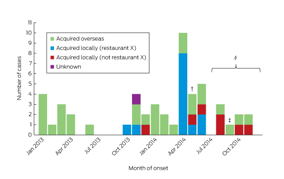
Between January 2013 and December 2014, 55 cases of HEV infection were notified (Box 1). The median age of the patients was 45 years (range, 4–77 years), 36 (65%) were male, and all but one (98%) lived in metropolitan Sydney. Twenty-four (44%) required hospitalisation, with a reported median length of stay (where known) of 7 days (range, 1–67 days). Three people (identified as co-diners of notified patients) were asymptomatic, and details about symptoms were unknown in one case. ALT levels were elevated in 33 of the 37 patients for whom they were recorded, with a median value of 1058 IU/L (range, 26–4868 IU/L; reference interval, 10–40 IU/L). None were pregnant.

Of the 55 patients, 30 (55%) reported a history of overseas travel during their incubation periods: to South Asia (17), East Asia (six), South-East Asia (two), Africa (two), Europe (two), or the Middle East (one). One patient could not be contacted; the remaining 24 (44%) did not report overseas travel.

Restaurant X outbreak

Restaurant X mainly served dishes suitable for sharing by a group. The menu included more than 28 meat, seafood and vegetarian options. Seventeen cases of HEV infection in nine separate groups who dined between October 2013 and May 2014 were linked to restaurant X. Of these 17, seven were identified by routine surveillance, eight by testing co-diners, and two by the retrospective serosurveys. Two people refused further interview; food histories were collected from the remaining 15 infected persons and from seven dining companions who tested HEV-negative by serology.

The demographic data for the diners is summarised in Box 2; the food items most commonly consumed are listed in Box 3. The highest attack rates were in those who consumed pork liver pâté, pork chorizo or roast pork. All 15 patients who provided a food history reported consuming pork liver pâté, compared with four of the seven uninfected co-diners (P < 0.05).

Locally acquired cases not linked to restaurant X

During interviews, the seven infected persons not linked to restaurant X reported eating a number of pork products during their incubation periods, including supermarket ham, prosciutto, pork liver, homemade pork liver sausage, pork chops and pork belly.

Retrospective serological surveys

Of 136 serosurvey samples (31 in survey 1, 105 in survey 2), nine (6.6%) were IgG-positive, four (2.9%) were IgM-positive, and four (2.9%) were both IgM- and IgG-positive for HEV. Of the eight people who were IgM-positive, HEV RNA was detected in four; sequencing confirmed infection with genotype 3. Two of these four people reported eating at restaurant X but not overseas travel, one reported travel to an HEV-endemic country, and one could not be contacted.

Laboratory investigation

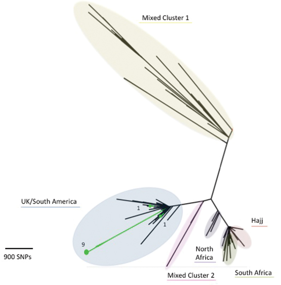
HEV RNA was detected in samples from ten of the 17 restaurant X cases; of the others, five with mild or no symptoms were PCR-negative, one was PCR-negative but showed seroconversion, and a sample was unavailable in one case. Sequencing of the ORF2 region was successful for all ten samples, and the HEV isolate was classified as genotype 3. There was at least 99% between-sample sequence homology in the targeted portion of ORF2 among restaurant X isolates.

HEV RNA was also detected in six of the seven locally acquired infections not linked to restaurant X (the specimen supplied by one person was insufficient for testing): five were genotype 3, and one sample was insufficient for genotyping. The viral sequence of these samples was about 90% homologous with samples from the restaurant-linked cases.

Environmental investigation

Investigation of restaurant X

Restaurant X was found to be well managed; no breaches in food safety or handling were identified. Staff were trained in handwashing and general food safety, including understanding cross-contamination and temperature control. During the observed cooking process, the internal temperature of the pork livers reached 51°C at 3 minutes, and between 82°C and 97°C at 4 minutes.

The livers used for pâté preparation were traced to a single farm. The pork shoulder, jowl and chorizo products were all sourced from different suppliers to the pork livers. HEV was not detected in any of the food samples obtained from the restaurant.

Investigation of pork products of locally acquired cases not linked to restaurant X

Pork products eaten by the seven infected persons not linked to restaurant X were bought from four different butchers and three different supermarkets. Pork livers from two of these butcheries could be traced back to two abattoirs supplied by several farms; further tracing was not undertaken. Pork liver sausages still held by one patient were found to contain very low levels of HEV RNA; the levels were too low for sequencing.

Public health interventions

NSW Health convened an expert panel involving public health, clinical, laboratory, agricultural and industry experts to assess the risks and to guide the investigation. On 15 May 2014, restaurant X was informed about its possible link with a number of cases of HEV infection. The importance of thorough cooking of pork products, including of pork liver pâté, was stressed, and the restaurant voluntarily removed this item from its menu. No further cases of HEV infection were linked to restaurant X.

As part of case finding, NSW Health issued an alert to gastroenterologists and public and private pathology laboratories in May 2014. The information garnered was then used to inform general practitioners in an alert, issued in September 2014, which requested that they consider HEV infection in people with a compatible illness, regardless of overseas travel. A joint media release with the New South Wales Food Authority, also issued in September 2014, urged the public to cook pork products thoroughly and, in particular, to cook pork livers to 75°C at the thickest part for 2 minutes.19

Discussion

This is the first reported Australian outbreak of locally acquired HEV infection and one of the largest linked with a restaurant reported anywhere. Seventeen cases were linked to consuming pork liver pâté at restaurant X during a 9-month period, and seven cases were linked to eating pork products bought from four butchers and three supermarkets with at least two different suppliers.

Retrospective serological testing identified a further eight previously undiagnosed cases of HEV infection (anti-HEV IgM). In two of these cases, HEV RNA was detected in people who reported no overseas travel but who had dined at restaurant X during their incubation periods. A further six cases were notified after the restaurant outbreak, probably as a result of increased vigilance and testing by clinicians. Data from a large public health laboratory confirmed this, with more than triple the number of HEV tests requested and carried out from July to December 2014 (after the laboratory began testing for HEV in people without a travel history) than during the same period in 2013 (unpublished data).

Active case finding among co-diners of restaurant cases detected locally acquired HEV infections that were either asymptomatic or mildly symptomatic, suggesting under-recognition and under-diagnosis of infection. A recent HEV serosurvey of blood donors by the Australian Blood Service identified past HEV infection in 14 of 194 blood donors without a history of overseas travel (7%).20 A case report in the Northern Territory21 and a study in Victoria22 each described single cases of HEV infection in which overseas travel was not implicated and no other risk factors were identified.

Common source outbreaks of HEV infection in high-income countries are rare. However, our investigation concurs with previous French,5 English10 and Japanese11 studies that have linked HEV infection with consumption of undercooked pork products. In these countries, locally acquired HEV infections predominate, and in 2013 accounted for 99% of all cases in France23 and almost 70% of cases in the UK.24

HEV is inactivated by heating to 71°C.19 Review of pork liver pâté preparation at restaurant X found that it was adequately cooked at the time of inspection, and testing available pork samples did not detect HEV RNA. It is nevertheless possible that, at the time of the restaurant infections (some weeks earlier), pork livers contaminated with HEV could have been undercooked at the thickest part before blending into pâté. This may explain the relatively low proportion of patrons infected with HEV at this popular restaurant. While we did not have access to leftover pâté samples from meals served to people infected at restaurant X that could be tested for HEV RNA, it was detected in pork liver sausages retained by one of the non-restaurant X patients.

Most fresh pork products in Australia are locally produced. The presence of HEV in Australian pigs was first noted in 1999 by a study that reported seropositivity rates of 17% in wild-caught pigs and more than 90% in commercial pigs by 16 weeks of age.25 To our knowledge, no further studies investigating the epidemiology of HEV in Australian pigs have been conducted. Despite the link between HEV outbreaks and pork products overseas, this discovery of HEV in Australian pigs did not translate into clinical practice, perhaps because HEV was not widely recognised as being endemic to Australian pigs, and because of a lack of awareness among Australian clinicians of the veterinary literature.

A limitation to this investigation was the time lag between some infected persons and co-diners being exposed, interviewed and tested for HEV, particularly co-diners of symptomatic persons from restaurant X. A lag in interviewing some infected persons and co-diners, coupled with the long incubation period of HEV (15–64 days), may have led to a recall bias in responses to the questionnaires and providing food histories. The limited sample size made it difficult to achieve statistically significant results. However, our findings are biologically plausible, and important associations could be deduced.

This study adds to our current understanding of the potential for HEV to be a food-borne illness in developed countries. Clinicians should request HEV testing in patients with acute hepatitis, irrespective of travel history, particularly where no aetiology has been determined. Laboratories should test for HEV where indicated to prevent under-recognition of infection. Health departments must be aware of the possibility of underestimating the prevalence of hepatitis E when using surveillance data. Pork products, particularly pork livers, should be cooked until they reach 75°C at the thickest part for 2 minutes.

Increased awareness, ongoing research and collaboration between primary industries, animal and human health authorities should help detect and prevent this and other emerging infectious diseases in Australia.

Box 1 –
Notifications of hepatitis E virus infections in New South Wales with onset dates between January 2013 and December 2014, by likely source of acquisition*


* Excludes three asymptomatic cases and one case with unknown symptom history. † May 2014: restaurant X was inspected, and pork pâté identified as the possible source of infection; restaurant voluntarily removed pork pâté from their menu. An alert was issued to gastroenterologists and pathology laboratories. ‡ September 2014: alert issued to general practitioners and the general public. § July–December 2014: increased HEV testing reported by the main public pathology laboratory.

Box 2 –
Characteristics of infected diners and healthy co-diners at restaurant X, October 2013 – May 2014

Infected persons (cases)

Healthy co-diners


Number

17

7

Median age (range), years

48 (29–75)

45 (29–47)

≤ 39 years

5 (29%)

1 (14%)

40–59 years

6 (35%)

5 (71%)

≥ 60 years

6 (35%)

0

Unknown

0

1 (14%)

Sex: men

12 (71%)

4 (57%)


Box 3 –
Commonly reported food items consumed by infected diners and healthy co-diners at restaurant X between October 2013 and May 2014*

Number of people who ate the item


Number of people who did not eat the item


Risk ratio (95% CI)

P

Infected persons (cases)

Healthy co-diners

Attack rate (%)

Infected persons (cases)

Healthy co-diners

Attack rate (%)


Brussel sprouts

5

3

63%

8

4

67%

1 (0.5–1.8)

1.00

Calamari

3

2

60%

10

5

67%

1 (0.5–2.0)

1.00

Eggplant

7

5

58%

6

2

75%

0.8 (0.5–1.5)

0.66

Pork chorizo

7

2

78%

6

5

55%

1.5 (0.8–2.7)

0.36

Pork pâté

15

4

79%

0

3

0

Undefined

0.02

Roast pork

9

4

69%

4

3

57%

1.2 (0.6–2.6)

0.64


* Food histories were available for 15 of the 17 infected persons (13 were complete and two were incomplete) and for all seven well co-diners.

Dengue fever in travellers: are we missing warning signs of severe dengue in a non-endemic setting?

Worldwide, there are an estimated 50–100 million cases of dengue virus infection each year. Far North Queensland has experienced dengue epidemics, with deaths reported in outbreaks in 2004 and 2008–2009.1

A 38-year-old man presented one day after returning from Colombo, Sri Lanka. He was a Sri Lankan-born Australian resident with no significant past medical history. He was admitted 10 days after the onset of a biphasic febrile illness: fever, chills, and generalised myalgia for 4 days, resolution of symptoms, then recurrence of symptoms on Day 7. On the day of admission, he developed diarrhoea and bloodstained vomiting. Dengue non-structural protein 1 (NS1) antigen was detected, and results of tests for dengue immunoglobulin (Ig) M and dengue IgG antibody were positive, suggesting secondary dengue virus infection. Persisting high fever, worsening thrombocytopenia (platelet count, < 50 × 109/L; reference interval, 150–400 × 109/L) and bloodstained vomitus led to a diagnosis of dengue fever (DF) with warning signs. The 2009 World Health Organization (WHO) guidelines for the management of dengue2 were followed (Box), with close monitoring of fluid status and haematocrit (HCT). On Day 4 of admission, the fever resolved, heralding the critical phase of DF. Haemoconcentration was noted, with HCT rising to 0.51 (> 20% above the baseline). Within 2 days of defervescence, a new pruritic rash was noted on the arms and legs that was characteristic of the convalescent phase of DF. There was slow resolution of the HCT, and intravenous fluid infusions were ceased. The patient was discharged 7 days after admission.

The revised 2009 WHO guidelines are based on validation studies from DF-endemic countries,3 and classify cases into DF, DF with warning signs and severe DF.2 In travellers, warning signs may also predict progression to severe dengue.4,5

Our patient’s case of DF with warning signs prompted a retrospective study of DF admissions at our institution. From 2012 to 2014, we identified 35 confirmed cases (median age of patients, 31 years). All cases were in returned travellers from dengue-endemic countries. Assessment for dengue severity was not well documented. No cases met the definition for severe DF and there were no deaths. Over 50% had warning signs for severe DF, including minor bleeding, abdominal pain and persistent vomiting. Warning signs were recognised in less than 30% of cases, and less than 10% of cases were managed according to WHO guidelines with strict fluid balance and HCT monitoring.

In conclusion, many returned travellers admitted with DF have warning signs, which predict the development of severe conditions with life-threatening endpoints, such as severe organ dysfunction and refractory shock. Hospitals in non-endemic areas should develop protocols for diagnosing and managing DF based on the WHO guidelines. Further research into the utility of warning signs in travellers with DF for predicting severe disease is needed.

Box –
Suggested dengue case classification and levels of severity


Reprinted from World Health Organization. Dengue: guidelines for diagnosis, treatment, prevention and control. New edition 2009. Geneva: WHO, 2009.

ALT = alanine aminotransferase. AST = aspartate aminotransferase. CNS = central nervous system. DSS = dengue shock syndrome. HCT = haematocrit.

Rising incidence of invasive meningococcal disease caused by Neisseria meningitidis serogroup W in Victoria

Invasive meningococcal disease (IMD) caused by Neisseria meningitidis is one of the most rapidly progressive sepsis syndromes, often resulting in significant morbidity and mortality. Since the introduction of meningococcal C conjugate vaccine in 2003, IMD in Victoria has decreased from 2.5/100 000 to 0.6/100 000 population.1 Epidemiological typing of N. meningitidis isolates is by serogroup, multilocus sequence typing and finetyping.

In Victoria, from January 2014 to September 2015, the number of cases of IMD caused by N. meningitidis serogroup W (MenW) increased. Previously uncommon (< 5% of IMD overall in the period from 2008 to 2013 [n = 260]), MenW as a proportion of IMD has increased: four of 33 cases in 2014 and 12 of 41 cases in 2015 (Microbiological Diagnostic Unit, University of Melbourne, unpublished data). Over this period, the median age of cases was 55 years, compared with 19 years for serogroup B, with many non-classical presentations including pneumonia, epiglottitis, septic arthritis and pericarditis. There has been one death in a healthy young adult. No epidemiological links between cases have been observed (Victorian Government Department of Health and Human Services [DHHS], unpublished data).

Globally, MenW has been responsible for an increasing proportion of IMD since outbreaks associated with the Hajj pilgrimage in 2000.2 Large outbreaks predominantly due to MenW strain type P1.5-2: F1-1: ST11 have been reported in South America and the United Kingdom.2,3 In the UK, MenW cases doubled year on year from < 2% of IMD prior to 2009–10 to 25% in 2014–15, prompting a change in vaccination guidelines.4 Initially, almost 25% of these IMD cases were older adults with non-classical presentations.3,4

From 1 January 2014 to 30 September 2015, molecular characterisation, including whole-genome sequencing (WGS), of Victorian MenW strains was undertaken at the Microbiological Diagnostic Unit. Of the 16 MenW isolates, 11 were strain type P1.5-2: F1-1: ST11; two were ST184; one was ST22; one was a new type; and one was polymerase chain reaction-positive only and thus unable to undergo WGS. Comparison of these 11 isolates with international strains using the PubMLST Neisseria database (http://pubmlst.org/neisseria) revealed that the Victorian isolates fall within a cluster formed by UK–South American outbreak strains and are distinct from Hajj outbreak strains (Box). Within the UK–South American cluster, nine Victorian isolates appear as an exclusive group of taxa. The close genetic relationship between the nine isolates, long branch length compared with other UK–South American cluster isolates, and lack of identified epidemiological links between cases suggest that these isolates may be representative of a N. meningitidis clone arising from a single introduction event that is undergoing widespread endemic transmission in Victoria. The location of the remaining two UK–South American cluster isolates in the tree indicates independent introduction events into Victoria.

While IMD due to MenW in Victoria remains low in absolute case numbers, the rise in incidence is concerning. The Victorian DHHS has instigated enhanced surveillance measures with full molecular characterisation of future isolates to inform ongoing public health responses. National surveillance with enhanced molecular characterisation will improve understanding of the current epidemiology of meningococcus in Australia.

Box –
Phylogenetic tree for Victorian and international meningococcal isolates


The diagram illustrates the phylogenetic relationship between isolates based on core genome single nucleotide polymorphisms (SNPs). Victorian MenW ST11 isolates demonstrate region-specific clustering, and the close relationship of Victorian MenW strains (in green) to the United Kingdom–South American cluster is shown. (Numerical values for the Victorian isolates represent the number of isolates, while the underlined description refers to geographical origin of isolates.) Note: Mixed Cluster 1 refers to isolates from the UK 1975–2007 and South Africa 2003–2013; Mixed Cluster 2 refers to isolates from UK 1996–2000, South Africa 2003–2004 and North Africa 1996–1999.