×

Ipilimumab-induced hypophysitis: early Australian experience

To the Editor: We report two men aged in their 60s receiving ipilimumab for metastatic melanoma who presented with headache and constitutional symptoms after the third 3-weekly dose, and were diagnosed with ipilimumab-induced hypophysitis. Ipilimumab is a monoclonal antibody that binds to cytotoxic T lymphocyte-associated antigen 4, resulting in T-cell activation and proliferation. It was the first therapy to yield a survival benefit in metastatic melanoma,1 but at the cost of frequent immune-related adverse events.2

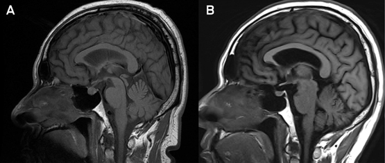
Patient 1 experienced headache, fatigue, postural lightheadedness, anorexia and asthenia. Morning blood test results before steroid administration were consistent with central hypocortisolism (serum cortisol, < 28 nmol/L [reference interval (RI), 70–650 nmol/L]; inappropriately normal adrenocorticotropic hormone [ACTH], 1.2 pmol/L [RI, 0–12.0 pmol/L]), hypothyroidism (low thyroid-stimulating hormone [TSH], 0.2 mIU/L [RI, 0.4–4.0 mIU/L]; low free thyroxine [FT4], 8.9 pmol/L [RI, 9.0–19.0 pmol/L]; normal free triiodothyronine [FT3], 4.7 pmol/L [RI, 2.6–6.0 pmol/L]), and hypogonadism (low testosterone, 2.8 nmol/L [9.5–28.0 nmol/L]; insufficiently raised follicle-stimulating hormone [FSH], 14.0 IU/L [RI, 1.0–12.0 IU/L]; luteinising hormone [LH] within RI, 3.7 IU/L [RI, 0.6–12 IU/L]). Insulin-like growth factor-1, growth hormone and prolactin levels were within RIs. Magnetic resonance imaging (MRI) scans of the brain excluded metastatic disease and demonstrated pituitary enlargement without chiasmal or infundibular involvement (Box). The patient was initially commenced on replacement hydrocortisone.

Patient 2 presented with severe headaches and emesis. Morning blood test results demonstrated central hypothyroidism (low TSH, 0.24 mIU/L [RI, 0.27–4.20 mIU/L]; FT4, 11.4 pmol/L [RI, 12.0–22.0 pmol/L]; and FT3, 2.8 pmol/L [RI, 3.0–7.8 pmol/L]) and hypogonadism (low testosterone [2.1 nmol/L] and inappropriately normal FSH [8.1 IU/L] and LH [3.9 IU/L]). Although morning cortisol and ACTH levels were initially adequate, the patient had been placed on high-dose dexamethasone because of suspicion of brain metastases, which were later excluded by lumbar puncture and brain MRI. The pituitary was not enlarged, but superior convexity was evident.

Both patients received a 3-week tapering course of prednisolone starting at 30 mg daily and weaned to replacement doses. They received thyroxine and, later, testosterone replacement. Ipilimumab was continued to include the standard fourth and final dose. The endocrine abnormalities persisted in both men at 3–6-month follow-up, despite normalisation of MRI findings (Box). For Patient 2, a subsequent insulin-tolerance test demonstrated a flat cortisol response consistent with corticotropic insufficiency due to hypophysitis, although suppression from exogenous steroids may have contributed. Both patients experienced tumour progression over the follow-up period.

Ipilimumab was registered by the Therapeutic Goods Administration in 2011 and funded by the Pharmaceutical Benefits Scheme from August 2013. Ipilimumab-induced thyroiditis and adrenalitis are common differential diagnoses to consider.4 Suprasellar extension is seen frequently,5 requiring urgent ophthalmic evaluation. There are no prospective data on whether high-dose steroids improve pituitary recovery or abrogate the tumour response to ipilimumab. Our cases demonstrated no short-term advantage. Interdisciplinary awareness of this condition is required to ensure that replacement glucocorticoids are started promptly on diagnosis and increased during periods of physiological stress, including surgery or chemotherapy.

Sagittal magnetic resonance imaging (MRI) scans


T1-weighted sagittal MRI scans of Patient 1, showing symmetrical pituitary enlargement up to 9.3 mm in height with superior convexity and loss of the posterior pituitary bright spot at presentation (A) consistent with a diagnosis of autoimmune hypophysitis,3 followed by normalisation 6 months later (B).

Ant venom immunotherapy in Australia: the unmet need

Jack jumper ant (JJA) venom extract is available through the Therapeutic Goods Administration (TGA) Special Access Scheme, and it is now time to provide this treatment alongside other insect venom immunotherapies (VITs) to allergic patients in areas of Australia where JJAs are prevalent.

The risk of insect sting anaphylaxis depends on complex interactions between the likelihood of human contact, insect aggression and regional distribution of the causative insect. Whereas venom allergy prevalence and presentation rates may be relatively low in highly urbanised areas, in rural areas and highly exposed populations, the prevalence of previous systemic reactions to stings can approach 3%–4%,1,2 and up to 30% of cases of anaphylaxis presenting to emergency departments (EDs) are triggered by insect stings.3 There were 20 deaths attributed to insect stings in Australia between 1997 and 2005.4

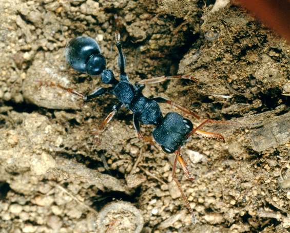
The Australian JJA (Myrmecia pilosula) (Box) is an aggressive species with patchy distribution around Australia, causing severe anaphylaxis, mainly in the south-eastern states.5 A Tasmanian allergist, Paul Clarke, first highlighted the extent of this problem in the Journal in 1986,6 and the first detailed description of four JJA sting anaphylaxis deaths was published in 2000.7 In Tasmania, the prevalence of JJA venom allergy is around 2.7%, and the rate of ED presentation with anaphylaxis for JJA is twice that for honeybees.1,3 Attempts to desensitise JJA-allergic patients with a crushed whole-body preparation produced by CSL were ineffective and that product was withdrawn in the 1990s.8

Management of sting anaphylaxis centres on identifying the causative insect, avoidance strategies where possible (eg, nest removal, moving to a location where the species is absent or less common), providing an emergency action plan and specific VIT. Although cost-effectiveness is difficult to quantify,9 VIT for wasps and honeybees is a well established procedure associated with lower rates of recurrent anaphylaxis and higher quality of life compared with carrying an adrenalin autoinjector alone.10 The efficacy of VIT directed against JJA has been established recently in a randomised, double-blind, placebo-controlled trial.11 The method was further refined in another randomised comparison of different treatment regimens,12 and a diagnostic framework has been developed to identify suitable candidates for treatment with this extract in mainland Australia as well as in Tasmania.5 Real-world efficacy is also being monitored; treatment tolerability and compliance are comparable with honeybee VIT, and in 478 patients receiving JJA VIT between 2002 and 2012, six of 132 individuals who experienced accidental stings (4.5%) had systemic allergic reactions, and only one individual was given adrenalin (own unpublished data).

Purified JJA venom for VIT is not commercially available, but is prepared by the Tasmanian Jack Jumper Allergy Program at the Royal Hobart Hospital as an active pharmaceutical ingredient (ie, concentrate), and use requires prior approval through the TGA Special Access Scheme. Although the evidence base supporting JJA VIT is at least equivalent to that supporting VIT for the honeybee, European wasp and paper wasp, which is subsidised through the Pharmaceutical Benefits Scheme, there is no equivalent subsidy for JJA venom supplies and the cost must be covered in full by the hospitals and/or patients. The cost of supplying JJA venom extract currently ranges from $1000 to $3000 per patient per year, depending on the dose schedule. This is more than for other insects because of the relatively small market volume. On the basis of our experience in Tasmania with enrolment rates, dropouts and treatment completion, we expect that subsidising the treatment nationally would result in a “steady state” of 500 to 1000 patients receiving treatment at any one time.

In response to this issue, the Australasian Society for Clinical Immunology and Allergy (ASCIA) has released a literature review and position statement on ant venom immunotherapy in Australia.13 ASCIA recognises that JJA VIT is unlikely to generate commercial venom development and, in the interest of equity, calls on federal and state governments to recognise the clinical need for this uniquely Australian problem and to allocate specific funding for hospital clinical services to provide this treatment. The statement also recognises the need for funding for ongoing research to investigate the use of lower dose protocols, immunological adjuvants and pharmaceutical research to investigate more stable formulations for distribution and supply to clinicians. However, ASCIA’s key messages are that the evidence base supporting JJA VIT now matches if not exceeds the evidence base supporting VIT for other species, and so it is now time to provide this treatment alongside other insect VITs to allergic patients in areas of Australia where JJAs are prevalent.

Australian jack jumper ant, Myrmecia pilosula, found mainly in the south-eastern states of Australia

Listeria monocytogenes in a healthy young adult

To the Editor: Meningitis caused by Listeria monocytogenes is rare in immunocompetent adults.1 In the absence of risk factors for
L. monocytogenes infection, Australian antibiotic guidelines recommend a third-generation cephalosporin alone for management of community-acquired meningitis, which has no activity against
L. monocytogenes
.2

A 22-year-old immunocompetent woman presented to our hospital with 3 days of fever, headache, photophobia and neck stiffness.
This had been preceded by a 2-day prodrome of diarrhoea. Medications on admission were cephalexin
for 1 day and a combined oral contraceptive. The patient had a history of coeliac disease, controlled on a gluten-free diet, and penicillin allergy. On examination, she was febrile (temperature, 38.7°C), with nuchal rigidity and positive Kernig’s sign, but with no rash. Heart rate (80 beats/min), respiratory rate
(18 beats/min) and blood pressure (145/80 mmHg) were within normal range. White cell count (WCC) (16 × 109/L; reference interval [RI], 5–10 × 109/L) and C-reactive protein concentration (206 mg/L; RI, < 2 mg/L) were elevated. HIV serology and β-human chorionic gonadotropin results were negative. Computed tomography of the brain gave normal results. A lumbar puncture revealed clear cerebrospinal fluid (CSF) with opening pressure > 35 mmH2O
(RI, 10–20 mmH2O), a glucose level of 1.5 mmol/L (RI, 2.0–3.9 mmol/L), a protein level of 0.96 g/L (RI, 0.15–0.45 g/L), and a WCC of 1000 (polymorphonuclear cells, 80; mononuclear cells, 920) × 106/L
(RI, < 5 × 106/L). Cryptococcal antigen was not detected in her CSF.

The patient commenced taking ceftriaxone, combined trimethoprim and sulfamethoxazole
(co-trimoxazole) and aciclovir. CSF culture was positive after 2 days, showing catalase-positive,
β-haemolytic colonies with gram-positive bacilli exhibiting tumbling motility at room temperature. These were identified as L. monocytogenes (99% on Vitek MS v2.0 and Vitek 2 Compact systems, bioMérieux), susceptible to ampicillin and
co-trimoxazole (Etest, bioMérieux). Antibiotics were rationalised to
co-trimoxazole alone, and the Victorian Department of Health was notified. Treatment ceased on Day 19 of 21, due to co-trimoxazole-related myelosuppression (WCC, 1.0 × 109/L; neutrophils, 0.8 × 109/L). Four weeks later, the patient’s full blood count was normal, and she was well, with no complications or immunodeficiency.

L. monocytogenes meningitis accounts for 5%–10% of bacterial meningitis and has a mortality rate up to 62%.3,4 It mostly occurs in extremes of ages, pregnant women and immunocompromised patients.5 As a cause of lymphocytic meningitis, it can be confused with viral meningitis.3 In our patient, differentiating factors were her high CSF opening pressure, low glucose level, and degree of elevated lymphocyte count and protein concentration. Other differential diagnoses included partially
treated bacterial, cryptococcal and tuberculous meningitis.3 The latter two were less likely, given the
short clinical history, a negative cryptococcal antigen and absence
of exposure to tuberculosis.

We recommend a low threshold for empiric prescription of benzylpenicillin (or co-trimoxazole
if the patient is allergic to penicillin) in patients with lymphocytic meningitis, regardless of underlying risk factors for L. monocytogenes infection.

Methylisothiazolinone in baby wipes: a rising star among causes of contact dermatitis

To the Editor: Methylisothia-zolinone (MI) is a preservative that has been used alone in cosmetic and personal products since the early 2000s.1 Before that time, MI was combined with methylchloroiso-thiazolinone in the widely used preservative Kathon CG (Dow Chemical Company), in a 1:3 ratio with the concentration of MI limited to 3.75 parts per million. This limit was subsequently increased to 100 parts per million, and MI is now being widely used in consumer products.

MI has been included in our baseline patch test series since 2011, following European reports of an increasing prevalence of MI allergy.1 We have subsequently seen a rapid increase in the number of patients with contact allergy to MI. Our rate
of positive reactions on MI patch tests to November 2013 was 11.3% (40 patients who had relevant reactions
of a total 353), compared with a rate
of 3.5% (15/428) in 2011 and 8.4% (38/454) in 2012. MI is now the most common cause of allergic contact dermatitis in our patient population.

Our Australian experience is reflected overseas. An increase in the frequency of allergic reactions to MI has been reported in Europe2,3 and, in the United States, MI was named the 2013 “Contact Allergen of the Year” by the American Contact Dermatitis Society, which bestows this “award” to highlight important and emerging allergens.

Among our patient population,
the most common source of MI is disposable wet wipes,4 now commonly used in nappy changing. MI is present in many popular
brands of wipes used in Australia. Interestingly, it is parents who use baby wipes on their children who
are presenting with hand dermatitis, although it is likely that allergic contact dermatitis involving the groin in children may not be diagnosed accurately.

Other common consumer sources of MI include make-up removal wipes, shampoos, conditioners, body washes, moisturisers, sunscreens and deodorants. Occupational sources include paints, cooling tower water and cutting oils.

The use of disposable wipes in nappy changing, personal care, cosmetic removal and cleaning has increased rapidly. Preservatives are required to prevent bacterial contamination in moist wipes. Medical practitioners and consumers should be aware of the potential for allergic contact dermatitis to develop to MI from wipes, in particular causing persistent hand dermatitis.

Perceptions of precautionary labelling among parents of children with food allergy and anaphylaxis

Precautionary labelling for food allergens such as “may contain traces of” are now present on more than half of all packaged processed foods in Australian supermarkets.1 Recent European and United States studies have shown that this high prevalence of precautionary labelling as well as consumers’ understanding that these statements are voluntary may have led to consumers not heeding precautionary statements. This might expose food-allergic consumers to the risk of allergen contamination, which may lead to life-threatening reactions such as anaphylaxis.2,3

The Australian manufacturing industry has undertaken to develop a new standard known as VITAL (voluntary incidental trace allergen labelling), to help improve standards for declaring the presence of allergens within foods. In 2007, a new precautionary statement “may be present” was introduced. The VITAL process has attracted international interest, but, to date, the “may be present” statement is rarely used.1,4

In this study, we aimed to gain an understanding of the behaviour, perceptions and opinions of parents about precautionary labelling, stratified by whether or not their food-allergic child had a history of anaphylaxis. We also aimed to understand consumers’ perceptions of the “may be present” statement advocated by VITAL.

Methods

Participants were recruited in the Department of Allergy and Immunology at the Royal Children’s Hospital, Melbourne, from 1 August to 31 October 2011. The parents (one only) of a consecutive series of children who had an appointment for a skin-prick test to investigate possible or established food allergy were asked to complete a self-administered questionnaire.

All analyses were restricted to children with current medically diagnosed food allergy, and grouped based on the following:

  • children with a past history of anaphylaxis, defined as a past history of respiratory or cardiovascular compromise in the setting of an acute allergic reaction;5 and
  • children with a past history of mild to moderate IgE-mediated reactions, defined as immediate reactions involving the skin or gastrointestinal system (eg, vomiting, abdominal pain, diarrhoea, hives, and swelling of the eyes, lips or face) without involvement of the airway or circulatory systems.

We compared responses between the parents of children with and without a history of anaphylaxis using Pearson χ2 tests.

This study was approved by the Royal Children’s Hospital Human Research Ethics Committee (RCH HREC 31140A).

Results

The parents of 535 children were approached, and those of 497 children (93%) agreed to participate. Two-thirds of the children (66%) were boys and 84% were aged under 10 years. Food allergy had been medically diagnosed in 293 (59%) of these children. The most common food allergies were to peanut (152 children; 52%), egg (142; 48%), tree nuts (112; 38%) and milk (81; 28%). Of the 293 children with food allergy, 246 (84%) had sufficient information provided to allow past reactions to be classified as either a past history of anaphylaxis (113 children) or a past history of mild to moderate IgE-mediated reactions (133 children). Among children with a history of anaphylaxis, the parents of 84 (74%) reported that their child had been treated at an emergency department or been hospitalised.

Difference in knowledge between parents of children with and without a past history of anaphylaxis

There were no differences between the parents of children with and without a past history of anaphylaxis in reading of food labels or whether they would give their child a product if the food they were allergic to was listed in the precautionary labelling section (Box 1). However, parents of children with a past history of anaphylaxis were more likely to remove from the house any food products containing the food to which their child was allergic (Box 1).

Perception, opinions and behaviour relating to precautionary food labels

In total, the parents of 54 children (48%) with a history of anaphylaxis felt that the ingredient list information of food labels was easy to understand and use and six (5.4%) felt that they could trust food labels (Appendix 1). The parents of 88 and 106 children with a history of anaphylaxis (78% and 84%, respectively) thought precautionary labels were not useful and that they did not know whether the food was safe to eat irrespective of the wording of the labels (Appendix 2). The vast majority of parents of food-allergic children felt that there should be better government regulation imposed on manufacturers in the way that they use precautionary labels (Appendix 1).

Avoidance of foods by type of precautionary labelling

The proportion of participants with children with a history of anaphylaxis who would avoid feeding the child a particular food product with a precautionary label varied depending on the wording of the precautionary label, with 74 parents (65%) reporting that they would ignore the statement “made in the same factory” compared with 24 (22%) reporting that they would ignore the statement “may be present” (Box 2). However, 68 (60%) would allow their child to consume foods labelled with this statement if their doctor said it was safe to do so (Appendix 1).

There was no significant difference between parents of children with a history of anaphylaxis compared with those with mild to moderate reactions who reported that they would allow their child to consume foods with precautionary labelling (Pearson χ2 test; Box 1).

There was no difference in parents’ self-reported behaviours according to whether or not their child had a history of anaphylaxis (Box 2). However, the precautionary statement “may be present” was perceived as less useful by participants with a child with a history of anaphylaxis compared with a child without a history of anaphylaxis (53 [47%] v 109 [82%]: P < 0.05; Appendix 1).

Discussion

Our results show that parents of food-allergic children, including those with a child with a past history of anaphylaxis who might be considered to be at greater risk of an adverse reaction, appear complacent about precautionary labelling. In addition, we found that parents of food-allergic children are assuming a gradient level of risk based on the wording of the precautionary statements; this has also been seen previously.2 It is also of interest that the VITAL statement “may be present” was seen as most useful, and was taken more seriously despite there being no training or education provided to the participants about the VITAL process.

Our results may be at least partly explained by the ubiquitous nature of precautionary labelling in Australia, where more than 65% of all edible packaged foodstuffs have some form of precautionary labelling.1

Similar to our findings, two previous studies reported that patients with allergies were taking risks by disregarding some forms of advisory labelling because they were incorrectly assuming that statements such as “shared facility” and “may contain” indicated different levels of risk.2,6 In fact, detectable peanut residues were more common in products that had “shared facility” as a precautionary statement compared with those with a “may contain” statement.6 However, previous studies that have investigated consumers’ perceptions of precautionary labelling did not stratify people with allergies by the severity of their reaction as we did in this study.7,8

The key strengths of our study are the response rate of 93% and the fact that parents of all children attending for a skin-prick test were asked to participate in the study. A possible limitation is that we relied on parents’ self-report that their child had medically diagnosed food allergy and a past history of anaphylaxis. However, we believe this is appropriate for this type of study as parents’ perceptions and attitudes are likely to drive their choices when making decisions on behalf of their children. Our high participation rate makes a bias towards participation of more anxious families in the study unlikely; however, our results are not necessarily representative of the wider community, as all participants were recruited from a single hospital-based allergy clinic. Furthermore this study pertains to parent choice for food-allergic children as opposed to older food-allergic patients making decisions for themselves.

In conclusion, the attitudes of parents of food-allergic children to precautionary labelling appear to be complacent, whether or not children had a past history of anaphylaxis. Policies that promote the use of fewer precautionary statements or more effective labelling strategies may lead to less consumer complacency.

1 Behaviour relating to food labels among parents of food-allergic children with a history of anaphylaxis (113) and with a history of mild to moderate IgE-mediated reactions (133)

Question and possible responses

Anaphylaxis

Mild–moderate reactions

P*

If your child has a specific food allergy (eg, peanuts) do you intentionally remove food products containing the specific food from the house?


No

39 (35%)

65 (49%)


Not sure

1 (1%)

3 (2%)


Yes

73 (55%)

64 (48%)

0.04

When you buy a food product which part of the label do you check for an allergen?


Ingredients only

27 (24%)

32 (25%)


Precautionary information only

1 (1%)

1 (2%)


Both

82 (73%)

95 (74%)


Neither

2 (2%)

1 (1%)

0.88

Would you give your child a food if the food he or she was allergic to was listed in the precautionary labelling section?


No

64 (58%)

81 (62%)


Not sure

8 (7%)

11 (8%)


Yes

38 (35%)

39 (30%)

0.72

How often do you look at precautionary food labels?


Only when I buy a product for the first time

33 (30%)

44 (34%)


Only occasionally when I buy a product

10 (9%)

14 (11%)


Most of the times when I buy a product

31 (28%)

34 (26%)


Every time I buy a product

36 (33%)

38 (29%)

0.85


* Calculated using the Pearson χ2 test.

2 Percentage of parents of food-allergic children who reported that they would ignore a particular precautionary label, stratified by risk


* Vomiting, abdominal pain, diarrhoea, hives, and swelling of the eyes, lips or face.

Salicylate elimination diets in children: is food restriction supported by the evidence?

When a food is identified as causing allergic symptoms, that food will usually be removed from the diet. However, inappropriate use of extensive food elimination can be harmful. Salicylate elimination or “low salicylate” diets — which remove foods deemed to contain natural salicylates — can be particularly restrictive, especially as they are often implemented with restriction of other foods such as those containing amines, glutamates, synthetic food additives, gluten and dairy. These diets appear to be commonly used in New South Wales, but to our knowledge are not widely used outside of the state or in other countries. We discuss our own experiences with children who were referred for care to the allergy clinics of three public hospitals, and who had previously used these diets, and review the evidence for using low salicylate diets in treating a variety of disease indications.

For which conditions are low salicylate diets prescribed in Sydney?

We sought to identify the indications for which salicylate elimination is prescribed in Sydney by conducting a retrospective case note review of children attending the allergy clinics of the two main children’s hospitals, Sydney Children’s Hospital and the Children’s Hospital at Westmead, as well as a major regional allergy clinic at Campbelltown Hospital, between 1 January 2003 and 31 December 2011. We confirmed any missing details through a single telephone conversation between an immunologist or allergist and the child’s carer. Approval for the study was obtained from the South Eastern Sydney Local Health District, Human Research Ethics Committee – Northern Sector.

We identified 74 children who had at some point in their lives been on a low natural salicylate diet. The most common indication for initiation of the diet, reported by the patient’s carer, was eczema in 34/74, followed by a behavioural abnormality (eg, attention deficit hyperactivity disorder [ADHD] or unsettled infant behaviour) in 17/74 and gastrointestinal disturbances (eg, abdominal pain or gastro-oesophageal reflux disease) in 12/74 (Box).

What is the evidence supporting the role of low salicylate diets for these indications?

We reviewed the literature using MEDLINE and PubMed, combining search terms “salicylate”, “elimination diet” or “exclusion diet” with “food allergy”, “food intolerance”, “eczema”, “atopic dermatitis”, “chronic urticaria”, “ADHD”, “behaviour” or “gastrointestinal”. We found no evidence in the peer-reviewed literature to suggest a role for salicylates in any of the diseases for which the diet is prescribed.

In the absence of an overt type I hypersensitivity clinical response, food is an uncommon precipitant of eczema. A 2008 Cochrane review concluded that, with the exception of egg exclusion in patients who have positive specific IgE antibodies to egg, there is little evidence to support restriction of tolerated foods in eczema.1

On the other hand, there is good evidence that food exclusion can ameliorate the hyperkinesis symptoms of ADHD, with numerous studies showing a benefit for broad-based food exclusion diets.2 However, a recent randomised controlled trial suggests that much of this effect is caused by artificial food additives, and we were unable to identify any peer-reviewed evidence that natural salicylates can cause hyperactive behaviour.3 One published letter referred to challenge with salicylates precipitating behavioural symptoms, however the authors did not stipulate whether the challenge substance was natural salicylate or acetylsalicylic acid (aspirin)4 — aspirin being known to cause significant symptoms when natural salicylates have no effect.5

Finally, while foods are well known to cause a variety of gastrointestinal symptoms, from coeliac disease to irritable bowel syndrome, there is no good peer-reviewed evidence that natural salicylates cause any gastrointestinal symptoms.

Do salicylate elimination diets cause harm?

Although food elimination diets used to treat allergy have been associated with side effects including micronutrient deficiency,68 protein or energy malnutrition,9 eating disorders,10 food aversion,11 and the development of allergic reactions including fatal anaphylaxis to the excluded food on reintroduction,12,13 we were unable to identify any evidence regarding the safety or otherwise of salicylate elimination diets in children. This is of concern given that many of the patients attending our clinics had started the diets at a young age (median, 24 months; range, 6 weeks to 15 years), and continued for an extended period (> 1 year in 30/61 children).

Among our patients, where details were available, we identified a high occurrence of possible adverse outcomes among children who had been on low salicylate diets, with 31 out of 66 children suffering one or more possible adverse events. Symptoms and problems experienced included weight loss or failure to thrive in 13/66 children, eating disorders (including three cases of anorexia nervosa) in 4/66, specific nutrient deficiency in 2/66 (one case of vitamin C deficiency, one case of protein, iron and zinc deficiency), food aversion in 6/66, alopecia in 2/66 and unplanned weaning in 3/66. Four out of 13 mothers who went on the diet to benefit their breastfeeding infant suffered significant weight loss, which they perceived as problematic.

While we acknowledge that our cohort has an inherent selection bias and that without a control group it is not possible to attribute the reported events to the diet, we are concerned that all adverse events were reported to have occurred after initiation of the diet.

Also, beyond the possible adverse events noted in our patients, we are additionally concerned about the use of broad-based empirical food elimination in early life, with increasing evidence suggesting that food elimination at this time predisposes to the development of food allergy to the excluded foods, particularly among children with eczema, which was the largest group identified here.14,15

Who prescribes salicylate elimination diets?

Among those patients where details were available, 47/69 were prescribed the diet through medical allergy services, with general paediatricians 7/69 and dietitians 7/69 prescribing less frequently, while 8/69 parents obtained the diet from friends or from the internet. We do not prescribe the diets in our practice.

In order to assess whether the diet was more widely used elsewhere, we surveyed overseas allergists. An online survey of members of the editorial boards of major European and North American allergy journals produced 23/125 responses, with none of the responding experts employing the diet for ADHD, and only 1/23 using a form of salicylate exclusion for eczema.

Does the available research support a role for natural salicylates in any disease causation?

As discussed above, there is no peer-reviewed evidence to support the use of low salicylate diets in treating eczema, behavioural symptoms or gastrointestinal symptoms.

One disease where the role of natural salicylates has been studied in more detail is aspirin-sensitive asthma, where doses of natural salicylic acid 10 times higher than the aspirin dose have no effect.5 The lack of importance of natural salicylates in this disease is well established in clinical practice, as reflected by the evidence-based clinical decision support website, UpToDate (http://www.uptodate.com/home), which states that “dietary salicylates do not cause symptoms in NSAID [non-steroidal anti-inflammatory drug] sensitive patients”.16

A second disease where low salicylate diets have been trialled is chronic idiopathic urticaria (CIU); however, while there is some evidence that synthetic food additives may play a role in a small proportion of adult CIU cases,17 the peer-reviewed evidence that salicylates play any role in this disease is largely limited to studies that used aspirin as the challenge substance.18 On the other hand, there are several reasons to question the idea that salicylate-containing foods play any role in CIU. First is the recent discovery that half of childhood CIU is autoimmune in nature, resulting from autoantibodies against the high-affinity IgE receptor.19 Second, evidence suggests that those few foods said to contain salicylates that may precipitate CIU (eg, tomatoes, wine, herbs) probably do so not because of their salicylate content, but because they contain volatile aromatic chemicals (eg, alcohol, ketones and aldehydes).20 Third, there is evidence that the foods removed in low salicylate diets may not actually contain significant levels of salicylates, with one group suggesting that many “high salicylate foods” contain no aspirin and only tiny amounts of natural salicylates.21

Finally, it is important to discuss local research on salicylate intolerance performed in the early-to-mid 1980s. Most of that work focused on CIU, with a lesser focus on a number of other symptom complexes.4,2224 The research involved placing patients on diets that removed foods containing salicylates, using food challenge to identify which constituents were responsible for any perceived improvement.22,24 However, teasing out which component of these broad-based elimination diets were responsible for any perceived benefit is difficult, given that the diets removed many food constituents, including those now known to cause symptoms, such as artificial food additives,3,17,25 and because the challenge substance was commonly aspirin,22,24 although sodium salicylate was said to have been used in some work.22 Moreover, most of the clinical data appeared in a non-peer-reviewed format,22 or with incomplete methodological details in review format in peer-reviewed journals.4,23 These non-peer-reviewed findings of disease associations of natural salicylates have not been reproduced by other investigators, and a recent British textbook of food hypersensitivity concluded “there are no effective diagnostic tests for salicylate intolerance, and no studies showing the efficacy of dietary exclusion”.26

Can salicylate elimination diets be recommended for use in children?

The use of low salicylate diets in children is not supported by current evidence or by expert opinion. There is also no evidence that these diets are safe, in particular for infants and their breastfeeding mothers, and for those at risk of developing eating disorders. While our retrospective case note review is insufficient to prove any risk associated with the diets, it is concerning that harm may occur when children and adolescents are placed on such restrictive diets, particularly if they stay on them for long periods.

We would invite any proponents and prescribers of the diet to produce evidence of the efficacy and safety for the disorders in which they consider such a restrictive diet is indicated. Pending such evidence, we cannot recommend the use of salicylate elimination diets.

Characteristics of 74 children prescribed salicylate elimination diets

Characteristic

No. of children/total*


Age at initiation of diet

Infancy (1 year or less)

26/67 

Early childhood (1–3 years)

22/67 

Childhood (4–10 years)

10/67 

Adolescence (11–18 years)

9/67 

Duration of diet

< 1 month

5/61 

1 month to 6 months

17/61 

> 6 months to 1 year

9/61 

> 1 year

30/61

Indication for diet

Eczema

34/74 

Behaviour (including ADHD)

17/4 

Gastrointestinal complaints

12/74 

Failure to thrive

4/74 

Acute allergic reaction

3/74 

Anaphylactoid reaction

2/74 

Urinary urgency

1/74 

Headache

1/74 

Adverse events

Failure to thrive or weight loss

13/66 

Food aversion

6/66 

Eating disorder

4/66

Infant weaned early

3/66

Alopecia

2/66

Nutrient deficiency

2/66

Constipation

1/66 

Total children with adverse events

31/66 

Breastfeeding mothers with complications of diet

4/13 

Prescribed by

Medical allergy clinics

47/69

Dietitian

7/69 

Paediatrician

7/69 

Friend or internet

8/69 


ADHD = attention deficit hyperactivity disorder. * Varying denominators reflect the completeness of available data. Including three cases of anorexia nervosa.

Salicylate elimination diets in children

With suitable guidance and monitoring, dietary modification is safe and can improve quality of life

Salicylates are a diverse family of 2-OH benzoic acid derivatives produced in all plants. The parent molecule, salicylic acid (SA), is a ubiquitous phytohormone responsible for pathogen resistance, regulation of growth, and a wide variety of other biological functions.1,2 The medicinal use of salicylate-containing plant extracts was first recorded about 3500 years ago, and was widespread until the mid 19th century, when SA was isolated from willow bark, and acetylsalicylic acid (ASA) was subsequently synthesised and marketed by Bayer as aspirin in 1899.2

The occurrence of urticaria and angioedema from SA was recognised by the 1890s, and the first reports of urticaria, angioedema and asthma induced by ASA began to appear in the early years of the 20th century. Although by then salicylates were known to be present in certain foods, it took over 50 years before low salicylate therapeutic diets were developed. Feingold, an allergist at Kaiser Permanente in the 1960s, was the first to attempt exclusion of natural salicylates in patients with aspirin sensitivity. His claims of efficacy of the “K-P” diet in children with attention deficit hyperactivity disorder aroused enormous controversy at the time but were eventually substantiated by randomised trials.3,4

In the 1970s, a flurry of reports on the use of similar elimination diets in patients with chronic urticaria led one of us (A R S) to undertake a comprehensive analysis of the salicylate content of more than 330 common foods.5 This analysis enabled more accurate diagnosis and management of patients with salicylate intolerance.6,7 Since intolerance to food chemicals is highly idiosyncratic, with protean clinical manifestations in children and adults (including eczema with urticarial features), the only reliable means of identifying the provoking substances is by elimination of all potential triggers for 2–6 weeks, followed by systematic challenges if there is significant improvement.7 An individually tailored diet can then be prescribed, after which patients are encouraged to gradually liberalise their intake to determine their tolerance threshold for each substance.

In this issue, Gray and colleagues report a retrospective chart review of 74 children seen over a 9-year period who at some point in their lives had been on a low salicylate diet, and call into question the safety, efficacy and evidence base for such dietary modifications.8 We believe that these concerns are misplaced for several reasons. First, the results from peer-reviewed publication of group data may not be transferrable to determine what dietary modifications are appropriate in individual cases; the diagnostic elimination–challenge process we use is essentially a single-patient (n-of-1) trial method which is generally considered to provide strong evidence in guiding clinical practice.9

Second, Gray and colleagues attempt to cast doubt on the analysis of salicylates in foods; however, a recent systematic review firmly established that they are present in biologically significant amounts.10 Further, they draw an inappropriate distinction between “natural salicylates” and “aspirin”. On analysis, most salicylate-containing foods contain both ASA and SA, and more than one-third contain ASA alone.11 Since there is extensive cross-reactivity between SA, ASA, benzoic acid and 4-OH benzoate in patients with chronic urticaria, ASA challenge reactivity is best regarded as a marker for intolerance to a range of natural salicylates and related dietary phenolics.11

Finally, Gray and colleagues list what they refer to as “adverse events” in 31 children, implying that these events might be a result of past exclusion of dietary salicylates. However, they do not demonstrate a causal relationship between these events and salicylate exclusion.

The possibility of nutritional deficiency is an issue of concern in any child on a modified diet. However, Gray and colleagues do not provide information about how “specific nutrient deficiencies” were evaluated in the cases they reviewed. Since estimation of nutrient intake from diet recall is notoriously unreliable, whenever such concerns arise, formal analysis of a 4-day food diary is warranted, followed by intervention where appropriate.

In our experience, intake of nutrients by children on a low chemical diet is usually at or above the recommended daily amount and is not significantly different from that of children on a normal diet (own unpublished data), confirming previously published studies of the nutritional adequacy of the Feingold diet.12,13 We believe that with suitable guidance, dietary modification based on n-of-1 testing is safe in children and can lead to significant improvements in quality of life.

Progressive multifocal leukoencephalopathy caused by BK virus?

The evidence is provocative but not definitive; nonetheless, it should serve as a stimulus to further research

In this issue of the Journal, Daveson and colleagues1 describe a case of progressive multifocal leukoencephalopathy (PML) possibly caused by BK virus rather than JC virus. This finding is potentially very significant. The polyomaviruses BK and JC commonly infect humans and remain latent in immunocomptent individuals. Both are associated with clinical disease in the setting of immunosuppression. However, only JC virus has been causally associated with PML; BK virus has been causally associated with nephropathy, ureteric stenosis and cystitis. There have been previous case reports of BK virus causing a meningoencephalitis and PML as detailed by Daveson et al, but the evidence for causality has been tenuous, largely because of the lack of confirmation in tissue. The data provided by Daveson et al are more convincing, although not definitive.

How could their evidence change clinical practice? First, the significance of PML being caused by BK virus is that the diagnosis of PML has until now only focused on the detection of JC virus.2,3 A large number of patients have or are presumed to have PML as a consequence of immunosuppression from cytotoxic chemotherapy (especially rituximab), from immunodeficiency related to HIV disease, or from immunomodulation related to the multiple sclerosis drug natalizumab.3 In a reasonable number of cases, JC virus is not detected in the cerebrospinal fluid or brain biopsy. This has been considered a consequence of insensitive assay tools, sampling error, or episodes of immune restoration inflammatory syndrome that may reduce viral DNA load before collection of cerebrospinal fluid.2,3 It is now conceivable that some of these PML cases may be caused by BK virus. Such cases would potentially be amenable to therapy: some evidence exists for efficacy with cidofovir, fluoroquinolones such as ciprofloxacin, and leflunomide.4,5 Further, risk stratification for PML in natalizumab-treated patients is currently heavily weighted towards the presence or absence of JC virus on serological testing.6 If BK virus truly can cause PML, such risk stratification strategies would need to include assessment for BK virus antibodies.

Given the potential importance of BK virus causing PML, how robust is the evidence of the causal link in Daveson et al’s report? While the authors did not find evidence for JC virus, it would have been helpful to have negative serology results for JC virus. The presence of enhancing lesions on magnetic resonance imaging is somewhat unusual for PML unless the patient has immune restoration inflammatory syndrome. Nonetheless, it can occur and has been recorded in about 10% of non-natalizumab-treated patients.2 The detection of BK virus in brain tissue, especially in the context of inflammation, raises the possibility that it was imported into the brain in inflammatory cells and that it is an “innocent bystander”. This is certainly possible but, on the other hand, no other cause was found and, in particular, JC virus was not detected by polymerase chain reaction. Further, it would be reassuring to know that the BK virus antibodies did not cross react with JC virus. Last, the case for BK virus causing PML would have been strengthened if there were data showing BK viraemia or viruria. Nonetheless, the BK viral DNA load in the cerebrospinal fluid was high, at 11 975 copies/mL; a level in the plasma of > 10 000 copies/mL is associated with a 93% specificity for presence of BK virus nephropathy.7 This level was quoted in recent guidelines by the Kidney Disease Improving Global Outcomes Transplant Work Group for the diagnosis of BK virus nephropathy.8 Given the implications of this observation, confirmation by an independent laboratory with validated and certified assays for these viruses could be reassuring.

The case for a causal link between BK virus and PML is still not solid, yet the details of the case report are highly suggestive. We consider the implications to be substantial. The report should prompt further research and meticulous analysis of future PML cases with particular attention to the issues we have outlined here.