×

Warfarin or excipient allergy: a clinical dilemma resolved

A 57-year-old Asian woman was referred for surgical ablation of atrial fibrillation (AF), coronary revascularisation and mitral valve surgery. She had a history of paroxysmal AF, previous stroke, coronary artery disease and severe mitral regurgitation. In the work-up for surgery, she developed recurrent, pruritic maculopapular rashes involving her trunk and upper limbs on two occasions within 3 days of initiating oral anticoagulation with warfarin (Marevan, Aspen) for AF. There were no other changes to the patient’s longstanding medication regimen during this period. Her regular medications consisted of aspirin 100 mg and perindopril arginine 2.5 mg in the morning, and atorvastatin 80 mg at night. Long-term anticoagulation with warfarin was necessary due to planned mechanical valve replacement and ongoing paroxysmal AF. Subsequently, she was referred to a clinical immunologist for assessment of her reaction to warfarin. Since allergic reactions to warfarin are rare, a reaction to one of the dyes in the tablet was considered.

The patient had been taking all three strengths of Marevan tablets (1 mg, 3 mg and 5 mg), which contain a number of excipients (Box). Initial drug challenge commenced with Coumadin 1 mg (Bristol-Myers Squibb) tablets, as these did not contain any shared excipients. She tolerated this for 1 week and subsequently the dose was increased to three 1 mg tablets of Coumadin. She remained symptom-free and reached a therapeutic international normalised ratio. It was therefore deemed safer to avoid Marevan 1 mg and 3 mg tablets and the Coumadin 2 mg tablet, which shared the colouring constituent indigo carmine (132). She later underwent mitral valve replacement with a mechanical valve, closure of the left atrial appendage, surgical ablation and coronary revascularisation. She currently remains rate-controlled at 7 months follow-up with therapeutic anticoagulation on Coumadin 1 mg tablets.

“Warfarin allergy” can be due to the coumarin structure or the excipients introduced for colouring and safety.1 While this has previously been documented, excipients are rarely considered as a possible cause of allergic reaction in common practice.1,2 Importantly, not all allergies are attributed to the coumarin structure, and it is possible to have dermatological adverse reactions to the colouring dyes in warfarin.1,3 In our patient, we strongly suspect indigo carmine (FD&C Blue No. 2). Blue dyes have previously been implicated as the cause of urticarial rashes in patients taking synthyroid tablets.4 Although there was no objective confirmation of causality (eg, patch testing), the clinical presentation coupled with a high score of 9 on the Naranjo probability scale is compelling, although not conclusive.5 With this in mind, we suggest that excipient allergy should be considered in patients with a reaction to warfarin, and dye-free preparations can be administered as a therapeutic alternative.


Excipient dyes used in Coumadin and Marevan brands

Brand/strength

Tablet colour

Active ingredient

Dye


Coumadin 1 mg

Light tan

Warfarin sodium

Amaranth (123)Quinoline Yellow (104)

Coumadin 2 mg

Lavender

Warfarin sodium

Amaranth (123)Indigo Carmine (132)

Coumadin 5 mg

Green

Warfarin sodium

Quinoline Yellow (104)Brilliant Blue FCF (133)

Marevan 1 mg

Brown

Warfarin sodium

Iron Oxide Yellow (172)Iron Oxide Red (142)Indigo Carmine (132)

Marevan 3 mg

Blue

Warfarin sodium

Indigo Carmine (132)

Marevan 5 mg

Pink

Warfarin sodium

Erythrosine (127)


Other excipients (all strengths): Coumadin — lactose, starch (tapioca, stearic acid and magnesium stearate); Marevan — lactose, starch (maize and pregelatinised maize, sodium starch and magnesium stearate).

Successful treatment of ACE inhibitor-induced angioedema with icatibant, a bradykinin B2 receptor antagonist

Clinical record

A 65-year-old woman of European ancestry presented to the emergency department (ED) of our tertiary hospital at 09:30 with isolated tongue swelling without rash, gastrointestinal symptoms or wheeze, which had progressed over the previous 4 hours and was non-responsive to adrenaline administered by her general practitioner at 08:15. She recalled a single episode of mild self-limiting tongue swelling several months earlier and had no family history of angioedema. The patient had well controlled hypertension managed with perindopril (5 mg daily) for the past 10 years. Other comorbidities included gastroesophageal reflux disease, hypercholesterolaemia and osteopaenia. She was an active smoker. Other medications included atorvastatin, calcium carbonate, dothiepin, esomeprazole and oestradiol valerate, and she denied over-the-counter medication use.

Despite treatment with intravenous dexamethasone (6 mg), intravenous glycopyrrolate (0.2 mg) and intramuscular promethazine (25 mg), the patient’s condition continued to deteriorate. Fibreoptic nasoendoscopy (FNE) revealed epiglottic and left arytenoid oedema. Her treating team planned awake fibreoptic intubation as a priority one case, with a surgical team on standby for emergency tracheostomy.

Icatibant, a competitive bradykinin B2 receptor (B2R) antagonist, was administered subcutaneously at a dose of 30 mg at 10:30, without development of a local injection-site reaction. On repeat FNE 10 minutes later, there was a significant improvement in her condition. Intubation was ultimately avoided and she was observed in the intensive care unit (ICU) for 2 hours before being transferred to a high-dependency ward area overnight. Complete resolution of symptoms was seen within 5 hours, and she was discharged the next morning after a normal FNE.

Perindopril was implicated in the patient’s angioedema. It was discontinued and replaced with amlodipine (5 mg daily) and hydrochlorothiazide (12.5 mg daily). At follow-up 9 months later, she reported no further episodes of angioedema. Despite thorough investigation, no other cause aside from angiotensin-converting enzyme (ACE) inhibitor-induced angioedema was identified. An infective focus was excluded, with urinalysis, chest x-ray and white cell count yielding normal results. Hereditary angioedema and acquired C1 esterase inhibitor deficiency were excluded by a normal C4 concentration of 0.4 g/L (reference interval, 0.16–0.52 g/L) at presentation. Quantitative and functional C1 inhibitor studies at a subsequent follow-up visit did not find reduced values.

ACE inhibitors are commonly used in the management of hypertension, and of cardiac and renal disease. Angioedema occurs in 0.1%–0.68% of patients,1 more commonly within 90 days of the introduction of ACE inhibitors, although it can occur later.2

After cessation of medication, patients can develop recurrent angioedema, typically within the first month but sometimes several months later.3 ACE inhibitors account for 30% of ED presentations with angioedema, and although most are discharged directly from ED, 11% require ICU management4 for an average of 2.9 days at considerable cost.5

ACE inhibitor-induced angioedema is mediated by bradykinin, and empiric therapy with adrenaline, steroids and antihistamines targeted at histaminergic responses is largely ineffective.1 Bradykinins bind B2Rs and induce potent vasodilation. ACE (also known as kininase II) promotes bradykinin degradation, so ACE inhibition prolongs the half-life of bradykinin.6

Dipeptidyl peptidase-4 (DPP-IV) is a secondary enzyme in bradykinin breakdown.6 Concomitant use of ACE inhibitors and DPP-IV inhibitors such as vildagliptin, used to treat type 2 diabetes, or DPP-IV modulation in the context of immunosuppression after transplant, puts patients at increased risk of angioedema.7,8 The use of other medications that can cause urticaria and angioedema, such as aspirin or non-steroidal anti-inflammatory drugs, is associated with increased risk of angioedema among patients taking ACE inhibitors.6

Various epidemiological factors are associated with increased incidence of angioedema with ACE inhibitor use. Patients of African descent have a threefold higher risk compared with those of European descent,8 while women have a 50% higher risk than men.2 Patients aged over 65 years, smokers and those subject to trauma, particularly involving the airway, are also at increased risk.6 ACE inhibitor use in heart failure is associated with higher risk compared with its use in hypertension and coronary artery disease, probably due to higher baseline levels of bradykinin.9 Our patient, apart from being a female smoker, did not have any additional risk factors. Factors predisposing to ACE inhibitor-induced angioedema are summarised in the Box.68

Icatibant, a synthetic decapeptide competitive inhibitor of B2R, is a treatment for hereditary angioedema listed on the Pharmaceutical Benefits Scheme. It is administered as a single 30 mg subcutaneous injection at a cost of about $2500 per dose.

Given the bradykinin-driven mechanism of ACE inhibitor-induced angioedema, icatibant offers an appealing therapeutic option.

There are no randomised controlled trials examining the use of icatibant in ACE-inhibitor induced angioedema. However, a small case series compared eight patients with ACE inhibitor-induced upper-airway angioedema treated with icatibant with 47 historical controls treated with steroids and antihistamines.10 Patients receiving icatibant avoided intubation and improved in a mean of 51 minutes, with complete relief at 4.4 hours, compared with 33 hours in the historical controls.10

To our knowledge, our case is the first documenting a positive response to icatibant in a patient with an imminently threatened airway. Icatibant is expensive, but the incidence of ACE inhibitor-induced angioedema requiring intubation is fortunately low. Review of local data from Fremantle Hospital, a 450-bed tertiary acute care hospital with 58 000 ED presentations annually, identified 120 presentations with angioedema from 2011 to 2013. Of these presentations, seven had severe airway-threatening ACE inhibitor-induced angioedema requiring critical care admission for 1–3 days at an estimated cost of $5000 per day. Two patients were intubated at considerable cost in terms of theatre time, equipment and involvement of multiple teams (Dr Yusuf Nagree, Emergency Physician and Academic, Fremantle Hospital, personal communication). Our patient was admitted to the ICU for only 2 hours for observation after receiving icatibant, and avoided intubation.

We propose that icatibant may be an effective treatment for severe airway-threatening ACE inhibitor-induced angioedema, a condition that can be associated with significant morbidity and is typically refractory to other therapies. However, further randomised controlled trial data are needed to confirm efficacy, and evaluation of cost-effectiveness is required before consideration of its use in routine clinical practice.

Lessons from practice

  • Angiotensin-converting enzyme (ACE) inhibitor-induced angioedema is mediated by bradykinin.
  • Empiric treatment with steroids, antihistamines and adrenaline is largely ineffective. Angioedema can occur at any time after ACE-inhibitor therapy is initiated.
  • Icatibant, a competitive inhibitor of the bradykinin B2 receptor, may be effective in selected patients with airway-threatening angioedema.

Risk factors for the development of angiotensin-converting enzyme (ACE) inhibitor-induced angioedema

Patient-related factors

  • African descent
  • Female
  • Age > 65 years
  • Smoking

Disease-related factors

  • Trauma, especially airway intubation
  • Heart failure

Drug-related factors

  • Non-steroidal anti-inflammatories and aspirin
  • Dipeptidyl peptidase-4 inhibitors, such as sitagliptin and vildagliptin
  • Immunosuppressants
  • ACE inhibitor-induced cough

Genetic factors

  • XPNPEP2 gene polymorphisms (APP gene)

BCG for the prevention of food allergy — exploring a new use for an old vaccine

BCG has profound immunomodulatory effects that may reduce the risk of food allergy in children

The prevalence of allergic disease in developed countries has risen dramatically since the mid 20th century and Australia now has the highest documented prevalence of childhood food allergy in the world.1 Theories to explain this rise include changes in the timing of food introduction, epigenetic changes related to environmental factors, and alterations in micronutrient status (particularly of vitamin D). Interactions between the human microbiome, microbial exposures during infancy and the developing immune system are particularly important. According to this model, termed the “hygiene hypothesis” or, more recently, “biome depletion”,2 immune system development may be influenced not only by infections, but also by exposures to animals and antibiotics, and through birth by Caesarean section.

There are currently no interventions to combat the epidemic of food allergy. However, vaccination with live attenuated Mycobacterium bovis, also called bacille Calmette-Guérin (BCG), might offer a strategy to reduce the risk of allergic disease at the population level or in high-risk groups. Administered shortly after birth to protect against tuberculosis, BCG is the oldest vaccine still in routine use (since 1921), and is one of the most widely used vaccines in newborns and young infants worldwide. It has a long-established safety profile: with correct intradermal administration, local adverse effects occur in fewer than 1% of vaccinees. Disseminated BCG disease is rare, usually occurring only in the context of HIV infection or rare inherited immunodeficiencies. The ability of BCG to influence the immune response to diseases unrelated to tuberculosis has become a topic of great interest with broad, global implications.3 It is these immunomodulatory effects, distinct from the protection it provides against tuberculosis, that underlie the possibility that BCG might be useful in reducing the risk of allergic disease.

T-helper cells, atopy and heterologous immunity

For more than half a century, it has been recognised that natural infections can affect the immune response to unrelated pathogens by, for example, altering the balance in T-helper cells. T-helper 1 (Th1) and T-helper 2 (Th2) cells are subsets of adaptive T-lymphocytes that are distinguished on the basis of the cytokines they produce. In simple terms, Th1-mediated responses are associated with protection against intracellular pathogens (and also some autoimmune diseases), while Th2-mediated responses are linked to IgE production, atopy (elevated sensitivity to certain allergens), and protection against extracellular parasites. A balance between these two cell types is maintained by reciprocal inhibition and through regulation by T-helper 17 and regulatory T-cells.

Recent evidence suggests that BCG vaccination can alter T-helper cell polarisation in a similar manner to natural infection.4 In particular, after a natural infection or BCG vaccination, T-cells can cross-protect against other, unrelated pathogens in a process known as heterologous T-cell immunity.3,5 Further, the innate immune system, like the adaptive immune system, has the ability to learn from past experience in a phenomenon termed “trained immunity”.6 BCG vaccination enhances or trains the innate immune response to subsequent infection through epigenetic reprogramming of monocytes.5

It has been proposed that these immunological mechanisms explain the remarkable finding that, in high-mortality settings, certain infant vaccinations have an effect on child mortality that is not explained by their effect on the targeted disease (the concept of heterologous or non-specific effects of vaccines).3,7 There is evidence that live attenuated vaccines, such as BCG and the measles vaccine, reduce all-cause infant mortality in high-mortality settings, primarily by protecting against neonatal sepsis and respiratory infections caused by pathogens other than Mycobacterium tuberculosis and the measles virus.3,7 Further, recent evidence from Denmark suggests that the measles–mumps–rubella (MMR) vaccine reduces the risk of hospitalisation for infectious diseases caused by unrelated (non-targeted) pathogens for at least 6 months after vaccination,8 showing that there is also a direct relevance to high-income, low-mortality settings such as Australia.

The impact of childhood vaccinations on heterologous immunological development raises the question of whether other immune-mediated conditions might be affected by childhood vaccinations. Atopic disease is characterised by dysregulation of the immune system, particularly by Th2 cell polarisation, partly due to epigenetic changes related to exposure during early life to microorganisms, micronutrients and other modulators of the immune system.9

The first randomised trials of BCG in at-risk infants

Food allergy, in particular, is one of the earliest manifestations of the atopic phenotype and has been described as the “second wave” of the allergy epidemic.9 Its prevalence is rising at a time when the prevalence of other atopic diseases has plateaued, and it now affects about 10% of 12-month-old infants in Melbourne.1 As Th2 cell polarisation in these cases probably occurs very early in infancy, possibly in utero, neonatal BCG vaccination could potentially divert immune system development toward a less atopic phenotype and prevent subsequent clinical food allergy because of its marked Th1 cell polarising effect. Importantly, BCG is suitable for administration immediately after birth.4

Recent reviews suggest that vaccination with BCG protects against childhood asthma and eczema, but there have been few randomised controlled trials and the data from observational studies are inconsistent.10,11

Although the two randomised trials of BCG and atopic disease that have been reported had shortcomings, and neither was designed to specifically investigate food allergy, their results are nevertheless interesting. In the first study, 121 6-week-old infants at high risk of allergic disease (having a close relative with allergic disease) in the Netherlands were randomised to vaccination with BCG or placebo to determine whether BCG vaccination reduced the rate of subsequent allergic disease. At 18 months of age, BCG vaccination was associated with reduced prevalence of eczema (relative risk [RR], 0.72; 95% CI, 0.5–1.0; P = 0.07) and reduced use of eczema medication (RR, 0.58; 95% CI, 0.3–1.0; P = 0.04), but there was no effect on other allergic diseases.12 However, the study was stopped well before the target number of infants had been enrolled, so that it was underpowered and unable to definitively determine whether BCG vaccination had an overall effect on atopic disease. Further, infants were vaccinated at 6 weeks of age, but there is evidence that the protective effects against atopic sensitisation are greatest when BCG is given during the first week of life.13

The second randomised trial compared early (median age: 2 days) versus late (median age: 42 days) BCG vaccination in 281 low-birthweight infants in Guinea-Bissau, West Africa.There was no overall effect on atopic sensitisation (assessed by skin-prick testing) 3 to 9 years later. However, when children who had been given a vitamin A supplement (a potential effect modifier) were excluded from the analysis, early BCG appeared to have been protective (odds ratio [OR], 0.40; 95% CI, 0.15–1.06; P = 0.07). Interestingly, among all children, those who had responded to BCG vaccination by developing a scar had a diminished risk of sensitisation (OR, 0.42; 95% CI, 0.19–0.94; P = 0.03). However, the small size of the study made it impossible to determine a general effect of BCG vaccination on atopic sensitisation. The study was also limited by the absence of an unvaccinated control group. Finally, the high burden of infectious disease in the treated population and the low prevalence of atopic disease in Africa limit its relevance to children in Australia.13

The next steps

Neither of these studies investigated food allergy adequately, but definitive trials are underway elsewhere. In Melbourne, more than 1400 infants are being enrolled in a randomised controlled trial to investigate whether BCG at birth reduces allergy and infection during the first year of life (ClinicalTrials.gov identifier: NCT01906853). Extensive immunological studies as well as investigation of the gut and respiratory microbiome will help unravel the mechanisms underlying any observed effects.14 In Denmark, 4300 infants have been enrolled in a randomised controlled trial of BCG vaccination at birth, with the primary outcome being hospitalisations by 15 months of age (ClinicalTrials.gov identifier: NCT01694108). Secondary outcomes include allergic disease in general and food allergy in particular; immunological studies will complement the epidemiological investigation.15 Each study also includes the prevalence of non-tuberculosis infectious diseases as outcomes, based on the ability of BCG to confer protection against unrelated infections.

The World Health Organization has recently highlighted the potential importance of the heterologous effects of BCG vaccination and advocated that more studies be undertaken to identify the underlying immunological mechanisms.16 In addition to BCG, the possible heterologous effects of other vaccinations need further exploration, both in relation to doses and timing.13 If routine childhood vaccinations in younger infants leave profound, long-lasting impressions on the immune system that influence susceptibility to non-targeted infections,7,17,18 it is not inconceivable that other immune-mediated diseases, including food allergy and other atopic diseases, may also be affected. The possibility of using currently available vaccinations to alter the immune system in this beneficial manner is an exciting prospect in the battle against the epidemic of allergic disease.

Point-of-care testing for coeliac disease antibodies — what is the evidence?

To the Editor: The recent introduction of rapid point-of-care testing (PoCT) in Australian pharmacies to screen for coeliac disease has attracted controversy1 and provides an important opportunity to review the current literature.

PoCT provides a rapid (within 10 minutes) assessment of the presence or absence of coeliac disease-specific antibodies using a skin-prick blood sample. Based on lateral flow immunochromatography, circulating IgG and IgA antibodies to deamidated gliadin peptides, if present, bind to a membrane, which generates a coloured line of varying intensity.2 Total IgA antibodies are also assessed to detect the 3% of patients with coeliac disease who are IgA-deficient.

Coeliac Australia’s Medical Advisory Committee has developed a position statement, supported by the Royal College of Pathologists of Australasia, that reviews the evidence base for PoCT in coeliac disease and provides a detailed explanation of the technology used in currently available PoCT kits.3

The diagnostic accuracy of current assays to perform PoCT for coeliac antibodies is inferior to laboratory-based testing, particularly in the context of average-risk populations, where coeliac disease prevalence is relatively low.4,5 A positive PoCT result does not confer a definitive diagnosis of coeliac disease; nor does a negative test sufficiently exclude it. Diagnosis of coeliac disease still requires demonstrating the characteristic enteropathy in a small intestinal biopsy specimen.6

Interpretation of PoCT results requires a suitably trained practitioner, as it is inherently subjective and greater reader experience is associated with improved accuracy. Interpretation of results where antibody binding generates a “faint positive” line is challenging.4 Validated standards for reporting results, sound clinical governance, and protocols that establish regular control procedures will be important to ensure robust performance of PoCT.

Although it is an attractive technology, the accuracy and clinical utility of PoCT by community clinics, general practitioners and pharmacies have not been studied, and prospective data are required. Given the clinical implications of a positive or negative screen for coeliac disease and the multitude of differential diagnoses in patients presenting with a range of symptoms, professional medical review remains a crucial factor in the diagnostic work-up of coeliac disease.

An unusual neurological complication from a garden-variety organism: post-melioidosis parkinsonism

We report the first case of acute parkinsonism following disseminated melioidosis with multiorgan abscesses in a 62-year-old man. After 1 month of treatment with levodopa, the parkinsonism resolved completely. Melioidosis should be considered as a possible cause for parkinsonism in endemic areas.

A 62-year-old man presented to our tertiary hospital’s emergency department with a 4-week history of fever associated with lethargy and constitutional symptoms. For 9 days before admission, he had been vomiting two to three times per day. He had longstanding diabetes and hypertension and worked for the local city council as a truck driver, transporting water to local gardens and public areas. About 2 months previously, he had sustained an abrasion on his left foot that had healed completely at time of presentation.

On initial assessment, he had a blood pressure of 129/78 mmHg, a heart rate of 111 beats/min, an SpO2 of 96% in room air, and a respiratory rate of 16 breaths/min. He was clinically dehydrated and his body temperature was 38.8°C. His abdomen was soft and non-tender, with hepatomegaly of two fingers’ breadth. Respiratory examination revealed left basal lung crepitations. Results of the clinical assessment, including cardiovascular and neurological examinations, were otherwise normal.

The patient’s initial blood investigations revealed an elevated random blood glucose level (11.6 mmol/L; reference interval [RI], 4.4–6.1 mmol/L) and white cell count (12.1 × 109/L; RI, 4.0–11.0 × 109/L) with neutrophilia (93%). His haemoglobin level was low (117 g/L; RI, 130–170 g/L). His sodium concentration was low (115 mmol/L; RI, 135–145 mmol/L) and his potassium concentration was normal (3.5 mmol/L; RI, 3.5–5.0 mmol/L). His creatinine level was low (45 µmol/L; RI, 70–104 µmol/L) and C-reactive protein level was elevated (116.2 mg/L; RI, 0–100 mg/L). Platelet count (240 × 109/L; RI, 150–400 × 109/L) and urea levels (2.9 mmol/L; RI, 2.5–6.7 mmol/L) were normal. Urine analysis, including culture and sensitivity tests, yielded normal results. Leptospirosis IgG and IgM test results were negative. Results of serological testing for hepatitis B, hepatitis C, syphilis and HIV were negative. However, blood culture tested positive for Burkholderia pseudomallei.

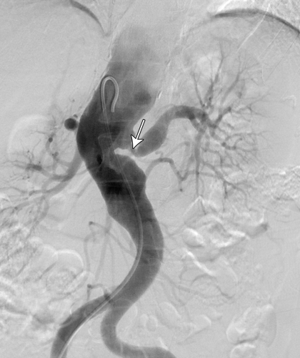
A computed tomography (CT) scan of the thorax, abdomen and pelvis showed right pleural effusion and liver and prostate abscesses (Box). The patient was diagnosed with disseminated melioidosis with multiorgan abscesses, and he was started on intravenous imipenem for a planned duration of 6 weeks. Supportive therapy with intravenous normal saline was instituted to resolve his dehydration. Therapeutic drainage of the liver abscess and right pleural effusion was performed under ultrasound guidance.

The patient’s condition responded well to treatment, showing clinical improvement after 3 days. He became afebrile, and his blood parameters normalised with a gradual increase in his serum sodium level to 122 mmol/L over 3 days.

However, on Day 7 of admission, he started feeling weak, requiring help to ambulate. He was noted to be slow in his movements and in answering questions, with slurred speech. He complained that his upper limbs and trunk felt stiff. There were multiple new skin abscesses on his forehead. On neurological examination, he was alert, with negative Kernig’s and Brudzinski’s signs. Results of cranial nerve examination were normal. There was generalised rigidity of the neck, trunk and limbs. He had mask-like facies, bradykinesia and bradyphrenia, with monotonous speech and fine resting tremor of both hands. Medical Research Council muscle power grading of all four limbs was 4/5 with normal reflexes. Sensations were otherwise normal, and he had no cerebellar signs. He had not been given any antidopaminergic medications.

A CT scan and magnetic resonance imaging (MRI) of the brain was normal. A lumbar puncture revealed clear cerebrospinal fluid (CSF) with a cell count of 20 cells/mm3, predominantly neutrophils (RI, < 5 cells/mm3, predominantly lymphocytes). His total CSF protein level was 405 mg/L (RI, 150–450 mg/L), and CSF glucose level was 3.1 mmol/L (RI, 2.8–4.2 mmol/L) with a CSF to blood glucose ratio of > 0.5 (RI, > 0.5). CSF Ziehl–Neelsen smear and polymerase chain reaction results were negative for tuberculosis. The CSF culture was negative for B. pseudomallei.

A diagnosis of parkinsonism secondary to melioidosis was made after excluding other causes of parkinsonism, including drug-induced parkinsonism and extrapontine myelinolysis. Extrapontine myelinolysis was unlikely in our patient as the correction of hyponatraemia was gradual, and there were no supportive MRI changes.

The patient was treated symptomatically with levodopa/benserazide 50/12.5 mg twice daily for a month. The intravenous antibiotic for melioidosis was continued. After 1 month, his parkinsonism symptoms resolved and his antiparkinson medication was stopped. An ultrasound of his abdomen showed resolution of the abscess. Repeated blood culture showed no growth. He was subsequently discharged after a 1.5-month stay, and prescribed oral co-trimoxazole (trimethoprim–sulfamethoxazole 320/1600 mg) 12-hourly and oral doxycycline (100 mg 12-hourly) for 3 months.

Discussion

To our knowledge, this is the first reported case of parkinsonism secondary to melioidosis. Melioidosis is an infection caused by B. pseudomallei, a gram-negative bacterium transmitted through direct skin contact with contaminated soil. It is endemic in the Asia–Pacific region, with a reported incidence of 4.4 per 100 000 person-years in north-eastern Thailand and 50.2 per 100 000 person-years in the Top End of the Northern Territory.1,2

Neurological complications of melioidosis are rare. In the Darwin Prospective Melioidosis Study, only 14 of 540 patients (3%) developed neurological complications following melioidosis over a 20-year study period.2,3 The clinical features reported include unilateral limb weakness, cerebellar signs, brainstem signs and flaccid paraparesis.2,4 Parkinsonism and extrapyramidal signs have not been reported in previous case series.

Various infective organisms have been reported to cause post-infectious parkinsonism, including dengue virus,5 Japanese encephalitis B virus,6 West Nile virus,7 encephalitis lethargica8 and Streptococcus species.9 It is postulated that infective organisms can cause parkinsonism by three different mechanisms.

The most widely accepted mechanism is via direct infiltration of the causative organism into the central nervous system. Patients usually have pathological changes on imaging of the central nervous system and abnormal CSF findings. In the Darwin Melioidosis Prospective Study, of the 14 patients who developed neurological complications, 10 had meningoencephalitis, two had myelitis and two had cerebral abscesses.2 All were noted on MRI to have abnormal T2-weighted hyperintensities and had abnormal results of CSF analysis, with mononuclear pleocytosis and elevated protein levels.2

The second mechanism involves endotoxin lipopolysaccharide released from the gram-negative bacterial cell wall causing damage to the blood–brain barrier. There is subsequent microglia and macrophage activation, as well as the release of cytokines and oxygen radicals. This results in dopaminergic neurone damage. This pathophysiological mechanism has been postulated as a possible model for development of Parkinson disease based on animal studies.10

The final possible mechanism involves the development of antibasal ganglia antibodies with resultant insult to the basal ganglia. Antibasal ganglia antibodies are commonly implicated in many movement disorders, including chorea and tics.11 Acute parkinsonism with antibasal ganglia antibodies following streptococcal infection has been reported.9

Our patient did not have MRI changes to suggest a pathophysiological mechanism of direct invasion of the infective organism into the central nervous system. Although the CSF culture was negative and the protein level was normal, there was pleocytosis with predominant neutrophils, suggesting an ongoing inflammatory process in the central nervous system.

The onset of parkinsonism was delayed and developed when the patient was recovering from the bacteraemia, as evidenced by improving blood indices and vital signs. This suggests, at least in our patient, that the most probable mechanism for the parkinsonism was an immune-mediated process, either by liposaccharide endotoxins or antibasal ganglia antibodies. Unfortunately, we do not have a facility to test for antibasal ganglia antibodies at our centre.

The recommended treatment for neurological melioidosis includes parenteral ceftazidime or a carbapenem for 6 to 8 weeks, followed by maintenance treatment with oral doxycycline or co-trimoxazole.12 However, to date, there is no standard guideline for managing post-infectious parkinsonism. Previous cases of post-infectious parkinsonism were treated symptomatically with levodopa and other anti-parkinson agents.13 Evidence for immunotherapy for post-infectious parkinsonism is anecdotal at best.5,13

In conclusion, parkinsonism could be a neurological complication of melioidosis. Despite its rarity, melioidosis should be considered as a differential diagnosis of parkinsonism, particularly in endemic areas. In our case, the pathophysiological mechanism appears to be secondary to immunological response rather than direct CNS infiltration. Little is known about the treatment of post-infectious parkinsonism. However, at least in our patient, it was self-limiting and responded well to symptomatic treatment.

Abscesses due to melioidosis in a 62-year-old man


Liver (A) and prostate (B) abscesses.

Optimising treatment for Australian melanoma patients can save taxpayers millions of dollars annually

To the Editor: Patients with BRAF-mutated metastatic melanoma benefit greatly from the novel BRAF inhibitor dabrafenib and the checkpoint inhibitor ipilimumab, recently listed under the Australian Pharmaceutical Benefits Scheme (PBS). Dabrafenib results in rapid responses; but the cancer later adapts, and patients relapse rapidly. Conversely, ipilimumab slowly reactivates anticancer immunity, meaningfully improving long-term survival (up to around 20% at 5 years).

The costs of these drugs are substantial. Dabrafenib costs A$8759 per month, and median duration of therapy is 9.4 months.1 The Australian price of ipilimumab is confidential, but potentially up to A$190 000 per patient, depending on weight. The 2014 year-to-September PBS cost of ipilimumab was $68 456 890 (data from https://www.medicareaustralia.gov.au/statistics/pbs_item.shtml).

Current PBS approval mandates that dabrafenib may only be used as first-line therapy. Commencing treatment with dabrafenib then switching to ipilimumab when the disease progresses may inadvertently deliver worse outcomes than using the slower but longer acting immunotherapy followed by the potent but impermanent BRAF inhibitor.2,3 After a BRAF inhibitor fails, there is often insufficient time to deliver the full course of ipilimumab, let alone for the immune system to reactivate. While some patients’ symptoms necessitate a BRAF inhibitor upfront, most are well enough to take ipilimumab first, with dabrafenib in reserve.4

Formal randomised trials of these sequences are underway (NCT01940809, NCT01673854, NCT02224781) but our clinical experience and two international case series reinforce that administering ipilimumab then a BRAF inhibitor is very likely to be superior. (Patients treated with BRAF inhibitor then ipilimumab: objective response rate, 0; stable disease, 6%; progressive disease, 94%.3 BRAF inhibitor then ipilimumab v ipilimumab then BRAF inhibitor: overall survival, 9.9 months v 14.5 months; P = 0.04.2)

In the absence of a formal economic analysis, we assume most of the approximately 1500 Australian patients who will die with metastatic melanoma every year accept treatment.5 Forty-six per cent of these patients have BRAF-mutated melanoma;6 and of these, 85% are well enough to defer treatment with a BRAF inhibitor.4 Thus, about 585 patients receive a potentially inferior treatment sequence, and around $36 million per annum of otherwise effective treatment is administered inefficiently.

Other countries with publicly funded health care (eg, the United Kingdom) do not restrict treatment sequences in metastatic melanoma. We argue that, until randomised trials identify inferior or superior sequences, Australian melanoma patients and taxpayers would benefit from flexibility in prescribing these breakthrough treatments.

Infliximab therapy in two cases of severe neurotuberculosis paradoxical reaction

Clinical record

Patient 1

A 60-year-old HIV-negative woman presented with a week’s history of fever, vomiting and confusion, followed by progressive personality change. On admission, she was noted to have urinary retention, left oculomotor nerve palsy and an upgoing right plantar response. A magnetic resonance image (MRI) of the brain showed leptomeningeal enhancement with gyral swelling and subtle cortical T2 signal hyperintensity in the right frontal lobe, suggesting meningoencephalitis. Cerebrospinal fluid (CSF) cultures grew fully susceptible Mycobacterium tuberculosis. Antituberculous therapy was started with isoniazid, rifampicin, ethambutol and pyrazinamide, plus dexamethasone. CSF cultures tested negative by Week 1. Over the next month, she had ongoing fevers and fluctuating conscious state. High CSF pressures necessitated ventriculoperitoneal (VP) shunting. An MRI 3 months into therapy showed numerous granulomas, microabscesses and infarcts. Her condition failed to improve with a further course of dexamethasone, and an MRI at 5 months showed increasing size and number of granulomas, with worsening oedema and midline shift (Figure 1, A). She was given a trial of three doses of infliximab 10 mg/kg, 1 month apart, resulting in marked improvement in neurological status and radiological findings (Figure 1, B). She regained movement of her limbs, opened her eyes spontaneously and was able to articulate a few words. After completing 2 months of four-drug therapy, she received isoniazid and rifampicin for 10 months, with ongoing improvement. She was left with mild cognitive deficit and required some assistance with activities of daily living.

Patient 2

A 32-year-old HIV-negative woman presented with delirium and back pain. A chest radiograph suggested miliary tuberculosis. A computed tomography brain scan was unremarkable. Results of CSF molecular testing were positive for M. tuberculosis complex, and cultures from CSF, blood and a laryngeal swab grew fully susceptible M. tuberculosis. Isoniazid, rifampicin, ethambutol, pyrazinamide and prednisolone 50 mg were commenced. One month into therapy, she developed headache in the context of weaning from prednisolone. An MRI showed multiple rim-enhancing nodules in the CSF spaces, with leptomeningeal enhancement and enhancing lesions in the right cerebellum and hemipons. CSF cultures tested negative.

The prednisolone dose was increased to 60 mg, with little response, then converted to dexamethasone 12 mg/day. One month later, while steroid tapering, she developed diplopia. An MRI showed worsening tuberculomas with increasing oedema (Figure 2, A). Dexamethasone was reinitiated at 12 mg/day. Three months into therapy, she developed obstructive hydrocephalus requiring VP shunting. Over the following weeks, she developed peripheral visual field loss. After 4 months of tuberculosis therapy, a trial of infliximab 5 mg/kg was initiated. The steroid dose was tapered over the next week without worsening of symptoms, and she was discharged. One month later, an MRI showed moderate improvement (Figure 2, B). Two further doses of infliximab were given over the subsequent 6 weeks, with complete resolution of visual symptoms. She completed 2 months of four-drug therapy, followed by 10 months of isoniazid and rifampicin. The course was complicated by a seizure at Month 8, necessitating antiepileptic therapy, but she made an otherwise full neurological recovery.

A paradoxical reaction (PR) in tuberculosis (TB) is the worsening of disease after starting TB therapy, usually despite microbiological response. It may represent an inflammatory response to the release of antigen from dying bacilli.1 Such disease exacerbation has also been observed in people with HIV when antiretrovirals are started,2 and in individuals with TB when tumour necrosis factor alpha (TNF-α) antagonists are discontinued.3 A PR may manifest with new pulmonary lesions or lymphadenopathy3 and can be life-threatening, especially in patients with neurotuberculosis.1 Management involves high-dose corticosteroids, but in intractable cases success has been reported with TNF-α blockade.1,35

Our two patients had severe neurotuberculosis PRs unresponsive to dexamethasone, which abated after administration of the anti-TNF-α antibody infliximab. Before these two cases, there was only one report of therapeutic use of infliximab for TB PR in an individual without prior history of TNF-α antagonist use.1 Our two cases add weight to this approach being safe and effective in patients with steroid-refractory TB PR. The previously reported patient had steroid-refractory neurotuberculosis that did not respond to a trial of cyclophosphamide. Radiological and neurological parameters improved only after infliximab was given.1 In that case and ours, cultures tested negative soon after antituberculous therapy was started, suggesting that ongoing disease was due to an immunologically mediated PR rather than inadequate microbiological control.

These cases highlight the potentially devastating effects of central nervous system (CNS) TB, which, despite contemporary therapeutic approaches, still results in permanent disability or death in half of those treated.6 Much of this morbidity can be attributed to the inflammatory response. A key inflammatory cytokine is TNF-α, which plays an integral role in granuloma formation to contain TB infection. However, in mouse models of neurotuberculosis, TNF-α has been shown to increase blood–brain barrier permeability, resulting in increased CSF leukocytosis and CNS inflammation.7

Attenuation of the inflammatory response with routine administration of corticosteroids in patients with neurotuberculosis has been shown to reduce mortality.6 However, in cases of PR, outcomes are often poor despite steroids. There is growing evidence that medications with anti-TNF-α activity may have a role in controlling this inflammatory response, without compromising microbiological response.1,35

Thalidomide, a potent TNF-α inhibitor, was administered to two patients with steroid-refractory neurotuberculosis, with apparent improvement.8 While also showing promise in rabbit models and a small pilot study, it was poorly tolerated and failed to show clinical benefit when used as adjunctive therapy for childhood TB meningitis in a randomised trial.9

There are accumulating data on the role of the anti-TNF-α monoclonal antibodies infliximab and adalimumab and the soluble TNF-α receptor etanercept. They have potent anti-inflammatory properties and are well tolerated, but have been associated with increased risk of TB in those taking them for autoimmune conditions.10 TB developing in patients receiving TNF-α antagonists is more likely to be extrapulmonary or disseminated,11 and early reports suggested that it was more refractory to treatment.12 However, as experience with these agents grew, it became apparent that the poor response could be a PR to the TNF-α antagonist withdrawal. As the immunosuppressive effect of the TNF-α antagonist wanes, the recovering immune system can generate an intense inflammatory reaction against mycobacterial antigens. Two patients with steroid-refractory disease were successfully treated with reintroduction of the offending TNF-α antagonist.4,5

Our report supports an additional role for TNF-α inhibition in severe PRs in immunocompetent individuals. Given that TNF-α antagonists appear to be safe in TB PR, further studies of their role in management are warranted.

Lessons from practice

  • Central nervous system tuberculosis remains a potentially devastating disease that, despite contemporary therapeutic approaches, still results in permanent disability or death in half of those treated.
  • A paradoxical reaction is an inflammatory reaction that can cause disease progression and complications after initiation of antituberculous therapy.
  • High-dose corticosteroids are recommended but if these are ineffective, there is mounting evidence for the use of tumour necrosis factor alpha antagonists such as infliximab.

Vasculitis or fibromuscular dysplasia?

Clinical record

A 45-year-old previously well woman of Filipina descent was admitted to hospital following an episode of vertigo that spontaneously resolved after 2 minutes. The patient denied experiencing any associated symptoms. Her past history included hypertension, diagnosed when she was 27 years old. She did not smoke or drink alcohol. Her brother suffered a stroke at age 46 years.

On physical examination, the patient’s blood pressure was 160/100 mmHg in the right arm and 140/95 mmHg in the left arm. Her blood glucose level was 6.8 mmol/L (reference interval [RI], 4–11 mmol/L). There were loud bruits in both carotid arteries and the left renal artery. The right radial pulse was stronger than the left. Both lower limb pulses were palpable. Results of the remainder of the physical examination were normal.

Results of a full blood count, tests for urea and electrolyte levels, liver function tests, thyroid function tests, and tests for troponin and C-reactive protein levels were normal. Erythrocyte sedimentation rate was slightly elevated at 22 mm/h (RI, < 20 mm/h). Results of vasculitis screening — tests for antinuclear antibodies, extractable nuclear antigens, antibodies to double-stranded DNA and antineutrophil cytoplasmic antibodies — were normal. Results of thrombophilia screening — tests for anticardiolipin IgG, antithrombin III, protein C function, free protein S, prothrombin gene (20210) mutation, factor V Leiden and anti-β2 glycoprotein 1 antibodies — were also normal. Total cholesterol level was 2.6 mmol/L (RI, < 5.5 mmol/L), low-density lipoprotein level was 1.5 mmol/L (RI, < 3.5 mmol/L) and high-density lipoprotein was 0.9 mmol/L (RI, > 1.0 mmol/L).

A computed tomography (CT) scan of the brain was unremarkable. Magnetic resonance imaging of the brain showed supratentorial T2-weighted and fluid-attenuated inversion recovery (FLAIR) white matter hyperintensity, suggesting moderate chronic small-vessel ischaemic disease. Carotid and vertebral duplex ultrasound confirmed high-grade stenosis in both internal and external carotid arteries. CT angiography showed extensive atherosclerosis for the patient’s age and tight stenosis in the right internal carotid artery and distal aspect of the left common carotid artery. CT angiography of the thoracic aorta revealed a descending aortic aneurysm and thrombosis of the left subclavian artery. A CT renal angiogram revealed a stenosis at the origin of the left renal artery (Figure, arrow). A full body positron emission tomography scan could not be interpreted because of physiological uptake of contrast medium by brown fat, which limited the ability to evaluate for large-vessel vasculitis.

The combination of both stenotic and aneurysmal changes in multiple vascular beds makes Takayasu arteritis (TA) a compelling diagnosis in this patient. Given the absence of disease activity, late-stage TA is a probable diagnosis. Patients with fibromuscular dysplasia (FMD) can also present in this manner, but FMD is unlikely to involve several vascular territories (Box).

TA is an inflammatory vasculopathy of the aorta and its main branches.1 It was once thought to be a disorder that affected only Asian women, but it has since been identified in men and women from many ethnic groups,2 usually in the second or third decade of life.3

The pathogenesis of TA is poorly understood. Vessel inflammation results in wall thickening, fibrosis, stenosis and thrombus formation.2 Panarteritis with adventitial and intimal scarring extends distally from the aorta into its branches, causing narrowing and occlusions. Inflammation can destroy the arterial media and lead to aneurysm formation.2 Morbidity due to vascular occlusion and aneurysm formation is high.

TA has a triphasic pattern of disease progression. The clinical features vary depending on the degree of arterial dilation, narrowing and occlusion. Phase I is the inflammatory period, which is characterised by non-specific symptoms such as fever and weight loss. Phase II involves vessel tenderness due to inflammation. Phase III is the burned-out (or late) stage, in which bruits and ischaemia predominate in multiple organs.4 Phase III does not occur in all patients. In patients who are deemed to be in remission, relapses can occur.

FMD is an uncommon disease that mimics vasculitis. It is a non-inflammatory, non-atherosclerotic angiopathy that occurs most commonly in women of child-bearing age.5 It has been observed in nearly every arterial bed.

The clinical manifestations of FMD depend on the arterial distribution affected. FMD may cause stenosis, aneurysm, dissection and occlusion of arteries.6 Common manifestations include dizziness, hypertension and stroke.7 Patients with FMD commonly have involvement of the renal and carotid arteries.7,8 FMD has three subtypes, according to the dominant arterial wall layer involved.

There are instances when it is difficult to differentiate FMD from vasculitis. As FMD is a non-inflammatory process, it is not associated with anaemia, thrombocytopenia or elevated levels of acute phase reactants.9 If histological proof of FMD or inflammation is not available, distinguishing between these conditions may be difficult because the angiographic appearances are similar.9

Diagnostic confirmation for our patient could only be achieved with arterial biopsy. This was not an option owing to procedural risk. Given the number of vascular beds involved, TA was the favoured diagnosis. The absence of constitutional symptoms and low levels of inflammatory markers suggested that late-phase TA was likely, but active TA could not be excluded. Distinguishing between these entities is clinically important as the therapy differs.

The management of patients with TA can be challenging. The lack of a single gold standard measure of disease activity often results in the diagnosis being established from a combination of clinical history, pathology tests and imaging. There may be poor correlation between the results of such investigations, leading to uncertainty regarding diagnosis and appropriate therapy.

The goal of treatment in TA is to minimise long-term adverse effects while controlling disease activity and preserving vascular competence. In active TA, this is achieved with corticosteroids and cytotoxic medications. Use of these medications in late-phase TA has been contentious. The lack of a measurable inflammatory response may cause uncertainty regarding whether to aggressively immunosuppress the patient. Despite this, patients who are deemed to have late-phase TA may still have histological evidence of ongoing disease. Moreover, although the 10-year mortality is low, morbidity due to arterial occlusion is high in TA.10 Since inflammation is a risk factor for atherosclerosis, traditional cardiovascular risk factors should be screened for routinely and managed with antiplatelet agents, antihypertensive medication and statin therapy, as deemed necessary. However, using angiotensin-converting enzyme inhibitors requires close monitoring in view of the prevalence of renal artery stenosis. Stenting and balloon angioplasty can be used to maintain vascular patency.

TA is a challenging disease to diagnose and manage. Identification of superior diagnostic tools, including imaging techniques, may facilitate our understanding of disease activity and guide therapeutic interventions. Given a shortage of evidence regarding therapy in late-phase TA, patients should be assessed on a case-by-case basis and those who have a good prognosis should not be put at risk by treatment that is more harmful than the disease itself. Ultimately, a multidisciplinary approach with long-term follow-up is needed.

Lessons from practice

  • Fibromuscular dysplasia can mimic Takayasu arteritis.
  • Various factors, including age, vascular territory involvement and inflammatory markers, can help distinguish Takayasu arteritis from other diseases.
  • Patients who have a good prognosis should not be put at risk by treatment that is more harmful than the disease itself.
  • Secondary prevention and management of cardiovascular risk factors are vital in patients with Takayasu arteritis.

Differential diagnoses that were considered for a 45-year-old woman who had stenotic and aneurysmal changes in multiple vascular beds

Consistent with diagnosis

Less consistent with diagnosis


Burned-out Takayasu arteritis


  • Numerous vascular territories involved
  • Low levels of inflammatory markers
  • No systemic symptoms
  • Female sex
  • Age likely < 40 years at time of disease onset
  • No preceding constitutional symptoms that suggest past history of active Takayasu arteritis

Active Takayasu arteritis


  • Numerous vascular territories involved
  • Female sex
  • Age likely < 40 years at time of disease onset
  • No current constitutional symptoms
  • Low levels of inflammatory markers

Fibromuscular dysplasia


  • Age 30–50 years
  • Female sex
  • Diffuse vascular territory involvement

Atherosclerosis


  • Family history of stroke
  • Past history of hypertension
  • Age < 45 years
  • Diffuse and severe vessel involvement
  • Lack of typical cardiovascular risk factors

Giant cell arteritis


  • Large-artery involvement
  • Age < 50 years
  • No localised headache
  • No temporal artery tenderness or decreased temporal artery pulse
  • Erythrocyte sedimentation rate not significantly elevated

Childhood food allergy and anaphylaxis: an educational priority

The challenge of higher rates of food allergy must be met through development of better models of care and education

IgE-mediated food allergy (FA) and anaphylaxis have become an increasing public and personal health burden in developed countries over the past decade, contributing to increased demand for specialty services, significant economic cost of care, and reduced quality of life for children with FA and their families.1 In the most accurate estimate in Australia to date, the Victorian HealthNuts study found the prevalence of challenge-proven FA at age 12 months to be 10% overall.2 Effective strategies for primary prevention of FA are lacking, and secondary prevention is limited to strategies to reduce the risk of unintentional exposure. Although food-specific immunotherapy appears promising, it remains at the investigational stage because of the infrastructure required, high rates of adverse reactions and lack of persistent tolerance when treatment ceases.3 While several risk factors for childhood FA have been proposed — such as early-onset atopic eczema, timing of solids introduction, vitamin D status and intestinal bacterial load3 — this area remains an active area of research.

Although other triggers for anaphylaxis exist (insect venom, medication or latex), the major strategies for avoidance focus on FA, due to its relatively high prevalence in childhood and higher rates of accidental exposure, particularly in school and childcare settings.4 Risk management requires patient and carer education on reducing the risk of exposure to allergic triggers, providing an emergency action plan if reactions occur, dealing with higher-risk situations (eg, exposure to unlabelled food, attendance at parties, school excursions and camps), and providing an adrenaline autoinjector to individuals considered to be at higher risk. As children age and approach their teenage years, self-management and concerns about parties (exposure to unlabelled food), alcohol exposure (reduced vigilance) and risks of exposure to food allergens by kissing5 should also be discussed.

The demand for evidence-based and nationally consistent education across jurisdictional boundaries on how best to care for individuals with FA and anaphylaxis continues to outstrip the current resources available for face-to-face training by community and hospital-based FA and anaphylaxis trainers. With most specialist services located in major cities, alternative models for education delivery are required to service the needs of rural areas.

To meet the challenge of increasing FA prevalence and demand for education, the Australasian Society of Clinical Immunology and Allergy (ASCIA) has developed a number of educational resources including national standardised emergency action plans, adrenaline autoinjector prescription guidelines (http://www.allergy.org.au/health-professionals/anaphylaxis-resources), and allergy prevention guidelines for schools and child care.4 ASCIA has also partnered with various state education and health departments to develop childcare and school e-training courses, which are available without charge from the ASCIA website (http://www.allergy.org.au). The content of these educational programs was developed after extensive consultation, with childcare versions approved by the Australian Children’s Education and Care Quality Authority. Since launching in March 2010, 178 000 school and childcare staff have registered for the e-training.

Additional separate modules on anaphylaxis and FA management have been specifically designed to meet the needs of medical practitioners, pharmacists, dietitians, first aid providers and the broader community, with Royal Australian College of General Practitioners accreditation of a 6-hour active learning module on allergy and anaphylaxis worth 40 (Category 1) continuing medical education points (https://alm.ascia.org.au).

Education of patients, caregivers and health professionals is recommended in FA and anaphylaxis guidelines, with the aims of improving patient care and reducing the risk of adverse outcomes.6 While the clinical outcome from provision of emergency action plans is yet to be investigated in controlled studies,7 educational training has been shown to result in more accurate recognition of symptoms of anaphylaxis by health professionals,8 and to improve knowledge of FA and change practice in catering staff.9 Finally, an evaluation of ASCIA pharmacist e-training demonstrated improved knowledge after training compared with baseline or no training, and long-term retention of knowledge 7 months after completion.10

Until specific strategies are available to reduce the health burden of FA and anaphylaxis, the challenges for our health care systems will be how best to develop evidence-based policies to reduce the risk of FA development, care for younger children presenting with new cases of FA or anaphylaxis, and manage the shifting burden on older teenagers and young adults, who carry the highest relative risk for fatal anaphylaxis. There will be an ongoing need to develop models of care to enhance access to specialist medical services, improve acute management and educate those charged with delivering care both within and outside the health care sector. We encourage health professionals involved in the care of patients with FA and anaphylaxis to update their skills in this area.

A diagnosis that will go down in history

A well described case in the Christmas tradition is that of patient R, reported to have been afflicted with a very shiny nose. We believe his presentation was consistent with one of the cutaneous forms of sarcoidosis: lupus pernio.

A well described case in the Christmas tradition is that of patient R, reported to have been afflicted with a very shiny nose. The lesion was described as having a lustrous, glowing appearance and initially exerting a significant burden on quality of life — the patient’s peers engaged in name-calling and excluded him from social games. It would seem that the case proved a challenge diagnostically, with the disease managed expectantly. Fortuitously, it availed itself to a particularly coveted job prospect which in time eventuated in acceptance by his peers and, notably, this transpired with glee. The case has remained a diagnostic dilemma through the generations. It has been proposed that the cutaneous lesion might be attributable to variations in nasal microcirculation noted in specific genetic populations.1 However, the uniqueness of patient R’s lesion is what has made him a legend and, accordingly, a diagnosis of variations in nasal microcirculation seems unlikely. Rather, a pathological aetiology is more in keeping with his clinical picture. We believe patient R was afflicted with one of the cutaneous forms of sarcoidosis: lupus pernio.

The clinical presentation of lupus pernio is that of violaceous papules, nodules or plaques. The lesions have an indurated, shiny, and somewhat bright character to them and are classically located on the nose, but also the cheeks and ears: in those areas most sensitive to the cold, or “pernio”, that one might experience one foggy Christmas Eve. In lupus pernio, histological findings are remarkable for the presence of sarcoidal granulomas, with birefringent material observed in up to 50% of cases (you could even say they glow).2

An important clinical consideration in patients with lupus pernio is the association with pulmonary sarcoidosis. It has been reported that 74% of patients will have intrathoracic disease and 54% will have upper respiratory tract involvement.3 This form of cutaneous sarcoidosis portends a more aggressive clinical course with a higher likelihood of visceral involvement and recalcitrance to therapy. Cutaneous sarcoidosis is known for its protean clinical presentations, including erythema nodosum, macules, papules and plaques, scar sarcoidosis, ichthyosis, Darier–Roussy lesions, alopecia and onychodystrophy.4 Physicians should be aware of cutaneous signs of sarcoidosis because they lend themselves to biopsy facilitating diagnosis, many have prognostic significance, and they can impose a significant burden on quality of life.5

Based on morphology alone, a wide range of differential diagnoses could account for patient R’s clinical presentation (Box). However, considering the history and examination findings together, lupus pernio represents a unifying diagnosis. This patient’s story represents an instructive case and is one that will go down in history.

Differential diagnoses for Patient R

Aetiology

Diagnoses


Inflammatory

Rosacea (rhinophyma)

Infiltrates


Cellular


Lymphocytic

Tumid lupus, pseudolymphoma, Jessner lymphocytic infiltration

Neutrophilic

Sweet syndrome

Eosinophilic

Angiolymphoid hyperplasia with eosinophilia

Granulomatous

Xanthogranuloma

Mixed

Granuloma faciale

Acellular


Mucin

Cutaneous mucinosis

Amyloid

Cutaneous amyloidosis

Tumours

Basal cell carcinoma, amelanotic melanoma, cutaneous lymphoma, Merkel cell carcinoma, angiosarcoma

Infection

Syphilis (Treponema pallidum), lupus vulgaris (Mycobacterium tuberculosis), leishmaniasis (Leishmania spp)

Vascular malformations

Non-involuting congenital haemangioma, arteriovenous malformation (especially Wyburn-Mason syndrome)