×

Co-habiting influences person’s immune system more than illness

A Belgian study has found that co-habiting and specifically raising a child together can have a bigger impact on a person’s immunity than getting the flu shot or contracting gastro.

The research, published in Nature Immunology, found that people who lived together had immune systems that became 50% more similar compared to two non-related people in the wider community.

670 healthy people aged 2 to 86 were studied over a period of three years to “provide a description of the population-level heterogeneity in the cellular composition of the circulating immune system,” the authors wrote.

They found that the immune system was quite elastic, with it bouncing back to its original state after a bout of gastro or receiving the flu vaccine.

The result that was most interesting to them was the impact of co-parenting on immune systems.

Related: How modern life has damaged our internal ecosystems

“One of the most surprising results from our study was the degree to which immune profiles were more similar between parents than unrelated people living in different households. This suggests that a shared environment acts in some way to bring immunoprofiles toward a convergent equilibrium,” they wrote.

They also said there was little effect of gender on the immune landscape, which is at odds with the longstanding observation that autoimmune diseases are more common in women than men.

“Notably, sex-based differences are more limited at the cellular level than at the molecular level. The incomplete correlation between gene signature and cell type suggests that the discrepancy can be resolved by a model where high diversity in molecular expression is largely compensated for at the cellular level,” they wrote.

Latest news:

Shiitake dermatitis: the tale of an under-recognised, undercooked fungus

Clinical records

Case 1

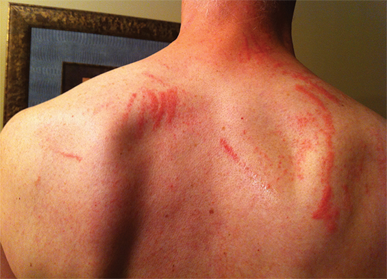
A 55-year-old man with no medical history or allergies presented in May 2015 with a widespread, pruritic, whiplash-like rash (Figure 1). On examination, multiple erythematous papules in a linear distribution, corresponding to areas of excoriation, were noted on his trunk, limbs, forehead and scalp. The rash had developed 12 hours after a meal containing fresh shiitake mushrooms. There were no associated systemic symptoms and the patient’s condition did not improve with topical administration of betamethasone dipropionate 0.05%. Based on dietary history and the characteristic appearance of the rash, a diagnosis of shiitake dermatitis was made. The rash resolved in 3 weeks without further treatment. The patient experienced a recurrence 5 months later, after eating another meal containing shiitake mushrooms.

Case 2

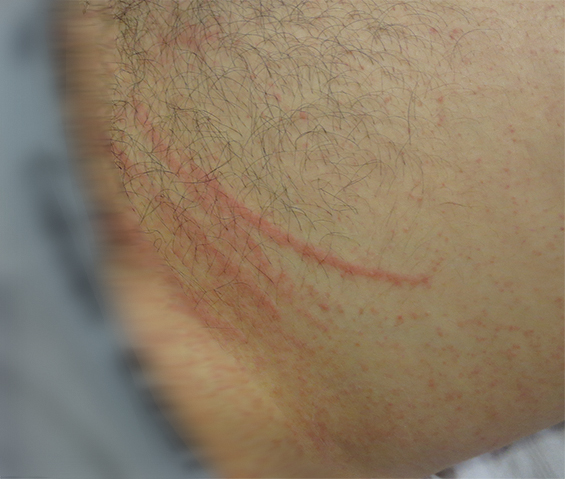
A 30-year-old man with no comorbidities or allergies presented in March 2015 with a 2-day history of a striking, linear, erythematous eruption (Figure 2). He was systemically well, but reported associated perioral tingling and pharyngitis, which were unresponsive to 10 mg oral cetirizine. Examination revealed linear groupings of papules on an erythematous urticated base on his forehead and trunk. Despite denying pruritus or excoriations, relative sparing of the central back (out of the patient’s reach) was noted. Further questioning revealed ingestion of lightly cooked shiitake mushrooms the night before the onset of symptoms. No relatives who had eaten the same meal were affected. The patient was diagnosed with shiitake dermatitis, and the rash resolved completely without treatment.

Case 3

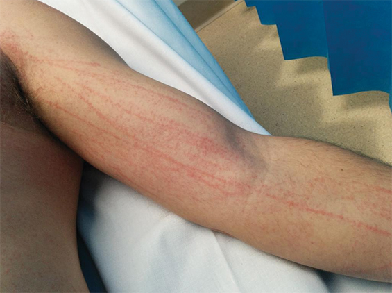
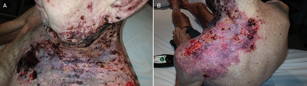
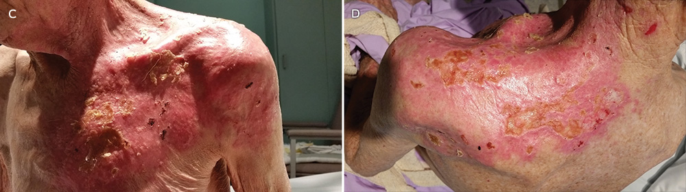
A 44-year-old man presented in August 2015 with a 2-day history of a flagellate erythematous rash (Figure 3). His past history included ischaemic heart disease, hypercholesterolaemia and rosacea. He had eaten raw shiitake mushrooms while cooking, and woke the next morning with a rash on his arms. Despite treatment with 25 mg oral prednisolone and topical betamethasone valerate 0.02%, prescribed by his general practitioner, the rash rapidly extended to his trunk, shins and scalp. His full blood count, C-reactive protein level and biochemistry results were unremarkable. A diagnosis of shiitake dermatitis was made. The patient’s wife, with whom he had shared the cooked meal, was asymptomatic, and the patient had previously eaten cooked shiitake without problems. Ten days later, the rash was resolving rapidly, with mild hyperpigmentation.

Shiitake mushrooms (Lentinus edodes) account for 20% of mushroom production worldwide1,2 and are the second most commonly eaten mushroom variety.1,3 Originally, most shiitake were log-grown in Japan. Since the late 1980s, most shiitake have been cultivated on a sawdust-based substrate in China.4,5 Shiitake are used in Asian medicine for their antihypertensive, anticarcinogenic and cholesterol-reducing qualities.1,5 Shiitake spores can cause IgE-mediated illness, such as hypersensitivity pneumonitis, asthma and allergic rhinitis.1,3,6 Shiitake can also cause contact urticaria, protein contact dermatitis and allergic contact dermatitis.1,3,6 In this article, we present the first three published cases of shiitake dermatitis in Australia. Systemic dermatitis caused by ingested allergens has also been reported with other foods, including spices, garlic, onions and root vegetables, metals, drugs, and preservatives.7,8

Shiitake dermatitis is also known as flagellate erythema, flagellate dermatitis and toxicodermia.3 It was first described in Japan in 1977, in Europe in 2006 and in the United States in 2010.1,2 Flagellate erythema occurs with all forms of shiitake — fresh, dried, powdered, boiled, baked, grilled and fried.2,5 It has been reported from adolescence to old age, and is more common in men.2

The highly distinctive rash develops following the ingestion of undercooked shiitake mushrooms by susceptible individuals. Onset usually occurs 12–48 hours after intake, although the reported range is 2 hours to 5 days.1 Intensely pruritic, linear, erythematous papules and petechiae develop on the trunk, limbs, head and neck,2 sparing the oral mucosa. The flagellate morphology is attributed to the Koebner phenomenon.2,3 There may be associated local oedema, fever, malaise, lip tingling, dysphagia and diarrhoea.1,9,10 Up to 47% of patients display ultraviolet A photosensitivity and photo-aggravation.3,9 Differential diagnoses include the flagellate rash associated with bleomycin, dermatomyositis, adult-onset Still disease, and HIV with hypereosinophilic syndrome.11

Lentinan, a thermolabile polysaccharide component of shiitake mushrooms, is the cause of shiitake dermatitis.13,5,9 Lentinan undergoes a conformational change at 130–145°C, so it is recommended that shiitake are cooked at temperatures of 150°C or greater to prevent toxicity.10 The pathogenesis remains unclear, but frequent case reports of single patients who had take part in shared meals and the delayed time course support the likelihood of an allergic reaction,2,10 with a possible role for exacerbating cofactors, such as use of angiotensin-converting enzyme inhibitors or diuretics, and exposure to ultraviolet A light.1,9 A trial of 519 patients in Japan who received intravenous lentinan chemotherapy yielded nine cases of flagellate erythema; extrapolating from these data, up to 2% of the population will be vulnerable to shiitake dermatitis.1,2

A case of shiitake dermatitis following ingestion of log-grown shiitake was recently reported in a patient who had previously tolerated substrate-grown shiitake from China.5 The authors of this report proposed that shiitake dermatitis may occur only with log-grown mushrooms. This hypothesis is supported by the low incidence of shiitake dermatitis in China, where the shiitake are substrate-grown, and the lower rates in Japan since 1992, when local production switched to substrate-grown mushrooms and the Japanese government began importing substrate-grown shiitake from China.4,5 Most cases in Japan were reported before 1998,9 typically during the harvest season for log-grown shiitake mushrooms.2 In Australia, shiitake are produced by both cultivation methods, and some shiitake are imported from China.12

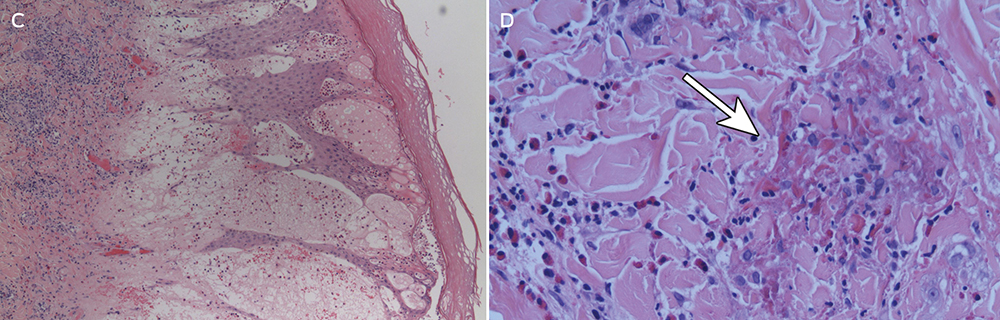
The diagnosis is made clinically, and may be confirmed by oral re-challenge. Histopathological testing of skin biopsy specimens does not provide specific diagnostic information; it generally reveals elongation of rete ridges, spongiosis, degenerating keratinocytes, dermal oedema, and a superficial and perivascular lymphocytic infiltration with eosinophils and neutrophils.1,2 Prick and patch tests conducted by authors of previously published case reports have yielded conflicting results.1,2,10 Positive patch test results have also been obtained in control subjects, further limiting their usefulness in flagellate erythema.2,10

No treatments have been shown to be effective for shiitake dermatitis, although oral antihistamines and topical and oral corticosteroids are often prescribed. Given the high reported rate of ultraviolet A photosensitivity, patients should be given guidance on careful photoprotection.1 The prognosis is excellent, with improvement from 2 days and resolution by 3 weeks.1,2

In Australia, only four confirmed cases and one possible case of shiitake dermatitis were reported in 11 years (Appendix); these were reported to the NSW Poisons Information Centre between January 2004 and April 2015. No cases have been reported to the poisons information centres in Western Australia, Victoria and Queensland. Given our experience of three cases at a tertiary hospital within 12 months, and the prevalence of shiitake consumption in Australia, we believe that shiitake dermatitis is under-recognised. The mushroom industry heavily endorses the health benefits of shiitake as a low kilojoule source of dietary fibre, protein, vitamins, antioxidants and minerals. Considering the availability of shiitake-based health supplements on the internet,1 the popularity of Asian cuisine, and the production of log-grown shiitake in Australia today,12 we anticipate an increasing incidence of shiitake dermatitis. Documenting the preparation temperature, cultivation method and source of the shiitake mushrooms in future cases may help elucidate the pathogenesis.

Lessons from practice

  • Shiitake dermatitis is probably under-recognised and under-reported in Australia

  • The diagnosis is made clinically, based on dietary history and a characteristic erythematous flagellate rash

  • The prognosis is excellent, but patients should be advised to avoid shiitake or to ensure it has been cooked at temperatures of 150°C or greater to prevent further episodes

  • Primary care practitioners, such as general practitioners and emergency physicians, should be aware of the diagnosis to avoid unnecessary investigations and treatments

Figure 1 –


Figure 1: Widespread, characteristic flagellate erythema of shiitake dermatitis in a 55-year-old man

Figure 2 –


Figure 2: Linear grouped papules on an erythematous urticated base in a 30-year-old man with shiitake dermatitis

Figure 3 –


Figure 3: Intensely pruritic, linear, erythematous rash in a 44-year-old man with shiitake dermatitis

The early bird and the worm: a case of cercarial dermatitis

Clinical record

A 36-year-old man presented to our dermatology department with a 5-day history of an intensely pruritic papular eruption on the dorsum of both feet, associated with significant swelling and discomfort. This began the morning after a night-time fishing trip on the central coast of New South Wales at the mouth of the Lake Tuggerah estuary, a system of brackish tidal estuaries.

The patient reported standing ankle-deep in water for 6 hours from dusk (9 pm) until early morning (3 am). He noted that the water was particularly warm after a daytime temperature of 33°C with seaward tidal flows. He awoke the following morning with multiple discrete nodules, oedema and intense pruritus to the dorsa of the feet. He reported previous episodes of “bites” after fishing trips that spontaneously resolved after 24–48 hours.

The next 12 hours saw an increase in pruritus and the size of lesions. The patient presented to a general practitioner, who diagnosed pelican itch with secondary infection and prescribed cephalexin 500 mg four times a day for 5 days. Pain and pruritus continued to increase, resulting in the patient presenting to a metropolitan emergency department where he was diagnosed with bilateral foot cellulitis and administered intravenous cefazolin 1 g twice daily for 5 days. Investigations showed leucocytosis (white cell count, 10.1 × 10⁹/L; reference interval [RI], 4.0–10.0 × 10⁹/L) with eosinophilia (0.8 × 10⁹/L; RI, 0–0.5 × 10⁹/L) and negative blood cultures. On Day 3 of intravenous antibiotics, he was referred to our dermatology department for reassessment of diagnosis.

On presentation, the patient was in significant discomfort with swelling, difficulty ambulating and inability to wear shoes. He did not describe any fever, vomiting, diarrhoea, abdominal cramping or haematuria. Multiple violaceous papules were noted over the dorsum of each foot, with sparing of the plantar surfaces, web spaces of the toes and distal third of the lower limb (Figure, A). Dusky oedema was evident surrounding the papules, with no evidence of lymphangitis. Popliteal and inguinal lymph nodes were palpable.

Histopathological examination (Figure, C and D) revealed epidermal spongiosis and prominent dermal oedema. A heavy inflammatory infiltrate predominately of eosinophils involving all layers of skin down to the subcutis with scattered flame figures was also noted. No parasites were identified. Bacterial, fungal and mycobacterial tissue cultures were negative. This histology supported a markedly pronounced hypersensitivity reaction in a patient clinically suspected to have cercarial dermatitis, and was not compatible with other clinical differential diagnoses such as hypertrophic lichen planus, Sweet syndrome and reticulohistiocytosis.

Our patient was successfully treated with 3 days of oral prednisolone therapy (25 mg daily) with complete resolution of lesions and residual post-inflammatory pigmentation by Day 7 (Figure, B).

Cercarial dermatitis (commonly known as swimmer’s itch, pelican itch or duck itch) conventionally refers to a cutaneous hypersensitivity reaction to non-human trematode larvae (cercariae) of schistasome flukes after penetration of human skin.1,2 In the classic avian form of the disease, the flukes undergo a two-host (digenetic) life cycle with egg development in freshwater snails and release of the larvae into open water (in temperatures above 23°C) to infest migratory aquatic birds (ducks and seagulls, the fluke’s definitive host).2 Human infections with avian cercariae represent a disruption of the natural schistasome life cycle. The fluke was previously thought to die within human skin within 48 hours; however, animal models have shown migration and survival of the trematodes in distant organs weeks after initial infection.1,2

Cercarial dermatitis is a common condition found worldwide1,2 but is under-recognised and under-reported in Australia owing to its self-limiting nature.1 High concentrations of cercariae occur during days of intense sunlight and higher air and sea surface temperatures, with coastal currents concentrating cercariae around shallow estuary outlets.2 Cases have been reported from Queensland,3,4 Terrigal on the central coast of NSW (eponymous for the schistasome Austrobilharzia terrigalensis5), and the Swan River estuary of Western Australia.1,6

The condition manifests as a monomorphic, papular, pruritic dermatitis confined to water-submerged areas that begins to spontaneously regress within 72 hours.1,2 Recurrent exposure can result in sensitisation phenomena precipitating a florid hypersensitivity reaction as seen in our case. Cercarial dermatitis can be differentiated from sea-bather’s eruption, as the latter condition affects areas of skin covered by swimwear, commonly as a swimmer exits the water or after swimwear has dried. It is caused by discharging nematocysts of sea anemones and jellyfish, and can be associated with systemic symptoms of fevers, vomiting and malaise.

Avian schistasomes are homologous to (but not to be confused with) human schistosomiasis, an endemic and potentially fatal tropical disease, manifesting in portal hypertension, bladder carcinoma, granulomatous central nervous system disease, pulmonary fibrosis and glomerulonephritis.7,8 In endemic tropical areas, cercarial dermatitis and schistosomiasis can be difficult to differentiate, although the early cercarial stage of human schistosomiasis is less inflammatory than that of avian schistosomiasis.2 The histopathology of cercarial dermatitis in humans is poorly documented owing to the acute self-limiting nature of the disease.2 Where descriptions are available, they are briefly described as a hypersensitivity reaction with oedema and eosinophils (plus or minus neutrophils). Specificity is provided by finding cercariae in the specimen (located within the epidermis) but they are usually not seen,7 presumably due to the limited life span of the cercariae within non-definitive hosts and the difficulty in finding isolated small (0.75–0.95 mm long) organisms within a disproportionately large area of inflammatory reaction. It is surmised that without identification of the organism, some cases could easily be misclassified as arthropod bite reactions in the absence of awareness of the specific clinical context.

Treatment for mild cercarial dermatitis includes reassurance and symptomatic management with potent topical corticosteroids (betamethasone dipropionate 0.05% ointment twice daily) and emollients. Oedema can be relieved with intermittent cold compress, rest and elevation. Exuberant reactions may require oral prednisolone (0.5 mg/kg up to a maximum dose of 25 mg daily for 3 consecutive days). Some authors recommend routine treatment with oral ivermectin (200 μg/kg up to a maximum of 12 mg per dose)9 until possible systemic complications arising from visceral migration of trematodes in humans are better understood.

It is important for physicians to be aware of this common and under-recognised entity, as well as the potential for schistosomiasis with similar presentations in returning travellers. Although the cutaneous manifestations of this condition spontaneously resolve, further research will clarify the potential for chronic infestation and the role of routine antihelminthic therapy.

Lessons from practice

  • Cercarial dermatitis is a common, under-reported condition caused by a hypersensitivity reaction to the penetration of human skin by the trematode fluke of avian schistosomiasis.

  • It commonly occurs due to submergence of unprotected skin in warm waters of freshwater tidal estuaries.

  • Clinicians should be aware of similar presentations in returning travellers, which may represent the potentially fatal tropical disease of human schistosomiasis.

  • Although most cases spontaneously resolve, topical and systemic corticosteroids are treatment options for exuberant hypersensitivity reactions.

Figure


A: Grouped violaceous papules on the dorsolateral aspect of the foot with associated oedema. B: Significant improvement in foot oedema with healing blisters and erosions after 3 days of oral prednisolone (25 mg). C: Low-power image of upper dermis and epidermis with marked subepidermal oedema and spongiosis responsible for the bullous clinical appearance (pseudobullous change) (objective magnification, ×4; haematoxylin–eosin stain). D: High-power image showing prominent interstitial dermal infiltrate of eosinophils with flame figure (arrow) at top of image (objective magnification, ×20; haematoxylin–eosin stain).

Severe ulcerative herpes zoster

A 78-year-old man presented to a regional emergency department with a severe progressive rash, on a background history of chronic lymphocytic leukaemia, dementia and malnourishment.

The rash was multidermatomal, with patchy areas of ulceration, crusting, excoriation and necrosis (Figure, A and B). Active bleeding, seborrhoeic discharge and occasional vesicles were also noted, extending to the left pelvis. Subsequently, the patient developed concurrent Pseudomonas aeruginosa cellulitis and bacteraemia.

Punch biopsies were non-specific with dermal necrosis, excoriation and possible lichenoid reactivity. However, swabs revealed varicella-zoster virus. The patient was successfully treated with intravenous piperacillin–tazobactam, intravenous acyclovir, normal saline (0.9% sodium chloride) washes, and 50% liquid paraffin with 50% white soft paraffin cream (Figure, C and D). Multifactorial immunodeficiency was deemed to be the aetiology.

Figure

Figure

Not just loading and age: the dynamics of osteoarthritis, obesity and inflammation

Body fat is not an inert structure

Obesity is a well recognised risk factor for osteoarthritis (OA).1 It is commonly believed that obesity affects joints through loading. However, there must be additional mechanisms since, for decades, obesity has been known to be a strong risk factor for hand OA. Given that we do not walk on our hands, an effect of obesity through loading of the joints cannot be the whole explanation. An understanding of the potential mechanisms by which obesity affects joints is important for optimising the treatment and prevention of OA.

Work over the past decade using magnetic resonance imaging has enabled the assessment of factors affecting joints across the spectrum of the disease, from normal asymptomatic joints to symptomatic OA.1 This has made it possible to examine the effect of obesity on joints, and to untangle the issue of whether obesity causes OA or whether OA-related pain causes obesity through modification of lifestyle behaviours and consequent weight gain. This work has shown that obesity is a causative factor in the development of OA, with increased weight being associated with early articular cartilage damage, well before symptoms develop.1 Obesity is an important risk factor for OA across a wide range of joints, including hands, back, hip and knee.

Having established the importance of obesity as a causative factor for OA, it is important to consider potential mechanisms and to recognise that measures of obesity, such as weight and body mass index, have limited usefulness because they do not provide information regarding body composition. For example, body composition may be very different in two men with an identical body mass index of 30 kg/m2: one may have a very high proportion of muscle, while the other may have a very high proportion of fat. Several studies have examined the effect of body composition, particularly fat mass, on joint health.1 A large body of evidence has shown that an increase in fat mass is associated with pre-clinical OA.1 Increased fat mass is also associated with faster loss of knee cartilage and an increased likelihood of joint replacement. An increase in fat mass is also associated with more back pain and disability2 and foot pain.3

The findings that increased fat mass is associated with early through to late OA, independent of obesity, suggest that the effect of obesity on the joint may be via metabolically driven inflammation. It is well recognised that body fat is not an inert structure but rather a highly metabolically active tissue that produces inflammatory molecules, including cytokines and adipokines, that have been shown to damage joints.4 Circulating levels of inflammatory cytokines5 and low-grade synovitis are associated with cartilage loss.6 Higher levels of the adipokine leptin are also independently related to increased cartilage loss, suggesting a systemic mechanism for the effect of obesity on knee cartilage.7 Thus the old paradigm of OA being a degenerative, wear-and-tear disease of older age, and not an inflammatory disease, has been challenged.

So what are the implications of these findings? For weight-bearing joints, as obesity affects joints through both mechanical loading and metabolically driven inflammation, the effects are synergistic; the joint that is being loaded, rather than being healthy, is also subjected to low-grade inflammation — a double “hit”. Thus if a patient is carrying 20 kg extra weight, they are not carrying 20 kg of inert fat. The individual is carrying 20 kg of metabolically active tissue that is not only overloading the joint, but also producing inflammatory molecules resulting in a more vulnerable joint being loaded. The inflammatory mechanisms may also contribute to some of the obesity-related risk for non-weight-bearing joints.

The inflammatory mechanism for obesity-related damage to joints highlights the importance of preventing obesity in early life to avoid early joint damage. Such damage sets up a vicious cycle of further joint damage through both inflammation and loading. It may also contribute to the increased risk of cardiovascular disease seen in those with OA.9 Preventing early weight gain is potentially a more achievable and effective option than weight loss in later life.9,10 Once disease is established, weight maintenance may be a more feasible goal than weight loss for minimising pain and structural progression in joints such as the knee.1,9 With our increasingly obese population and its associated burden of osteoarthritis, novel therapies aimed at targeting inflammatory pathways warrant further investigation.

[Perspectives] Samson Kinyanjui: pressing for more science in Africa, by Africans

It was during the years 2004 to 2006, while working in the Mill Hill laboratories of the UK National Institute for Medical Research, that Samson Kinyanjui began to think seriously beyond the study of malaria immunology that had brought him from Kenya to London. “We have a lot of African scientists working abroad. I looked at the facilities in Mill Hill and I asked myself why we couldn’t have similar facilities in Africa.” He’d already developed an interest in the need for training and capacity building back in his home country.

[Perspectives] Salmaan Keshavjee: tackling tuberculosis (without rocket science)

Salmaan Keshavjee’s CV is a puzzling document. A first degree in biochemistry from Queen’s University in Ontario, Canada, is followed by a move to the USA and a master’s in immunology and infectious diseases from the then Harvard School of Public Health. The next 5 years find him still at Harvard, but now doing a course in Middle Eastern Studies, and then writing a doctoral thesis in anthropology. 3 years on he’s graduating from Stanford University, this time with a medical degree. Is this a man who can’t make up his mind?

New treatment to overcome peanut allergies in children

By Jane Trembath, Southern Health News / 4th of November, 2015

This story was first published by The Lead, South Australia.

A new study is successfully helping children to overcome peanut allergies by exposing them to peanuts and desensitising them to their allergy.

For the past four years, paediatric allergist Dr Billy Tao has been developing a novel two-step desensitisation process at Flinders Medical Centre (FMC) in South Australia.

The first step involves boiling peanuts for an extended length of time to make them less allergenic.

The boiled peanuts are given to patients to partially desensitise them, and then once the patient shows no signs of allergic reaction, roasted peanuts are given to the children to increase their tolerance in the second step of the process.

Dr Tao said the low-cost and effective two-step process resulted in less adverse events than previously used single-step desensitisation methods – also known as oral immunotherapy.

“With traditional methods, a lot of people ingesting increasing amounts of roasted peanut flour or similar products start to react – so much so that many have to drop out and can’t finish the treatment,” Dr Tao said.

The FMC trial is carried out over a year or longer and includes patients aged between 10 and 15 years.

Of the 14 participants, 10 have already completed the first step and are now eating varying amounts of roasted peanuts, while four continue to eat boiled peanuts and are progressing well.

“One patient who had to be administered three adrenaline injections after consuming peanuts is now eating several roasted peanuts every day without problems,” Dr Tao said.

Studies show the number of children living with peanut allergy appears to have tripled between 1997 and 2008, and as many as one in every 200 children will have severe allergy to nuts.

Allergy symptoms can vary from very mild (including tingling mouth, puffy lips and welts around the mouth) to moderate symptoms (facial swelling, body rash, runny nose and red eyes, abdominal pains and vomiting); while severe reactions include trouble breathing, looking pale and unwell, and anaphylaxis.  Very occasionally death may result from a most severe reaction. 

Dr Tao’s idea for hypo-allergenic (less allergenic) nuts to be consumed first was based on an observation by German researcher Professor Kirsten Beyer, who in 2001 noted that peanut allergies were less prevalent in China than the western world because the Chinese ate boiled peanuts rather than peanut butter or roasted peanuts. She found that boiling peanuts for 20 minutes made them less allergenic than roasted peanuts.

Dr Tao said that a partnership with Dr Tim Chataway, Head of the Flinders Proteomics Facility, and Professor Kevin Forsyth from the FMC Paediatrics Department, proved that peanuts boiled for at least two hours were less allergenic and the pair designed a study using this immunotherapy approach.

Dr Tao hopes his research could one day be carried out in a doctor’s clinic and then at home and avoid the need for hospital-based treatment.

However he strongly warned people against ‘do-it-yourself’ desensitisation at home and stressed that patients should be seen by an allergist and individual care plans developed.

Among those who have already undergone Dr Tao’s new desensitisation method is 16-year-old Shehan Nanayakkara, who was diagnosed with a severe peanut allergy at the age of three.

“We first realised Shehan had an allergy when friends gave him a peanut butter sandwich and he had to be rushed to hospital…there have been many accidents since then,” father Asanka said.

“During one round of allergy testing he ended up in the Intensive Care Unit – that time I thought I’d lost him.

“I approached Dr Tao to help and at first Shehan ate boiled peanuts, working his way up to consuming 13 a day, and now he eats five normal roasted peanuts daily, mixed in with his meals.

“It’s been a big relief because children and teenagers don’t care too much about what they eat and just eat whatever, and there has always been that worry that something might happen – now we can relax a bit because Shehan has some tolerance.”


Bad times for good bacteria: how modern life has damaged our internal ecosystems

Human actions damage ecosystems on a global scale. Our influence is so great we’ve triggered a new geological epoch, called the Anthropocene, simply because of the changes we’ve brought about. But it’s not just the outside environment we’ve changed, we’ve also damaged the ecosystems inside us.

Our activities alter natural processes, such as weather patterns, and the way nutrients, such as nitrogen and phosphorus, move within ecosystems. We cause declines in species diversity, trigger extinctions and introduce weeds and pests.

All this comes with costs, caused by the increasing unpredictability of both physical and biological systems. Our infrastructure and agriculture rely on a consistent climate, but that’s now becoming increasingly unreliable. And it’s not just the outside world that’s unpredictable; it may come as a surprise to some that we have internal ecosystems, and that these have also been damaged.

Shrinking population

Every adult is made up of 100 million, million human cells (that’s a one followed by 14 zeroes). But the human body is also home to ten times this number of bacterial cells, which, collectively, are called the microbiota. Biologists have only been exploring this internal ecosystem for a decade or so, but surprising and important results are already emerging.

Bad times for good bacteria: how modern life has damaged our internal ecosystems - Featured Image

Humans damage ecosystems on an epic scale. Global Water Forum/Flickr, CC BY

Because the laboratory where I work is interested in how humans affect evolutionary processes, it was natural for us to ask how much humans might affect microbial ecosystems. The answer turns out to be quite a lot.

Possibly the most direct and personal effects are on our own microbiota. And these changes come with consequences for health and well-being. Exactly the same processes we see in external ecosystems – loss of diversity, extinction, and introduction of invasive species – are happening to our own microbiota. And damaged ecosystems don’t function as well as they should.

Scientists have tried to “go back in time” and ask what the original human microbiota might have looked like. There are three ways of doing this: biologists can look at the microbiota of our nearest relatives, the great apes; we can examine DNA from fossils; or we can look at the microbiota of modern-day humans who still have a hunter-gatherer lifestyle.

All these approaches tell the same story. Modern humans have a lower diversity of microbiota than our ancestors, and there’s been a consistent decline in this diversity across ancient and recent human history.

There are a number of reasons for the decline. The widespread use of fire from 350,000 years ago increased the calories we could obtain from food. This probably decreased our need for a big gut, and a smaller gut means less room for microbes.

The invention of agriculture between 8,000 and 10,000 years ago changed our diet, and with it, our microbiota. The end result was the extinction of some components of the microbiota in farming populations. Even today, hunter-gatherers and subsistence societies have many bacterial species in their gut that are never found in the guts of people from westernised societies.

Bad times for good bacteria: how modern life has damaged our internal ecosystems - Featured Image

One of the last hunter-gatherer societies in the world, the Yanomami people of South America, have a highly diverse and stable microbiota, and don’t suffer from diseases common in the developed world. christian caron/Flickr, CC BY-ND

Modern onslaught

Changes in microbiota have been tracked using bacteria preserved on the teeth of skeletons, and this showed falls in diversity linked to dietary changes, as well as a shift to microbial species associated with disease.

The changes are particularly apparent after the Industrial Revolution, when processed flour and sugar became widely available. And diet continues to have a major influence on our microbiota.

But the greatest disruption probably happened after the 1950s. This time period corresponds to a number of changes that directly affect the composition of the human microbiota. One involves the opportunity for microbiota to colonise newborns and infants. Normally, babies obtain some microbiota from their mother during childbirth, but caesarean births interrupt this opportunity. Bottle feeding, increased sanitation, and eating processed, sterile foods also limit opportunities to acquire microbiota.

Modern medicine has been very successful at controlling bacterial diseases with antibiotics. Unfortunately, antibiotics cause considerable collateral damage to innocent and beneficial bacteria. After antibiotic therapy, the microbiota may never return to their original abundance, and genetic diversity is reduced in those bacteria that remain.

Bad times for good bacteria: how modern life has damaged our internal ecosystems - Featured Image


Antibiotics can also damage beneficial bacteria. Photo: Shutterstock

Collectively, these changes mean that our microbial ecosystems have become degraded, much like natural ecosystems globally. The microbiota are less functional and resilient than they should be. And it turns out they have essential roles in developing our immune systems, and in regulating metabolism. So it shouldn’t be surprising that altered microbiota are now being associated with many diseases of the modern world.

These diseases include obesity, allergic reactions, chronic inflammatory conditions and autoimmune disorders. More recently, it’s also been suggested that psychological conditions, such as depression and anxiety, are linked to the bacteria that live inside us.

In some cases, the parallels with more conventional ecosystems are clear. Clostridium difficile is a bacterium that can grow out of control in our gut, like an invasive weed. And, like a weed invading degraded land, it often spreads rapidly after other bacteria have been eliminated from the gut by antibiotics. The most effective cure is similar to bush regeneration; donating microbiota from healthy volunteers (a “poo transplant”) helps restore a healthy ecosystem.

But, for many diseases associated with our microbiota, there are no immediate cures. Like most ecosystems, our gut bacteria are complex and dynamic. The challenge now is to understand this system and how to acquire and maintain a healthy microbiota, so that in the future, a microbiota check-up might be a routine part of a visit to the doctor.

In such a future, hunter-gatherers such as the Yanomami of the Amazon may turn out to be the custodians of valuable species that are extinct in the microbiota of the developed world.


This article was originally published on The Conversation. Read the original article.

[Clinical Picture] DOCK8 primary immunodeficiency syndrome

A 3-year-old boy presented to our dermatology department in July, 2013, with a history of moderate atopic dermatitis, food allergy to cow’s milk protein and hen’s egg, and a 4 week history of very extensive molluscum contagiosum. Initial treatment with topical cantharidin and imiquimod had been unsuccessful. The family had no history of immunodeficiency. Investigations showed lymphopenia with abnormal T-cell subsets (CD4 lymphocyte count of 0·384 × 109 cells per L [normal range 0·5–2·4 × 109 cells per L], CD3 lymphocyte count of 0·462 × 109 cells per L [0·9–4·5 × 109 cells per L], and CD8 lymphocyte count of 0·074 × 109 cells per L [0·3–1·6 × 109 cells per L]), increased IgE (2567 kIU/L [<0·35 kIU/L]), low IgM (210 mg/L [450–1400 mg/L]), and peripheral eosinophilia (1·08 × 109 cells per L [0·04–0·4 × 109 cells per L]).