×

Biological models of mental illness: implications for therapy development

Systems approaches are needed to recognise the complexity of the biological bases of psychiatric disease

The bases of mental disorders can best be understood as a complex interplay between biological, psychological, social and lifestyle factors: a classic bio-psycho-social-lifestyle model. There are undoubtedly some disorders where a biological model alone is more appropriate — this applies particularly to the psychotic disorders — but even in such cases it must be acknowledged that these illnesses are strongly influenced by psychosocial and lifestyle factors. What makes a biological understanding of mental illnesses necessary, however, is that it opens the way for the development of rational treatments. This has been the quest since antiquity, with treatments predicated on the putative underlying biological causes: purging and bleeding patients to correct imbalances in the humours to treat melancholia, which was attributed to an excess of black bile, or removing sources of focal infection, such as the teeth, tonsils and even the colon, that were once regarded as causing mental disorders. While these models perhaps now seem far-fetched, they were not entirely implausible when one considers contemporary neuro-endocrine and neuro-inflammatory models of mental illness.

Biological explanations of mental disorders gained momentum in the early 1950s through a series of fortuitous discoveries in psychopharmacology coupled with “reverse engineering”. Firstly, chlorpromazine, initially synthesised as an antihistamine, unexpectedly alleviated hallucinations and other symptoms of schizophrenia. Similarly, it was noticed that iproniazid, originally used to treat tuberculosis, made some lucky patients inappropriately happy; imipramine (considered to be another antihistamine) and structurally similar to chlorpromazine, was also found to have antidepressant activity.

As chemical neuroanatomy advanced from the late 1950s, models of mental illness were developed. For example, the dopamine hypothesis of schizophrenia was based, in part, on the discovery that chlorpromazine inhibited dopaminergic transmission, leading to further drugs being developed that blocked dopamine D2 receptors. It was subsequently found that imipramine and monoamine oxidase inhibitors with antidepressant properties also modified catecholaminergic transmission, and depression was consequently seen as the result of reduced catecholamine levels in the brain. This concept was refined by incorporating serotonin (5-HT) and the complex regulation of multiple monoamine receptor types into the model, and the development of agents that specifically targeted serotonin transmission, the selective serotonin re-uptake inhibitors (SSRIs). This focus on the monoamines and other neurotransmitters led to new me-too medications, the key elements of which remained blocking D2 receptors in schizophrenia and stimulating serotonin and noradrenaline receptors in depressive and anxiety disorders. The robust antipsychotic properties of clozapine, despite its being only a weak D2 receptor antagonist, spurred exploration of other neurotransmitters implicated in schizophrenia, including roles for 5-HT2A and 5-HT2C receptors, leading to the development of further second generation antipsychotics.

The winding road to new therapies

While these neurotransmitter hypotheses are of great heuristic value, they do not sufficiently explain mental disorders, nor does targeting these transmitter systems completely ameliorate their symptoms. Further, more recent findings have implicated several other pathways.

So how do we develop the next generation of therapies for people with psychiatric disorders? The traditional route was to identify a singular molecular focus, which could then be targeted. While this approach has intrinsic scientific rigour, is tightly hypothesis-driven, and has mechanistic appeal, it has not been a fruitful approach for developing truly novel therapies. It is also problematic for mental disorders, for which there is no clearly identified final common functional pathway, and where the patterns of biomarker abnormalities in seemingly different conditions overlap to a significant degree. This problem is partially the product of existing phenomenologically based diagnostic systems, such as the Diagnostic and statistical manual of mental disorders (DSM), which cannot accurately define phenotypes that are based on phenomenology rather than biological sub-categories. To overcome the problem, the National Institute of Mental Health (United States) has adopted a shift in emphasis in its Research Domain Criteria (RDoC) from diagnosis to symptom domains.1 These symptom domains — negative valence, positive valence, cognitive function, arousal, and social process systems — are mapped against the underlying genes, molecules, neural circuits and neurophysiology that putatively underpin them. While this new approach is a welcome injection of novel thinking, it matches clinical needs poorly, and we are yet to see positive outcomes from its application. There are nevertheless hopes that it will help elucidate brain processes and models of mental illness that can be used to identify therapeutic targets.

One can also adopt systems-based philosophies that are more broadly directed at networks involved in the pathophysiology of mental illness.2 This approach is gaining traction with the identification of a number of non-monoaminergic systems and processes that have been implicated in the pathogenesis of several psychiatric disorders, including inflammation, dysregulated oxidative signalling, neurogenesis, apoptosis and mitochondrial dysfunction. These functionally interacting cellular pathways combine in contributing to the dysregulation of systems and networks in the genesis of many non-communicable disorders, which are rarely the outcome of an abnormality in a single element. The gut microbiome has recently been identified as a critical system in its own right, with profound impacts on immune regulation and other systems. As it can be influenced by diet, it is correspondingly being seen as a plastic target in the development of novel therapies.3

A number of studies have examined such systems, and this approach remains one of the more promising avenues for therapeutic development. It must be noted that many of the known drivers of psychopathology, as varied as stress, poor diet, smoking, physical inactivity, sleep disturbance and vitamin D insufficiency, have common effects upon inflammation and oxidative stress, for example. While such drivers of psychopathology are deemed to be lifestyle factors, they exert their effects through their impact on recognised biological systems. By the same token, seemingly psychological risk factors, such as early trauma, can lead to biological changes through epigenetic effects on the reactivity of the hypothalamic–pituitary–adrenal axis and the activity of the immune system.

Putting it all together

An integrative model incorporating lifestyle and risk variants, operative neurobiological pathways, and the impact of those pathways on brain structure and function is thus beginning to emerge. The systems biology approach emphasises the fact that these are not siloed systems and that they interact intimately in complex and sometimes unpredictable ways. To capture the underlying pathophysiology of such multisystem dysfunction, novel molecular techniques, including the “omics” platforms, are needed, buttressed by big data analytic techniques. Researchers have leveraged these systems approaches to productively target inflammation, and a number of leads are being followed up, including the therapeutic benefits of agents such as celecoxib, aspirin, statins, minocycline, and antibodies to immune factors (such as tumor necrosis factor [TNF-α]).4 Given the ubiquity of oxidative stress in neuropsychiatric disorders, the first generation of investigations of therapies that modulate redox biology have been promising.5 In addition, the first studies targeting mitochondrial dysfunction are underway. Disruptions of the circadian system are also found in mental disorders, and novel treatments for re-synchronising these systems (bright light, melatonin receptor agonists) offer new approaches to therapy.6

A key element underpinning biological models of mental illness is the genetic component. High hopes of identifying a single gene for specific psychiatric disorders have evaporated with the advent of molecular genetics. In the disorders with the greatest heritability, numerous single nucleotide polymorphisms (SNPs) have been identified, each of which alone has only a very weak effect. More than 100 SNPs associated with schizophrenia have been identified by genome-wide association studies. A genetic relationship with the major histocompatibility complex locus has been described, and complement component 4 (C4) alleles that affect the expression of C4A and C4B proteins have recently been associated with schizophrenia;7 functionally, these findings may offer an explanation for the loss of cortical grey matter in people with schizophrenia. They also concur with much earlier biomarker findings that implicated abnormal C4 expression in the pathophysiology of depression.8 This illustrates that bottom-up biomarker and top-down genetic approaches can complement each other in clarifying the pathogenesis of psychiatric disorders. Additionally, in silico approaches have been used in hypothesis generation for detecting potential therapeutic agents.9

However, it needs to be stressed that almost all current therapies arose by exploiting serendipitous clinical findings, and it makes sense not to abandon this avenue of drug discovery.10 It remains crucial that clinical acuity and research platforms such as epidemiology continue to be utilised, to facilitate the detection of unexpected associations between treatment and clinical disease burden. A clear understanding of biological models of psychiatric dysfunction and how they interact and complement each other, using the full array of neuroscientific approaches available, remains essential for developing more effective therapies for disabling mental disorders. But this needs to be an iterative and bi-directional process, with back translation of clinical findings to reverse engineer neurobiology, historically a fruitful avenue for uncovering the underlying pathophysiology of these disorders.

Bat super immunity to lethal disease could help protect people

For the first time, researchers have uncovered a unique ability in bats which allows them to carry but remain unaffected by lethal diseases.

Bats are a natural host for more than 100 viruses, some of which are lethal to people, including Middle East respiratory syndrome coronavirus, Ebola virus and Hendra virus; however, bats do not get sick or show signs of disease from these viruses.

Published in the journal Proceedings of the National Academy of Sciences, the new research examines the genes and immune system of the Australian black flying fox, with surprising results (doi: 10.1073/pnas 1518240113).

“We focused on innate immunity of bats, in particular the role of interferons — which are integral for innate immune responses in mammals — to understand what’s special about how bats respond to invading viruses,” leading CSIRO bat immunologist Dr Michelle Baker said.

“Interestingly, we have shown that bats only have three interferon α genes, which is about a quarter of the number of interferon α genes we find in people.

“This is surprising given bats have this unique ability to control viral infections that are lethal in people and yet they can do this with a lower number of interferons.”

The research showed that bats express a heightened innate immune response even when they were not infected with any detectable virus.

“Unlike people and mice, who activate their immune systems only in response to infection, bats’ interferon α is constantly ‘switched on’, acting as a 24/7 frontline defence against diseases,” Dr Baker said.

“If we can redirect other species’ immune responses to behave in a similar manner to that of bats, then the high death rate associated with diseases, such as Ebola, could be a thing of the past.”

Led by the CSIRO, this international research effort included expertise from the CSIRO, Duke-NUS Medical School and the Burnet Institute.

Explainer: what autoimmune disorder is newly linked to Zika?

The ongoing Zika virus outbreak in South America has brought media and research attention to several rare neurological disorders.

Early reports suggested links between Zika infection and microcephaly (abnormal smallness of the head) in newborn infants. These were quickly followed by reports of increased rates of auto-immune disorder Guillain-Barre syndrome in Zika-infected adults. The most recent reports propose a connection between Zika infection and acute disseminated encephalomyelitis (ADEM).

These three rare conditions all cause damage to the nervous system, which includes the brain, spinal cord and nerves. Our nervous system functions to send signals throughout the body to co-ordinate movement, sense our environment and regulate body function.

What is acute disseminated encephalomyelitis (ADEM)?

ADEM is a rare autoimmune disease that causes lesions in the brain and spinal cord. Disease is usually triggered by a previous infection or vaccination, although why this occurs is not well understood. For example, one in 1,000 people infected with measles goes on to develop ADEM. Rates of disease have decreased in developed countries, due to reduced rates of infection.

Immune cells normally protect our body against disease, by killing invading viruses and bacteria. In ADEM, these immune cells attack our nerves within the brain and spinal cord and cause damage. Damage destroys the insulating coating on our nerve cells, called myelin, and interferes with signalling in the nervous system.

Early symptoms include fever, low energy, headache nausea and vomiting. Within several days, symptoms escalate and can range from low energy to coma, with weakness along one side of the body or in the legs (hemiparesis/paraparesis). Symptoms can also include loss of control of body movements (ataxia) and other movement disorders. Anti-inflammatory drugs are usually used to try to reduce damage.

Symptoms can start to improve quickly (within days) and people usually fully recover within six months. Most people experience no long-term symptoms. However, some individuals do “relapse”, meaning they go on to experience another round of symptoms.

If patients continue to having recurring symptoms, they may be diagnosed with multiple sclerosis. Multiple sclerosis causes similar damage and symptoms to ADEM and is a life-long disease. It is currently unclear whether ADEM leads to multiple sclerosis in some people or whether ADEM is simply confused for the first episode of multiple sclerosis.

How do you show Zika causes neurological disease?

Last week, the Centers for Disease Control and Prevention (CDC) concluded that Zika infection causes microcephaly. Microcephaly is a birth defect that causes abnormally small head size in infants and is associated with brain defects.

CDC researchers have also provided evidence for a strong link between Zika infection and Guillain-Barre syndrome. Guillain-Barre syndrome, which causes temporary paralysis and can lead to death, has increased in 12 countries currently experiencing a Zika virus outbreak.

Explainer: what autoimmune disorder is newly linked to Zika? - Featured Image

Aedes aegypti – the mosquito responsible for spreading the Zika virus. jentavery/Flickr, CC BY

Demonstrating Zika infection actually causes disease requires extensive study and consideration of a range of data. This was done in the recent publication connecting Zika infection and microcephaly. These conclusions required data showing individuals who develop disease are infected with Zika, as well as population data showing that rates of disease increased in Zika-affected areas and ruling out other possible causes.

The conclusions were also supported by laboratory data demonstrating that Zika virus can infect and kill nerve cells. These functional studies in the lab provide a logical connection between Zika virus and neurological disease.

Is ADEM linked to Zika virus infection?

A possible link between Zika infection and ADEM has been proposed based on recent study findings from Brazil. In 151 patients with confirmed arbovirus infection (a group of viruses that includes Zika), six developed neurological symptoms.

All six patients were infected by Zika virus and four developed Guillain-Barre syndrome. The remaining two developed ADEM. ADEM is well known to develop after infection by a range of viruses, so it’s entirely possible Zika virus will be added to this list. However, it should be noted that even among people infected with Zika virus, a very small number will develop ADEM.

While a link between Zika infection and ADEM is cause for concern, it remains to be formally proven. It may take months or years of study to confirm whether Zika actually causes ADEM. The hope is that through continued study we can better understand both Zika infection and ADEM disease and develop better ways of treating both.

The Conversation

Steven Maltby, Post-doctoral Fellow in Immunology & Genetics / Research Academic for Centre of Excellence in Severe Asthma, University of Newcastle

This article was originally published on The Conversation. Read the original article.

Vaccination objection rates haven’t changed: study

Despite media reports to the contrary, the overall level of vaccination objection has remained largely unchanged since 2001.

Research published in the Medical Journal of Australia looked at the trends in registered vaccination objection and estimated the contribution of unregistered objection to incomplete vaccination among Australian children.

Dr Frank Beard and colleagues from the National Centre for Immunisation Research and Surveillance at The Children’s Hospital at Westmead and the University of Sydney found that registered objectors affecting children from 1 – 6 had increased from 1.1% in 2002 to 2.0% in 2013.

However the proportion of children with incomplete vaccinations but no objection recorded declined during this period.

Related: Doctors get carrot, anti-vax parents the stick, in immunisation boost

The authors also found that more than half of the 2.4% of children with no vaccinations recorded were born overseas.

It’s suggested that most of these children are likely to be vaccinated however they haven’t been recorded on the Australian Childhood Immunisation Register.

“We recommend that primary care clinicians pay close attention to ensuring that the vaccination history of overseas-born children is correctly recorded in the ACIR,” the authors urged.

Related: Punishing families not the way to boost vaccination rates

The authors estimate 1.3% of children were incompletely vaccinated due to unregistered parenting vaccination objection. In total, an estimation of 3.3% of children were affected by registered or unregistered objection.

A 2001 survey found that 2.5-3.0% of children had parents who had registered an objection, suggesting “that there has been little change in the overall impact of vaccination objection since 2001”.

The authors urged GPs to be on the lookout for appropriate catch up opportunities for under vaccinated children.

Latest news:

Unexpected cause of urticaria

A 30-year-old man presented with acute generalised urticaria (Figure, A) 6 hours after ingestion of raw fish. While most adult food allergies occur within 1–2 hours of ingestion, allergy to the live fish parasite Anisakis simplex which has penetrated the gastrointestinal mucosa manifests 1 hour to a few days after ingestion.1 Emergency upper endoscopy detected the live larva in the stomach (Figure, B) and disinfestation resolved his symptoms. Allergy to A. simplex can be confirmed by testing for allergen-specific IgE or skin prick testing with Anisakis extract. Gastrointestinal anisakidosis can be an unexpected cause of urticaria owing to delay between the ingestion of fish and the appearance of symptoms. While most cases of anisakidosis currently occur in Japan, this may become an increasing problem in Australia due to increasing consumption of sushi and sashimi.2

Figure

It’s official: Zika causes birth defects

The United States’ Centers for Disease Control and Prevention has declared that the Zika virus is a cause of microcephaly and other severe foetal brain defects, confirming long-held suspicions about the infection’s link to serious neurological disorders.

As the US gears up for outbreaks of the potentially deadly virus, the CDC has reported that an accumulation of evidence proves Zika can cause birth defects and pregnant women living in or travelling to areas of where it is prevalent should strictly follow steps to avoid mosquito bites and prevent sexual transmission of the virus.

“This study marks a turning point,” CDC Director Dr Tom Frieden said. “It is now clear that the virus causes microcephaly. We’ve now confirmed what mounting evidence has suggested, affirming our early guidance to pregnant women and their partners to take steps to avoid Zika infection.”

The CDC report, published in the New England Journal of Medicine, said its conclusion was not based on any one discovery but rather an accumulation of evidence from a number of recently published studies and a careful evaluation using established scientific criteria.

The CDC announcement came as the Australasian Society for Infectious Diseases reminded GPs to be on heightened alert for tropical diseases in patients with febrile illnesses – particularly those who have recently travelled overseas.

Society President Professor Cheryl Jones said serious tropical diseases including Zika, multi-drug resistant malaria and dengue were endemic in many overseas destinations popular with Australians, including Thailand, Vietnam, Myanmar, Laos and Cambodia, and there was also a local outbreak of dengue in northern Queensland.

“There has never been a more critical time for Australian health professionals to get up to speed with developments in tropical medicine,” Professor Jones said. “With malaria resistance growing and no antiviral treatment available for dengue, Zika and other mosquito-borne viruses, it is imperative that Australian doctors are able to identify these diseases and refer patients swiftly.”

Her warning came as a senior US public health official, Dr Anne Schuchat, told a White House briefing that the virus “seems to be a bit scarier than we initially thought”.

Dr Schuchat, who is a deputy director of the US Centers for Disease Control and Prevention, said that initially it was thought the species of mosquito primarily associated with carrying the disease was only present in about 12 states, but that had now been revised up to 30 states.

Authorities are particularly concerned about the US territory of Puerto Rico, where they fear there may be hundreds of thousands of infections, but the speed of the disease’s spread has them concerned it may soon appear in continental US as temperatures rise.

“While we absolutely hope we don’t see widespread local transmission in the continental US, we need the states to be ready for that,” Dr Schuchat said.

While the Zika virus has been documented in 61 countries since 2007, the World Health Organization said its transmission has really taken off since it was first detected in Brazil in May last year, and it is now confirmed in 33 countries in Central and South America, as well as 17 countries and territories in the Western Pacific, including New Zealand (one case of sexual transmission), Fiji, Samoa, Tonga, American Samoa, Micronesia and the Marshall Islands.

Its appearance has been linked to a big jump in cases of microcephaly, Guillian-Barre syndrome (GBS) and other birth defects and neurological disorders, and the WHO said that there was now “a strong scientific consensus” that the virus was the cause.

In Brazil, there were 6776 cases of microcephaly or central nervous system malformation (including 208 deaths) reported between October last year and the end of March. Before this, an average of just 163 cases of microcephaly were reported in the country each year.

The WHO reported 13 countries or territories where there has been an increased incidence of GBS linked to the Zika virus. French Polynesia experienced its first-ever Zika outbreak in late 2013, during which 42 patients were admitted to hospital with GBS – a 20-fold increase compared with the previous four years. All 42 cases were confirmed for Zika virus infection.

Similar increases in the incidence of GBS cases have been recorded in other countries where there is Zika transmission, including Brazil, Colombia, El Salvador, Venezuela, Suriname and the Dominican Republic.

Scientists have also detected potential links between the infection and other neurological disorders. In Guadeloupe, a 15-year-old girl infected with Zika developed acute myelitis, while an elderly man with the virus developed meningoencephalitis. Meanwhile, Brazilian scientists believe Zika is associated with an autoimmune syndrome, acute disseminated encephalomyelitis.

Scientists worldwide are working to develop a vaccine for the virus, and an official with the US National Institute of Allergy and Infectious Diseases said initial clinical trials of a vaccine might begin as soon as September.

Meanwhile, research on other aspects of Zika, including its link with neurological disorders, sexual transmission and ways to control the mosquitos that spread the disease is being coordinated internationally.

So far, the only confirmed cases of Zika in Australia have involved people who were infected while travelling overseas, and authorities are advising any women who are pregnant or seeking to get pregnant to defer travelling to any country where there is ongoing transmission of the virus.

Adrian Rollins

US gears up for ‘scary’ Zika

The United States is gearing up for outbreaks of the potentially deadly Zika virus amid concerns the mosquito-borne infection can also be sexually transmitted and may cause neurological disorders in adults as well as children.

As Australian health authorities monitor the appearance of the disease, particularly in areas of the country where mosquito vectors are present, a senior US public health official, Dr Anne Schuchat, told a White House briefing that the virus “seems to be a bit scarier than we initially thought”, and health authorities are ramping up efforts to research the disease and raise public awareness of the threat.

Dr Schuchat, who is a deputy director of the US Centers for Disease Control and Prevention, said that initially it was thought the species of mosquito primarily associated with carrying the disease was only present in about 12 states, but that had now been revised up to 30 states.

Authorities are particularly concerned about the US territory of Puerto Rico, where they fear there may be hundreds of thousands of infections, but the speed of the disease’s spread has them concerned it may soon appear in continental US as temperatures rise.

“While we absolutely hope we don’t see widespread local transmission in the continental US, we need the states to be ready for that,” Dr Schuchat said.

Related: MJA – First report of Zika virus infection in a returned traveller from the Solomon Islands

While the Zika virus has been documented in 61 countries since 2007, the World Health Organization said its transmission has really taken off since it was first detected in Brazil in May last year, and it is now confirmed in 33 countries in Central and South America, as well as 17 countries and territories in the Western Pacific, including New Zealand (one case of sexual transmission), Fiji, Samoa, Tonga, American Samoa, Micronesia and the Marshall Islands.

Its appearance has been linked to a big jump in cases of microcephaly, Guillian-Barre syndrome (GBS) and other birth defects and neurological disorders, and the WHO said that there was now “a strong scientific consensus” that the virus was the cause.

In Brazil, there were 6776 cases of microcephaly or central nervous system malformation (including 208 deaths) reported between October last year and the end of March. Before this, an average of just 163 cases of microcephaly were reported in the country each year.

The WHO reported 13 countries or territories where there has been an increased incidence of GBS linked to the Zika virus. French Polynesia experienced its first-ever Zika outbreak in late 2013, during which 42 patients were admitted to hospital with GBS – a 20-fold increase compared with the previous four years. All 42 cases were confirmed for Zika virus infection.

Similar increases in the incidence of GBS cases have been recorded in other countries where there is Zika transmission, including Brazil, Colombia, El Salvador, Venezuela, Suriname and the Dominican Republic.

Related: Zika app, registry for pregnant women

Scientists have also detected potential links between the infection and other neurological disorders. In Guadeloupe, a 15-year-old girl infected with Zika developed acute myelitis, while an elderly man with the virus developed meningoencephalitis. Meanwhile, Brazilian scientists believe Zika is associated with an autoimmune syndrome, acute disseminated encephalomyelitis.

Scientists worldwide are working to develop a vaccine for the virus, and an official with the US National Institute of Allergy and Infectious Diseases said initial clinical trials of a vaccine might begin as soon as September.

Meanwhile, research on other aspects of Zika, including its link with neurological disorders, sexual transmission and ways to control the mosquitos that spread the disease is being coordinated internationally.

So far, the only confirmed cases of Zika in Australia have involved people who were infected while travelling overseas, and authorities are advising any women who are pregnant or seeking to get pregnant to defer travelling to any country where there is ongoing transmission of the virus.

Adrian Rollins

Latest news:

Old but not forgotten: Antibiotic allergies in General Medicine (the AGM Study)

The prevalence of antibiotic allergy labels (AAL) has been estimated to be 10–20%.1,2 AALs have been shown to have a significant impact on the use of antimicrobial drugs, including their appropriateness, and on microbiological outcomes for patients.3,4 Many reported antibiotic allergies are, in fact, drug intolerances or side effects, or non-recent “unknown” reactions of questionable clinical significance. Incorrect classification of patient AALs is exacerbated by variations in clinicians’ knowledge about antibiotic allergies and the recording of allergies in electronic medical records.57 The prevalence of AALs in particular subgroups, such as the elderly, remains unknown; the same applies to the accuracy of AAL descriptions and their impact on antimicrobial stewardship. While models of antibiotic allergy care have been proposed8,9 and protocols for oral re-challenge in patients with “low risk allergies” successfully employed,10 the feasibility of a risk-stratified direct oral re-challenge approach remains ill defined. In this multicentre, cross-sectional study of general medical inpatients, we assessed the prevalence of AALs, their impact on prescribing practices, the accuracy of their recording, and the feasibility of an oral antibiotic re-challenge study.

Methods

Study design, setting and population

Austin Health and Alfred Health are tertiary referral centres located in north-eastern and central Melbourne respectively. This was a multicentre, cross-sectional study of general medical inpatients admitted between 18 May 2015 and 5 June 2015; those admitted to an intensive care unit (ICU), emergency unit or short stay unit were excluded from analysis.

At 08:00 (Monday to Friday) during the study period, a list of all general medical inpatients was generated. Baseline demographics, comorbidities (age-adjusted Charlson comorbidity index11), infection diagnoses, and inpatient antibiotic medications (name, route, frequency) were recorded. Patients with an AAL were identified from drug charts, medical admission notes, or electronic medical records (EMRs). A patient questionnaire was administered to clarify AAL history (Appendix), followed by correlation of the responses with allergy descriptions in the patient’s drug chart, EMR and medical admission record. To maintain consistency, this questionnaire was administered by pharmacy and medical staff trained at each site. Patients with a history of dementia or delirium who were unable to provide informed consent were excluded only from the patient questionnaire component of the study. A hypothetical oral antibiotic re-challenge in a supervised setting was offered to patients with a low risk allergy phenotype (Appendix).

Definitions

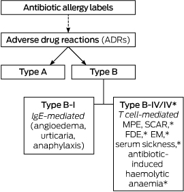
An AAL was defined as any reported antibiotic allergy or adverse drug reaction (ADR) recorded in the allergy section of the EMR, drug chart, or medical admission note. AALs were classified as either type A or type B ADRs according to previously published definitions (Box 1):12,13

  • type A: non-immune-mediated ADR consistent with a known drug side effect (eg, gastrointestinal upset);

  • type B: immune-mediated reactions consistent with an IgE-mediated (eg, angioedema, anaphylaxis, or urticaria = type B-I) or a T cell-mediated response (type B-IV):

    • Type B-IV: delayed benign maculopapular exanthema (MPE);

    • Type B-IV* (life-threatening in nature): severe cutaneous adverse reactions (SCAR),14 erythema multiforme (EM), fixed drug eruption (FDE), serum sickness, and antibiotic-induced haemolytic anaemia.

Study investigators JAT and AKA categorised AALs; if consensus could not be reached, a third investigator (LG) was recruited to adjudicate.

An AAL was defined as a “low risk phenotype” if it was consistent with a non-immune-mediated reaction (type A), delayed benign MPE without mucosal involvement that had occurred more than 10 years earlier (type B-IV), or an unknown reaction that had occurred more than 10 years earlier. Unknown reactions in patients who could not recall when the reaction had occurred were also classified as low risk phenotypes. All low risk phenotypes were ADRs that did not require hospitalisation. A “moderate risk phenotype” included an MPE or unknown reaction that had occurred within the past 10 years. A “high risk phenotype” was defined as any ADR reflecting an immediate reaction (type B-I) or non-MPE delayed hypersensitivity (type B-IV*).

AAL mismatch was defined as non-concordance between a patient’s self-reported description of an antibiotic ADR in the questionnaire and the recorded description in any of the medical record platforms (drug charts, medical admission notes, EMR). Infection diagnosis was classified according to Centers for Disease Control/National Healthcare Safety definitions.15

Statistical analysis

Statistical analyses were performed in Stata 12.0 (StataCorp). Variables of interest in the AAL and no antibiotic allergy label (NAAL) groups were compared. Categorical variables were compared in χ2 tests, and continuous variables with the Wilcoxon rank sum test. P < 0.05 (two-sided) was deemed statistically significant.

Ethics approval

The human research ethics committees of both Austin (LNR/15/Austin/93) and Alfred Health (project 184/15) approved the study.

Results

Antibiotic allergy label description and classification

The baseline patient demographics for the AAL and NAAL groups are shown in Box 2. Of the 453 patients initially identified, 107 (24%) had an AAL. A total of 160 individual AALs were recorded: 27 were type A (17%), 26 were type B-I (16%), 45 were type B-IV (28%), and 62 were of unknown type (39%) (Box 3). Sixteen of the type B-IV reactions (35%) were consistent with more severe phenotypes (type B-IV*). When the time frame criterion (more than 10 years v 10 years or less since the index reaction) was applied to phenotype definitions, this translated to 63% low risk (101 of 160), 4% moderate risk (7 of 160), and 32% high risk (52 of 160) phenotypes. The antibiotics implicated in AALs and their ADR classifications are summarised in Box 3; 34% of reactions were to simple penicillins, 13% to sulfonamide antimicrobials, and 11% to cephalosporins. Three AAL patients (2.8%) were referred to an allergy specialist for assessment (one with type A, two with type B-I reactions). No recorded AALs were associated with admission to an ICU, while eight either ended or occurred during the index hospital admission (two type A, five type B-I, and one type B-IV).

Antibiotic use

Ceftriaxone was prescribed more frequently for patients with AALs (29 of 89 [32%]) than for those in the NAAL group (74 of 368 [20%]; P = 0.02); flucloxacillin was prescribed less frequently (0 v 21 of 368 [5.7%]; P = 0.02). The rate of prescription of other restricted antibiotics, including carbapenems, monobactams, quinolones, glycopeptides and lincosamides, was low in both groups (Box 4).

Antibiotic cross-reactivity

Seventy patients had a documented reaction to a penicillin (a total of 72 penicillin AALs: 55 to penicillin V or G, eight to aminopenicillins, nine to anti-staphylococcal penicillins), including two patients with two separate penicillin allergy labels to members of different β-lactam classes. Of these, 23 (32.9%) were prescribed and tolerated cephalosporins (Box 5). Of the 55 patients with a penicillin V/G AAL, β-lactam antibiotics were prescribed for 19 patients (34%); one patient received aminopenicillins (1.8%), four first generation cephalosporins (7%), two second generation cephalosporins (3.6%), and 12 received third generation cephalosporins (21.8%). Conversely, 18 patients had documented ADRs to cephalosporins, with a total of 19 AALs (14 to first generation, one to second generation, two to third generation cephalosporins, and two to cephalosporins of unknown generation). Of these, five patients (27.8%) were again prescribed cephalosporins without any reaction, and a further five (27.8%) tolerated any penicillin (Box 5).

Eight patients with AALs (7%) were administered an antibiotic from the same antibiotic class. No adverse events were noted in any of the patients inadvertently re-challenged. Eighty-six AAL patients (77%) reported a history of taking any antibiotic after their index ADR event. Thirteen patients (12%) believed they had previously received an antibiotic to which they were considered allergic, 62 had not (58%), and 32 were unsure (30%).

Recording of AALs

Almost all AALs (156 of 160 [98%]) were documented in medication charts, but only 115 (72%) were documented in admission notes and 81 (51%) in the EMR. Twenty-five per cent of patients had an AAL mismatch. No patients received the exact antibiotic recorded in the AAL.

Hypothetical oral antibiotic re-challenge

Fifty-eight AAL patients (54%) were willing to undergo a hypothetical oral antibiotic re-challenge in a supervised environment, of whom 28 (48%) had a low risk phenotype, seven a moderate risk phenotype (12%), and 23 a high risk phenotype (40%). If patients had received and tolerated an antibiotic to which they were previously considered allergic, they were more likely to accept a hypothetical re-challenge than those who had not (9 of 12 [75%] v 3 of 12 [25%]; P = 0.04).

Discussion

The major users of antibiotics in community and hospital settings remain our expanding geriatric population.16 An accumulation of AALs, reflecting both genuine allergies (immune-mediated) and drug side effects or intolerances, follows years of antibiotic prescribing. This is reflected in the high AAL prevalence (24%) in our cohort of older Australian general medical inpatients, notably higher than the national average (18%) and closer to that reported for immune-compromised patients (20–23%).4,17

To understand the high prevalence of AALs and the predominance of low risk phenotypes in our study group requires an understanding of “penicillin past”, as many AALs are confounded by the impurity of early penicillin formulations and later penicillin contamination of cephalosporin products.18,19 Re-examining non-recent AALs of general medical inpatients is therefore potentially both a high yield and a low risk task, considering the low pre-test probability of a persistent genuine penicillin allergy.2022 While the definition of a low risk allergy phenotype is hypothetical, it is based upon findings that indicate the loss of allergy reactivity over time,20,21,23 the low rate of adverse responses to challenges in patients with mild delayed hypersensitivities,20,22,23 and the safety of oral challenge in patients with similar phenotypes.24

The high rate of type A, non-severe MPE and of non-recent unknown reactions in our patients (74% of all AALs; 63% low risk phenotypes) provides a large sample size to explore further, while the higher use of antibiotics that are the target of antimicrobial stewardship programs (eg, ceftriaxone) in AAL patients provides an impetus for change. The increased use of restricted antibiotics (eg, ceftriaxone and fluoroquinolones) and the reduced use of simple penicillins (eg, flucloxacillin) in patients with an AAL were marked. The effects of AALs on antibiotic prescribing have been described in large hospital cohorts and in specialist subgroups (eg, cancer patients).3,4 Associations between AALs and inferior patient outcomes, higher hospital costs and microbiological resistance have also been recently noted.24,8,17,25 Re-examining AALs in older patients from an antimicrobial stewardship viewpoint is therefore essential, particularly in an era when multidrug-resistant (MDR) organisms are being isolated more frequently in Australia.26 The fact that third generation cephalosporins and fluoroquinolones are associated with MDR organisms and with Clostridium difficile infection generation further supports the need for re-examining AALs, especially in those with easily resolved non-genuine allergies.2730

The high rate of potential patient acceptance of an oral re-challenge (54%), especially by those with low risk phenotypes (48%), suggests that this should be explored in prospective studies. The idea of an antibiotic allergy re-challenge of low risk phenotypes is a practical extension of the work by Blumenthal and colleagues,24 who found a sevenfold increase in β-lactam uptake and a low rate of adverse reactions. Another group found that oral re-challenge was safe in children with a history of delayed allergy.23 These are both important advances; while skin-prick allergy testing is sensitive for immediate penicillin hypersensitivity, skin testing (delayed intradermal and patch) lacks sensitivity for delayed hypersensitivities.8,22,31 Incident-free accidental re-challenge with the culprit antibiotic or a drug from a similar class had occurred in some of our patients, adding further support for exploring this approach. A structured oral re-challenge strategy is attractive, as skin-prick testing is potentially expensive and inaccessible for most people.8

Analysing the high rate of AAL mismatch may be a more pragmatic low-cost approach, as not only were AAL labels absent from a number of medical records, the EMR AAL often differed from patients’ reports. Incorrect and absent AALs in other centres have been raised as a concern from a drug safety viewpoint.6,7,10 Education programs aimed at improving clinicians’ (pharmacy and medical) understanding of allergy pathogenesis could also assist antibiotic prescribing in the presence of AALs.5,10 Interrogation of the patient and their relatives about allergy history and examination of blood investigations at the time of the ADR for evidence of end organ dysfunction or eosinophilia may also provide greater accuracy in phenotyping and severity assessment. Many accumulated childhood allergies reflect the infectious syndrome that resulted in the implicated antibiotic being prescribed, rather than an immunologically mediated drug hypersensitivity.21,23 Referral to allergy specialists at the time of drug hypersensitivity may also reduce over-labelling.

That a clinician questionnaire about antibiotic prescribing attitudes was not administered is a limitation of this study, as was the inability to obtain AAL information from all patients (eg, because of dementia or delirium) or to further clarify “unknown” reactions. Some AAL descriptions are also likely to be affected by recall bias; however, this reflects real world attitudes and prescribing in the presence of AALs. While the prevalence of AALs in younger patients is probably lower than found in this study, the distribution of genuine, non-genuine and low risk allergies may well be the same. In a group of paediatric patients with an AAL for β-lactam antibiotics following non-immediate mild cutaneous reactions without systemic symptoms, none experienced severe reactions after undergoing oral re-challenge.23

Conclusion

AALs were highly prevalent in our older inpatients, with a significant proportion involving non-genuine allergies (eg, drug side effects) and low risk phenotypes. Most patients were willing to undergo a supervised oral re-challenge if their allergy was deemed low risk. AALs were sometimes associated with inadvertent class re-challenges, facilitated by poor allergy documentation, without ill effect. AALs were also associated with increased prescribing of ceftriaxone and fluoroquinolone, antibiotics commonly restricted by antimicrobial stewardship programs. These findings inform a mandate to assess AALs in the interests of appropriate antibiotic use and drug safety. Prospective studies incorporating AALs into antimicrobial stewardship and clinical practice are required.

Box 1 –
Classification of reported antibiotic allergy labels into adverse drug reaction groups12,13


EM=erythema multiforme; FDE=fixed drug eruption; MPE=maculopapular exanthema; SCAR=severe cutaneous adverse reactions (includes Stevens–Johnson syndrome, toxic epidermal necrolysis, drug rash with eosinophilia and systemic symptoms, and acute generalised exathematous pustulosis). *These adverse reactions are classified as type B-IV* in this study, denoting their potentially life-threatening nature.

Box 2 –
Baseline demographics for patients with and without antibiotic allergy labels

Characteristic

Patients with an antibiotic allergy label

Patients with no antibiotic allergy label

P


Number

107

346

Median age [IQR], years

82 [74–87]

80 [71–88]

0.32

Sex, men*

38 (36%)

194 (56%)

< 0.001

Immunosuppressed

25 (23%)

29 (8%)

< 0.001

Median age-adjusted Charlson Comorbidity Index score [IQR]

6 [4–7]

6 [4–7]

0.17

Ethnicity

0.38

European

106 (99%)

334 (97%)

African

0

2 (1%)

Asian

1 (1%)

10 (3%)

Infection diagnosis

50 (47%)

140 (41%)

0.25

Infections (205 patients)

56

151

0.002

Cardiovascular system

0

2 (1%)

Central nervous system

1 (2%)

3 (2%)

Gastrointestinal

9 (16%)

9 (6%)

Eyes, ears, nose and throat

0

3 (2%)

Upper respiratory tract

7 (13%)

30 (20%)

Lower respiratory tract (including pneumonia)

12 (21%)

54 (36%)

Skin and soft tissue

7 (13%)

14 (9%)

Urinary system

11 (20%)

21 (14%)

Pyrexia (no source)

3 (5%)

4 (3%)

Sepsis (unspecified)

5 (9%)

8 (5%)

Other

0

2 (1%)

Received antibiotics

45 (42%)

162 (46%)

0.43


* There were a total of 232 men and 221 women in the study.

Box 3 –
Spectrum of implicated antibiotics linked with reported antibiotic allergy labels according to adverse drug reaction classification

Implicated antibiotics

Antibiotic allergy labels: adverse drug reactions


Type A

Type B


Unknown

Total

Type B-I

Type B-IV

Type B-IV*


All antibiotics

27 (17%)

26 (16%)

29 (18%)

16 (10%)

62 (39%)

160

Simple penicillins*

7 (26%)

14 (54%)

16 (55%)

4 (25%)

14 (23%)

55 (34%)

Aminopenicillins

1 (4%)

2 (8%)

2 (7%)

1 (6%)

2 (3%)

8 (5%)

Anti-staphylococcal penicillins

0

0

1 (3%)

5 (31%)

3 (5%)

9 (6%)

Cephalosporins

3 (11%)

1 (4%)

1 (3%)

2 (13%)

11 (18%)

18 (11%)

Carbapenems§

0

0

0

0

1 (2%)

1 (0.6%)

Monobactam

0

0

0

0

0

0

Fluoroquinolones

2 (7%)

0

2 (7%)

0

3 (5%)

7 (4%)

Glycopeptides

0

0

1 (3%)

1 (6%)

1 (2%)

3 (2%)

Lincosamides

0

0

1 (3%)

0

2 (3%)

3 (2%)

Tetracyclines

4 (15%)

1 (4%)

0

1 (6%)

5 (8%)

11 (7%)

Macrolides

1 (4%)

2 (8%)

1 (3%)

1 (6%)

6 (10%)

11 (7%)

Aminoglycosides

0

0

1 (3%)

0

0

1 (0.6%)

Sulfonamides

4 (15%)

4 (15%)

3 (10%)

1 (6%)

9 (15%)

21 (13%)

Others

5 (19%)

2 (8%)

0

0

5 (8%)

12 (8%)


All percentages are column percentages, except for the “all antibiotics” row. * Benzylpenicillin, phenoxymethylpenicillin, benzathine penicillin. † Amoxicillin, amoxicillin–clavulanate, ampicillin. ‡ Flucloxacillin, dicloxacillin, piperacillin–tazobactam, ticarcillin–clavulanate. § Meropenem, imipenem, ertapenem. ¶ Trimethoprim–sulfamethoxazole, sulfadiazine.

Box 4 –
Antibiotic use in patients with and without an antibiotic allergy label

Antibiotic class prescribed

Antibiotic prescriptions


P

Antibiotic allergy label group

No antibiotic allergy label group


Total number of patients

89

368

β-Lactam penicillins

14 (16%)

120 (35%)

0.02

Simple penicillins*

4 (5%)

32 (9%)

0.27

Aminopenicillins

8 (9%)

52 (14%)

0.22

Anti-staphylococcal penicillins

2 (2%)

36 (10%)

0.02

Carbapenems§

2 (2%)

5 (1%)

0.63

Cephalosporins (first/second generation)

8 (9%)

20 (5%)

0.22

Cephalosporins (third or later generation)

29 (33%)

82 (22%)

0.05

Monobactam

0

0

NA

Fluoroquinolones

5 (6%)

6 (2%)

0.04

Glycopeptides

3 (3%)

12 (3%)

1

Tetracyclines

6 (7%)

46 (13%)

0.14

Lincosamides

0

0

NA

Others

26 (29%)

109 (30%)

1


NA = not applicable. * Benzylpenicillin, phenoxymethylpenicillin, benzathine penicillin. † Amoxicillin, amoxicillin–clavulanate, ampicillin. ‡ Flucloxacillin, dicloxacillin, piperacillin–tazobactam, ticarcillin–clavulanate. § Meropenem, imipenem, ertapenem. Some patients received more than one antibiotic.

Box 5 –
Antibiotic use in patients with penicillin and cephalosporin antibiotic allergy labels


Patients with documented allergy to penicillins* (n = 70)

Antibiotics prescribed:

Any antibiotics

28 (40%)

More than one class of antibiotic

31 (44%)

Culprit group penicillins

1 (1.4%)

Non-culprit group penicillins

2 (2.9%)

First generation cephalosporins

4 (5.7%)

Second generation cephalosporins

2 (2.9%)

Third generation cephalosporins

17 (24%)

Carbapenems

2 (2.9%)

Fluoroquinolones

4 (5.7%)

Glycopeptides

2 (2.9%)

Aminoglycosides

2 (2.9%)

Lincosamides

0

Patients with documented allergy to cephalosporins (n = 18)

Antibiotics prescribed:

Any antibiotics

10 (56%)

More than one class of antibiotic

7 (39%)

Culprit generation cephalosporins

1 (5.6%)

Non-culprit generation cephalosporins

3 (17%)

Other

1 (5.6%)

Any penicillins*

5 (28%)

Carbapenems

1 (5.6%)

Fluoroquinolones

1 (5.6%)

Glycopeptides

1 (5.6%)

Aminoglycosides

1 (5.6%)

Lincosamides

0


* Penicillins (benzylpenicillin, phenoxymethylpenicillin, benzathine penicillin); aminopenicillins (amoxicillin, amoxicillin–clavulanate, ampicillin), and anti-staphylococcal penicillins (flucloxacillin, dicloxacillin, ticarcillin–clavulanate and piperacillin–tazobactam). † Prescription of culprit group penicillin: received any penicillin from the same group as that to which the patient is allergic. This patient had a documented allergy to an unknown generation of cephalosporin, and received ceftriaxone.

Your season of birth is stamped on your DNA and can affect your risk of allergies

If you work in healthcare and have a blog topic you would like to write for doctorportal, please get in touch.

People born in autumn or winter are more likely to suffer from allergies than people born in spring or summer. Nobody is certain why this is, but there are several theories. These include seasonal variations in sunlight (which could affect vitamin D levels), levels of allergens such as pollen and house dust mite (which vary by season), the timing of the baby’s first chest infection (colds tend to be more common in winter), and maternal diet (price and availability of fruit and vegetables vary by season).

But no matter which of these exposures causes changes to the risk of developing an allergy, until now nobody knew how these early environmental influences were so long lasting.

Our study tested whether epigenetic marks on a person’s DNA could be a mechanism behind these birth season effects. Of course, your genome doesn’t change depending on which season you’re born in, but there are epigenetic marks attached to your DNA that can influence gene expression – the process where specific genes are activated to produce a certain protein. This may result in different responses to immune triggers and hence different susceptibility to diseases.

Unlike DNA, which is inherited from your parents, epigenetic marks can change in response to the environment and allow gene expression to respond to environmental exposures. And they can also be very long-lasting.

Epigenetic imprint

We scanned DNA methylation (one type of epigenetic mark) profiles of 367 people from the Isle of Wight and found, for the first time, that the season in which a person is born leaves an epigenetic print on the genome that is still visible at the age of 18. This discovery means that these marks on the genome could be how season of birth is able to influence the risk of having allergies later in life.

We went on to test whether these DNA methylation differences that varied by season of birth were also associated with allergic disease. We found that two of them appeared to be influencing the risk of allergy in the participants. As well as allergies, other studies have shown that season of birth is associated with a number of things such as height, lifespan, reproductive performance, and the risks of diseases including heart conditions and schizophrenia. It is possible that the birth season-associated DNA methylation that we discovered might also influence these other outcomes but this will need further investigation.

The marks that we found in the DNA samples collected from the 18-year-olds were mostly similar to the epigenetic marks found in a group of Dutch eight-year-olds that we used to validate our findings. But when we looked at another cohort – a group of newborn babies – the marks were not there. This suggests that these DNA methylation changes occur after birth, not during pregnancy.

There’s something about the seasons

We are not advising women to change the timing of their pregnancy, but if we understood exactly what it was about birth season that causes these effects, this could potentially be changed to reduce the risk of allergy in children. For example, if the birth season effect on allergies was found to be driven by sunlight levels experienced by the mother during pregnancy or breastfeeding, then the increased risk of allergies among babies born in autumn and winter might be lessened by giving the expectant or breastfeeding mother vitamin D supplements. You wouldn’t need to time births with the seasons to get the benefits.

Our study reports the first discovery of a mechanism through which birth season could influence disease risk, though we still don’t know exactly which seasonal stimuli cause these effects. Future studies are needed to pinpoint these, as well as to investigate the relationship between DNA methylation and allergic disease, and what other environmental exposures have an effect.

With the considerable burden allergic disease places not only on individual sufferers but also on society, any step towards reducing allergy is a step in the right direction.The Conversation

Gabrielle A Lockett, Postdoctoral research associate, University of Southampton and John W Holloway, , University of Southampton

This article was originally published on The Conversation. Read the original article.

Other doctorportal blogs

[Seminar] Atopic dermatitis

Atopic dermatitis (also known as atopic eczema) is a chronic inflammatory skin disease that is characterised by intense itching and recurrent eczematous lesions. Although it most often starts in infancy and affects two of ten children, it is also highly prevalent in adults. It is the leading non-fatal health burden attributable to skin diseases, inflicts a substantial psychosocial burden on patients and their relatives, and increases the risk of food allergy, asthma, allergic rhinitis, other immune-mediated inflammatory diseases, and mental health disorders.