×

New recommendations for Hepatitis C treatment

New recommendations have been released for the management of hepatitis C virus (HCV) infection in a consensus statement.

The statement was drawn up by Gastroenterological Society of Australia, the Australasian Society of Infectious Diseases, the Australasian Hepatology Association, the Australasian Society for HIV, Viral Hepatitis and Sexual Health Medicine, Hepatitis Australia and the Royal Australian College of General Practitioners.

A summary, published in the Medical Journal of Australia, says that the recommendations for Hepatitis C treatment were drawn up in the wake of the new direct-acting antiviral therapies that were added to the Pharmaceutical Benefits Scheme earlier this month.

Related: 5 things you need to know about the new Hepatitis C medicines on the PBS

“The introduction of DAA therapies for HCV that are highly effective and well tolerated is a major medical advance,” said Professor Alexander Thompson, director of gastroenterology at St Vincent’s Hospital in Melbourne.

“All Australians living with HCV should now be considered for antiviral therapy.”

Recommendations in the consensus statement include:

  • All individuals with a risk factor for HCV infection should be tested.
  • Annual HCV serological testing is recommended for seronegative individuals with risk factors for HCV transmission.
  • People with confirmed HCV infection should be tested for HCV genotype (Gt).
  • All concomitant medications should be reviewed before starting treatment, using the University of Liverpool’s Hepatitis Drug Interactions website.
  • The use of any DAA regimen during pregnancy is not recommended.
  • People who are not cured by a first-line interferon-free treatment regimen should be referred to a specialist centre.
  • All people with decompensated liver disease, extra-hepatic manifestations of HCV, HCV–HIV or HCV–HBV co-infection, renal impairment or acute HCV infection, as well as people who have had a liver transplant should be referred for management by a specialist who is experienced in the relevant areas.
  • All people living with HCV infection should have a liver fibrosis assessment before treatment to evaluate for the presence of cirrhosis.
  • People with no cirrhosis can be treated by general practitioners working in consultation with specialists.

Read the full recommendations on the Gastroenterological Society of Australia’s website.

Latest news:

’Twas the night before Grand Round …

’Twas the night before Grand Round, when all through the Walton
not a creature was stirring, not even the matron.
The nurses were waiting on the ward in despair
with hopes that the registrar soon would be there.

This educated, thirty-three-year-old chap
had just been admitted after a minor mishap.
The gent had fallen whilst out for a jog
and awoke the next morning, his left leg like a log.

Before his admission he had been so well
With no past history on which we can dwell.
No family history for us to try to link
but had eleven pack-years and took the occasional drink.

A pain in his groin he was suffering
He described as dull and not like a sting
“What a miserable way to spend my Christmas vacation.”
The man remarked using “double air quotation”.

He talked of his issue with sensory supply
noting the numbness of his anterior thigh.
Spreading over his knee and down to his calf
I asked “Which side?”, he said “The medial half.”

Then out on the ward there arose such a clatter
I sprang from my exam to see what was t’matter.
Away to the door it’s our neuro main man
pulling back the curtain, to reveal Dr Doran.

His eyes — how they twinkled! His dimples, how merry!
His cheeks were like roses, his nose like a cherry!
Around his neck was a tie drawn up like a bow
and the ’tache on his lip was as white as the snow.

“We must examine” Dr Doran ordained
“or else this man’s symptoms go unexplained.”
So on thorough neurological examination
it confirmed the described loss of pinprick sensation.

Tone grossly normal, his reflexes intact
except the left knee jerk, it appeared that he lacked.
No wasting or fascic’ to find on inspection
there was clear weakness of left hip into flexion.

3 out of 5 knee extension was detected
but internal rotation was left unaffected.
And finally of note in an exam that’s so formal
was to say that his left A-D-ductors are normal.

I laid down my tendon hammer, the exam at its close,
I thought to myself, “What will we diagnose?”
“Your exam’s out of order”, Doran did chime.
“Well you try to do it whilst keeping in rhyme.”

I gathered my thoughts with flare and cohesion
To let us know where and what is the lesion?
I straightened my tie and said it with verve
“This fits with a lesion of the femoral nerve.”

“Now wait just a minute, don’t be so hurried.
What are causes that we should be worried?”
More rapid than Broca, the differential came
and I whistled and shouted and called them by name:

“Direct injury! A tumour! And radiation to the pelvis!
Pelvic fracture! Vasculitis!” Keep going Dr Ellis!
My mind went blank, my confidence misplaced!
But then I remembered, “Haemorrhage, retrofascial space!”

An MRI arrived of his abdo and pelvis
revealing to us all his lesional diagnosis.
Isointense on T1 and hyper- on T2
a subacute haematoma appeared in our view.

Left iliopsoas muscle its location reported
Clearly the femoral nerve would be distorted.
Second to fourth branches of t’lumbar plexus
This neurological correlate will ne’er again vex us.

And anymore results for us to see?
His APTT’s elevated by a factor of three.
“Should we now check this man’s clotting line?”
“It appears that he’s deficient in factor nine.”

It would seem the odd cause of his admission
Arose from an inherited bleeding predisposition.
And if you know your haemophilia A’s from your haemophilia B’s
You’ll know that the diagnosis is … Christmas Disease!

So Doran packed up his bag, to his team gave a whistle
and off he flew like the down of a thistle.
But I heard him exclaim, as he head out of sight
“Happy Christmas to all, and to all a good night!”

Splenic abscess complicating gastroenteritis due to Salmonella Virchow in an immunocompetent host

Clinical record

A 20-year-old man was admitted to a regional hospital with fevers, rigors, anorexia and left upper quadrant pain. It was his fourth presentation to the emergency department in the preceding 10 days. On the first two presentations, he had been sent home with a provisional diagnosis of renal colic. After review by his general practitioner, he had undergone outpatient imaging that identified filling defects in the pulmonary arteries of his left lower lobe, which were reported as being consistent with pulmonary emboli. In addition, two hypodense splenic lesions were identified, as well as collapse and possible consolidation of the left lower lobe. His GP had referred him to the emergency department for further review (his third presentation), after which he had commenced therapeutic anticoagulation for a presumed diagnosis of pulmonary emboli.

The patient’s history was notable for a self-limiting episode of gastroenteritis 6 weeks before his initial presentation, with sick family contacts. On his fourth presentation, he described progressive left upper quadrant and flank pain over the preceding 10 days, with intermittent fevers and rigors. He had no other focal infective symptoms on review.

On examination, he was found to have a fever (temperature, 39.3°C), sinus tachycardia (heart rate, 154 beats/min), tachypnoea (respiratory rate, 28 breaths/min), hypotension (blood pressure, 97/66 mmHg), decreased breath sounds at the left base of his lung fields and mild left upper quadrant tenderness. Investigations showed a white cell count of 16 × 109/L (reference interval [RI], 4.0–11.0 × 10⁹/L), with a predominant neutrophilia (neutrophils, 14 × 109/L [RI, 2.0–7.0 × 10⁹/L]). Results of his liver function tests and electrolyte, urea and creatinine levels were all within reference intervals.

A computed tomography scan of the chest and upper abdomen again showed two low-density lesions of unclear aetiology in the spleen, as well as a left-sided pleural effusion and collapse of the left lower lobe. Given the possibility that the hypodense splenic lesions represented septic emboli from a cardiac source, the patient was treated empirically with benzylpenicillin, flucloxacillin and gentamicin for a provisional diagnosis of endocarditis. However, a transthoracic echocardiogram performed the next day did not support this diagnosis, with no abnormalities detected. Beyond the radiological findings described, there were no other clinical grounds to support a diagnosis of endocarditis.

Blood cultures taken on Day 1 of admission were positive for gram-negative bacilli, with confirmation of a non-typhoidal Salmonella species (later confirmed as Salmonella Virchow) the following day. This allowed targeted antibiotic therapy, once susceptibilities were known, with ampicillin (2 g every 6 hours). Cultures of stool samples taken at admission were positive for the same isolate, consistent with the patient’s self-limiting episode of gastroenteritis 6 weeks before his first presentation.

Magnetic resonance imaging of the abdomen suggested that the two splenic lesions were likely to represent abscesses in this clinical context (Figure). Given our patient’s ongoing sepsis, a decision was made to perform a laparoscopic splenectomy for source control on Day 5 of admission. Surgical specimens tested positive for Salmonella Virchow. Histopathological testing identified cystic lymphangiomas of the spleen. Despite problems with postoperative pain and a prolonged ileus, the patient made a full recovery. He received appropriate post-splenectomy vaccinations, along with a total of 2 weeks’ intravenous ampicillin, followed by a 2-week course of oral amoxicillin.

Non-typhoidal salmonellae are common foodborne pathogens. In Australia, they are the second most frequent bacterial isolates identified in cases of acute gastroenteritis, after Campylobacter jejuni. In 2010, OzFoodNet sites reported 11 992 cases of Salmonella infection, a rate of 53.7 cases per 100 000.1 Salmonella Virchow was the third most common isolate, after Salmonella Typhimurium and Salmonella Enteritidis. Non-typhoidal Salmonella infection outbreaks are most commonly associated with consumption of poultry and eggs, but have also been linked to fresh produce and, increasingly, contact with pet reptiles.2

Up to 8% of patients with gastroenteritis secondary to non-typhoidal Salmonella infection develop bacteraemia.3 Risk factors for invasive infection include extremes of age, immunosuppressed states, malignancy, HIV infection and use of tumour necrosis factor-blocking medication.4 Our case is unusual in that bacteraemia occurred in an otherwise immunocompetent host.

Extraintestinal focal infections have been reported to occur in 5% to 10% of patients with non-typhoidal Salmonella bacteraemia.3 The best recognised complications are endovascular infections, most commonly involving the aorta, that result from seeding of atherosclerotic plaques and aneurysms.5 However, focal infections of almost all organ systems have been reported.

Splenic abscesses are most commonly seen as a complication of infective endocarditis, occurring in about 5% of patients.6,7 They are also found as a rare complication of non-typhoidal Salmonella infections. In one case series of 49 patients from southern Taiwan, Salmonella species were the third most common pathogens isolated from splenic abscesses, accounting for 11% of cases.8 The most common presentations among the 49 patients with splenic abscesses were fever (47 patients), abdominal pain confined to the left upper quadrant (33 patients), left pleural effusion and splenomegaly (both 27 patients), all of which were present in our patient.

About 50% of patients presenting with splenic abscesses have pre-existing anatomical abnormalities.9 The cystic lymphangiomas identified in our patient almost certainly predisposed him to developing splenic abscesses.

According to the literature, the mainstay of treatment for splenic abscesses is splenectomy. Data from 287 cases published between 1987 and 1995 suggested that non-operative management, which included invasive treatment with percutaneous aspiration and catheter drainage, had a success rate of less than 65%.10 The same series suggested that antimicrobial therapy alone had a success rate of less than 50%. Salvage splenectomy, however, was not shown to result in increased mortality. Another retrospective study of 51 patients in a tertiary hospital between 1998 and 2003 reported survival rates of 48% with antimicrobial therapy alone, 45% with pigtail catheter insertion and drainage in addition to antimicrobial therapy, and 100% with splenectomy and antimicrobial therapy.11 These results may be influenced by selection bias but do suggest improved outcomes with splenectomy over less invasive strategies.

Lessons from practice

  • Splenic abscesses are a rare but potentially life-threatening complication of non-typhoidal Salmonella bacteraemia.
  • Splenic abscesses should be considered as a possible source of infection in patients presenting with unexplained fevers and left upper quadrant or left flank pain.
  • Splenectomy plus appropriate antimicrobial therapy remains the mainstay of treatment for splenic abscesses.
  • Interventional radiological techniques should be considered as a spleen-preserving strategy on a case-by-case basis and where experienced radiologists are available.

Splenic abscesses are a rare but serious complication of non-typhoidal Salmonella bacteraemia that may occur in otherwise immunocompetent individuals. Splenic abscesses should be suspected in patients with unexplained fevers and left upper quadrant pain. The mainstay of treatment is splenectomy with appropriate antimicrobial therapy.


A: Axial T2-weighted magnetic resonance image (MRI) of the abdomen, without contrast, showing an abscess in the inferior pole of the spleen (circle).


B: Saggital T2-weighted MRI of the abdomen, without contrast, showing two splenic abscesses (circles).


The spectacular recent trials of urgent neurointervention for acute stroke: fuel for a revolution

How should we redesign our stroke services in light of neurointerventional advances?

In 2013, neutral results from three trials of neurointervention for treating ischaemic stroke were simultaneously published — a triad of gloom.13 In just over 2 years since, five positive trials have been reported.48 What explains this extraordinary turnaround, and what are the implications for stroke services in Australia and around the world? The answers to these questions are surprising and reflect a mixture of science, technology and policy.

The roles of science, technology and policy

The science involved is the culmination of a decade of work on proving that brain imaging can identify the ischaemic penumbra — the area of the brain that has shut down and is on the path to infarction but, with successful reperfusion, is potentially salvageable. By recruiting patients with a favourable profile for reperfusion therapy (so-called target mismatch, where the ratio of perfusion lesion to established infarct is > 1.8, the perfusion lesion volume is > 15 mL, and the established infarct volume is < 70 mL),9 we are now able to identify those who are likely to respond well. In addition, computed tomography (CT) angiography is now widely available and can demonstrate major cerebral vessel occlusion — a clear target for therapy.9

In contrast to the neutral trials, the recent trials all used either advanced imaging to identify patients with the “reperfusion responder” profile or angiography to prove major vessel occlusion, or both, then randomly assigned this population of likely responders to receive endovascular reperfusion (usually in addition to alteplase thrombolysis) or standard acute stroke care. The combination therapy resulted in potent reperfusion and a dramatic treatment effect (Appendix), such that three of the five neurointervention trials were stopped early.

The technology is all about the device. In today’s fast-moving world, it is almost impossible to design, fund and complete a trial of a device without it becoming obsolete by the time the trial has finished — the fate of the previous studies.13 Unlike in coronary intervention, the thrombus or embolus in ischaemic stroke must be physically extracted, and the new generation of retrievable stents are a major advance in this regard. One of the recent trials, MR CLEAN, demonstrated that carotid stenting (for extracranial occlusion) was also required for 13% of the patients receiving intra-arterial treatment.4

Finally, the unanticipated influence of policy can have profound effects. One of the neutral trials, IMS III, was conducted in the United States at a time when neurointervention was generously compensated.1 This, together with the attractiveness of the technology (despite its lack of evidence), meant that most people were treated outside the trial. The difficulties in recruitment (only one or two patients per centre per year) and a possible selection bias of recruiting only “difficult” patients might have had an effect on the results of IMS III.

In contrast, the Dutch MR CLEAN trial provides an important lesson.4 All the neurointervention centres in the Netherlands participated in this trial, and from 2013 there was no reimbursement for people treated outside the trial. This allowed some 500 patients to be recruited from 16 sites in just over 3 years, compared with IMS III, which needed 7 years to recruit 656 patients from 58 sites. If trial-only reimbursement for unproven devices were enforced, it is likely that reliable data on efficacy would have been available much earlier, potentially saving hundreds, if not thousands, of lives.

Implications for practice

Implementing intravenous thrombolysis has been a difficult and protracted affair, and we are still researching the best ways to achieve it.10 However, there has been progress in Australia, thanks to the hard work of clinicians in the state stroke networks. For example, in New South Wales, the Agency for Clinical Innovation led the establishment of a state-wide stroke reperfusion strategy that involved training paramedics to screen for potential thrombolysis candidates with the FAST (Face, Arms, Speech, Time) test and fast-tracking potentially eligible patients to 24-hour thrombolysis centres.

Our challenge is how to redesign our stroke services and how best to build capacity in the neurointerventional workforce. What is the required infrastructure, support and training in advanced imaging selection that would work in our hospitals? The London model of hyperacute stroke centres might work in our capital cities, but the “drip and ship” model of starting thrombolysis followed by urgent transfer to a comprehensive stroke centre may be a solution for outer metropolitan and regional thrombolysis centres. It is abundantly clear that we will never be able to provide on-site neurointervention at all stroke thrombolysis-capable hospitals, nor achieve complete equity of access to endovascular therapy for stroke patients from rural and remote communities.

A potential solution to the shortage of neurointerventionalists is the emerging model to train neurologists in interventional neuroradiological skills. Given there is broad acceptance that a stroke physician (with appropriate training) does not necessarily have to be a neurologist, there is growing support in the US and Australia for the position that a neurointerventionalist does not have to be a radiologist, provided he or she has had appropriate training.11

The exact shape of future neurointerventional stroke centres is still uncertain, but the endovascular revolution has arrived, and the stroke community needs to work quickly to redesign stroke care services and build the workforce of specialists trained in endovascular therapies. Drivers for change will include the new national stroke care standard (launched on 10 June 2015)12 and the next revision of the national Clinical guidelines for stroke management.

Stroke medicine has come a long way from the nihilism of two decades ago, with numerous interventions now supported by high-level evidence. Immediate brain imaging will identify strokes that are due to haemorrhage, and rapid blood pressure lowering and stroke unit (or intensive) care are the mainstay of treatment for these patients, with surgery needed only for a select few. For patients with ischaemic stroke, revascularisation with appropriate intravenous thrombolysis should be sought, followed by advanced brain imaging to identify patients suitable for additional endovascular therapy.

What is the future? Colleagues in the US and Germany are exploring the utility of an ambulance with an onboard CT scanner (Box), with anecdotal reports of excellent responses to alteplase when given within minutes of major stroke onset. The search is also on for more effective intravenous thrombolytic drugs, with Australia leading an international trial of tenecteplase versus the current standard, alteplase.13 However, none of this will be effective without further public health interventions to improve awareness of stroke and the importance of immediately calling 000 for any suspected stroke patient. When it comes to stroke, time is brain.

Ambulance with a computed tomography scanner

Warfarin-induced skin necrosis following recommencement of warfarin after perioperative Prothrombinex-VF

Clinical record

A 62-year-old man with thrombophilia was receiving warfarin for recurrent venous and arterial thrombosis, and had a known 48 mm diameter infrarenal abdominal aortic aneurysm (AAA). He presented with collapse at home after 2 days of increasing pain in the left flank. A left-sided retroperitoneal haematoma was identified by computed tomography angiography. Increasing abdominal pain and a decline in haemoglobin levels from 125 g/L to 88 g/L made it necessary to transfer the patient urgently to theatre for exploration and open repair of a presumed ruptured AAA.

The patient had been taking 1.5 mg warfarin each day for 20 years without complication. He was known to be heterozygous for both the factor V Leiden and the prothrombin G2021A mutations. He was a current smoker with a 40-pack-year history who also had mild rheumatoid arthritis, insulin-dependent type 2 diabetes mellitus, stage 3A chronic kidney disease, moderate aortic stenosis and hypertension.

Before surgery, anticoagulation with therapeutic warfarin (international normalised ratio [INR] 2.5) was reversed according to our unit protocol with 5000 IU Prothrombinex-VF (CSL Behring Australia). A posterior rupture of the AAA was confirmed during the operation. Sodium heparin (5000 U) was administered before aortic cross-clamping, and its action was fully reversed at the end of surgery with 50 mg protamine sulphate.

Recovery was initially uneventful, and therapy with 1.5 mg warfarin was resumed on postoperative day 1, together with a renally adjusted dose of enoxaparin sodium (40 mg twice daily).

On postoperative day 5, the patient experienced increasing abdominal pain and was returned to theatre for an exploratory laparotomy; nothing significant was found. His INR was 3.1, and reversal of anticoagulation was not performed.

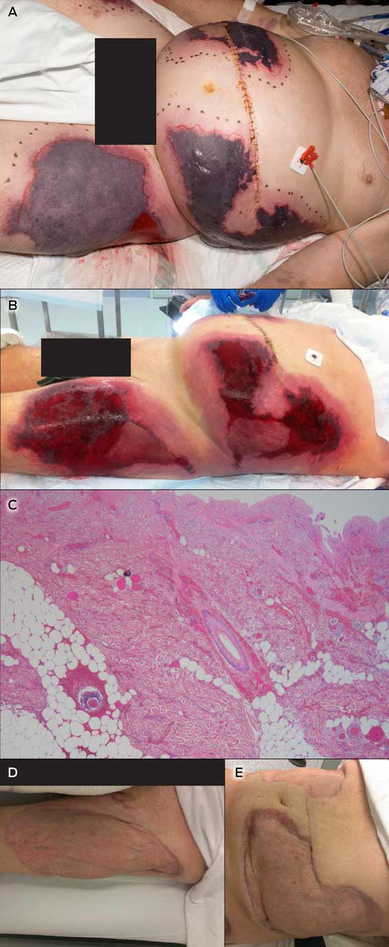
On postoperative day 8, he was transferred to the intensive care unit because of deteriorating gas exchange, hypotension and an evolving coagulopathy. Large and painful areas of skin necrosis had developed on the abdomen, flanks and thighs (Figure, A). His INR was 6.2, activated partial thromboplastin time (APTT) 64 seconds, fibrinogen levels 1.0 g/L, and platelet numbers had dropped from 265 × 109/L to 123 × 109/L.

Seven units of fresh frozen plasma and 10 units of cryoprecipitate were infused. Warfarin treatment was withdrawn, and anticoagulation therapy with intravenous heparin initiated (target APTT: 65–100 seconds) to treat the presumed warfarin-induced skin necrosis (WISN).

The results of an enzyme-linked immunosorbent assay (ELISA) test for heparin-induced thrombocytopenia were negative, as was screening for vasculitis-related antibodies. Anti-cardiolipin and anti-β2 glycoprotein I antibodies were not detected, nor was lupus anticoagulant. Protein C and S levels were low (0.45 U/mL and 0.53 U/mL, respectively).

The skin lesions continued to demarcate over the next 2 days, and were debrided on postoperative day 10 (Figure, B). Histopathological findings were consistent with WISN (Figure, C).

Anticoagulation treatment with intravenous heparin continued for 2 weeks, and was then changed to enoxaparin sodium (100 mg twice daily).

The skin lesions were regularly debrided and negative pressure dressings applied during the following months. Autologous split skin grafts were later performed with excellent results (Figure, D, E), and the patient was transferred to our rehabilitation facility on postoperative day 63. Treatment with oral rivaroxaban was initiated when he was discharged from hospital, and is to continue indefinitely at a dose of 20 mg daily.

Warfarin-induced skin necrosis (WISN) is a rare complication of a commonly used medication. The underlying mechanism is unclear, but it is thought that WISN is induced by a transient paradoxical hypercoagulable state.

Warfarin inhibits certain vitamin K-dependent factors more quickly than it does others, producing a transient imbalance in procoagulant and anticoagulant activity.1 The anticoagulant activity of protein C is rapidly reduced (within 24 hours) because of its short half-life (5–8 hours). The levels of other vitamin K-dependent coagulation factors (II, IX and X) decline at slower rates because they have longer half-lives (24–72 hours). The initial result, therefore, is a relative increase in thrombin generation and a transient hypercoagulable state that may lead to thrombotic occlusion of the microvasculature and thus tissue necrosis.

Our patient had several risk factors for WISN, including his age, obesity and a history of thrombophilia. Further, he was heterozygous for the factor V Leiden mutation (resulting in activated protein C resistance and a functional protein C deficiency) and for the prothrombin G2021A mutation (resulting in elevated prothrombin levels).2 Hypercoagulable conditions more commonly associated with WISN include deficiencies of protein C, protein S and antithrombin III. Other recognised predisposing factors include being female and being given higher loading doses of warfarin.3

The potential role of Prothrombinex-VF in the development of WISN in our patient warrants further consideration. In Australia, immediate warfarin reversal is achieved by using prothrombin complex concentrates or fresh frozen plasma. Prothrombinex-VF is the only prothrombin complex concentrate routinely used in Australia and New Zealand. It is a three-factor concentrate (prothrombin II, IX and X), including low levels of factor VII, but does not contain proteins C or S.

Our unit protocol for immediate warfarin reversal at the time of this patient’s admission reflected the recommendations published in 2009 by Chiu and colleagues.4 Our patient received Prothrombinex-VF alone; neither vitamin K nor fresh frozen plasma were used during reversal. We hypothesise that protein C and S levels were low at the time of his operation, as Prothrombinex-VF does not reverse the reduction of protein C and S levels caused by warfarin. Perioperative blood loss would have reduced their levels further, and the resumption of warfarin treatment immediately after the operation would have depleted them even more. We therefore suggest that very low levels of proteins C and S, together with his pre-existing thrombophilia, are likely to have tipped the balance in favour of thrombosis.

A recent update of the consensus guidelines for warfarin reversal in Australia suggested that 5–10 mg vitamin K1 be given parenterally at the same time as Prothrombinex-VF.5 The half-lives of the infused clotting factors are similar to those of endogenous clotting factors, but the addition of vitamin K1 (as a cofactor in their synthesis) would sustain the reversal effect. It may also increase protein C and S levels, and thereby avoid a transient prothrombotic state when treatment with warfarin is resumed.

Our case highlights the importance of being aware of WISN as a rare complication of warfarin therapy. Consideration of individual patient factors, including a history of thrombosis, before initiating warfarin reversal is critical for ensuring that appropriate adjuvant therapy is provided and an optimal outcome achieved. Vitamin K1 should be administered with Prothrombinex-VF during warfarin reversal, as it sustains the reversal effect, may increase the levels of proteins C and S, and thereby avert thrombotic complications.

Lessons from practice

  • Individual patient factors, including a history of thrombosis, must be considered before warfarin reversal.
  • Updated consensus guidelines for warfarin reversal suggest giving vitamin K1 with Prothrombinex-VF.
  • By increasing protein C and S levels, vitamin K1 may prevent a transient hypercoagulable state after resuming warfarin therapy.

Warfarin-induced skin necrosis and results following autologous skin grafts


A Bilateral flank and thigh skin necrosis.


B: Wound debridement.


C Diffuse dermal ischaemic necrosis, haemorrhage and oedema. A small number of platelet thrombi are evident in the small veins of the dermis. There is no evidence of vasculitis. Haematoxylin-eosin stain; magnification × 200.


D: Left thigh at 6-month follow-up.


E: Abdomen at 6-month follow-up.

Intravenous OxyContin-associated thrombotic microangiopathy treated successfully without plasma exchange

In April 2014, in response to intravenous misuse of oral extended-release oxycodone hydrochloride, a new tamper-resistant formulation was released in Australia. We report a case of thrombotic microangiopathy after intravenous misuse of the new tamper-resistant formulation that was successfully managed without plasma exchange.

Clinical record

A 56-year-old man of European ancestry with no clinically significant medical history presented with a 3-day history of periumbilical abdominal pain. He admitted to daily intravenous (IV) misuse of oral extended-release oxycodone hydrochloride (OxyContin; Mundipharma) over a period of months. For the 5 weeks before presentation, he had been injecting the new tamper-resistant formulation because he was unable to access the discontinued crushable form.

On presentation, the patient was afebrile, had a pulse of 95 beats/min, blood pressure of 154/85 mmHg, a respiratory rate of 14 breaths/min and oxygen saturation of 97%. On examination, he had mild periumbilical tenderness. Results of cardiovascular and respiratory examinations were unremarkable. No murmur was detected. There was no injection site infection or axillary lymphadenopathy.

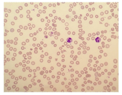
Laboratory investigations showed a haemoglobin level of 87 g/L (reference interval [RI], 135–180 g/L), a total white cell count of 15.0 × 109/L (RI, 4–11 × 109/L), a neutrophil count of 10.84 × 109/L (RI, 2–8 × 109/L), a monocyte count of 1.47 × 109/L (RI, 0.1–1.0 × 109/L) and a platelet count of 53 × 109/L (RI, 140–400 × 109/L). Electrolyte levels were normal, and serum creatinine level was normal at 66 µmol/L. The patient’s unconjugated bilirubin level was 34 µmol/L (RI, < 20 µmol/L) and lactate dehydrogenase level was 769 U/L (RI, 150–280 U/L); other liver function test results were normal. His reticulocyte count was 168 × 109/L (RI, 10–100 × 109/L), haptoglobin level was 0.04 g/L (RI, 0.36–1.95 g/L) and Coombs test result was negative. Three per cent of his red blood cells were fragmented and polychromasia was present, consistent with microangiopathic haemolytic anaemia (Box 1). ADAMTS13 activity was 70% (RI, 40%–130%).

The patient’s serological test results for hepatitis B, hepatitis C and HIV were negative. Vitamin B12, folate, lupus anticoagulant, anticardiolipin, anti-β2 glycoprotein I, antinuclear antibody, extractable nuclear antigen and complement levels were normal. His ferritin level was elevated at 442 µg/L (RI, 30–300 µg/L), and transferrin saturation was normal at 18%. Activated partial thromboplastin time and prothrombin time were normal and his fibrinogen level was elevated (5.4 g/L [RI, 1.7–4.5 g/L]). A random urine test showed a proteinuria level of 340 mg/L (RI, < 100 mg/L) and a protein-to-creatinine ratio of 66 g/mol (RI, < 15 g/mol).

We elected to manage the patient’s condition conservatively without the use of plasmapheresis, steroids or antiplatelet agents. Spontaneous resolution of the microangiopathic haemolysis followed, and subsequent outpatient review showed that his parameters continued to normalise (Box 2).

To our knowledge, this is the first case of thrombotic microangiopathy (TMA) associated with IV-administered reformulated OxyContin.

Discussion

Misuse of prescription opioids is an increasing problem in Australia. Morphine, oxycodone and methadone liquid are the most commonly misused prescription opioids, with injection being the route of administration in 75% of misusers.1 In response to these concerns, the National Pharmaceutical Drug Misuse Framework for Action (2012–2015) (http://www.nationaldrugstrategy.gov.au/internet/drugstrategy/Publishing.nsf/content/drug-mu-frm-action) has been developed, outlining various strategies to reduce misuse of pharmaceutical medications.

In April 2014, in response to IV misuse of OxyContin, a new crush-resistant formulation with the intent to deter inappropriate tampering and misuse of the drug was released in Australia, and supply of the old formulation was discontinued. The new tablets, which are embossed with “OP” (the original formulation was embossed with “OC”), are difficult to break, cut, crush or chew, and when added to water, form a viscous hydrogel, which cannot readily pass through a needle.

However, in the United States, where tamper-resistant OxyContin was introduced in August 2010, 24% of misusers reportedly found ways to inject the new formulation.2 Similarly, since its introduction in Australia, misusers presenting to the Sydney Medically Supervised Injection Centre have successfully injected the tamper-resistant formulation.3

TMA is a rare but serious blood disorder characterised by thrombosis in arterioles and capillaries that manifests clinically as a microangiopathic haemolytic anaemia and thrombocytopenia. Types of TMA include thrombotic thrombocytopenic purpura (TTP) or TMA associated with ADAMTS13 deficiency; haemolytic–uraemic syndrome (HUS) or infection-induced TMA; atypical HUS or TMA associated with disorders of complement; drug-induced TMA (Box 3); and TMA associated with other conditions including transplantation, lupus, glomerulopathies and malignant hypertension.4 For patients with TMA, HIV and other active viral infections should also be excluded.

Laboratory findings in TMA are consistent with non-immune haemolysis, including anaemia with high reticulocyte counts, elevated bilirubin and lactate dehydrogenase levels, reduced serum haptoglobin levels, red cell fragmentation and negative Coombs test results.

Treatment of TMA varies depending on the cause. Patients with TTP require plasma exchange, whereas HUS treatment is largely supportive. Atypical HUS is treated with plasma exchange and eculizumab, a monoclonal antibody that prevents C5 activation. Of the drug-induced TMAs, only ticlopidine-induced TMA has been shown to respond to plasma exchange. While determining ADAMTS13 levels assists in making a diagnosis, the decision to treat with plasma exchange is usually based on clinical history and other laboratory parameters, owing to the delay in obtaining this result and the necessity to start plasma exchange promptly.

Here, we report the first case of reformulated OxyContin-associated TMA and demonstrate that conservative management is a valid approach. While drug rechallenge would be required to prove a causal association, the temporal association with IV misuse of the reformulated OxyContin, the lack of alternative aetiologies, and the disease resolution with drug cessation indicate that the TMA was induced by the reformulated OxyContin. In addition, reformulated OxyContin is not the first reformulated drug to be associated with TMA. In August 2013, the US Food and Drug Administration and Centers for Disease Control and Prevention issued warnings regarding reformulated oral extended-release oxymorphone hydrochloride (Opana ER; Endo Pharmaceuticals; not available in Australia) after a link between IV Opana ER misuse and TMA was identified.5,6 Initial cases of reformulated Opana ER-induced TMA were treated with plasma exchange;7,8 however, subsequent reports have demonstrated that plasma exchange is unnecessary.9,10

In contrast to our case of OxyContin-associated TMA, acute kidney injury requiring haemodialysis was a prominent feature among patients injecting reformulated Opana ER.8 Our patient had a somewhat atypical presentation, with normal serum creatinine levels, minor proteinuria and no definitive evidence of other end-organ damage.

It is unclear what components of tamper-resistant OxyContin or Opana ER might trigger TMA and whether different methods of preparation can increase or decrease this risk. The new formulation of OxyContin contains inactive ingredients not found in the original formulation, including polyethylene oxide (PEO), butylated hydroxytoluene and magnesium stearate. Opana ER also contains PEO, and notably, in one study, rats intravenously injected with PEO subsequently became thrombocytopenic.11 Interaction with other prescription or illicit drugs is also a possibility. Our patient was taking sertraline 200 mg once daily and diazepam 5 mg as needed, and admitted to chronic daily marijuana use but denied any other substance misuse or medications.

Given the recent switch to tamper-resistant OxyContin in Australia, further cases of OxyContin-associated TMA may occur. A high degree of clinical suspicion is required to make this diagnosis, and we recommend that all patients presenting with TMA be questioned about the use of IV drugs. If OxyContin misuse is acknowledged, clinicians should consider conservative management, which avoids the morbidity and costs associated with plasma exchange.

1 Peripheral blood smear showing microangiopathic haemolysis with red blood cell fragmentation


Wright stain; original magnification, × 40.

2 Improvement in parameters throughout conservative management of thrombotic microangiopathy

Parameter

RI

Day 1

Day 2

Day 3

Day 4

Day 5

Day 6

Day 9

3 months


Haemoglobin (g/L)

135–180

87

86

87

88

87

88

92

164

Platelet count (× 109/L)

140–400

53

34

42

55

80

123

281

358

Unconjugated bilirubin (µmol/L)

< 20

34

22

30

24

18

8

10

10

Lactate dehydrogenase (U/L)

150–280

769

870

889

858

688

611

439

221


RI = reference interval.

3 Drugs that may induce thrombotic microangiopathy

  • Thienopyridines: eg, ticlopidine, clopidogrel, prasugrel
  • Calcineurin inhibitors: eg, cyclosporine, tacrolimus
  • Nucleoside analogues: eg, gemcitabine, mitomycin C
  • Anti-vascular endothelial growth factor therapies: eg, bevacizumab, sunitinib
  • Imatinib
  • Quinine
  • Interferon beta
  • Tamper-resistant extended-release oxymorphone hydrochloride

Compliance with Australian splenectomy guidelines in patients undergoing post-traumatic splenectomy at a tertiary centre

To the Editor: The lack of a functioning spleen is associated with a lifelong risk of overwhelming post-splenectomy infection (OPSI). Historically, mortality rates associated with OPSI have been in excess of 50%.13 OPSI is a preventable illness through vaccination, education, prophylactic antibiotic use and other measures, as summarised in the national Australasian Society for Infectious Diseases (ASID)-endorsed guidelines for prevention of sepsis in asplenic and hyposplenic patients.4

We performed a retrospective cohort study among adult patients who had undergone post-traumatic splenectomy at a tertiary referral centre in Sydney, to assess compliance by health professionals and identify factors that could improve uptake of ASID recommendations. We reviewed hospital medical records and discharge summaries to assess compliance with recommendations before and after the publication of the ASID guidelines.

The Research and Ethics Office of the South Western Sydney Local Health District granted site-specific approval on the basis of low and negligible risk.

A total of 79 patients were identified, 37 in the preguideline group (January 2003 – June 2008) and 42 in the postguideline group (July 2008 – December 2013). Our findings are summarised in the Box.

Overall, compliance with the recommendations was poor, except for the rate of first vaccination against Streptococcus pneumoniae, Neisseria meningitidis and Haemophilus influenzae type b (Box). At discharge, most patients were advised to follow up with their general practitioner; however, GPs were neither provided with the information on the type of vaccination given in the hospital, nor with the appropriate recommendation on follow-up vaccinations.

Our study highlights gaps in best practice and areas for quality improvement and education. Lack of awareness of the guidelines among the surgical teams was found to be a notable factor in the poor compliance with the 2008 ASID guidelines. Asplenia and hyposplenia care should involve a multidisciplinary approach with involvement of surgeons, infectious diseases physicians, haematologists, pharmacists and clinical nurse coordinators.

We recommend the ASID 2008 guidelines be updated, as there have been changes in the vaccination recommendations since publication. A national spleen registry could be considered, for sending vaccination reminders and providing long-term follow-up and ongoing support. This would also allow prospective data collection for assessing compliance and measuring rates of OPSI.

Compliance with recommendations in the ASID management guidelines for prevention of sepsis in patients with asplenia or hyposplenia,4 before and after guideline publication in 2008


Number of patients*


Areas of compliance

Preguideline (n = 37)

Postguideline (n = 42)

P


Patient education

8

22

0.08

First vaccination after surgery


Pneumococcal

34

39

0.87

Meningococcal

34

39

0.87

Haemophilus influenzae type b

34

38

0.82

Influenza

2

5

0.31

Day of first vaccination, median (range)

7 (− 7 to 44)

7 (1 to 45)

0.47

Prophylactic antibiotic use

11

17

0.32

Reserve antibiotic supply

1

5

0.20

Risk-reduction measures


Patient alerts (eg, bracelet)

1

9

0.04

Splenic salvage

0

0

Risk of sepsis included in histology report

0/36

0/42

Risk of sepsis reported if Howell-Jolly body seen in peripheral blood smear

0/15

0/19

Meningococcal vaccination for travellers to high-risk areas

0

0

Informing the patient of malaria risks

1

3

0.61

Informing the patient of Babesia risks

0

1

> 0.99

Patient warned of risks associated with animal bites

1

1

Spleen registry referral

0

3

0.24


ASID = Australasian Society for Infectious Diseases. * Unless otherwise indicated. † Optimal timing uncertain; ideally 14 days after emergency splenectomy, or earlier if there is a risk of loss of the patient to follow-up. ‡ Amoxicillin 250 mg daily was the most commonly prescribed prophylactic antibiotic, with a variable duration of recommendation (1 year to lifelong).


First reported case of transfusion-transmitted Ross River virus infection

We describe the first documented case of Ross River virus (RRV) infection transmitted by blood transfusion. The recipient had a clinically compatible illness, and RRV infection was confirmed by serological tests. The implicated donation was positive for RRV RNA. We discuss the risk to blood recipients and the implications for blood donation in Australia.

Clinical record

In May 2014, the Australian Red Cross Blood Service (the Blood Service) in Western Australia received a delayed notification from a donor who had developed fatigue and arthralgia 2 days after giving blood in March 2014 and was subsequently diagnosed with acute Ross River virus (RRV) infection (Box).

PathWest Laboratory Medicine WA detected RRV IgM antibodies using an inhouse indirect immunofluorescence antibody (IFA) test, but no RRV antibodies were detected using an inhouse haemagglutination inhibition (HI) antibody test 10 days after blood donation. RRV IgM antibodies are detected by IFA testing within a few days of onset of illness and routinely persist for several weeks or, occasionally, months or years. IFA tests are less prone to false-positive results compared with enzyme immunoassays. The HI antibody test primarily detects IgG antibodies, which appear within several weeks but after the IgM response.

Blood Service procedure stipulates that donors with a diagnosed RRV infection are unable to donate fresh components for 4 weeks after recovery. Moreover, fresh components donated from 4 weeks before illness onset to 4 weeks after recovery must be recalled.

The components from the implicated donation were identified: the red blood cell (RBC) component had been transfused to a patient on 12 March 2014, the plasma had been pooled for the manufacture of plasma-derived products and the platelet component had not been used. The treating clinician of the RBC recipient was notified as part of the recall procedure.

The recipient was having regular blood transfusions due to myelodysplastic syndrome that was associated with chronic fatigue and joint pains. The recipient reported a worsening of symptoms in the months after transfusion of the infected blood; however, there was not a clear exacerbation of these symptoms consistent with the incubation period of RRV.

On notification from the Blood Service, the recipient’s treating clinician requested serological testing for RRV on 28 May 2014, which found detectable IgM antibodies using the IFA assay and a high titre of antibodies by HI testing (antibody titre, > 1 : 640). The detection of both IgM and HI antibodies indicates RRV infection in recent months. Previous testing for RRV IgM and HI antibodies in 2006 and August 2013 had been negative. Subsequent inhouse reverse transcriptase polymerase chain reaction (RT-PCR) analysis for RRV RNA performed on stored serum from 28 May gave a negative result. These results are consistent with RRV illness several months before 28 May, with resolution of the transient viraemic phase. No samples from the recipient in March 2014 were available for serological or PCR testing.

In response to this possible case of transfusion-transmitted RRV, the associated archived donor sample was retrieved and sent to the Victorian Infectious Diseases Reference Laboratory for RRV serological tests and RT-PCR analysis. This sample tested negative for RRV IgM and IgG but RRV RNA was detected by two inhouse RT-PCR tests and verified by sequencing. These results are consistent with the blood donation being collected during the pre-seroconversion but transient viraemic phase of RRV illness.

Discussion

Since the isolation of RRV from humans was first reported in 1972,1 our understanding of the epidemiology of the disease has increased considerably. RRV is now known to be the most common mosquito-borne disease of humans in Australia,2 and is endemic in several regions. An average of around 5000 cases of RRV disease are notified annually in Australia, with considerable yearly, seasonal and regional variability due to differences in environmental conditions that affect the mosquito vectors and native animal hosts of the virus.2 The incubation period averages 7 to 9 days with a range of 2 to 21 days.3 Symptoms of RRV most commonly include joint manifestations, which are usually symmetrical and acute in onset, with rash and fever being less common.3 As many as 55% to 75% of RRV infections are asymptomatic.3

Arboviruses such as dengue viruses and West Nile virus are known to be transfusion transmissible,4 and the potential of RRV to be transfusion transmissible was raised in this Journal in 1995.5

Although not previously documented, transfusion transmission of RRV has been considered theoretically possible, given a likely period of asymptomatic viraemia before the onset of symptoms.4,6 This risk is supported by the observation of asymptomatic viraemia typically lasting 5 days after RRV infection in a mouse model.7 On the assumption that transfused blood could transmit RRV, this study estimated the risk of RRV transfusion transmission during a 2004 outbreak in Cairns as one in 13 542 donations. This risk was of the same order of magnitude as that estimated for dengue virus transmission by transfusion during a contemporaneous dengue fever outbreak in Cairns.7

The donor we describe developed an illness clinically compatible with RRV infection 2 days after donating blood and was shown to have a serological profile consistent with acute RRV infection 10 days after donating. The donated blood was subsequently shown to contain RRV RNA by two inhouse RT-PCR tests, and this was confirmed by sequencing.

While the exacerbation of the chronically ill recipient’s fatigue and muscle and joint pains was not clearly consistent with the incubation period of RRV subsequent to the transfusion, the results of RRV serological tests performed about 2 months after transfusion were consistent with infection within this 2-month period. Unfortunately, there were no stored blood specimens collected from the recipient shortly after receiving the blood donation, and hence it was not possible to compare sequences with the donor virus to confirm transmission.

Surveillance by the WA Department of Health showed that the recipient was the only person for whom RRV infection was reported between 1 July 2013 and 30 June 2014 from the local government area in which she resided. The recipient also spent most of her time indoors and could not recall being bitten by mosquitoes. Taken together, these lines of evidence strongly support the likelihood that the recipient’s RRV infection was transmitted by transfusion. Thus, this is the first report of transfusion-transmitted RRV.

Laboratory testing for RRV is not done for Australian blood donors during the donation process, and there is no validated blood screening test for RRV. To manage the risk of transfusion transmission, the Blood Service does not permit donors with symptoms compatible with RRV to donate until they are fully recovered. However, given that most RRV infections are asymptomatic and viraemia is present during the incubation period, excluding donors based on symptoms will not prevent all potentially infectious donations entering the blood supply. Provided infected donors report subsequent illness immediately to the Blood Service, the recall process should prevent the proportion of donations from symptomatic RRV infected donors from being used. Unfortunately, in this case, where notification was delayed for 2 months, the blood component had already been transfused. In response to this, the Blood Service is taking steps to strengthen its messaging to donors regarding development of post-donation illnesses.

In 2012, the Blood Service established a sample archive of every blood donation to meet regulatory standards and assist in investigation and lookback (tracing and notifying patients who may have received infected blood components and investigating donations and donors when a patient has a suspected transfusion-transmissible infection). This archive provides the ability to perform further testing on samples from past donations, as in this case, providing data on the actual risk associated with transfused donations from implicated donors and for investigations where an infection is reported in a recipient.

Transfusion transmission of RRV no longer appears to be only a theoretical risk. However, with about 5000 mosquito-related RRV notifications per year, transfusion transmission of RRV — or the related Barmah Forest virus, which has a lower incidence — is likely to remain a rare event. Any actions taken to prevent infected components entering the blood supply need to take into account the cost, the impact on supply and the severity of the infection in recipients. Laboratory screening is not a feasible option, given that RRV nucleic acid testing is not validated for blood donation screening or available for the large-scale nucleic acid detection equipment used by the Blood Service. In addition, the cost of individual testing is unlikely to be cost-effective and, although RRV can cause debilitating symptoms in some patients, most infections are either asymptomatic or mild and self-limiting.8

Identifying donors who are at risk of exposure and temporarily excluding them from donating fresh blood components in areas and times of RRV outbreaks is one potential risk-mitigation option. When this strategy was applied to dengue fever, it was estimated to cost the Blood Service around $1.0–$3.8 million.9 However, irrespective of the financial cost, this option is unlikely to be feasible, since RRV is endemic in many parts of Australia and such restrictions might have a critical impact on supply. Pathogen reduction technology (PRT) is an alternative risk-management option that would not have an impact on supply. The Blood Service is investigating the effectiveness of PRT for the prevention of arboviral transfusion transmission, including RRV, but further research is needed.10

The Australian blood supply is one of the safest in the world with respect to transfusion-transmitted infections. Yet, it is important to remember that blood transfusion is not without risk and should only be undertaken when the efficacy of the transfusion and improved clinical outcome outweigh the risks.11

Timeline of major events related to the case of Ross River virus (RRV) transfusion transmission, 2014


PCR = polymerase chain reaction.

Domestically acquired hepatitis E successfully treated with ribavirin in an Australian liver transplant recipient

We describe a rare case of domestically acquired hepatitis E in Australia and the first in an Australian liver transplant recipient. The infection was successfully treated with ribavirin.

Clinical record

A 48-year-old Australian man of European ancestry received his third liver transplant in February 2013 for hepatic failure precipitated by ischaemic cholangiopathy and secondary biliary cirrhosis. His first liver transplant was performed 10 years earlier for complications of cirrhosis arising from autoimmune hepatitis – primary sclerosing cholangitis overlap syndrome, but required retransplantation after 3 months due to hepatic vein thrombosis and hepatic infarction. The second liver transplant was complicated by hepatic artery thrombosis, resulting in ischaemic cholangiopathy.

For his third transplant, from 13 days before to 13 days after transplantation, the patient received blood products from 22 individual donors. His initial immunosuppressive regimen comprised cyclosporin 150 mg twice daily, prednisolone 20 mg once daily and mycophenolate mofetil (MMF) 500 mg twice daily. Subsequently, moderate renal impairment (creatinine, 170 µmol/L; reference interval [RI], 60–110 µmol/L) and cytopaenias prompted gradual cyclosporin and MMF dose reductions. Blood products were not required, and results of liver function tests (LFTs) remained normal.

Ten weeks after transplantation, an elevation in LFT levels occurred (alanine aminotransferase [ALT], 131 U/L [RI, < 40 U/L]; aspartate aminotransferase [AST], 53 U/L [RI, < 45 U/L]; γ-glutamyltransferase [GGT], 76 U/L [RI, < 60 U/L]). Abdominal ultrasound was unremarkable, and the raised LFT results gradually settled without adjustment of the immunosuppressive regimen.

Twenty-two weeks after the patient’s transplantation, he developed significant transaminitis (ALT, 338 U/L; AST, 215 U/L; GGT, 183 U/L; alkaline phosphatase, 126 U/L [RI, 35–135 U/L]; total bilirubin, 13 µmol/L [RI, < 20 µmol/L]) (Box 1).

The patient adhered to his immunosuppressive regimen and had not commenced other medications. Physical examination was unremarkable and abdominal ultrasound did not show biliary or hepatic vascular abnormalities. A liver biopsy was performed and reported as consistent with moderately active acute rejection. This prompted methylprednisolone therapy (500 mg once daily for 3 days) followed by prednisolone (50 mg once daily) and replacement of cyclosporin with tacrolimus (3 mg twice daily). MMF was continued unchanged. A second liver biopsy was performed 1 week later due to a further rise in the ALT level (456 U/L), which demonstrated non-specific hepatitis without definite features of rejection or an autoimmune aetiology (Box 2). Subsequent comparison of these biopsies by a specialist histopathologist confirmed acute hepatitis in both samples, without significant features of rejection on either biopsy.

Investigation for infectious causes excluded hepatitis A, hepatitis B, hepatitis C, Epstein–Barr virus, cytomegalovirus, and human herpesvirus 6. Anti-hepatitis E IgG antibody (HEV ELISA, MP Biomedicals Asia Pacific) was not detected, but an in-house hepatitis E virus (HEV) reverse transcription polymerase chain reaction (RT-PCR) assay (Appendix) detected HEV RNA in the patient’s blood. HEV RNA was also detected at the Victorian Infectious Diseases Reference Laboratory (VIDRL), and nucleotide sequencing demonstrated genotype 3 HEV. Paraffin-embedded liver tissue from the biopsy collected 22 weeks after transplantation was HEV RNA positive, as were blood samples collected 22 and 23 weeks after transplantation. Retrospective testing of the patient’s and liver donor’s blood at the time of transplantation was negative for anti-HEV IgG antibody and HEV RNA. A biopsy of the donor liver, collected at the time of transplantation, was also HEV RNA negative.

Stored blood from all donors of the blood products were tested for anti-HEV IgG antibody by two assays (HEV IgG ELISA, Genelabs Diagnostics; HEV-IgG ELISA, Beijing Wantai), anti-HEV IgM antibody (HEV-IgM ELISA, Beijing Wantai) and by in-house (VIDRL) and commercial (RealStar HEV RT-PCR 1.0, Altona Diagnostics) HEV RT-PCR. All 22 donors’ samples were negative for anti-HEV IgM antibody and HEV RT-PCR. Three donors were found to have detectable anti-HEV IgG antibody levels. Two of these donors were found to have detectable IgG anti-HEV antibody levels in both assays; one was born in South Africa but had not left Australia in 6 years, and the second had travelled frequently to India in the past 5 years, most recently 8 months before donation. The third donor had never travelled outside of Australia, but had worked in a piggery. Repeat samples collected 9 months after donation from these donors and 18 of the 19 seronegative donors produced the same serological results.

The patient was born in Australia, had not recently travelled overseas, and worked in a city office. He had no contact with overseas travellers, and had not visited rural areas, farms or had any livestock exposure. He consumed pork regularly, which was sourced from local supermarkets.

Despite reducing the patient’s immunosuppression, the hepatitis continued to worsen over the next 5 weeks (ALT, 669 U/L) and HEV RNA remained detectable (Box 1). Ribavirin at a dose adjusted for his renal impairment (200 mg once daily) was commenced with an immediate improvement of his liver function. HEV RNA was last detected 17 days after commencement, and HEV RT-PCR was negative after 24 days. Pegylated interferon alfa was not used due to pre-existing cytopaenia and the risk of precipitating acute rejection. The patient received a 12-week course of ribavirin and remained HEV RNA negative 15 weeks after cessation. The patient had not developed anti-HEV IgG antibody 7 months after onset of hepatitis.

Discussion

HEV is a non-enveloped RNA virus identified in 1980 as the cause of “epidemic, non-A, non-B hepatitis”, a waterborne illness similar to hepatitis A.1 After an incubation period of 2–9 weeks2 the illness is usually self-limiting, but can progress to severe disease, particularly during advanced pregnancy, and among very young children and those with pre-existing chronic liver disease.3

There are four human HEV genotypes. Genotypes 1 and 2 cause large outbreaks in Asia, Africa and Central America via contaminated water. Genotypes 3 and 4 are predominantly swine viruses causing sporadic zoonotic disease in Europe, the United States and Eastern Asia.4

Acute hepatitis E is most reliably diagnosed either serologically by IgG anti-HEV antibody seroconversion,1,3 or by detection of HEV RNA in blood or faeces. HEV RNA is detectable in blood samples from up to 2 weeks before and 1 week after the onset of jaundice, and in stool it is detectable for up to 3 weeks after the onset of jaundice.

In developed countries, genotype 3 HEV is mostly transmitted by the consumption of undercooked pork or raw offal,3 and occasionally from animal contact or blood transfusion.1,4 The seroprevalence in Europe and the US is lower than for hepatitis A, but higher than for hepatitis B and hepatitis C.1,3 However, the incidence of acute hepatitis E in developed countries is unknown, with only five US cases of domestically acquired acute hepatitis E reported from 1997 to 2006.3

The source for HEV infection in our patient was unknown, but the donor liver, blood products, or contaminated food or water could have been responsible. Solid organ donors are not routinely screened for HEV infection in Australia, although this is recommended where HEV is endemic.4 Donor liver transmission of HEV, presenting 5 months after transplantation from an anti-HEV antibody negative but HEV RNA positive donor has occurred,5 but in our case both donor blood and liver tested negative for anti-HEV IgG antibody and HEV RNA. The blood donor who had travelled to India is a possible source as this donor’s anti-HEV IgG antibody sample-to-cut-off ratio was high in both IgG assays, which has been correlated with a recent illness compatible with hepatitis E and overseas travel.2 A contaminated food source cannot be excluded, as anti-HEV seropositive pigs have been found in Australian piggeries6 and, although processed, up to 80% of the ham, bacon and smallgoods sold in Australia is made from imported pig meat, mostly from the US, Canada and the European Union.7

Chronic hepatitis E can develop in solid organ transplant recipients, patients receiving cancer chemotherapy, and people with HIV. About two-thirds of solid organ transplant recipients infected with HEV develop chronic disease,8 which can be severe and cause significant inflammation and fibrosis.1 In our patient, the undetectable anti-HEV IgG antibody is likely a reflection of his immunosuppression.9

Management options for hepatitis E in solid organ transplant recipients include reducing immune suppression, pegylated interferon alfa or ribavirin therapy, or a combination of these. Reduction of immunosuppression alone can clear HEV in a minority of solid organ transplant recipients and pegylated interferon alfa has been used effectively to treat chronic hepatitis E after transplantation, but may precipitate donor organ rejection.4,8 Although not approved for this use in Australia, ribavirin for at least 3 months has been shown to produce sustained virological responses in at least two-thirds of patients with chronic hepatitis E,8 and is recommended as first-line treatment in solid organ transplant recipients who do not clear the virus despite reducing the immunosuppression.4

Domestically acquired hepatitis E has been reported rarely in Australia since the mid 1990s.1012 To our knowledge, this is the first case of an Australian organ transplant recipient with hepatitis E successfully managed with antiviral therapy. Domestically acquired cases in Australia may be missed due to infrequent HEV serological testing in the absence of a travel history and the relative unavailability of HEV RNA testing. Hepatitis E should be considered in patients with unexplained hepatitis, and solid organ transplant recipients or those with compromised immune systems with hepatitis should be tested for HEV RNA because anti-HEV antibody tests may be negative in these patients.

1 Timeline of laboratory test results and treatment


HEV = hepatitis E virus. PCR = polymerase chain reaction. RI = reference interval, < 40 U/L.

2 Liver biopsy findings 5 months after transplant


A: Mild portal tract inflammation with interface and lobular hepatitis (haematoxylin and eosin stain [H & E], x20). B: Lobular disarray with spotty necrosis (H & E, x40). C: Portal tract expansion by early fibrosis with extension into the lobule (Masson trichrome, x10).

An update of consensus guidelines for warfarin reversal

Correction

Incorrect box cross-references and incorrect number range: In the full online version of “An update of consensus guidelines for warfarin reversal” in the 4 March 2013 issue of the Journal, three box cross-references were incorrect. In Box 5 and Box 8, all references to Box 6 should have been to Box 4, which gives the suggested dose of Prothrombinex-VF according to INR.

In the third paragraph under the final heading “Pre- and postoperative management of warfarin anticoagulation”, an incorrect range was given in the definition of low-risk atrial fibrillation: “CHADS2 score 0–1” should read “CHADS2 score 0–2”.

In the short print-only version, the reference to Box 3 that appears in Box 2 is redundant.