×

Government’s Brain Cancer Mission

The Federal Government has announced a $100 million funding plan to rapidly increase brain cancer survival by bolstering patients’ access to clinical trials and accelerating the discovery of new therapies.

This will be done by expanding research platforms and technologies, and equipping researchers with the best tools and infrastructure.

The Australian Brain Cancer Mission is a partnership between the Federal Government, philanthropists, medical experts, patients and their families.

As a first step, the Government is providing $50 million through the Medical Research Future Fund (MRFF), combined with $10 million from the Minderoo Foundation’s Eliminate Cancer Initiative and a commitment of $20 million from Cure Brain Cancer Foundation. 

The Government is expected to announce the remaining $20 million in the coming months. 

Health Minister Greg Hunt said the commitment was made with the aim of halving deaths from brain cancer over the next decade and to “imagine the potential in our lives to eliminate brain cancer as a fatal disease”.

The Mission is underpinned by a research roadmap developed by Australian and international experts in brain cancer treatment and research, and those affected by brain cancer, their advocates and philanthropic interests. 

Cure Brain Cancer Foundation chief executive officer Michelle Stewart said the announcement: “makes a massive difference in the new activities that can be started up, but also in terms of providing a spotlight for brain cancer.

“We’ve never had an overall strategic framework or a plan for tackling brain cancer and now we have a national plan.”

Ms Stewart said brain cancer killed more than 30 children in Australia each year, more than any other disease. It also kills more people aged over 40 than any other type of cancer.

A key objective of the Australian Brain Cancer Mission is to ensure every patient, adult and child in Australia has the opportunity to participate in clinical trials. 

“We want to get every Australian who has brain cancer the opportunity to be part of a clinical trial to address their particular type of brain cancer, there are more than 100 subtypes, and at the end of the day our goal is to halve mortality rates over the course of the next decade, but ultimately to defeat it as part of a global initiative,” Minister Hunt said.

Prioritised first investments include the establishment of an Australian arm of the GBM AGILE, an international adaptive trial platform for adults with glioblastoma, which will be co-funded by the Turnbull Government, the Minderoo Foundation’s Eliminate Cancer Initiative and Cure Brain Cancer Foundation. 

Other priorities include new funding for Australian and New Zealand Children’s Haematology Oncology Group (ANZCHOG) clinical trial centres, and support for the consolidation of the national ZERO Children’s Cancer initiative.

There will be opportunities for new research grant projects, scholarships, fellowships and biopharmaceutical industry partnerships to collaborate on drug discovery.

Cancer Australia will administer the Mission, supported by a Strategic Advisory Group. 

MEREDITH HORNE

Emerging infectious disease agents and blood safety in Australia: spotlight on Zika virus

The ongoing threat of new transfusion hazards requires vigilance to protect the safety of the blood supply. In Australia, the estimated transfusion transmission residual risks (RRs) for the major transfusion-relevant viruses — hepatitis B virus, human immunodeficiency virus, hepatitis C virus and human T-lymphotropic virus — have now been reduced to very small probabilities. The current RRs are less than one in 1 000 000 per unit transfused for each virus.1 These very low risk levels are due to a combination of a pre-donation questionnaire designed to identify donors at risk of infection, and universal donor screening for these viruses. However, over the past 20 years, there has been an increased awareness, both internationally and in Australia, of the potential threat to blood safety due to emerging infectious disease (EID) agents.2,3 A number of EID agents of relevance to blood safety in Australia are listed in Box 1. The public health implications of EID agents have received a renewed focus due to the 2015–16 outbreak of Zika virus (ZIKV) in the Americas, which has gained an extraordinary level of media coverage; has been the subject of numerous position statements, recommendations and risk assessments from public health organisations and government health departments; and has elicited considerable expert commentary.4 In addition, on 1 February 2016, the World Health Organization (WHO) Emergency Committee advised that the cluster of microcephaly cases and other neurological disorders associated with ZIKV infection reported in Brazil since 2015 constituted a Public Health Emergency of International Concern.5

In this review, we provide an overview of EID agents from a blood safety perspective and describe the strategies used to mitigate the potential risks posed by EID agents to blood safety in Australia. We also summarise how the potential risk from ZIKV to blood safety in Australia has been assessed and managed in light of the recent outbreak in the Americas. It is acknowledged that a number of EID agents may also be transmitted by organ and tissue transplantation, and many of the concerns highlighted here are therefore also applicable to a transplantation context.6,7 However, a detailed discussion is outside the scope of this review.

We searched PubMed for reviews and consensus conference reports relating to EIDs, focusing on prediction of future outbreaks, transfusion transmission, blood safety and donor management; reviews and studies on public perception of transfusion risk; and studies, reviews and commentaries on ZIKV epidemiology, disease association and impact on blood safety. We also accessed information on the websites of the WHO, the European Centre for Disease Prevention and Control, the United States Centers for Disease Control and Prevention, and the Australian Department of Health.

Emerging infectious diseases

A widely accepted definition of EIDs is “those [infectious diseases] whose incidence in humans has increased within the past 2 decades or threatens to increase in the near future”.8 EID agents can include agents that are newly arisen or previously present but undetected, as well as known agents for which a disease association has only recently been established.9,10 In contrast to the major transfusion-relevant viruses noted above, for most EID agents, suitable tests for blood donor screening have either not been developed or are not yet approved. In addition, EIDs are often not well characterised, giving rise to considerable uncertainty about their epidemiology, outbreak potential and public health risk.

Why emerging infectious disease outbreaks occur

International EID experts agree that we can continue to expect more EID outbreaks, primarily because of known zoonotic infections crossing over from animals to humans with the potential for subsequent enhancement of transmission due to pathogen mutation.2,8,9,11,12 Human activity may also enhance pathogen transmission from human to human (eg, international travel and large-scale human movements), as well as vector or reservoir to humans (eg, climate change; human population growth, with increasing encroachment onto animal habitats; intensive farming practices and the breakdown of public health measures). In addition, there is the possibility of completely new EIDs in humans, which are also expected to be primarily caused by zoonotic infections.8,1315

Assessing the risk to blood safety from emerging infectious diseases

Given the uncertainties associated with many EIDs, it is essential that health authorities in general, and blood services in particular, carry out surveillance and monitoring and employ appropriate risk assessment methodologies to answer key questions:11

  • When does an EID agent represent a potential risk to blood safety?

  • What is the estimated level of any potential risk?

  • What are the implications for public confidence in the safety of the blood supply?

The first threshold question may be dealt with by using the criteria outlined in Box 2. The second question may be managed by risk modelling that provides a quantitative estimate of risk level.16,17 The final question requires a more complex assessment against the mandate of blood services to provide a sufficient and safe blood supply. In Australia, this involves a number of stakeholders, including state and federal governments, the industry regulator (ie, the Therapeutic Goods Administration), clinicians and the general public.

Risk perception by the general public is complex, relying more on intuition rather than on probability-based assessments.1820 It has been noted that non-experts tend to be swayed by events that are widely publicised, dramatised and image-laden, suggesting that extensive media coverage may influence risk perception, which in turn may affect the likelihood of patients being prepared to accept a transfusion and the willingness of donors to provide blood.18 Therefore, it is important that the public confidence in the safety of the national blood supply is maintained by ongoing surveillance of EIDs, robust risk assessments and effective communication with stakeholders and the general public.

Strategies to reduce the transfusion transmission risk of emerging infectious disease agents

There are a number of strategies to reduce the risk to blood safety from EIDs (Box 3). These are based on extensive surveillance, risk assessment (which may include risk modelling), donor deferrals related to recent travel (ie, temporarily restricting the use of donations) and targeted screening.3,21 It is worth noting that the Australian Red Cross Blood Service (Blood Service) donor travel deferrals are primarily based on recent travel to countries endemic for malaria (Plasmodium spp), dengue virus, West Nile virus or chikungunya virus. However, as a number of these countries are also endemic for other EIDs, these deferrals may also reduce the risk from additional EIDs. Variant Creutzfeldt–Jakob disease (vCJD) is an unusual infectious disease as the aetiological agent is a misfolded protein, not a microorganism.22 Most vCJD cases have been reported in the United Kingdom and have been associated with the consumption of beef contaminated with the bovine form of the disease (ie, bovine spongiform encephalopathy).23 In addition, four cases of transfusion-transmitted vCJD have been reported in the UK.24,25 To mitigate the potential risk to blood safety in Australia from vCJD, donors who spent a total cumulative period of ≥ 6 months in the UK between 1 January 1980 and 31 December 1996 — the period of the bovine spongiform encephalopathy epidemic in the UK — are permanently deferred from donating blood.

While universal or targeted donor screening would appear to be an effective approach to reducing the potential risk from EIDs, as shown by the implementation of the West Nile virus screening in the US,26 it may not always be feasible or appropriate. First, as previously noted, suitable and approved screening tests are not available for most EID agents. Second, even if a screening test is available, donor screening may not necessarily be a cost-effective approach if the cost of implementing a new screening test is high and the level of risk posed by the agent in question is low.

Assessing the risk of Zika virus to blood safety in Australia

The recent ZIKV outbreak in the Americas, the largest ever reported ZIKV outbreak, provided a very topical example for assessing the potential risk to blood safety in Australia from an EID agent. The outbreak, which was accompanied by extensive media coverage, was the latest reminder of the unpredictable nature of EIDs, how quickly they can become a public health problem and how rapidly information (reliable or otherwise)27 can be disseminated.

Epidemiology

ZIKV is a mosquito-borne flavivirus for which there has been a number of previous, albeit smaller, outbreaks.28 The recent outbreak in the Americas was due to what has been described as “a perfect storm” of temporal and geographical factors.29 After being imported to Brazil — possibly in 2013 or 2014 from French Polynesia28,30 — the presence of competent vectors, ZIKV-naive populations, a suitable climate and high urban population density and mobility resulted in an unexpected and explosive outbreak. At the same time, the extensive media coverage and focus from public health authorities were driven by a combination of factors. These included:

  • the potential spread of the virus to the continental US;

  • the unprecedented size of the outbreak, with over 554 479 suspected and 207 557 confirmed cases reported to 6 April 2017;31

  • the reported disease association with microcephaly in newborns;32

  • a previously recognised association with Guillain–Barré syndrome;33,34

  • the reporting of several cases of probable sexual transmission of ZIKV, suggesting that sexual intercourse may be an important mode of non-vector transmission;3537 and

  • concerns in the lead-up to the 2016 Summer Olympic Games in Brazil.38

Moreover, as noted, on 1 February 2016 the WHO Emergency Committee advised that the ZIKV outbreak in Brazil constituted a Public Health Emergency of International Concern.5 Subsequently, on 18 November 2016, the Emergency Committee issued a statement noting that the association of ZIKV with microcephaly was established. Therefore, the committee felt that while ZIKV remained a significant and enduring public health challenge requiring intense action, it no longer represented a Public Health Emergency of International Concern as defined under the International Health Regulations.39

Transfusion transmission risk

Before the 2015–16 outbreak in the Americas, ZIKV had been regarded as a potential threat to blood safety40 as it met a number of criteria (Box 2):

  • ZIKV is able to establish infection in humans and spread within local populations, giving rise to outbreaks;28

  • infection includes a viraemic phase — albeit typically brief and with relatively low levels of virus — and most infections appear to be asymptomatic, while symptomatic infection includes a pre-symptomatic viraemic phase;41,42 and

  • in addition to a previously noted association with Guillain–Barré syndrome, based on reports from the outbreak in the Americas, there is accumulating evidence and a general consensus that ZIKV infection is associated with microcephaly in newborns, although a causal relationship has not been conclusively proven.3335,43

Moreover, the phylogenetically related flaviviruses, dengue virus44 and West Nile virus,45 have been shown to be transfusion transmissible. However, the first reported case of apparent transfusion-transmitted ZIKV was not notified until the recent outbreak in the Americas, when a case was reported in the Brazilian media in mid-December 2015.46 A peer-reviewed report of this case, which involved transfusion of a pooled platelet concentrate, was published in 2016.47 Transfusion transmission was considered the most likely cause of infection because ZIKV RNA was retrospectively detected in a stored sample from the implicated donor, which was taken at the time of donation; the ZIKV RNA was detected in the recipient 4 days after the transfusion; there was a high degree of sequence homology between the viral isolates from the implicated donor and the infected recipient; and there was no ZIKV outbreak in the local area. It is also of interest to note that the donor was asymptomatic at the time of donation. In addition, in early 2016, the media reported a possible second case of transfusion-transmitted ZIKV in Brazil.48 Later, two cases of transfusion-transmitted ZIKV infection associated with a pooled platelet concentrate from a single donor were reported.49 Given the size and extent of the 2015–16 outbreak and the potential transfusion transmissibility of ZIKV, it is not surprising that there have been renewed concerns about the impact of ZIKV on blood safety.5055 Although current evidence suggests that ZIKV may not be efficiently transmitted by transfusion, some caution is required. The lack of reported cases may, in part, be related to underreporting owing to a high proportion of asymptomatic infections in recipients, a lack of surveillance and reporting systems in many endemic countries, and misdiagnosis due to co-infection or clinical similarities with cocirculating arboviruses, such as dengue virus.28 In response to concerns about the potential transfusion transmissibility of ZIKV, the US Food and Drug Administration approved two investigational tests to screen blood donations for ZIKV in areas with active mosquito-borne transmission of ZIKV.56,57 Moreover, it subsequently mandated universal ZIKV screening for all US blood centres.58

Is Zika virus a threat to blood safety in Australia?

Local transmission of ZIKV has not been reported in Australia, and only a relatively small number of imported cases have been notified, although there was a substantial increase in 2016. The annual number of imported confirmed or probable ZIKV cases reported in Australia for the period 2012–2015 was one, one, 13 and ten respectively.59 The number of reported cases increased in 2016, with 102 confirmed or probable. Of these 102 cases, 54 were acquired in the Pacific region and 47 in the Americas (the origin of one case was not specified).59 Consistent with the decrease in the number of reported cases in the Western Pacific region and the Americas during the latter part of 2016 and early 2017, only two imported cases of probable or confirmed ZIKV have been reported in Australia in 2017 — as at 25 March. This increase in imported cases, along with the presence of Aedes mosquito vectors (although primarily restricted to northern Queensland),60 indicates the potential for local transmission. However, a recent study of potential mosquito vectors in Australia has indicated that the risk of a local outbreak of ZIKV infection in Australia is relatively low.61 The study found that A. aegypti is the most likely, and possibly only, potential mosquito vector for ZIKV in Australia. While several other Aedes mosquitoes were permissible to ZIKV infection, they were unable to transmit the virus, and the Culex mosquitoes examined in the study were either refractory to ZIKV infection or did not develop systemic infection. Moreover, a high viral load of ZIKV in humans is required to infect a feeding A. aegypti mosquito (higher than the reported levels in the blood of symptomatic patients during recent outbreaks in the Western Pacific), and a high viral load in mosquitoes is required for virus transmission to humans. The low risk of a ZIKV outbreak in Australia is further indicated by the effective mosquito surveillance and control programs, the Department of Health recommendations to prevent the sexual transmission of ZIKV, advice for travellers to outbreak areas and ongoing monitoring of outbreaks.60,62

As at 7 April 2017, all countries that had reported autochthonous cases of ZIKV transmission in the recent outbreaks in the Western Pacific and the Americas63 were already subject to donor travel deferrals related to malaria, dengue virus or chikungunya virus. However, to further mitigate the already low risk of ZIKV to blood safety in Australia, the Blood Service has implemented a 4-month deferral from the date of recovery for donors with a current ZIKV infection, and a 6-month deferral from the date of last contact for donors who have had sexual contact with someone infected with ZIKV. With the geographical spread of ZIKV, it is possible that local transmission may be reported in countries without current donor travel deferrals. Therefore, the Blood Service has also implemented a 4-week deferral for donors who may have travelled to countries where ZIKV transmission has been reported but which do not have travel deferrals relating to other EIDs.

When assessing the ZIKV risk to blood safety in Australia, accurate risk modelling is difficult due to the unreliable reporting of ZIKV cases in many countries in the Americas — most reported cases are classified as suspected because they are not subject to laboratory confirmation — and there is uncertainty about a number of viral characteristics, including the ratio of symptomatic to asymptomatic infections and the duration of the pre-symptomatic incubation period.54 However, our risk assessment based on epidemiological data and known transmission modes (Box 4) indicates that, despite the extent of the 2015–16 outbreak in the Americas and growing public health concerns worldwide, ZIKV currently represents a low risk to blood safety in Australia.

Box 1 –
Some emerging infectious disease agents relevant to blood safety in Australia

EID agent

Transfusion transmission

Comments


CHIKV

Potential

Asymptomatic viraemic period; ongoing outbreak in Western Pacific region, including Papua New Guinea; imported cases reported in Australia each year; mosquito vector present in northern Queensland

DENV

Demonstrated

Seasonal outbreaks in northern Queensland due to returning travellers infected overseas with subsequent local transmission

HeV

Potential

Suspected asymptomatic viraemic period based on related NiV

HAV

Demonstrated

Periodic community outbreaks associated with contaminated food

HEV

Demonstrated

Increasing number of cases reported worldwide; 6% seroprevalence rate in Australian donors

MVEV

Potential

Infection includes an asymptomatic viraemic phase; transfusion transmission of related flaviviruses has been reported; endemic in northern Western Australia and the Northern Territory, and occasional outbreaks have occurred in south-eastern Australia

NiV

Potential

Infection appears to include an asymptomatic viraemic phase; outbreaks have occurred in Bangladesh, India and Malaysia; it may have pandemic potential

RRV

Potential

Several thousand cases are reported each year in Australia, predominately in Queensland and WA; the first suspected case of transfusion transmission was reported by the Australian Red Cross Blood Service in 2014

WNV

Demonstrated in strains circulating in the United States; potential for Australian strains (WNV-Kunjin strains)

Mosquito vector present in Australia; potential for exotic WNV strains to become established; human cases of WNV-Kunjin strain are relatively rare and predominately reported in Queensland


CHIKV = chikungunya virus. DENV = dengue virus. EID = emerging infectious disease. HAV = hepatitis A virus. HeV = hendra virus. HEV = hepatitis E virus. MVEV = Murray Valley encephalitis virus. NiV = Nipah virus. RRV = Ross River virus. WNV = West Nile virus.

Box 2 –
Criteria to determine whether an emerging infectious disease agent represents a potential threat to blood safety


  • Ability to establish infection in humans and spread within a local population
  • Transfusion transmissible
  • Infection includes an asymptomatic blood phase (it is assumed that donors with symptomatic infection would not attend to donate or would be deferred, based on a pre-donation interview and assessment process)
  • In the case of vector-borne agents, competent vectors must be present
  • Ability to survive during blood processing and subsequent blood storage conditions
  • Associated with a clinically apparent disease in at least a proportion of recipients

Box 3 –
Strategies used by the Australian Red Cross Blood Service to manage emerging infectious disease threats to blood safety


Surveillance and monitoring

  • Monitoring the peer-reviewed scientific literature and alerts from infectious disease networks (eg, ProMED) and important websites, including the United States CDC, the ECDC and the WHO
  • Membership of international collaborative groups, such as the Alliance of Blood Operators, the Asia Pacific Blood Network, the Pacific Public Health Surveillance Network and the European Blood Alliance

Risk assessment

  • Performed in collaboration with the industry regulator (TGA) and CSL Behring (national plasma fractionator)
  • Each identified EID agent regularly reviewed and risk assessment performed based on up-to-date information
  • Risk modelling may be carried out

Donor deferral (based on recent travel history or residency)

Examples of current deferrals in place for EID agents include:

  • Internationally: donors recently returned from countries endemic for malaria (Plasmodium species), DENV, WNV or CHIKV are restricted to donating for plasma for fractionated products for a specified period after leaving the endemic country — 120 days for malaria-endemic countries, and 4 weeks for countries endemic for DENV, WNV or CHIKV but not endemic for malaria. To mitigate the potential risk associated with vCJD, donors who have spent a total cumulative period of ≥ 6 months in the United Kingdom between 1 January 1980 and 31 December 1996 are permanently deferred from donating blood
  • Locally: during outbreaks of local transmission of DENV in northern Queensland, donations from donors in affected areas are restricted to plasma for fractionated products during the outbreak period

Targeted donor screening

A current example is targeted donor screening for Plasmodium antibodies for donors returning from malaria-endemic countries. This is performed following the 120-day restriction period noted above


CDC = Centers for Disease Control and Prevention. CHIKV = chikungunya virus. DENV = dengue virus. ECDC = European Centre for Disease Prevention and Control. EID = emerging infectious disease. TGA = Therapeutic Goods Administration. vCJD = variant Creutzfeldt–Jakob disease. WHO = World Health Organization. WNV = West Nile virus.

Box 4 –
Assessing the risk of Zika virus (ZIKV) to blood safety in Australia


Based on the following criteria, ZIKV currently represents a very low risk to blood safety in Australia:

  • a relatively small number of imported ZIKV infections have been reported in Australia;
  • cases of local ZIKV transmission have not been reported in Australia;
  • geographical distribution of the potential ZIKV mosquito vector in Australia (Aedes aegypti) is limited to northern Queensland;
  • the Australian Government’s effective mosquito surveillance and control programs will minimise the risk of spread of potential mosquito vectors;
  • reported cases of transfusion-transmitted ZIKV worldwide are rare, suggesting that transfusion may not be an efficient mode of transmission; and
  • donors returning from ZIKV outbreak countries are restricted to donating plasma for fractionated products for 120 days after leaving the outbreak country if also endemic for malaria, or 4 weeks if not endemic for malaria

Raccoon eyes in systemic light chain amyloidosis

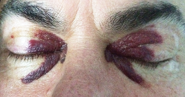
A 51-year-old man presented with a 3-year history of recurrent upper eyelid haematomas. He had a low level IgA lambda paraprotein in blood, Bence–Jones protein and a lambda predominance on serum free light chain ratio. Biopsies of his eyelids showed vascular infiltration by amyloid fibrils (lambda light chains) establishing a diagnosis of light chain (AL) amyloidosis. The patient had cutaneous involvement only at diagnosis. He underwent an autologous stem cell transplant 4 years after the initial presentation, at which time there was evidence of early cardiac involvement by AL amyloidosis manifest by increased B-type natriuretic peptide and confirmed on cardiac magnetic resonance imaging. While thrombocytopenic during his transplant, the patient developed bilateral periorbital haematomas, which resolved with platelet engraftment.

First confirmed case of transfusion-transmitted hepatitis E in Australia

Clinical record

In July 2014, a 6-year-old boy underwent a split liver transplant following liver failure of unknown cause and received 18 blood components peri-operatively. In January 2015, routine monitoring revealed elevated levels of serum liver enzymes (alanine aminotransferase, 289 U/L; reference interval, < 30 U/L). Two biopsies showed possible but inconclusive evidence of rejection, and alanine aminotransferase levels continued to rise, reaching 1170 U/L, despite anti-rejection treatment. Hepatitis E virus (HEV) testing was performed on a third biopsy sample and HEV RNA was detected by reverse transcription polymerase chain reaction. Retrospective testing of the patient’s blood and liver samples showed that he was HEV RNA negative before transplantation, but HEV RNA positive in post-transplant blood from September 2014. After 3 months of ribavirin therapy, the patient’s liver enzyme levels normalised and HEV RNA became undetectable.

The patient had not consumed uncooked pork products and had no history of contact with swine, a known zoonotic HEV source, or overseas travel. HEV RNA was not detected in donor liver samples tested retrospectively. In July 2015, the case was referred to the Australian Red Cross Blood Service (Blood Service) for investigation into possible transmission by transfusion. HEV RNA and IgG testing was performed on archived samples from all 18 blood donations manufactured into the transfused components. HEV RNA was detected in one donation manufactured into a transfused fresh frozen plasma component. The donor of this component reported no symptoms but had travelled to the south of France, a known high HEV prevalence area,1 in the 2 months before donation and had eaten local pork products. The timing of travel was consistent with overseas-acquired infection and donation during the infectious period. HEV IgG was detected in a subsequent donation with RNA clearance, demonstrating seroconversion. This, together with molecular characterisation of patient and blood product HEV (see the Appendix at mja.com.au), strongly supported transmission by transfusion.

Hepatitis E virus (HEV) is a single-stranded RNA virus spread by various routes — including faecal–oral, foodborne, bloodborne, mother to child during pregnancy or birth, and animals to humans — typically causing a self-limiting viral hepatitis after an incubation period of 2–6 weeks.1 Chronic HEV infection has been identified almost exclusively among immunocompromised people and has been found to lead to cirrhosis in liver transplant patients.1 However, early recognition and addition of ribavirin treatment has generally good outcomes with viral clearance.2

Hepatitis E is a disease of emerging importance in developed nations,1 especially in the context of blood donation. HEV is a known transfusion-transmission agent,3 and the prevalence of asymptomatic blood donor viraemia internationally has been found to be considerably higher than expected. England has reported a viraemic prevalence of 1 in 28483 blood donors, and the Netherlands has reported a prevalence of 1 in 762.4 In addition, the potential for adverse outcomes is highest in immunosuppressed recipients who typically receive blood components. Laboratory testing for HEV is not performed on Australian blood donors during the donation process.

We report the first confirmed case of the transmission of HEV by transfusion in Australia; although transmission by transfusion was not definitely excluded in a previously described case.5 The risk that HEV poses to blood safety is specific to the Australian context. Hepatitis E is a rarely notified disease in Australia with about 30–40 notifications to health authorities each year.6 The vast majority of HEV infections notified in Australia are acquired overseas. Asymptomatic infections from most overseas-acquired infections are expected to be covered by existing Blood Service malarial risk travel deferrals, which prevent donation for fresh component manufacture for 4 months.7 However, the assumption that locally acquired infections are rare may be influenced by past external laboratory practice, where testing for HEV infection only occurred in individuals with a history of overseas travel.6 Given that infection with HEV genotype 3 has a high asymptomatic proportion — reportedly as high as 98%1 — the true infection burden in the Australian population remains unknown.

A preliminary Blood Service study found a rate of HEV viraemia of 1 in 14 799 donations,8 which is considerably lower than in other countries such as the United Kingdom that supply an HEV-safe inventory for high risk recipients. Compared internationally, adverse outcomes from transfusion-transmitted HEV in Australia are likely to be a rare event and Australia’s blood supply is at considerably lower risk. In Australia, potential risk management approaches include options such as accepting the risk as tolerable, HEV-specific travel deferrals, universal screening, and targeted screening for high risk recipients. Quarantine and donor deferrals have very limited effectiveness for infections with a high asymptomatic proportion. To guide risk management, the Blood Service has commenced a large Australia-wide HEV RNA prevalence study in blood donors. In the interim, given the uncertain incidence of HEV infection in Australia, we suggest that clinicians remain alert to the possibility of HEV infection, especially in immunosuppressed patients.

Lessons from practice

  • Hepatitis E is a disease of emerging importance for blood safety in developed nations, with the published prevalence of blood donor viraemia reported to be approximately 1 in 750 in the Netherlands, 1 in 3000 in England and 1 in 15 000 in Australia.

  • HEV is a known transfusion-transmissible agent; while the risk in Australia is low compared with other countries, we report the first confirmed Australian case of transmission by transfusion.

  • Chronic infection can occur in immunocompromised individuals and may lead to cirrhosis; however, early recognition and treatment generally results in viral clearance.

  • Clinicians should remain alert to the possibility of HEV infection, particularly in immunocompromised patients.

The slow climb from innovation to cure: treating anaemia with gene editing

The ability to precisely edit DNA via CRISPR technology has emerged as the one of the most powerful advances in biology. A new paper showing repair of a genetic mutation in human blood cells represents an important step towards treating the common, debilitating and expensive-to-treat blood disease known as sickle cell anaemia.

Gene therapy has been a long time coming.

Other breakthroughs – such as vitamins, antibiotics, and vaccines – were translated into medical remedies very quickly. Advances in diagnosis and the combination of different therapies have gradually improved cancer survival rates, and HIV can often be controlled with combination therapy.

But inherited genetic mutations that lead to unrelenting and life-long disease have stumped us. It has proved much harder than expected to put replacement genes into cells. Too often our genome seems to recognise the new DNA as foreign and shuts it down.

But CRISPR technology offers a completely new approach. We can now repair a gene, whereas in the past gene therapy involved adding a replacement gene.

A common, debilitating and expensive disease

Sickle cell anaemia results from a mutation in a gene that encodes haemoglobin – the protein in red blood cells that carries oxygen around our bodies. The mutation not only impairs the function of the protein, but also causes it to aggregate and distort cell shape. This leads to clumps of cells that block blood vessels, with devastating effects. Vital organs are damaged, strokes and episodes of great pain occur, and life span is reduced by around 30 years.

Blood transfusions can help but ultimately excess iron – a key component of haemoglobin – accumulates and tissues are further damaged. Life-long treatments are estimated to cost a million dollars per patient.

Sadly, this disease is common. Mutations in globin genes are the most prevalent of all single gene disorders. In the US there are around 70,000 patients at any given time, and across the world about 200,000 children with sickle cell anaemia are born each year. The total costs in terms of human suffering and the ultimately ineffective spending on health care is colossal.

Targeting the mutation

Researchers led by Jacob Corn at Berkeley – the home of Jennifer Doudna, a CRISPR pioneer – have introduced several innovations to bring CRISPR therapy for Sickle Cell Anaemia closer to the clinic.

As described in their newest paper, their basic strategy was to purify immature blood cells (before they lose their nucleus, a normal part of red blood cell development), correct the mutation using a CRISPR system, and then graft the cells back into a recipient – in this case a laboratory mouse.

The team developed a number of innovations. They first synthesised all the components of the CRISPR machinery in the lab, then assembled them and delivered the parcel into cells using an electric shock process termed “electroporation”. The parcel contained bespoke molecular tools to find, cut and replace the target mutation in the haemoglobin gene.

The authors targeted the gene-editing parcel to blood cells that subsequently divide and give rise to many generations of blood cells. This feature is essential for long-term treatment, since red blood cells quickly wear out as they are being pumped around our bodies.

Using their technique, the team was able to successfully correct the target gene mutation in about 10% of cells. This might not sound much, but should be enough to have very real clinical benefits for patients.

How far off is use in the clinic?

While this work most certainly advances the use of CRISPR for editing human cells, a number of factors limit immediate applicability.

First, this paper concerned the correction of human blood cells that were grafted into laboratory mice. Although the same cells could be re-introduced into human patients, a lot more cells would be required to treat a human, since humans are much bigger than mice.

Also, the choice of “blood progenitor cells” to target is interesting. Blood progenitor cells are cells that have begun to develop down the pathway that forms blood. They aren’t self-renewing in the same way immortal stem cells are, so the supply of corrected blood will eventually run out.

It would have been better to use actual blood stem cells and sustain the cell renewal for longer, but those cells are much rarer and are difficult to recover in large numbers from patients. Also, some researchers have struggled to achieve gene correction in blood stem cells, and they wonder if these cells have all the required repair pathways.

Nevertheless, even corrected progenitor blood cells could be effective over reasonable periods, so this could represent a new treatment if not a cure.

Ethical and political factors

When considering this work it is worth noting that we are talking about correcting blood cells. This is quite different from earlier work by a separate team that used donated, non-viable human embryos for their research.

Previous CRISPR experiments focused on the use of gene editing to change the genome of the entire body, which would be included in the eggs or sperm of any offspring if it came to fruition. Such gene therapy remains highly controversial and is considered unethical by many – partly because it could affect future generations who cannot consent to the treatment.

The work on blood progenitor cells is called “somatic” gene therapy, because only somatic or body cells are altered, and this is widely accepted as appropriate.

Beyond the important ethical considerations, will expensive treatments like this actually enter the clinic?

In America, yes they may. With strong investment in medical companies and health care insurance processes it could be cost effective in the USA.

In other developing countries it could be much harder. Consequently, a great many laboratories – including my own – continue to work on understanding the fundamental biology of the haemoglobin gene. The goal is to find affordable drugs that could treat the disease and could be made available throughout the world.The Conversation

Merlin Crossley, Deputy Vice-Chancellor Education and Professor of Molecular Biology, UNSW Australia

This article was originally published on The Conversation. Read the original article.

Latest news

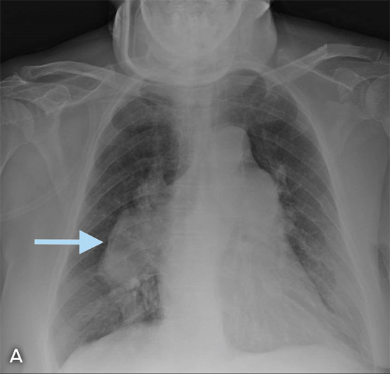
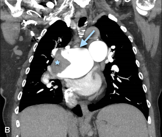
Fleischner sign with pulmonary hypertension complicated by thrombosis

A 73-year-old woman with chronic obstructive pulmonary disease presented with 4 weeks of exertional dyspnoea. A plain chest x-ray revealed a right hilar enlargement with enlarged pulmonary outflow tract (Fleischner sign; Figure, A [arrow]) and mild cardiomegaly. A computed tomography pulmonary angiogram confirmed the presence of marked dilatation of the pulmonary trunk (Figure, B [arrow]) and arteries with a large mural thrombus (Figure, B [star]). An echocardiogram indicated pulmonary hypertension with an elevated pulmonary artery pressure of 88 mmHg.

Figure A –

Figure B –

Stroke in a young man with untreated HIV infection and neurosyphilis

Clinical record

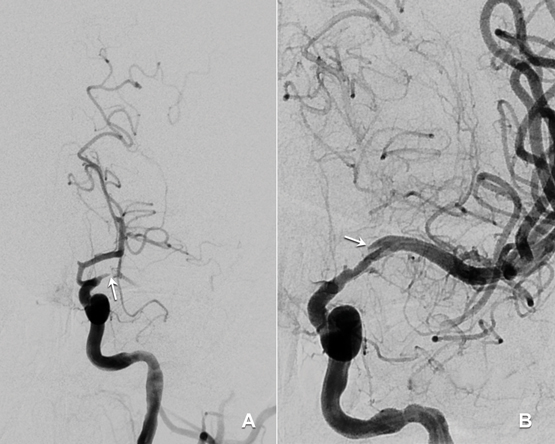
A 33-year-old man presented to an emergency department with acute dysphasia and a dense right hemiparesis. His National Institute Health Stroke Scale score was 12, indicating a moderate severity stroke (score range 0–42, with increasing values indicating increasing severity). His computed tomography (CT) brain scan was normal. A CT angiogram showed a filling defect in the left intracranial internal carotid artery. Intravenous thrombolysis was commenced 2.5 hours after stroke onset and completed during urgent transit to our hospital for endovascular thrombectomy. Combined stent retrieval and suction thrombectomy of the left internal carotid occlusion restored flow 4.5 hours after stroke onset. A small dissection in the left intracranial internal carotid artery was the source of the thrombotic occlusion (Figure). A magnetic resonance imaging scan of the brain showed small scattered infarctions in the left middle cerebral arterial territory.

The patient was later found to have a human immunodeficiency virus (HIV) infection that had been diagnosed 5 years earlier but for which he had not sought or received treatment. There was no history of screening for syphilis. He had a remote and brief history of recreational use of methamphetamines and cocaine (more than 12 months previously). He had no other vascular risk factors (non-smoker, normal fasting lipid and blood glucose levels, negative autoimmune serology). His CD4 cell count was 220 × 106 cells/L (reference interval [RI], ≥ 360 × 106 cells/L) and serum quantitative HIV RNA testing revealed 77 400 copies/mL. Hepatitis serology results were negative. Syphilis serology results were positive: reactive rapid plasmin reagin (RPR) with a titre of 1:256; reactive Treponema pallidum particle agglutination (TPPA) assay; and positive syphilis antibody enzyme immunoassay. His cerebrospinal fluid (CSF) protein level was 1.17 g (RI, < 0.45 g/L) and his white cell count was elevated at 62 μ/L (RI, < 5 μ/L), predominantly due to monocytosis (84%). CSF syphilis serology was positive, with reactive results from the venereal disease research laboratory, TPPA and fluorescent treponemal absorption antibody tests, confirming neurosyphilis. There were no other clinical or radiological features of tertiary syphilis. CSF polymerase chain reaction test results were negative for other pathogens including varicella-zoster virus, John Cunningham virus and tuberculosis. Cryptococcal antigen test results were negative. Other stroke investigations, including transoesophageal echocardiogram, returned negative results.

A 15-day course of intravenous benzylpenicillin (1.8 g, 4-hourly) with prednisone cover (three doses of 20 mg twice daily to prevent Jarisch–Herxheimer reaction) was completed as treatment for neurosyphilis. He received counselling and was commenced on antiretroviral therapy including abacavir–dolutegravir–lamivudine. Contact tracing was performed. The 3-month outcome was excellent, with only minor persistent dysphasic speech errors and a modified Rankin scale score of 1 (range 0–6, with increasing values indicating worse deficit and 6 for death). Progress serum RPR titres were significantly reduced (1:64), indicating a serological treatment response. A recent progress CD4 cell count was 630 × 106/L and quantitative HIV RNA testing revealed < 20 copies/mL.

Studies indicate that HIV infection increases the risk of ischaemic stroke, particularly in young patients (≤ 45 years) with low CD4 cell counts (< 350 × 106 cells/L).1,2 It is important for clinicians to recognise the various mechanisms by which HIV infection predisposes to stroke. These include a direct HIV-induced vasculopathy, and an indirect opportunistic co-infection-related arteritis with organisms such as tuberculosis, syphilis and varicella-zoster virus.2 HIV vasculopathy has been reported in the extracranial and intracranial cerebral circulations and may cause aneurysmal fusiform or saccular dilatation of vessels or a non-aneurysmal vasculopathy manifest by stenosis, occlusion or vasculitis.24 Additional factors contributing to stroke risk in HIV include a more frequent smoking history, coagulopathy, increased homocysteine levels and metabolic syndromes associated with antiretroviral therapies, which may result in accelerated atherosclerosis.1,3 Descriptions of intracranial arterial dissection in patients with HIV infection are limited to rare case reports.5 We postulate that HIV and syphilis co-infection in our patient may have caused a vasculopathy-associated intracranial arterial dissection.

The role and safety of intravenous thrombolysis in patients with HIV infection is not established.2,4 Thrombolysis could be theoretically harmful in patients with HIV vasculopathy or co-infection-related arteritis owing to a potential increased bleeding risk from abnormal vessel wall integrity.4 Despite this, intravenous thrombolysis has been used successfully in patients with untreated HIV infection to treat myocardial infarction and, in our patient, to treat acute ischaemic stroke without adverse outcomes.2,4 Clinicians should be aware that endovascular thrombectomy of proximal anterior cerebral circulation clots after intravenous thrombolysis is now evidence-based treatment for acute ischaemic stroke.6 Our case illustrates the “drip, ship and retrieve” concept of acute stroke management; with intravenous thrombolysis (“drip”) commenced at an initial hospital and completed while the patient was transferred (“shipped”) to another hospital for endovascular thrombectomy (clot “retrieval”). At present, only a limited number of stroke centres provide an endovascular thrombectomy service. Reorganisation of existing systems is required to allow rapid access to endovascular thrombectomy for all appropriate patients in Australia.6

This case presents an important reminder that HIV infection is a risk factor for stroke and that HIV testing should be performed in all young stroke patients. A lumbar puncture is recommended for diagnosis or exclusion of co-existing infections including tuberculosis, syphilis and varicella-zoster, which are all associated with vasculopathy in patients with HIV infection.

Lessons from practice

  • HIV infection is an important risk factor for stroke and HIV testing should be performed in all young stroke patients.

  • Patients with HIV infection and stroke should have a lumbar puncture to examine for co-existing opportunistic infections.

  • A diagnosis of neurosyphilis requires a cerebrospinal fluid (CSF) cell count and protein measurement and serological testing on serum and CSF.

  • There is evidence that the “drip, ship and retrieve” management approach to managing acute ischaemic stroke is effective. However, in patients with known HIV infection, acute stroke should be managed on a case-by-case basis.

Figure


Digital subtraction angiography: A: Pre-clot retrieval showing left internal carotid occlusion (arrow). B: Post-clot retrieval showing dissection (arrow) and restoration of flow.

[Perspectives] Julie Makani: at the cutting edge of sickle-cell disease

A passion for science is not unusual among medical researchers; rather less common is the capacity to step back from the seductive pleasures of discovery and take a more strategic view of what you’re about. Julie Makani has spent a decade and half in the Department of Haematology and Blood Transfusion at Tanzania’s Muhimbili University of Health and Allied Sciences in Dar es Salaam. During this time she has shown how choosing the right strategy can foster the kind of invigorating purpose that now underpins her department’s programme of work on sickle-cell disease.

Antenatal haemoglobinopathy screening in Australia

Haemoglobinopathies are inherited conditions caused by defects in globin chain synthesis. Worldwide, haemoglobinopathies are the most common genetic defect in humans — about 7% of the world’s population are carriers.1 In Australia, haemoglobinopathies are becoming more prevalent because of recent immigration of people from countries where these disorders are endemic.

Haemoglobinopathy disorders include α– and β-thalassaemia, sickle-cell disease and globin chain variants. Screening is complex and has limitations due to lack of evidence; ambiguities in the results of screening tests; health practitioners assessing patients’ risks with incomplete education in this evolving area; poor correlation between the genotype and phenotype in affected people leading to difficulty in predicting outcomes in an unborn child; definitive testing being expensive and not covered by Medicare; and generally lower health literacy in the target population.

Screening is predominantly initiated by general practitioners, obstetricians and midwives. Subsequently, haematologists, geneticists and laboratory scientists may become involved. In Australia, there is no national screening program and wide variation in testing practices. Many pregnant women are not being screened in a timely manner.2

In this article, we aim to provide a brief overview of the pathophysiology of haemoglobinopathies, a rationale for screening, and a description of the steps involved in screening and managing abnormal results. The term “haemoglobinopathy” is used broadly, rather than being restricted to variant haemoglobins.

Geographical distribution of haemoglobinopathies

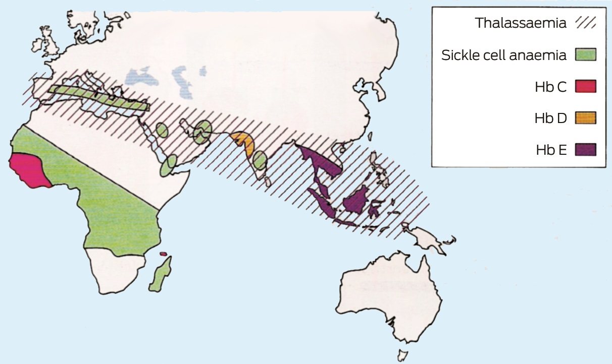
The incidence of α– and β-thalassaemia is high in Mediterranean countries, the Middle East, on the Indian subcontinent, in South-East Asia, and in parts of Africa. Severe forms of α-thalassaemia (αo) are more prevalent in South-East Asia. Sickle-cell anaemia has the highest incidence in tropical Africa. Haemoglobin E (HbE) is prevalent in South-East Asia and on the Indian subcontinent.1 Box 1 shows the geographical distribution of haemoglobinopathies.3 Migration from these regions to Australia is increasing.

Pathophysiology and clinical impact of haemoglobinopathies

Haemoglobin carries the oxygen in red blood cells. Structurally, it is a four globin chain tetramer (HbA, two α-globin chains and two β-globin chains). There are four α– and two β-globin genes. In adults, the major form of haemoglobin is HbA (α2β2) (about 95%); HbF (α2γ2) and HbA2 (α2δ2) are minor components (< 1% and 2–3% respectively). At birth, the major form of haemoglobin is HbF, which comprises 60–80% of total haemoglobin. At one year of age, adult levels of HbF and HbA are obtained.4 This makes the β-thalassaemia trait difficult to detect during infancy.

Decreased synthesis of globin chains results in thalassaemia (a quantitative defect). In α-thalassemia, reduced synthesis of α-globin chains leads to an excess of β-globin chains, and the situation is reversed in β-thalassaemia. The imbalance of globin chains causes haemolysis and impairs erythropoiesis. Conversely, structural changes in the globin chains are known as variant haemoglobins (qualitative defects). Box 2 summarises the clinical phenotypes and genotypes, and highlights the haemoglobinopathies that have significant clinical implications in the fetus, identifying those with transfusion dependency, shortened life expectancy or sickle-cell disease. The haemoglobinopathy trait is often asymptomatic and undetected, being clinically significant only if a woman is pregnant and her partner is also a carrier. Thalassaemia major and sickle-cell disease are managed in specialist centres.

The form of α-thalassaemia known as haemoglobin H disease produces a variety of symptoms ranging from none to transfusion dependency. Haemoglobin Barts leads to a non-viable fetus that dies in utero and significant maternal complications.5 Termination of pregnancy may be recommended, but there have been cases of intrauterine blood transfusions in Barts disease leading to a thalassaemia major phenotype after birth.6

β-Thalassaemia major renders an individual transfusion-dependent, which subsequently requires removal of excess iron (chelation). Without transfusions these patients die as children. Without iron chelation, death occurs in the second decade.7 Inadequate chelation leads to the complications of cardiomyopathy, cirrhosis, diabetes, hypothalamic hypogonadism, hypothyroidism and osteoporosis.8

The effects of sickle-cell disease are highly variable. The abnormal haemoglobin leads to “sickling” of the red blood cells, which causes occlusion of small blood vessels, leading to painful crises, acute chest syndrome (acute dyspnoea and hypoxia), splenic or bone infarcts and cerebrovascular thrombosis. Other problems include haemolytic crises and overwhelming pneumococcal infection from functional asplenia. This disease causes significant morbidity and the highest mortality rate is among 1–3-year-old children.7

Why and when should we screen?

Opportunities for screening include before conception, and the antenatal and neonatal periods.

Ideally, women should be screened before they conceive to allow those at risk to make informed decisions. Pre-conception screening also allows alternatives to termination, such as pre-implantation genetic diagnosis, gamete donation and adoption.9 Several countries with high carrier frequencies, such as Iran, Saudi Arabia, the Palestinian territories and Cyprus, have implemented mandatory pre-marital screening.6

Antenatal haemoglobinopathy screening aims to detect pregnancies at risk of carrying affected offspring, and should be performed early in pregnancy. In Australia, there is no national screening program and practice varies within states and local area health districts. In the United Kingdom, the National Health Service (NHS) Sickle-Cell and Thalassaemia Screening Programme aims to offer antenatal screening by 10 weeks’ gestation to enable prenatal diagnostic testing to be completed by 12 weeks’ gestation.10 Screening at the time that pregnancy is confirmed may require additional resources, but increases the number of pregnancies screened before 10 weeks’ gestation.11

A retrospective analysis of data of 462 patients of a large Australian antenatal service found that screening was performed in women at a median gestation of 15 weeks.2 A British study in a high prevalence area similarly found that 74% of women visited their GP early (at a mean gestation of 7.6 weeks); however, screening was delayed until a median gestation of 15.3 weeks, with only 4.4% being screened by 10 weeks.12 Evidence suggests that health professionals, rather than pregnant women, are responsible for the delay in screening. The barriers identified include the lack of time within the 10-minute consultation, delays in arranging blood tests, language problems and a lack of awareness among health professionals.13

Should any abnormality be identified, partner screening, genetic counselling and prenatal testing can be offered. Women in the earlier stages of pregnancy are more likely to accept prenatal diagnosis.14 By offering it sufficiently early in pregnancy, women can make informed, less time-pressured decisions, which might include termination of pregnancy. Termination can be offered for affected pregnancies up to 20 weeks’ gestation.7,15

In the UK, neonatal haemoglobinopathy screening occurs within the NHS Newborn Blood Spot Screening Programme16,17 aiming to identify infants with sickle-cell disease and β-thalassaemia major to improve outcomes through early treatment and care. New diagnoses lead to prenatal counselling before future pregnancies. In Australia, haemoglobinopathy screening is generally not part of the routine newborn screening program because of the lower prevalence of these disorders in this country. Effective maternal screening should abrogate the need for newborn screening.

The antenatal haemoglobinopathy screening process

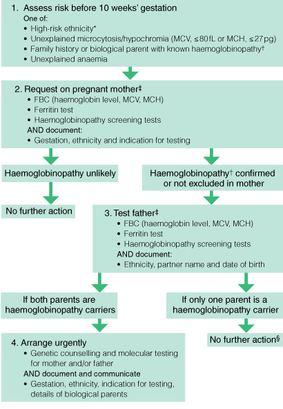
Box 3 shows a proposed algorithm for antenatal haemoglobinopathy screening.2 The diagnostic sequence for haemoglobinopathy is a multistep process. Local general practice guidelines state that all women should be screened with a full blood count and ferritin test during their first antenatal visit or during the pre-conception phase.18 Haemoglobinopathy screening should be performed on women:

  • with a mean corpuscular volume (MCV) of less than 80 fL, a mean corpuscular haemoglobin (MCH) mass of less than 27 pg, and a normal ferritin level (> 30 μg/L); or

  • with high risk features, such as belonging to a high risk ethnic group, having a family history of haemoglobinopathy, being the partner of a haemoglobinopathy carrier, or consanguinity with the partner.19

Screening tests for haemoglobinopathy are subsequently performed by various complementary methods. The use of molecular studies is determined by the maternal and paternal risk; however, there is significant variation in access and testing between health regions.

Full blood count and ferritin test

The haemoglobin level and MCH and MCV values can be used as a guide, but are not the sole indicators of the need for further screening. Most people with the α-thalassaemia trait (two gene deletions) have an MCH of less than 26 pg.19 Rarely, individuals may have the thalassaemia trait and a normal MCH, which could be due to a silent β-thalassaemia mutation, single α-gene deletion, or co-inheritance of both α– and β-thalassaemias.19 Structural variants, such as haemoglobin S (HbS; sickle-cell trait or disease) can be missed if a diagnosis is based on full blood count indices alone because people with HbS trait can have normal MCV and MCH values.

Although a low MCV is seen as a driver for further testing, MCH is more reliable because the MCV can increase artefactually because of red cell swelling if sample processing is delayed. Further, the MCV reference range differs between automated cell counters20 and it rises during pregnancy and in people with liver disease, and those with vitamin B12 or folate deficiency.

Ferritin testing helps with the interpretation of red cell indices, but iron deficiency cannot be used to exclude further screening, as thalassaemia can be found in iron-deficient women.19

Haemoglobinopathy screening tests

These include haemoglobin electrophoresis (HbEPG), high performance liquid chromatography (HPLC), and capillary electrophoresis (CE). They are described in Box 4.

In Australia, each laboratory performs at least two screening tests (most commonly HbEPG and HPLC) to identify and quantify HbA2, HbA, HbF and variant haemoglobins. HPLC and CE are the more accurate quantitative techniques.21

An elevated HbA2 level of 3.5–7.0% is diagnostic of β-thalassaemia trait.22 This is because reduced β-globin chain synthesis promotes the combining of excess α-globin chains with δ-globin chains. Rarely, silent β-thalassaemia can be missed because affected people have a normal or borderline HbA2 level.9 Iron deficiency has been thought to falsely lower the HbA2 level and obscure the diagnosis of β-thalassaemia. Data on this are conflicting, with one study concluding that there is a small but significant decrease in HbA2 level in iron-deficient patients, with no impact on making the diagnosis of the β-thalassaemia trait.20,23 Ideally, screening should be performed when patients are iron-replete. However, if time does not permit, screening tests can be performed and their results interpreted with caution. Further testing, either with genetic studies or partner screening, may be indicated in this setting.

Partner screening is offered to identified carriers or equivocal cases. Cost-effectiveness studies support screening of fathers sequentially rather than upfront testing,24 but this can lead to a time delay that may be critical in the antenatal setting.

Some laboratories offer combined couple risk assessment when results are available for both the mother and father, and if their relationship is clearly identified. This is clinically valuable to clinicians, but is time-consuming and assumes paternity.

Genetic diagnosis

A couple’s combined risk determines whether DNA testing for haemoglobinopathies is required. It is performed when α-thalassaemia cannot be excluded in both individuals, when there is uncertainty about the partner of a known β-thalassaemia carrier, and when both parents are affected and prenatal genetic diagnosis of the fetus is being pursued. The risk will change if there has been a change of partners between pregnancies. Genetic diagnosis is coupled with genetic counselling to discuss abnormal results.

Access to genetic testing for haemoglobinopathies and the costs and types of testing available vary. Genetic tests include multiplex ligation-dependent probe amplification, gap polymerase chain reaction and β-gene sequencing. Genetic testing does not have a Medicare Benefits Schedule item number, and the cost is borne by the patient, an institution, research grants, or the state government (Victoria only). It could be argued that α-thalassaemia gene testing should be publicly funded because thalassaemia carriers can have a low MCV and a normal HPLC result and ferritin level. Turnaround times are high (2–6 weeks), which reduces the clinical utility of antenatal testing if termination is being considered. There is no national registry for the results of genetic tests for haemoglobinopathies. In Victoria, only one laboratory performs the testing, which avoids duplicate testing in subsequent pregnancies, assuming identifiers are accurately correlated.

Prenatal diagnosis can be offered if there is a risk of the child being affected and if the parents’ genotypes are known or linkage analysis is possible. Fetal DNA obtained by chorionic villous sampling (CVS) at 11–13 weeks’ gestation or by amniocentesis from 15 weeks’ gestation onwards is tested. The risk of miscarriage is 0.5–1.0% for amniocentesis and 1–2% for CVS. In the future, it may be possible to perform prenatal diagnosis using free fetal DNA found in maternal plasma.25

Conclusion

Haemoglobinopathies are one of the most common genetic defects worldwide. In Australia, the number of carriers is increasing due to migration from countries with high carrier frequencies. Screening aims to reduce the burden of these disorders by identifying those at risk and managing their pregnancy choices, but there is currently no nationally coordinated screening program in this country.

To optimise clinical outcomes, screening should be performed before conception or early in pregnancy, and this requires awareness among patients and their doctors and carers, and short turnaround times for laboratories performing the tests.

Additionally, molecular testing should be more widely available. A national registry to record results is much needed to reduce duplicate testing and provide access to results. We envisage a future with cost-effective, accurate and accessible diagnostic testing. In the interim, prompt screening must be offered to at-risk pregnant women.

Box 1 –
Geographical distribution of haemoglobinopathies


Source: Hoffbrand AV, Moss PAH, Pettit JE. Essential haematology. 5th ed. Oxford:Wiley-Blackwell.3 Reproduced with permission.

Box 2 –
Haemoglobinopathies with significant clinical implications for the fetus

A. Summary of clinical phenotypes and genotypes of α– and β-thalassaemia

Disorder

Genotype

Anaemia

Clinical characteristics


α-Thalassaemia*

Silent

α/αα

Absent

Nil; normal MCV

Minor (trait)

αα/− − or −α/−α

Mild

Asymptomatic; normal or reduced MCV

Haemoglobin H disease

α−/− −

Moderate

Variable; asymptomatic, microcytic anaemia, hepatosplenomegaly, may require transfusions

Major (haemoglobin Barts)

− −/− −

Severe

Usually death in utero, hydrops fetalis syndrome

β-Thalassaemia

Minor (trait)

β/β0, β/β+

Mild

Asymptomatic; normal or reduced MCV

Intermedia

β+/β+, β0/β+

Moderate

Wide clinical variability; features range between those of thalassaemia trait and thalassaemia major

Major

β0/β0

Severe

Transfusion-dependent to prevent growth retardation, hepatosplenomegaly, haemolytic anaemia; iron overload from chronic transfusions, which reduced life expectancy before the use of iron chelation therapy


MCV = mean corpuscular volume. * Genotype nomenclature reflects the number of absent genes: α−/αα indicates one absent gene; α−/α− and αα/− − indicate two absent genes; α−/− − indicates three absent genes; and − −/− − indicates all genes absent. † α-Thalassaemia major is commonly referred to as haemoglobin Barts, although other mutation combinations can produce a similar phenotype. ‡ In genotype, β0 indicates absent β-globin chains and β+ indicates reduced synthesis of β-globin chains.

B. Summary of clinical phenotypes and genotypes of common structural variants of thalassaemia

Disorder

Genotype

Anaemia

Clinical characteristics


Haemoglobin S (HbS)

Sickle-cell trait

AS

Absent to mild

Mostly asymptomatic; normal MCV

Sickle-cell disease

SS, SC, S/βthal, SD, S/O-Arab

Mild to moderate

Vaso-occlusive phenomena and haemolysis affecting brain, chest, kidneys, bones and spleen

Haemoglobin E (HbE)

Trait

AE

Normal

May have mild microcytosis; no anaemia

Homozygous E

EE

Mild

Mild anaemia and microcytosis

Compound heterozygous

E/βthal

Moderate to severe

Variable phenotype, thalassaemia intermedia or thalassaemia major

Haemoglobin Constant Spring (HbCS)*

Heterozygous

αα/αCSα

Mild

Asymptomatic

Homozygous

αCSα/αCSα

Moderate

Splenomegaly, moderate haemolytic anaemia

Compound heterozygous (in conjunction with α0 thalassaemia)

− −/αCSα

Moderate to severe

Wide clinical variability; often more severe than deletional haemoglobin H disease, with possible transfusion dependence


MCV = mean corpuscular volume. * First isolated in the Constant Spring district of Jamaica and characterised by a variant of the α-globin chain (designated αCS), which is abnormally long.

Box 3 –
A selective screening algorithm for antenatal haemoglobinopathy (adapted from Lavee et al2)


FBC = full blood count; MCH = mean corpuscular haemoglobin; MCV = mean corpuscular volume.

* Parent from southern Europe, Middle East, Africa, South-East Asia, Indian subcontinent or Pacific Islands.

β-Thalassaemia, β+-thalassaemia, δβ-thalassaemia, haemoglobin Lepore, haemoglobin E, αo-thalassaemia, haemoglobin H, haemoglobin S, haemoglobin C, haemoglobin D-Punjab, haemoglobin O-Arab.

‡ Steps 2 and 3 are interchangeable, depending on which parent has previously been tested.

§ No further action is needed during pregnancy, but the child may be a carrier and should be followed up later in life.

Box 4 –
Specialised laboratory methods for haemoglobinopathy screening

Test

Description


Haemoglobin electrophoresis (HbEPG)

  • Separation of haemoglobins with electrophoresis at pH 8.4 (alkaline) and pH 6.2 (acid)
  • Identifies variant haemoglobins, but is not useful for quantitative purposes
  • Does not detect α-thalassaemia trait
  • Cheap, laborious, operator-dependent

High performance liquid chromatography (HPLC)

  • Separates haemoglobins based on adsorption and ion exchange when a blood sample is in contact with a mobile liquid phase and a solid stationary phase
  • Identifies and quantifies HbS, HbA2, HbF and variant haemoglobins
  • A raised HbA2 level (3.5–7.0%) indicates the presence of β-thalassaemia trait
  • The HbA2 level may not be accurately quantified in the presence of HbS
  • Does not separate HbA2 from HbE
  • Does not detect α-thalassaemia trait
  • Automated, quick, small sample volume required (5 μL)

Capillary electrophoresis (CE)

  • Haemoglobin variants are separated by electro-osmotic flow with negatively charged silica capillaries and high voltage
  • Can be used as a complementary method to HPLC
  • Quantitative method
  • Separates HbE from HbA2
  • Does not detect α-thalassaemia trait

HbH inclusions

  • Red cells are stained to look for HbH inclusions (tetramers of β-globin chains)
  • Diagnostic for α-thalassaemia trait, HbH disease, α-thalassaemia and mental retardation (ATRX syndrome) or acquired HbH disease
  • HbH bodies are much easier to find in αo heterozygosity
  • α-thalassaemia trait is not excluded with a negative test result
  • Operator-dependent

Sickle solubility test

  • Sodium dithionite/metabisulfite reduces the amount of oxygen present in the blood sample, causing abnormal red cells to sickle
  • Gives a positive result in HbS trait and HbS disease
  • Not routinely performed; has been replaced by HPLC

ATRX = α-thalassemia mental retardation; Hb = haemoglobin.