×

Newborn bloodspot screening: setting the Australian national policy agenda

To the Editor: Maxwell and O’Leary’s article1 and Wiltshire and Cameron’s letter2 provide insights into the current issues facing newborn bloodspot screening (NBS) in Australia. It is clear that we have world-class NBS programs, but there is a lack of national policy guidance agreed on by governments. It has been argued that this has affected the programs’ capacity to respond to the changing environment in which they operate.1,3,4 There is a need for clear national policies to support the programs’ continued success and growth, and a way to assess the benefits and harms of screening additional conditions through NBS.5 Since the aforementioned articles were published, there has been substantial progress towards achieving these goals.

Australian governments have recently agreed to develop a national policy framework for NBS. This will include a decision-making pathway, against which congenital adrenal hyperplasia and other conditions can be assessed for inclusion in NBS. The policy framework is being developed under the auspices of the Standing Committee on Screening, through a multidisciplinary working group. The project is due for completion by early 2016, and will be informed by broad consultation, due to take place in 2015. Further information can be found at http://www.genomics.health.wa.gov.au.

The current process is a genuine opportunity for policymakers, NBS programs, clinicians, consumers and others to come together to put in place a framework that builds on the successes of NBS, safeguards the programs into the future, and enables transparent and consistent decision making.

Patients lose out in blue over genes

The Federal Court has upheld a private company’s claim to own the patent for the mutation of a human gene, fuelling fears life-saving diagnostic tests will be put out of the financial reach of many patients.

In a decision that stunned health campaigners, the full bench of the Federal Court early this month unanimously dismissed an attempt by lawyers acting for cancer survivor Yvonne D’Arcy to strip US company Myriad Genetics of its patent on a mutation of the BRCA1 gene, which forms the basis of common test for breast cancer.

Announcing its ruling, the full bench backed the original judgement of Justice John Nicholas that the process of isolating a gene from the body was an invention rather than a naturally occurring thing, and so could be patented.

The decision stands at odds with a ruling of the US Supreme Court last year that invalidated the Myriad patents, and has reignited concerns the patients could face much higher test costs.

“Australian women were only protected from an attempted commercial monopoly over the BRCA1 and BRCA2 tests in 2008 because the company that threatened to take those tests away withdrew its patent claims voluntarily,” said Cancer Council Australia Director of Advocacy Paul Grogan. “There was nothing in the law to protect consumers from the monopolisation of those diagnostic tests, and there still isn’t.”

The law firm acting for Ms D’Arcy, Maurice Blackburn, has flagged a possible appeal to the High Court, but legal experts said the decision appeared correct under the law, meaning any change would have to be through legislation.

Mr Grogan said the Federal Court decision meant there was now a “strong case” to change the law.

“The patents system should reward innovation and help deliver affordable health care, not stymie research and increase costs by allowing commercial entities to control the use of human genetic material,” he said.

Adrian Rollins

A genetic discovery that could be a bit easier to swallow

A mysterious disease that causes nerves in the oesophagus to disappear, preventing food from entering the stomach, has been found to be autoimmune in origin.

Researchers have long been confounded about what causes the rare complaint, which afflicts around one in every 100,000 people.

The condition, known as achalasia, causes nerve cells in the oesophagus – including those that control the opening and closing of the sphincter at the opening to the stomach – to gradually disappear. Eventually, the sphincter locks shut, causing food to accumulate in the oesophagus.

Treatments have typically involved either forcing the sphincter open using a balloon inserted endoscopically, or surgically cutting it, but understanding the cause has for years remained elusive.

But an investigation by researchers at KU Leuven in Belgium and Germany’s Bonn University, published in the journal Nature Genetics, has provided strong support for long-held suspicions the condition is autoimmune in origin.

Using DNA samples from 1506 achalasia patients and 5832 healthy volunteers, the researchers identified 196,524 tiny differences – single nucleotide polymorphisms (SNPs) – in the immune-related DNA of those with the condition, and compared them with those of the healthy subjects.

As a result, they identified 33 SNPs associated with achalasia, and all were located in the region of the human genome known to be associated with other autoimmune disorders such as multiple sclerosis and type 1 diabetes.

This evidence has led the researchers to conclude the condition is an autoimmune disease.

In the course of their investigations, the scientists pinpointed a string of amino acids inserted in the DNA of those with achalasia, opening a promising avenue for the development of a treatment and possible cure.

Adrian Rollins


The impact of genomics on the future of medicine and health

Precision genomic medicine will have a transformative impact on personal health and wellbeing, health economics and national productivity

In recent years, there has been an extraordinary leap in knowledge of the human genome and its role in health and disease. A decade ago, researchers were tentatively exploring the first reference human genome sequences, which cost over $1 billion to produce.1,2 Now, thousands of genomes from a cross-section of ethnic backgrounds have been sequenced. This explosion of activity has been enabled by unprecedented advances in sequencing technologies that can now sequence a person’s entire genome — more than 6000 million bases — in days, at a cost of US$1000,3 with costs expected to fall further in coming years.

Making sense of genomic data requires computational technologies and databases to evolve in parallel with sequencing technologies. Advances in both technologies enable an ever-increasing capacity for accurate diagnosis of existing disease, and development of effective and targeted treatment strategies. They also offer opportunities to assess predisposition to disease, potentially prompting more focused clinical monitoring and lifestyle changes.

Although our knowledge of the human genome is currently far from complete, accumulating examples demonstrate that even our limited genomic understanding can be powerful in the clinic. Currently, genome sequencing is having the greatest impact in stratifying cancer, characterising genetic disease, and providing information about an individual’s likely response to treatment.

Cancer: stratifying tumours for treatment

Genomic medicine has already shown benefit in refining diagnoses and guiding therapeutic approaches for cancer.4 Since the late 1990s, the clinician’s cancer “toolkit” of surgery, radiation and chemotherapy has been increasingly supplemented by therapies that target specific molecular pathways in cancer growth and development.5 Genomic information can now assist clinicians in deciding treatment strategies by classifying a tumour according to its mutations and corresponding drug sensitivities. In some cases, patients have been spared costly and complex procedures, such as bone marrow transplants, based on a molecular diagnosis.6 In other examples, a patient’s cancer development has been stabilised — for a time at least — by targeting specific molecules or pathways in the tumour cells.7 More controversially, clinicians are starting to use genomic information to refine a cancer diagnosis and prognosis and alter an individual’s quality of life, even when therapies are not currently available.8

Genome-wide sequencing is now also being applied to the analysis of circulating DNA in the plasma of cancer patients, as well as in individuals with other diseases.9 This technology enables non-invasive tumour detection and monitoring responses to therapy that promise to significantly improve patient management.

Drug prescription and development

Cancer treatment is also set to benefit from genomic information to predict how an individual will respond to drugs (known as pharmacogenomics) and inform prescription of the appropriate drug or dosage. Pharmacogenomic applications extend into many areas of clinical practice; for example, in the prescribing of drugs such as antidepressants, analgesics and anticoagulants.10

In the longer term, genomic information is expected to dramatically change the testing and use of pharmaceuticals through disease stratification.11 New research into molecular pathways underlying health and disease will continue to inform rational drug development and design. In parallel, researchers are using genomic data to suggest new therapeutic applications for existing drugs (repositioning) — with significant cost savings — and to better select individuals for clinical trials to find uses for drugs that failed earlier trials involving broader cohorts (rescue and repurposing).12 Better targeting of existing drug use avoids wasteful and risky therapy and can provide savings for the health care system and the economy at large.

Diagnosing and characterising genetic disease

With the exception of a few pharmacogenomic tests and cancer treatments, clinical genetic testing in Australia is at present limited to diagnosing hereditary and rare monogenic diseases. Every baby born in Australia is offered screening for about 30 genetic conditions in the Guthrie test, and more than 300 tests for genetic disorders are available through the health care system.13 However, clinicians worldwide are currently embracing far more comprehensive genome sequencing to search for variants implicated in undiagnosed genetic diseases14,15 and are using this information to guide treatment.16 As the field matures and our understanding of the affected pathways improves, there will be many more genomic loci implicated in rare disease, with improved treatment prospects. Further, genome sequencing is increasingly being used to assess genetic contributions to complex diseases, where multiple gene variants may be involved in disease development and progression.

Rather than sequencing the entire genome, rapid sequencing of the protein-coding portion (the exome, about 1.5% of the total) can offer comparatively cost-effective analysis of many genetic disorders. Although less effective in detecting chromosomal duplications, deletions and rearrangements, exome sequencing identifies most clinically relevant nucleotide variants in a single analysis, serving both as a diagnostic tool and a method to discover new genes and mutations that underlie these classes of disease. However, as the function of intergenic and intronic regions in disease-regulatory processes is further elaborated and variants in these regions are shown to have diagnostic utility, whole genome sequencing is likely to provide a clinically more robust, accurate and useful test that will replace exome sequencing as sequencing costs decrease.

From disease diagnosis to personalised genetic health

With genome sequencing becoming more accessible, it is becoming more common for individuals to seek personal genomic information through direct-to-consumer genetic testing services.17 However, these tests are not accredited for clinical diagnosis, and individuals will gain most benefit when their genomic sequence becomes integrated into their health care such that clinicians and other health professionals can provide considered advice and the portal to treatment options.

Inevitably, personal genome sequences, likely obtained at birth, will become an integral part of a patient’s electronic health record (EHR), where this information will be integrated with other clinical and environmental data and interrogated throughout the individual’s lifetime. Clinicians and patients may then query the sequence to accurately prescribe treatments, determine disease susceptibilities and identify drug sensitivities, and to determine a course of action to monitor, manage, ameliorate risk of or prevent the disease.

The idea of sequencing newborns remains controversial, because of questions about consent, potential stigmatisation and the value of the information to the individual.18 There are also practical questions about data storage and access, and the question of which results are returned when and to whom. Valuable insights are expected from projects recently funded in the United States exploring the use of genomic sequencing in paediatric medicine and the ethical issues that arise.19

Integrating genomic and clinical information

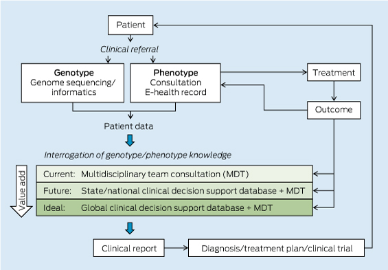
The pathway from patient DNA sequencing to a clinical treatment plan relies on the integration of the individual’s genomic information with knowledge databases that contain known genotype–phenotype correlations and genomic and clinical associations from large populations of individuals (Box 1).

Clinical decision making currently relies on the knowledge of individual practitioners and genetic counsellors. These practitioners work as a multidisciplinary team to assess the genomic variants identified through genomic sequencing and arrive at a treatment plan. Purpose-built, well curated and continuously updated evidence-based databases of human genotype–phenotype associations are urgently needed, with computational tools to interrogate the ever-increasing information in an automated way.

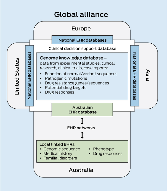
The inherent computational challenges in integrating genomic and clinical data necessitate a significant investment in bioinformatics capability. Given the considerable overheads of both storage and computational power, Australia will most effectively be served by a centralised genome knowledge repository linked to global repositories. Infrastructure support will also be critical for the development of e-systems and software interfaces. These must enable clinicians and health care providers to interrogate a patient’s genomic sequence against the clinical decision support databases to obtain an informative clinical report (Box 2).

Storing and sharing population data

The speed with which personalised genomic medicine becomes a reality for most of the population will be strongly influenced by the sharing of clinical information — in this case, an individual’s genomic sequence together with their medical record — as part of large population datasets that are accessible to clinicians and translational researchers. By aggregating and analysing large datasets, it will be possible to uncover patterns and relationships that would not otherwise be evident. The enormous value of data sharing in the acceleration of progress in genomic medicine is now recognised. A major role of the international Human Variome Project (http://www.humanvariomeproject.org) is to ensure that genetic variation information generated during routine diagnostic and predictive testing is collected and shared in the course of routine clinical practice. In addition, a consortium of 125 institutions in 40 countries have formed the Global Alliance for Genomics and Health to develop a framework for data sharing, including the technical standards required for storage and sharing of genomic and clinical data, and for the management of privacy, informed consent and security.20

Adding genomic information and linking large population datasets to a personally controlled EHR system would add significant value to the health care system through the acceleration of personalised genomic medicine. Typically, only a small subset of the genetic information yielded in a genomic test is used for clinical diagnosis, so this could become a potential treasure trove of information for translational researchers to further understand human variation and disease. Data mining of a national EHR database will allow evidence-based forward planning of health care needs and allocation of resources by government health departments. As a national resource, it is vital that such a database is access-controlled and that appropriate consent processes are in place.

Economic benefits of genomic medicine

Genomic medicine will transform health care and the national economy, especially in a population whose average lifespan is increasing. Personal economic benefits accrue from genomically informed restoration of health and consequent earning capacity. Higher precision in risk identification reduces health costs for an individual and the health care system by avoiding adverse reactions and unnecessary treatments.

Genomic medicine has the potential to make genetic diagnosis of disease a more efficient and cost-effective process, by reducing genetic testing to a single analysis, which then informs individuals throughout life. Although individuals will vary in their response to genomic information,21 personal identification of risk could be expected to result in uptake of more effective monitoring and preventive actions.

Genomic information and its application to technical developments, medical research and health care will also have a major impact on the national economy, not only by reducing productivity losses and decreasing costs of treating disease, but also by creating new medical information industries.

The rapid increase in the age of populations, accelerating costs of health care, and the growing burden of chronic disease present major challenges to health systems worldwide. The UK government has recognised this imperative, recently announcing its investment in a project, to be run by Genomics England, to sequence the genome of 100 000 patients over 5 years and to introduce genomic technology into its mainstream health system.22

Opportunities and challenges for the future

For clinical genomics to provide full benefit for the Australian community, major imperatives are to:

  • build the infrastructure for EHR databases that integrate patients’ genomic and medical information for clinical and research applications, with appropriate mechanisms for patient consent, protection of privacy and data security;
  • establish national and international knowledge-sharing platforms using a standardised approach for recording, sharing and interrogating fully integrated clinical and genomic databases to provide clinically useful reports; and
  • develop well designed and integrated public and professional education efforts nationwide to engage clinicians, the health workforce and the community in fully realising the medical potential of genomics.23

Genomic analysis provides opportunities for new approaches to therapeutic development, health care delivery and population health management. The medical and scientific communities around the world are just starting to seize the transformative opportunities that personalised, precision genomic medicine offers. With further investment in the infrastructure required to acquire and share clinical and genomic data, Australia will be positioned as one of the key leaders and major beneficiaries of the implementation of genomic medicine.

1 The patient pathway in personalised genomic medicine

2 Components of a clinical decision support database for genomic medicine


EHR = electronic health record.

Skin rash, a kidney mass and a family mystery dating back to World War II

This case of hereditary leiomyomatosis and renal cell cancer in a young man illustrates the importance of considering a hereditary basis for renal cancer in a young patient, and highlights how targeted therapy underpins modern personalised medicine in renal oncology.

Clinical record

A 25-year-old white man presented with a year-long history of malaise, a palpable right loin mass and a painless nodular rash over his back of indeterminate duration. Computed tomography (CT) showed a right renal mass with para-aortic and retroperitoneal lymphadenopathy. The patient underwent radical nephrectomy and para-aortic lymphadenectomy.

Examination of the kidney showed a 14 cm tumour centred in the renal medulla (Box, A). The tumour cells were organised in papillary fronds and featured abundant eosinophilic cytoplasm, large nuclei and prominent inclusion-like eosinophilic nucleoli (Box, B). There was widespread vascular space invasion and involvement of the renal sinus fat and hilar vein. All four para-aortic lymph nodes contained metastatic disease.

The patient’s longstanding skin rash (Box, C) was reviewed and confirmed on biopsy to represent multiple cutaneous leiomyomata.1 The patient’s mother was found to have had multiple cutaneous and uterine leiomyomata. The death certificate of his maternal grandfather stated that he died of metastatic renal carcinoma at the age of 44. He had returned from active service in World War II with a mysterious and longstanding rash, the pattern of which was consistent with cutaneous leiomyomatosis. On this basis a provisional diagnosis of hereditary leiomyomatosis and renal cell cancer (HLRCC) was made.1

After the patient underwent genetic counselling and provided informed consent, blood was drawn for molecular analysis of the fumarate hydratase (FH) gene locus by Sanger sequencing and multiplex ligation-dependent probe amplification assay (MLPA). MLPA showed complete deletion of the FH gene in one allele, confirmed by comparative genomic hybridisation on an Agilent custom oligonucleotide array (Agilent Technologies Inc) to be a 0.5 Mb deletion including FH and OPN3. No further cascade genetic testing has been undertaken in this family to date.

The patient’s postoperative recovery was uneventful. Fluorodeoxyglucose positron emission tomography 6 months after surgery showed multiple hepatic and left flank chest wall subcutaneous metastatic deposits. There was no evidence of recurrence in the nephrectomy bed. First-line therapy with sunitinib (50 mg daily, orally) was commenced and well tolerated.

Eighteen months after the patient’s surgery, CT imaging showed a reduction in the volume of the hepatic tumour and complete regression of the previously identified extra-hepatic disease. Subsequent planned subtotal hepatectomy was aborted due to the size and location of hepatic disease apparent only at exploratory laparotomy. Therapy was changed to everolimus (10 mg daily, orally). This resulted in symptomatic anaemia requiring transfusion. A restaging CT scan performed 8 months after commencement of therapy with everolimus showed progression in the size of the hepatic metastatic deposits.

Treatment was then changed to combination therapy with bevacizumab (15 mg/kg intravenous infusion every 3 weeks) and erlotinib (150 mg daily, orally) based on the treatment arm of a current National Cancer Institute phase II clinical trial (NCI-10-C-0114). The disease was stable for 16 months after commencement of the combination regimen, with the patient able to return to full-time employment. However he subsequently developed progressive disease and, despite a brief trial of sorafenib, died with widespread metastases 64 months after his initial presentation.

Discussion

At least 4% of renal cancers are hereditary, and there are known associations with germline mutations of VHL, c-MET, SDHx and FLCN loci and constitutional translocations of chromosome 3.2 Therefore, the possibility of hereditary disease should always be considered in people who present with renal carcinoma at a young age.

HLRCC is an autosomal dominant syndrome characterised by an inherited predisposition to cutaneous and uterine leiomyomata and renal cancer. Although multigenerational families with cutaneous and uterine leiomyomata were first described in the 1950s,3 the syndromic association with renal cancer was not appreciated until 2001 when two Finnish kindreds with a predisposition for cutaneous and uterine leiomyomata and early onset renal cancers were reported.4 Renal tumours occurred in four members of the index family in their 30s; all tumours were unilateral solitary lesions that had metastasised at the time of diagnosis, and displayed papillary architecture on histological examination. Using genome-wide linkage analysis, it was possible to map the genetic abnormality to the long arm of chromosome 1 (1q24). Subsequent studies have shown that this locus encodes FH, an enzyme involved in oxidative respiration through the Krebs cycle.5,6 The gene appears to function as a tumour suppressor in patients with HLRCC, in keeping with Knudson’s two-hit hypothesis.7

The distinctive histological features of HLRCC renal tumours first described in 20014 have recently been corroborated in a review of 40 tumours resected from members of HLRCC-affected families. It was primarily that we recognised this distinct morphology (a papillary architecture with strikingly prominent nucleoli)1 that subsequently allowed us to make a definitive diagnosis by molecular confirmation in this case.

Given the rarity of HLRCC, no consensus diagnostic criteria currently exist for the condition. The presence of cutaneous leiomyomata correlates strongly with the presence of germline mutations in the FH gene.8 Cutaneous leiomyomata tend to occur more commonly in women, are typically sensitive to cold or abrasion, and develop in the second and fourth decades of life as intradermal lesions with a disseminated or segmental distribution.6 Although uterine leiomyoma is a common incidental finding in adulthood, the presence of multiple large symptomatic uterine leiomyomata further increases the likelihood of HLRCC. The histology of these cutaneous and uterine smooth muscle tumours is typically bland. Although leiomyosarcomas have been described in patients with FH mutations, this clinical association is rare and appears predominantly confined to people of Finnish origin.9 In addition to renal tumours with distinctive HLRCC papillary morphology, collecting duct carcinomas have also been described in association with HLRCC.10

More than 120 families have been reported with presumed pathogenic mutations in the FH gene, with mutations being predominantly missense, nonsense and frameshift changes. No correlation between genotype and phenotype has been reported: mutations that result in reduced FH activity are anticipated to produce a similar phenotype irrespective of mutation class.6 Two whole-gene deletions, which were 2.4 Mb and 1.9 Mb in size, were identified in one cohort of patients, although no phenotype data was given.5 One other whole-gene deletion was recently reported, with three affected family members having a combination of cutaneous and uterine leiomyomata, but no renal tumours.11

In-vitro studies suggest that FH-mutated renal tumours and von Hippel–Lindau (VHL) renal tumours share a common tumorigenic pathway, via dysregulation of hypoxia-inducible factor (HIF).12 The VHL protein couples changes in oxygen availability to gene expression through the regulation of HIF. Inactivation of the VHL gene results in increased HIF activity which, through increased expression of vascular endothelial growth factor (VEGF), platelet-derived growth factor ß (PDGFß) and transforming growth factor α (TGFα), plays a critical role in tumorigenesis. Because of the similar downstream effects of FH and VHL mutations, these factors represent rational therapeutic targets in HLRCC.

Sunitinib is a small-molecule multitargeted tyrosine kinase inhibitor that acts on VEGF and PDGFß in the context of renal cancers, while bevacizumab is a humanised monoclonal antibody targeting VEGF. Everolimus inhibits the mammalian target of rapamycin (mTOR), which is involved in HIF regulation.13 The use of these targeted therapies in advanced renal cell carcinoma has resulted in significant gains in progression-free survival compared with cytokine therapy or placebo, providing proof of principle for exploiting HIF-dependent pathways therapeutically. Inhibition of the epidermal growth factor receptor, a receptor tyrosine kinase through which TGFα acts, is being explored as a means to overcome TGFα overexpression. Both antibodies and small-molecule inhibitors, such as erlotinib, are under investigation.14

In summary, this case highlights the importance of recognising the unique pathological and clinical features of hereditary renal cancer. Not only does this facilitate early detection and genetic counselling in family members, in this age of personalised medicine such recognition may also serve to guide therapy.

Anatomical and histological features of the patient’s renal cancer, and a photograph showing his skin rash


A: A 14 cm tumour in the renal medulla. B: Haematoxylin and eosin stained section of the renal tumour showing cells organised in papillary fronds with abundant eosinophilic cytoplasm, large nuclei and prominent inclusion-like eosinophilic nucleoli. C: The patient’s skin rash, which was confirmed on biopsy to represent multiple cutaneous leiomyomata.

Population-based genetic carrier screening for cystic fibrosis in Victoria

To the Editor: Cystic fibrosis (CF) is the most common inherited life-shortening condition affecting Australian children, with a carrier frequency of 1 in 25. Most children with CF (94%) have no family history of the condition.1 The Human Genetics Society of Australasia recommends that couples planning or in the early stages of pregnancy be made aware of the availability of CF carrier screening.2 In Victoria, since 2006, CF carrier screening has been available to individuals and couples as a fee-for-service program.3 The program initially screened for 12 mutations (2006–2012) and now screens for 38 mutations (2012–2013) at a cost of $150 per patient. The program was established through collaboration between Victorian Clinical Genetics Services, the CF clinic at the Royal Children’s Hospital in Melbourne, obstetricians and Cystic Fibrosis Victoria.

Over the past 7 years, the program has screened 10 489 individuals with no family history of CF, with 320 (3.05%; 1 in 33) found to be CF carriers. Of these, 267 (83.4%) were carriers of the common mutation p.F508del. Fifteen carrier couples were identified (four couples with non-p.F508del mutations). Eleven carrier couples were pregnant at the time of screening and nine had prenatal diagnosis (three affected, four carrier and two non-carrier fetuses). All three couples with affected pregnancies chose termination. All carrier couples
who had subsequent pregnancies chose prenatal diagnosis or preimplantation genetic diagnosis. Only one child with CF has been born to a mother who screened as low risk. The child had paternal uniparental disomy for a CFTR mutation, an extremely rare cause
of CF that cannot be predicted by screening of parents.

We have demonstrated that carrier screening for CF can be undertaken successfully before or in the early stages of pregnancy. The use of preimplantation genetic diagnosis for subsequent pregnancies emphasises the need to offer carrier screening before pregnancy, when more reproductive options are available.

Although it is not possible to ascertain the uptake of testing, significant barriers include cost and lack of knowledge and awareness.4 Government funding of CF carrier screening is essential in making screening broadly accessible. Professional and community education will be instrumental in raising awareness about the availability of carrier screening, particularly given that advances in genetic testing mean that carrier screening panels for multiple genetic conditions are now available.

The utility of genetics in inherited cancer

Clinical genetics is a small but important component of patient care

Actress Angelina Jolie’s recent public disclosure of
her BRCA1 gene mutation1 has highlighted the
role of genetic testing in cancer prevention and management. Her endorsement of the genetic counselling and BRCA1 predictive testing process as helping her to be “empowered” and enabling “informed choices” to pursue preventive surgery has energised many people to actively participate in clinical decision making.

Over the past two decades, genetic services have increasingly used cancer predisposition genetic data to deliver benefits to patients. But any benefit depends on two factors. First, any gene implicated in pathogenesis needs to be validated as a significant and reproducible component of heritability. Second, penetrance — the likelihood that the carrier of the gene mutation will develop cancer — is pivotal for disease risk analysis. This affects counselling, surveillance for disease and the surgical options available.

Cancer genetic services can deliver significant benefits
to both patients and families. For patients, it provides optimised management of both the sentinel cancer and future cancer risks. For instance, a colon cancer can be analysed for the expression of protein products of the mismatch repair genes that cause Lynch syndrome. Absent staining leads to expedited genetic testing, and the option of subtotal colectomy for mutation carriers to remove the high risk of a second cancer,2 and, for women, the option of risk-reducing surgery for gynaecological cancers. Similarly, detection of BRCA1 and BRCA2, TP53 or PTEN mutations in a breast cancer triggers risk management for a second cancer. Cancer predisposition gene testing in patients and their relatives has been the standard of care for many years in a number of other cancers: familial adenomatous polyposis (APC gene),3 hereditary retinoblastoma (RB1 gene), multiple endocrine neoplasia type 1 (MEN1 gene) and type 2 (RET gene), and von Hippel–Lindau syndrome (VHL gene). Testing is also standard for bowel cancer predisposition genes (APC, MLH1, MSH2, MSH6, PMS2), renal cancer predisposition genes (VHL, BHD, SDHB, FH, MET) and genes associated with paraganglioma-phaeochromocytoma syndrome (genes for SDH subunit A, B, C and D).

Detecting mutation carriers among the patient’s relatives enables disease risk management. For instance, risk-reducing salpingo-oophorectomy alone increases absolute survival in BRCA1 carriers by 15%, and by a further 6% with the addition of breast imaging.4 Finding an APC mutation in a patient with multiple colonic polyps allows preventive strategies for mutation-carrying relatives, saving lives and sparing non-carriers unnecessary burden and cost.3 Reproductive options including pre-implantation genetic diagnosis are discussed where appropriate; combined with in-vitro fertilisation, at-risk couples have the option of ensuring their offspring do not carry the family-specific mutation.

Understanding the biology of the genetic component of neoplastic processes can lead to appropriate disease surveillance in both sentinel cases and relatives carrying the mutation. For instance, the interval between colonoscopies in people with Lynch syndrome needs to be shorter than in the general population because of the associated accelerated malignant transformation of polyps.

In the absence of a significant known family history — an issue in Australia with its high proportion of immigrant families — certain histopathological characteristics of tumours can indicate mutation carriage.5 Immuno-histochemical analysis of colorectal cancers in patients under 50 years and of endometrial cancers in younger women frequently shows loss of expression of the proteins encoded by mismatch repair genes. It is now standard practice to perform BRCA1 and BRCA2 gene mutation analysis in women under 40 years with oestrogen receptor-, progestogen receptor- and human epidermal growth factor receptor 2-negative breast cancers, especially in the presence of high-grade tumours.

Clinical presentation alone is enough to necessitate genetic analysis in some cases, such as in patients under 40 years with central nervous system haemangiomas, those with bilateral or multiple schwannomas, and in patients under 50 years with phaeochromocytoma or paraganglioma.

In the future, whole genome testing of both the tumour and germline DNA in affected individuals may determine cellular pathways that are potentially targetable by therapeutic agents, improving outcomes. However, the advent of testing for panels of genes and whole genome sequencing raises new ethical and social dilemmas. These include unexpected mutations in genes unrelated to the cancer being investigated, and cases where cancer predisposition gene changes are identified “incidentally” while investigating other disorders. Awareness of and preparedness for addressing these issues is essential in this expanding area of investigating tumour and germline mutations for risk assessment, risk management and tailored treatment.

The Angelina Jolie effect

Media attention highlights the challenges faced by the rapidly developing field of familial cancer

amilial cancer centres (FCCs) have been established throughout Australia to investigate an individual’s personal and family history of cancer, with the goal of providing familial risk assessment and management advice, and ensuring that the limited resources available for genetic testing are used to maximum benefit. This highly specialised field recently enjoyed a brief moment
in the celebrity spotlight when Angelina Jolie, one of the world’s highest profile celebrities, disclosed that she carries a BRCA1 gene mutation and opted to have preventive bilateral mastectomy and breast reconstruction to manage her breast cancer risk. In a thoughtful opinion piece in the New York Times, Jolie discussed her extensive family history and explained how she had come to her own personal choices to manage her risk.1

The article was followed by a period of intense media interest locally: 233 print articles on this issue were published in Australia in the following week. The effect was felt immediately in the FCCs. Within days, the number of referrals of individuals concerned about their family history more than doubled, as shown by the activity data from two large hospital-based FCCs in Victoria for this period (Box). As expected, most of the contact involved people with personal and family histories of breast and ovarian cancer, most of whom had genuine issues around increased familial risk. Based on the broad risk categories described by Cancer Australia,2 the initial risk assessment showed that 64% (483/760) of these contacts and referrals involved patients at high risk (equivalent to more than a three-times increase in lifetime relative risk for breast cancer), with this proportion unchanged from 64% (229/357) in the preceding 6 weeks.

Genetic testing through a public clinic will be an option for some of these women. However, individuals are frequently unaware that the decision to offer testing follows a detailed analysis that includes verification of the family history, review of the tumour pathology, and consideration of which family member is the most appropriate person to be tested for the mutation. Private laboratory testing remains an option for women who are ineligible for a state government-funded test. This complex reality is at odds with the expectations created through media interest. This has resulted in increased waiting times for assessment across Australian FCCs. Three months after the announcement, referrals to the FCCs remain twice
as high as for the equivalent quarter the previous year.

For clinicians working in familial cancer, the extra activity is welcome, reflecting a genuine unmet need. However, the increased attention has highlighted the challenges in this rapidly developing field. The level of funding for genetic tests was insufficient to meet the rising demand even before recent events. The indications for genetic testing are expanding, and being increasingly interwoven with decisions around acute cancer management through treatment-focused testing.3 Indeed, the remarkable progress in sequencing technologies means that the capacity for genetic testing will soon be limited only by our ability to interpret the information in clinically meaningful ways. More broadly, if genetics is to achieve its potential and enter mainstream cancer care,
the current model of time- and information-intensive counselling may need to be revisited. If a more effective and streamlined approach does indeed emerge, Jolie’s announcement could come to be seen as an important watershed.

Weekly referrals to the FCCs at Royal Melbourne Hospital and Peter MacCallum Cancer Centre over 3 months before and after Angelina Jolie’s article in the New York Times on 14 May 2013

FCC = familial cancer clinic.

Preventing breast and ovarian cancers in high-risk BRCA1 and BRCA2 mutation carriers

Women with a mutation in the cancer predisposition genes BRCA1 or BRCA2 have a high lifetime risk of breast cancer and ovarian cancer (defined in this article as high-grade serous cancers of the ovary, fallopian tube or peritoneum). By the age of 70 years, the average risk for BRCA1 mutation carriers is 65% for breast cancer and 39% for ovarian cancer; these risks are 45% and 11%, respectively, for BRCA2 mutation carriers.1 Mutation carriers are at increased risk of breast cancer even in their 30s.1 By contrast, the risk of ovarian cancer does not increase above that of the general population until around the age of 40 years for BRCA1 mutation carriers, and about 50 years for BRCA2 mutation carriers.1 These cancer risks can be greatly reduced with risk-reducing surgery and medication (Box 1).

Multiple non-randomised studies have shown that risk-reducing mastectomy (RRM) and risk-reducing bilateral salpingo-oophorectomy (RRBSO) are associated with a reduction in risk of over 90% for breast and ovarian cancer, respectively.3 Furthermore, RRBSO in premenopausal women is associated with up to a 50% reduction in risk of breast cancer.3 Randomised trials have clearly shown that selective oestrogen receptor modulators (SERMs), such as tamoxifen and raloxifene, given for 5 years, reduce the risk of breast cancer by 38%.4 While the data for BRCA1 and BRCA2 mutation carriers from randomised trials are limited, a recent large non-randomised study showed a similar association between tamoxifen use and reduced breast cancer risk for mutation carriers.5 Use of the oral contraceptive pill is associated with about a 50% reduction in the risk of ovarian cancer for mutation carriers,6 although its association with breast cancer risk is unclear. Tubal ligation is also associated with about a 40% reduction in the risk of ovarian cancer for mutation carriers.7 Breast cancer screening is recommended for mutation carriers.2 Although screening does not reduce the risk of developing breast cancer, early diagnosis and treatment may improve the chance of cure. By contrast, screening for ovarian cancer is not recommended as it does not detect cancers at an early stage nor reduce mortality.8

Several studies have prospectively looked at the uptake of risk-reducing interventions by carriers of the BRCA1 or BRCA2 mutation and found wide variation.9 The only Australian data, published some years ago, showed a low uptake of risk-reducing surgery.10 Since then, evidence for the efficacy of risk-reducing interventions for mutation carriers has strengthened considerably, so contemporary examination of the uptake of risk-reducing surgery by women with mutations is warranted. In this study, we aimed to estimate the prevalence of risk-reducing surgery and medication in Australian carriers of BRCA1 and BRCA2 mutations. We hypothesised that a minority of mutation carriers would have undergone risk-reducing surgery or taken SERMs.

Methods

Participants were female members of families in which there were multiple cases of breast cancer who were enrolled in the Kathleen Cuningham Foundation Consortium for Research into Familial Breast Cancer (kConFab). kConFab is a resource of stored biospecimens, epidemiological and clinical data.11

Families were recruited into kConFab after an initial member attended a consultation in one of 16 family cancer clinics in Australia and New Zealand. Eligibility criteria included a strong family history of breast cancer and/or ovarian cancer, or being a documented carrier of a BRCA1 or BRCA2 mutation.12

Blood was drawn for BRCA1 and BRCA2 mutation analysis at enrolment into kConFab. When genetic test results became available for a family, all enrolled family members who consented to receive such information were notified that genetic information was available and given the option of attending for genetic counselling and clinical genetic testing (for the family mutation identified under the research protocol), and to then receive their result. All participants provided written informed consent, and the kConFab cohort study has Human Research Ethics Committee approval at all recruiting sites.

Women who were eligible for our analysis had no personal history of cancer (apart from non-melanoma skin cancer) at the time of recruitment into kConFab, carried a pathogenic mutation in BRCA1 or BRCA2 and reported that they had tested positive for that mutation when completing at least one 3-yearly follow-up questionnaire.

Demographic information, including age, ethnicity, country of birth, education level, marital status, parity, and family history, were collected at recruitment into kConFab by means of an interviewer-administered questionnaire. Follow-up data were systematically collected by means of a uniform self-reported questionnaire every 3 years after cohort entry;13 data collected included awareness of the result of the mutation test, date the result was disclosed, uptake of risk-reducing surgery including RRM and RRBSO, use of SERMs, any screening undertaken, hysterectomy, tubal ligation (reason not recorded) and cancer outcomes. Surgical and pathology reports were obtained to verify any reported risk-reducing surgery or new cancer. Data were collected between 3 November 1997 and 21 May 2012.

Statistical analysis

Women’s uptake of risk-reducing surgery and/or SERMs was assessed from the time they were enrolled into kConFab until the date of any cancer diagnosis, death or last follow-up. Exact binomial confidence intervals were calculated for proportions, and the Fisher exact test was used to compare them between groups. All statistical analyses were conducted using STATA, version 12.0 (StataCorp).

Results

Of 7112 women enrolled in kConFab at the time of our analysis, 4554 had no personal history of invasive cancer at cohort entry and 1317 of these were from families with a BRCA1 or BRCA2 mutation. All of these women had mutation testing; 870 did not carry the family mutation while 447 women were found to have pathogenic mutations and 325 of these women reported being aware of their genetic results. They comprised 175 with a BRCA1 mutation and 150 with a BRCA2 mutation. There were 2447 person-years of follow-up with a median of 9 years per woman.

Characteristics of the 325 study participants are summarised in Box 2, and uptake of risk-reducing interventions by these participants is summarised in Box 3. The median time between disclosure of their genetic test results and subsequent risk-reducing interventions was 1 year (range, 0–15 years).

RRM was undertaken by 69 women (21%) at a median age of 40 years (range, 26–67 years), including seven who had RRM before entry into the kConFab cohort. There was no significant difference in uptake of, or median age at, RRM between carriers of BRCA1 and BRCA2.

RRBSO was undertaken by 125 women (38%), including eight who had RRBSO before entry into the kConFab cohort. Their median age at the time of RRBSO was 44 years (range, 30–77 years). Separate analyses were performed, first restricted to those who knew their genetic test results before the age of 40 years and were followed up to at least the age of 40 years, and second, applying these same restrictions based on the age of 50 years (Box 3). Of the 62 women included in the first analysis, 16 underwent RRBSO by the age of 40 years, and 10 after the age of 40 years. Of the 44 women in the second analysis, 29 had RRBSO by the age of 50 years, and a further two had RRBSO when they were older than 50 years. There were no statistically significant differences in age of uptake of RRBSO between BRCA1 and BRCA2 carriers, although the power to address this was low.

Thirty-eight women (12%) underwent both RRM and RRBSO. Nine women (3%) reported participation in a clinical trial of risk-reducing medication at a median age of 36 years (range, 35–56 years) and one reported taking risk-reducing tamoxifen outside the setting of a clinical trial.

Tubal ligation was reported by 71 women (22%), at a median age of 32 years (range, 20–54 years), including 60 who had tubal ligation before cohort entry. Reasons for tubal ligation were not recorded, so it is uncertain how many of these were for contraception. Of these, 29 subsequently underwent RRBSO, including three who were found to have occult “ovarian” cancers at the time of their RRBSO.

Of the 325 women, 68 (21%) reported incident cancers. These included 52 breast cancers, nine ovarian cancers and three melanomas. A further four women reported bowel cancer, gastric cancer, pancreatic cancer and carcinoma with an unknown primary site, respectively. The median age of these women at cancer diagnosis was 45 years (range, 26–80 years). Six of the 52 women with breast cancer developed it after having had premenopausal RRBSO.

Discussion

We examined uptake of risk-reducing strategies in a large contemporary sample of Australian women who carry a mutation in BRCA1 or BRCA2. Uptake of SERMs was minimal, and only a relatively small proportion (21%) had undergone RRM. Overall, 38% of women had undergone RRBSO which, even accounting for the relatively young median age of our sample, was a lower proportion than expected given that there is no effective screening for ovarian cancers.8

This was a multicentre study with several strengths, including a relatively large sample size, long follow-up, prospective and systematic data collection and verification of surgical and pathology reports. Possible limitations include the potential for ascertainment bias and the self-reported nature of some data.

RRM is the most effective strategy for reducing breast cancer risk. It decreases worry about breast cancer for most women14 without adversely impacting overall quality of life. Potential disadvantages include risks of the surgery and alterations in body image.14 One study of 593 women who underwent RRM with reconstruction found 52% had unanticipated surgery after their original operation, most often for implant related problems, but also for post-operative complications, including bleeding, infection or haematoma, or aesthetic considerations.15 An uptake of 21% is consistent with the uptake of 11%–50% reported in other countries.9 The median age at which women had RRM in our study was 40 years. However, later uptake is associated with a higher risk of developing a breast cancer and a lower likelihood of benefit.16 Importantly, of the 79% who did not undergo RRM, most underwent breast screening (data not shown).

Wide variation in uptake of RRBSO by carriers has been reported internationally, ranging from 29% to 75%.9 These studies had heterogeneous samples that included mutation carriers and women with a family history of either breast or ovarian cancer who were untested, and are thus difficult to compare directly. Uptake of RRBSO varies with age,17 and the age distribution within the study sample influences interpretation. In our study, where the median age at last follow-up was 46 years, 38% of all women had undergone RRBSO, but 66% of those aged 50 years or older at last observation, who knew their genetic results at the time, had undergone RRBSO by the age of 50 years. Most experts strongly recommend RRBSO by the age of 50 years at the latest, to minimise the risk of ovarian cancer, but international guidelines recommend RRBSO as soon as childbearing is complete18 to maximise breast cancer risk reduction, particularly for BRCA1 mutation carriers whose ovarian cancer risk is elevated above the general population risk by age 40. Thus 66% uptake of RRBSO by age 50 years could be considered low.

Greater uptake of RRBSO is associated with lower educational level, parity, being married, having a family history of ovarian cancer, perceiving a greater cancer risk and cancer-related anxiety or fear.19 Fear of the adverse effects of premature menopause may explain the reluctance to undergo RRBSO. Menopausal symptoms are common after premenopausal RRBSO, and there is an increased risk of sexual dysfunction,20 osteoporosis and cardiovascular disease. Short-term (< 5 years) hormone replacement therapy is often used after premenopausal RRBSO to maintain bone density and ameliorate menopausal symptoms. This does not appear to increase breast cancer risk.21

Very few women in our study used SERMS for breast cancer prevention. During the study period, there were only limited data for the efficacy of SERMS for mutation carriers specifically,22 although there has been level 1 evidence for women at high risk in general for many years. New data suggest that SERMS are likely efficacious for mutation carriers,5 so uptake by carriers may increase in the future. Concern about potential side effects is a barrier to the use of preventive SERMs,23 but SERMS are usually well tolerated, and the risk of serious side effects is often overestimated.

Approaches to breast cancer prevention could perhaps be informed by those for cardiovascular disease prevention. Many Australian general practitioners use an online tool that assesses cardiovascular disease risk and puts the risks and benefits of various prevention strategies into perspective.24 A similar tool for breast cancer risk might promote greater uptake of SERMs.25 SERMs are not currently listed on the Pharmaceutical Benefits Scheme for breast cancer prevention, and this is also a barrier to prescribing them for this purpose.23

Optimal management of risk for a woman at high risk of breast or ovarian cancer tends to be dynamic over time because of changes in evidence for interventions, and because a woman’s age and childbearing status change. Australian family cancer clinics are generally focused on assessing cancer risk, genetic testing and disclosing genetic results. Risk management options are discussed at the time of disclosure, but few family cancer clinics undertake regular ongoing multidisciplinary specialist review. Thus many mutation carriers in our study may have had limited opportunities to further discuss the choices they made initially after learning their mutation status. This could explain the relatively low uptake of some risk-reducing strategies.

Most research has focused on predictors of uptake of prevention strategies, but few qualitative studies have explored the attitudes of women who choose not to opt for risk-reducing surgery or medication. Understanding the views of these women might inform strategies to improve the acceptance of appropriate interventions.

Managing cancer risk in mutation carriers is complex. More widespread use of existing decision-support systems16 might help both women and clinicians better understand risk management options and the best timing or sequence of these for individual women.

Over the past 20 years, major advances have been made in terms of identifying women with BRCA1 and BRCA2 mutations who are at high risk of breast and ovarian cancer, and investigating interventions to reduce risk. There will need to be greater uptake of these interventions to optimise the benefits of genetic testing, and reduce the incidence of potentially preventable cancers in women with BRCA1 and BRCA2 mutations.

1 Risk management strategies for breast and ovarian* cancers in BRCA1 and BRCA2 mutation carriers

Relative risk reduction


Strategy

Breast cancer

Ovarian cancer


Risk-reducing mastectomy

> 90%

Risk-reducing bilateral salpingo-oophorectomy

Up to 50%
(if premenopausal)

> 90%

Risk-reducing medication

38%
(tamoxifen/raloxifene)

About 50%
(oral contraceptive pill)

Screening

0 (mammography/MRI)

0 (ultrasound/Ca125)§

Tubal ligation

About 40%


* High-grade serous cancers of the ovary, fallopian tube or peritoneum. Estimate from meta-analysis of multiple randomised controlled trials in high-risk women; limited data suggest a similar benefit in mutation carriers. The effects of the oral contraceptive pill on breast cancer risk are uncertain. § Ineffective and not recommended.2

2 Characteristics of our sample of 325 women who were aware that they carried a BRCA1 or BRCA2 mutation

Characteristic

Number

Median age
(range)


Mutation

BRCA1

175 (54%)

BRCA2

150 (46%)

Age at cohort entry (years)

37 (18–78)

< 20

4 (1%)

20–30

84 (26%)

31–40

109 (34%)

41–50

68 (21%)

51–60

39 (12%)

61–70

16 (5%)

> 70

5 (2%)

Age at:

Disclosure of genetic results (years)

39 (18–78)

Last follow-up (years)

46 (22–86)

3 Uptake of risk-reducing interventions among 325 women who were aware that they carried a BRCA1 or BRCA2 mutation

Age at intervention (years)


Risk-reducing intervention

Number

Median

Range


RRM*

69 (21%)

40

26–67

RRBSO

125 (38%)

44

30–77

By age 40

16/62

BRCA1

12/35

BRCA2

4/27

By age 50§

29/44 

BRCA1

17/27

BRCA2

12/17

Both RRM and RRBSO

38 (12%)

Risk-reducing medication or placebo (on trial)

9 (3%)

36

35–56

Risk-reducing medication (off trial)

1 (< 1%)

Tubal ligation

71 (22%)

32

20–54


RRBSO = risk-reducing bilateral salpingo-oophorectomy. RRM = risk-reducing mastectomy.
* Seven before cohort entry. Eight before cohort entry. Restricted to 62 women who were followed to at least the age of 40 years and knew their genetic result before the age of 40 years. § Restricted to 44 women who were followed to at least the age of 50 years and knew their genetic result before the age of 50 years. 60 before cohort entry.

Gene test hopes chilled by legal action

Hopes that life-saving genetic tests will become cheaper and easier to access after a breakthrough United States Supreme Court ruling have been clouded by legal action launched by patent holder Myriad Genetics.

Two companies that offered cut-price screening tests for BRCA 1 and 2 genes in the wake of a Supreme Court judgement that human genes could not be patented have been sued by Myriad Genetics, alleging breach of its patents.

Myriad has filed complaints with the US District Court in the District of Utah against two companies, Ambry Genetics and Gen by Gene Limited, that began offering BRCA 1 and 2 diagnostic tests immediately following the Supreme Court’s decision.

In the action, Myriad claims the companies have infringed 10 of its patents related to tests of the BRCA 1 and 2 genes, mutations of which have been implicated in the occurrence of several cancers including breast cancer.

BRCA testing came to international prominence earlier this year when it was revealed actor Angelina Jolie had had a double mastectomy after tests revealed she was at high risk of developing breast cancer.

The legal action is expected to deter other potential test providers from entering the market until it is resolved, dimming hopes that the Supreme Court decision would quickly pave the way for cheaper and more readily accessible BRCA gene tests.

Under Myriad’s monopoly, the tests have cost between $3300 and $4500 in the United States, but following the Supreme Court ruling this had dropped as low as $1100.
Ambry Genetics said it would vigorously defend itself against the Myriad lawsuit.

“We have had an overwhelming response from our clients seeking an alternative laboratory to perform BRCA testing, and Ambry is fully committed to supporting our clients and patients moving forward,” Ambry Chief Executive Officer Charles Dunlop said in a statement.

But Myriad spokesman Ron Rogers told amednews.com its legal action was a “classic patent case” that had nothing to do with issue at the centre of the Supreme Court ruling.

“We consider this to be ordinary, classic patent cases,” Mr Rogers said. “They’re infringing our patents covering the use of primers, probes and arrays, as well as methods of testing.”

While the Supreme Court ruled that human genes could not be patented, Mr Rogers said it nonetheless affirmed the eligibility of patents on complementary DNA, synthetic genetic sequences and new applications for genes that had been discovered.

“Those are the claims that are really at issue in what we consider to be classic patent cases that we recently filed,” he told amednews.com.

The US Supreme Ruling, and the subsequent District Court action, have no legal standing in Australia, but they are likely to be taken into account by the Full Bench of the Federal Court when it rules, possibly within days, on an appeal against a court ruling that upheld Myriad’s patent claim on human genes.

Adrian Rollins