×

Progress in the care of familial hypercholesterolaemia: 2016

Familial hypercholesterolaemia (FH) is the most common autosomal dominant condition.1 FH reduces the catabolism of low-density lipoprotein cholesterol (LDL-c) and increases rates of premature atherosclerotic cardiovascular disease (CVD). This review focuses on recent advances in the management of FH, and the implications for both primary and secondary care, noting that the majority of individuals with FH remain undiagnosed.2

FH was previously considered to have a prevalence of one in 500 in the general community, including in Australia.3 Recent evidence, however, suggests the prevalence is between one in 200 and one in 350, which equates to over 30 million people estimated to have FH worldwide.4,5 These prevalence figures relate to the general population, and while FH is present in all ethnic groups, communities with gene founder effects and high rates of consanguinity, such as the Afrikaans, Christian Lebanese and Québécois populations, have a higher prevalence of the condition.

Further, the prevalence of homozygous or compound heterozygous FH has been demonstrated to be at least three times more common than previously reported, with a prevalence of about one in 300 000 people in the Netherlands.4 The detection and management of individuals with homozygous FH has been described in a consensus report from the European Atherosclerosis Society.6 Homozygous FH is a very severe disorder, with untreated people often developing severe atherosclerotic CVD before 20 years of age. Such individuals often have LDL-c concentrations > 13 mmol/L and severe cutaneous and tendon xanthomata. While diet and statins are the mainstays of therapy, early intervention (before 8 years of age) with LDL apheresis or novel lipid-lowering medication, such as proprotein convertase subtilisin/kexin type 9 (PCSK9) inhibitors or microsomal triglyceride transfer protein inhibitors, is indicated. Patients with suspected homozygous FH should be referred to a specialist centre.6

Several recent international guidelines on the care of FH have been published.3,710 These have focused on early detection and treatment of individuals with FH. However, there is still no international consensus on the diagnostic criteria for FH, or on the utility of genetic testing. The Dutch Lipid Clinic Network criteria (DLCNC) are preferred for diagnosing FH in index cases in Australia (Box 1).2 The International FH Foundation guidance acknowledges geographical differences in care, and recognises the need for countries to individualise service delivery. CVD risk in FH is dependent on classic CVD risk factors. However, FH is appropriately excluded from general absolute CVD algorithms, since these underestimate the absolute risk in FH.

FH guidelines provide therapeutic goals, which vary depending on the specific absolute CVD risk for patients with FH. In adults, the general LDL-c goal is a least a 50% reduction in pre-therapy LDL-c levels, followed by a target of LDL-c < 2.5 mmol/L, or < 1.8 mmol/L in individuals with CVD or other major CVD risk factors; these international targets update those of previously published Australian FH recommendations.2,3,10 Currently only about 20% of individuals with FH attain an LDL-c level < 2.5 mmol/L.11

Detecting FH in children

The European Atherosclerosis Society published a guideline focusing on paediatric aspects of the diagnosis and treatment of children with FH in 2015.8 This guideline outlined the benefit of early treatment of children with FH using statins. There is a significant difference in the carotid intima medial thickness (a measure of subclinical atherosclerosis) in children with FH and their unaffected siblings by 7 years of age, with implications for the value of early treatment. Lifestyle modifications and statins from 8 years of age can reduce the progression of atherosclerosis to the same rate as unaffected siblings over a 10-year period.8 Early treatment of children improves CVD-free survival by 30 years of age (100%) compared with their untreated parents (93%; P = 0.002).8 While further long term data on statin use in children are required, there are 10-year follow up data for children who were initiated on pravastatin between the ages of 8 and 18 years, which demonstrate that statin therapy is safe and effective.12 Hence, the balance of risk and benefit suggests that use of statins in children with FH is safe and efficacious, at least in the short to intermediate term, with all recommendations appropriately requiring that potential toxicity and adverse events be closely monitored.

Childhood is the optimal period for detecting FH, as LDL-c concentration is a better discriminator between affected and unaffected individuals in this age group. After excluding secondary causes and optimising lifestyle and repeating fasting LDL-c on two occasions, a child is considered likely to have FH if they have:

  • an LDL-c level ≥ 5.0 mmol/L;

  • a family history of premature CVD and an LDL-c level ≥ 4.0 mmol/L; or

  • a first-degree relative with genetically confirmed FH and an LDL-c level ≥ 3.5 mmol/L.

Universal screening for FH in children has been demonstrated to be effective in Slovenia, but experience is limited elsewhere.13 The therapeutic targets for children are less aggressive than for adults: a reduction in LDL-c of over 50% in children aged 8–10 years and an LDL-c level < 3.5 mmol/L from the age of 10 years.8

Cascade screening

Recent reports from Western Australia confirm that cascade screening is efficacious and cost-effective.11,14 The genetic cascade screening and risk notification process followed in Western Australia is shown in Box 2.11 Cascade screening involves testing close relatives of individuals diagnosed with FH. The autosomal dominant inheritance suggests 50% of first-degree relatives would be expected to have FH. Two new cases of FH were found by cascade screening for each index case in WA.11 These individuals were younger and had less atherosclerotic CVD than index cases. Interestingly, about 50% were already on lipid-lowering therapy, but they were not treated to the recommended goals and further lipid reductions were achieved overall. Over 90% of patients were satisfied with the cascade screening process and care provided by this service .11 However, for individuals who have not had genetic testing performed, or for individuals with clinical FH in whom a mutation has not been identified, cascade screening should also be undertaken using LDL-c alone.

Although we have recently shown that genetic testing is cost-effective in the cascade screening setting ($4155 per life year saved),14 only a few centres in Australia have this facility and testing is currently not Medicare rebatable. However, combining increased awareness of the benefits of identifying people with FH with the reducing analytical costs may increase the use of genetic testing. This in turn could guide advocacy and lobbying Medicare to support genetic testing for FH.

Detecting FH in the community

A novel approach that utilises the community laboratory to augment the detection of FH has recently been tested in Australia. The community laboratory is well placed to perform opportunistic screening, since they perform large numbers of lipid profiles, the majority (> 90%) of which are requested by general practitioners.15 Clinical biochemists can append an interpretive comment to the lipid profile reports of individuals at high risk of FH based on their LDL-c results. These interpretive comments on high risk individuals (LDL-c ≥ 6.5 mmol/L) led to a significant additional reduction in LDL-c and increased referral to a specialist clinic.16 A phone call from the clinical biochemist to the requesting GP improved both the referral rate of high risk (LDL-c ≥ 6.5 mmol/L) individuals to a lipid specialist and the subsequent confirmation of phenotypic FH in 70% of those referred, with genetic testing identifying a mutation in 30% of individuals.17 There is a list of specialists with an interest in lipids on the Australian Atherosclerosis Society website (http://www.athero.org.au/fh/health-professionals/fh-specialists).

In Australia, GPs consider they are the best placed health professionals to detect and treat individuals with FH in the community.18 A large primary care FH detection program in a rural community demonstrated that using pathology and GP practice databases was the most successful method to systematically detect people with FH in the community.19 However, a survey of GPs uncovered some key knowledge deficits in the prevalence, inheritance and clinical features of FH, which would need to be addressed before GPs can effectively detect and treat individuals with FH in the community.18 A primary care-centred FH model of care for Australia has recently been proposed to assist GPs with FH detection and management, but this requires validation.20 The model of care includes an algorithm that is initiated when an individual is found to have an LDL-c level ≥ 5.0 mmol/L, which could be highlighted as at risk of FH by either a laboratory or the GP practice software.20,21 The doctor is then directed to calculate the likelihood of FH using the DLCNC. Patients found to have probable or definite FH are assessed for clinical complexity and considered for cascade testing. See the Appendix at mja.com.au for the algorithm and definition of complexity categories. FH-possible patients should be treated according to general cardiovascular disease prevention guidelines.

Molecular aspects

There have also been advances in molecular aspects of FH. A recent community-based study in the United States confirmed that among patients with hypercholesterolaemia, the presence of a mutation was independently predictive of CVD, underscoring the value of genetic testing.22 The mutation spectrum of FH was described in an Australian population and was found to be similar to that in Europe and the United Kingdom.23 Mutation detection yields in Australia are comparable with the international literature; for example, 70% of individuals identified with clinically definite FH (DLCNC score > 8) had an identifiable mutation, whereas only 30% of those with clinically probable FH had a mutation.23

Polygenic hypercholesterolaemia (multiple genetic variants that each cause a small increase in LDL-c but collectively have a major effect in elevating LDL-c levels) is one explanation for not identifying an FH mutation. An LDL-c gene score has been described to differentiate individuals with FH (lower score) from those with polygenic hypercholesterolaemia (higher score), but this requires validation.24 About 30% of individuals with clinical FH are likely to have polygenic hypercholesterolaemia, and cascade screening their family members may not be justified.25

A further possible explanation for failure to detect a mutation causative of FH in an individual with clinically definite FH may lie in the limitations of current analytical methods such as restricting analysis to panels of known mutations. Further, FH is genetically heterogeneous and there may be unknown alleles and loci that cause FH. Next generation sequencing is capable of sequencing the whole genome or targeted exomes rapidly at a relatively low cost, and may improve mutation detection and identify novel genes causing FH, but further experience with its precise value in a clinical setting is required. Whole exome sequencing was able to identify a mutation causing FH in 20% of a cohort of “mutation negative” but clinically definite FH patients.25 However, when applied to patients with hypercholesterolaemia in a primary care setting, pathogenic mutations were only detected in 2% of individuals, with uncertain or non-pathogenic variants detected in a further 1.4%.26

Cardiovascular risk assessment

Absolute CVD risk assessment, employing risk factor counting, should be performed as atherosclerotic CVD risk is variable in FH.10,27 This involves appraisal of classic CVD risk factors including, age, sex, hypertension, diabetes, chronic kidney disease and smoking. The prevalence of classic CVD risk factors among Western Australians with recently identified FH was 13% for hypertension, 3% for diabetes and 16% for smokers, all of which were amenable to clinical intervention.11

Other non-classic CVD risk factors are also important for individuals with FH, especially chronic kidney disease and elevated levels of lipoprotein(a).28 Lipoprotein(a) is a circulating lipoprotein consisting of an LDL particle with a covalently linked apolipoprotein A. Its plasma concentration is genetically determined and it is a causal risk factor for CVD in both the general population and FH patients.29,30 Lipoprotein(a) concentrations are not affected by diet or lowered by statins.31

Management and new therapies

The past 2 years have also seen the development of new treatments for FH, but lifestyle modifications and statins remain the cornerstones of therapy for FH. Ezetimibe has been demonstrated to reduce coronary events against a background of simvastatin in non-FH patients with established CVD.32 PCSK9 inhibitors have recently been approved to treat individuals with FH or atherosclerotic CVD not meeting current LDL-c targets in Europe and America. PCSK9 is a hepatic convertase that controls the degradation and hence the lifespan of the LDL receptor. PSCK9 is secreted by the hepatocyte and binds to the LDL receptor on the surface of the hepatocyte. The LDL receptor–PCSK9–LDL-c complex is then internalised via clathrin-dependent endocytosis, but the PSCK9 directs the LDL receptor towards lysosomal degradation instead of recycling it back to the hepatocyte surface.33 A recent meta-analysis of early PCSK9 inhibition trials involving over 10 000 patients demonstrated a 50% reduction in LDL-c, a 25% reduction in lipoprotein(a), and significant reductions in all-cause and cardiovascular mortality.34

The PCSK9 inhibitors alirocumab and evolocumab were approved by the European Medicines Agency in 2016 for homozygous and heterozygous FH and non-FH individuals unable to reach LDL-c targets, and for individuals with hypercholesterolemia who are statin intolerant. In the US, the Food and Drug Administration has approved alirocumab for heterozygous FH and individuals with atherosclerotic CVD who require additional reduction of LDL-c levels. Evolocumab and alirocumab have recently been approved by the Therapeutic Goods Administration in Australia for people with FH. Adverse events are generally similar to placebo, but reported side effects include influenza-like reaction, nasopharyngitis, myalgia and raised creatine kinase levels, and there have been reports of neurocognitive side effects (confusion, perception, memory and attention disturbances).34 The cost of these agents is likely to be the major limitation to their clinical use. The indications and use of lipoprotein apheresis and other novel therapies, including lomitapide, a microsomal triglyceride transfer protein inhibitor, and mipomersen (an antisense oligonucleotide that targets apolipoprotein B), have been recently reviewed.35

Despite the advances reviewed, the implementation and optimisation of models of care for FH remain a major challenge for preventive medicine. Areas of future research should focus on better approaches for detecting FH in the young and on enhancing the integration of care between GPs and specialists. The value of genetic testing and imaging of pre-clinical atherosclerosis in stratifying risk and personalising therapy merits particular attention. Further, with families now living in a global community, more efficient methods of communication and data sharing are required. This may be enabled by international Web-based registries.36 Care for people with FH needs to be incorporated into health policy and planning in all countries.10

Conclusion

There have been significant advances in the care of individuals with FH over the past 3 years. An integrated model of care has been proposed for primary care in Australia. Progress has also been made in the treatment of FH with the emergence of PCSK9 inhibitors capable of allowing more patients already on statins to attain therapeutic LDL-c targets and hence redressing the residual risk of atherosclerotic CVD. Future research is required in the areas of models of care, population science and epidemiology, basic science (including genetics), clinical trials, and patient-centric studies.37 Finally, the onus rests on all health care professionals to improve the care of families with FH, in order to save lives, relieve suffering and reduce health care expenditure.

Box 1 –
Dutch Lipid Clinic Network Criteria score for the diagnosis of familial hypercholesterolaemia (FH)2

Criteria

Score


Family history

First-degree relative with known premature coronary and/or vascular disease (men aged < 55 years, women aged < 60 years); or

1

First-degree relative with known LDL-c > 95th percentile for age and sex

First-degree relative with tendon xanthomas and/or arcus cornealis; or

2

Children aged < 18 years with LDL-c > 95th percentile for age and sex

Clinical history

Patient with premature coronary artery disease (ages as above)

2

Patient with premature cerebral or peripheral vascular disease (ages as above)

1

Physical examination

Tendon xanthomata

6

Arcus cornealis at age < 45 years

4

LDL-c

≥ 8.5 mmol/L

8

6.5–8.4 mmol/L

5

5.0–6.4 mmol/L

3

4.0–4.9 mmol/L

1

DNA analysis: functional mutation in the LDL receptor, apolipoprotein B or PCSK9 gene

8

Stratification

Definite FH

> 8

Probable FH

6–8

Possible FH

3–5

Unlikely FH

< 3


LDL-c = low-density lipoprotein cholesterol. PCSK9 = proprotein convertase subtilisin/kexin type 9.

Box 2 –
Protocol for genetic cascade screening in Western Australia*


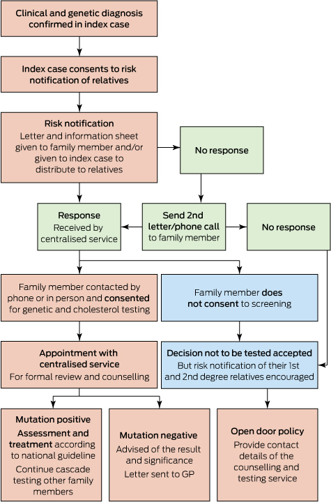
* Family cascade screening process performed according to national guidelines2 after obtaining written consent from the index case.11 This was undertaken by a trained nurse who contacted the family members and obtained verbal consent to contact further family members, after providing counselling and offering specialist review as indicated.

Next-generation sequencing for diagnosis of rare diseases in the neonatal intensive care unit [Research]

Background:

Rare diseases often present in the first days and weeks of life and may require complex management in the setting of a neonatal intensive care unit (NICU). Exhaustive consultations and traditional genetic or metabolic investigations are costly and often fail to arrive at a final diagnosis when no recognizable syndrome is suspected. For this pilot project, we assessed the feasibility of next-generation sequencing as a tool to improve the diagnosis of rare diseases in newborns in the NICU.

Methods:

We retrospectively identified and prospectively recruited newborns and infants admitted to the NICU of the Children’s Hospital of Eastern Ontario and the Ottawa Hospital, General Campus, who had been referred to the medical genetics or metabolics inpatient consult service and had features suggesting an underlying genetic or metabolic condition. DNA from the newborns and parents was enriched for a panel of clinically relevant genes and sequenced on a MiSeq sequencing platform (Illumina Inc.). The data were interpreted with a standard informatics pipeline and reported to care providers, who assessed the importance of genotype–phenotype correlations.

Results:

Of 20 newborns studied, 8 received a diagnosis on the basis of next-generation sequencing (diagnostic rate 40%). The diagnoses were renal tubular dysgenesis, SCN1A-related encephalopathy syndrome, myotubular myopathy, FTO deficiency syndrome, cranioectodermal dysplasia, congenital myasthenic syndrome, autosomal dominant intellectual disability syndrome type 7 and Denys–Drash syndrome.

Interpretation:

This pilot study highlighted the potential of next-generation sequencing to deliver molecular diagnoses rapidly with a high success rate. With broader use, this approach has the potential to alter health care delivery in the NICU.

Doctors criticise Australia’s oldest mum as ‘irresponsible’, ‘wrong’

News that a 62-year-old woman has become Australia’s oldest mum after giving birth to a daughter following IVF treatment has been criticised by some of Australia’s leading doctors.

The Tasmanian woman, who hasn’t been named, was implanted with a donor embryo in an overseas facility after undergoing several years of failed IVF procedures.

According to the Seven Network, the woman and her 78-year-old partner welcomed the arrival of the baby at Frances Perry House Private Hospital in Melbourne on Monday. It was previously reported that the woman was 63.

The baby was born at 34 weeks via a caesarean and is understood to be progressing well.

AMA President Dr Michael Gannon, an obstetrician and gynaecologist, condemned the birth on Twitter.

“This is a rights issue. Consider rights of the child, society, taxpayer. Madness. Not designed to have kids in 60s’,” he wrote in reference to a News Corp article about the topic.

Related: Don’t dismiss conflict-of-interest concerns in IVF, they have a basis

He followed up in another tweet: “63yo woman has baby. Greater priorities in . Child starts life in NICU. Anyone thought ahead to its teens? Selfish, wrong.”

Doctors criticise Australia's oldest mum as 'selfish, wrong' - Featured Image

The move was also criticised by IVF expert Gab Kovacs in News Corp newspapers.

“I think getting people of that age pregnant is irresponsible,” he said. “Our bodies weren’t designed to have children in our 60s. I don’t think any responsible IVF unit in Australia would treat someone of that age, and it’s not a standard of medicine I would condone.”

Most IVF clinics refused treatment once a woman turned 53, which is the “end of natural pregnancy”.

The previous record was held by a woman who had her first child at 60 in 2010. The oldest mother in the world was a 66 Romanian woman who gave birth in 2005.

Latest news:

Gene discovery sparks hope for type 2 diabetes

By Andrew Spece, The Lead South Australia
Researchers have identified the gene they believe is responsible for the onset of type 2 diabetes,
sparking hope for treatments to prevent and possibly reverse the progressive condition.
The researchers from Flinders University in South Australia collaborated with international research teams from the
United States, Sweden and the United Kingdom to narrow down the possible field of 5000 candidate genes to one,
known as RCAN1.
Study leader and Flinders University cell physiologist Professor Damien Keating said a cross-referencing approach
using genes from individuals with Down syndrome was crucial to the result.
Professor Keating said people with Down syndrome were prone to a range of health disorders, including diabetes,
resulting from the overexpression of particular genes because Down syndrome occurred when people had an extra
copy of chromosome 21.
“Many individuals with Down syndrome experience lower insulin secretion, mitochondrial dysfunction and increased
oxidative stress in the insulin-producing beta cells of the pancreas, which are all conditions that also appear in
people with type 2 diabetes,” Professor Keating said.
To identify genes duplicated in Down syndrome that contribute to problems with insulin secretion, scientists
screened the genes of four mouse models of the disorder – two had high blood sugar and two did not, with the
variation enabling a short-list of 38 implicated genes to be identified.
The scientists then narrowed down the list by comparing it to genes overexpressed in beta cells from humans with
type 2 diabetes.
“The comparison identified a single gene, RCAN1, which, when we overexpress it in mice, causes them to have
abnormal mitochondria in their beta cells, produce less cellular energy and secrete less insulin in the presence of
high glucose,” Professor Keating says.
According to the World Health Organisation, the number of people with diabetes in 2014 was 422 million, up from
108 million in 1980. In 2012, an estimated 1.5 million deaths were directly caused by diabetes and another 2.2
million deaths were attributable to high blood glucose.
Type 2 diabetes, which accounts for the majority of diabetes cases, is a progressive condition in which the body
becomes resistant to the normal effects of insulin and/or gradually loses the capacity to produce enough insulin in
the pancreas. The cause of type 2 diabetes is unknown.
“Given that we’ve identified this gene as important for reducing insulin secretion in type 2 diabetes, we are now at a
stage where we have a series of drugs that target RCAN1 and we are now going to test to find whether these drugs
can improve insulation secretion in type 2 diabetes,” Prof Keating said.
“We don’t understand what changes in our pancreas or in our insulin secreting beta cells to cause that transition
from just being insulin resistant and having metabolic syndrome to transitioning to full-blown type 2 diabetes.
“RCAN1, this gene we identified, is certainly a candidate now for that.”
Prof Keating said none of the available treatments for type 2 diabetes targeted the primary cause of the condition.
“All the drugs out there simply alleviate the symptoms,” he said.
“So we have to test these drugs that we have because we feel like that may be able to go straight to the cause of the
reduced insulin secretion that causes Type 2 diabetes … prevent it and possibly reverse it.”
The results of the study led by Prof Keating have been published in the international journal PLOS Genetics.

Rising incidence of invasive meningococcal disease caused by Neisseria meningitidis serogroup W in Victoria

Invasive meningococcal disease (IMD) caused by Neisseria meningitidis is one of the most rapidly progressive sepsis syndromes, often resulting in significant morbidity and mortality. Since the introduction of meningococcal C conjugate vaccine in 2003, IMD in Victoria has decreased from 2.5/100 000 to 0.6/100 000 population.1 Epidemiological typing of N. meningitidis isolates is by serogroup, multilocus sequence typing and finetyping.

In Victoria, from January 2014 to September 2015, the number of cases of IMD caused by N. meningitidis serogroup W (MenW) increased. Previously uncommon (< 5% of IMD overall in the period from 2008 to 2013 [n = 260]), MenW as a proportion of IMD has increased: four of 33 cases in 2014 and 12 of 41 cases in 2015 (Microbiological Diagnostic Unit, University of Melbourne, unpublished data). Over this period, the median age of cases was 55 years, compared with 19 years for serogroup B, with many non-classical presentations including pneumonia, epiglottitis, septic arthritis and pericarditis. There has been one death in a healthy young adult. No epidemiological links between cases have been observed (Victorian Government Department of Health and Human Services [DHHS], unpublished data).

Globally, MenW has been responsible for an increasing proportion of IMD since outbreaks associated with the Hajj pilgrimage in 2000.2 Large outbreaks predominantly due to MenW strain type P1.5-2: F1-1: ST11 have been reported in South America and the United Kingdom.2,3 In the UK, MenW cases doubled year on year from < 2% of IMD prior to 2009–10 to 25% in 2014–15, prompting a change in vaccination guidelines.4 Initially, almost 25% of these IMD cases were older adults with non-classical presentations.3,4

From 1 January 2014 to 30 September 2015, molecular characterisation, including whole-genome sequencing (WGS), of Victorian MenW strains was undertaken at the Microbiological Diagnostic Unit. Of the 16 MenW isolates, 11 were strain type P1.5-2: F1-1: ST11; two were ST184; one was ST22; one was a new type; and one was polymerase chain reaction-positive only and thus unable to undergo WGS. Comparison of these 11 isolates with international strains using the PubMLST Neisseria database (http://pubmlst.org/neisseria) revealed that the Victorian isolates fall within a cluster formed by UK–South American outbreak strains and are distinct from Hajj outbreak strains (Box). Within the UK–South American cluster, nine Victorian isolates appear as an exclusive group of taxa. The close genetic relationship between the nine isolates, long branch length compared with other UK–South American cluster isolates, and lack of identified epidemiological links between cases suggest that these isolates may be representative of a N. meningitidis clone arising from a single introduction event that is undergoing widespread endemic transmission in Victoria. The location of the remaining two UK–South American cluster isolates in the tree indicates independent introduction events into Victoria.

While IMD due to MenW in Victoria remains low in absolute case numbers, the rise in incidence is concerning. The Victorian DHHS has instigated enhanced surveillance measures with full molecular characterisation of future isolates to inform ongoing public health responses. National surveillance with enhanced molecular characterisation will improve understanding of the current epidemiology of meningococcus in Australia.

Box –
Phylogenetic tree for Victorian and international meningococcal isolates


The diagram illustrates the phylogenetic relationship between isolates based on core genome single nucleotide polymorphisms (SNPs). Victorian MenW ST11 isolates demonstrate region-specific clustering, and the close relationship of Victorian MenW strains (in green) to the United Kingdom–South American cluster is shown. (Numerical values for the Victorian isolates represent the number of isolates, while the underlined description refers to geographical origin of isolates.) Note: Mixed Cluster 1 refers to isolates from the UK 1975–2007 and South Africa 2003–2013; Mixed Cluster 2 refers to isolates from UK 1996–2000, South Africa 2003–2004 and North Africa 1996–1999.

Missing link in breast cancer gene mutations

Whilst genetic testing for breast cancer has become cheaper and more accessible, there still remains a missing link, experts write in the Medical Journal of Australia.

The BRCA1 and BRCA2 gene are responsible for 2% of breast cancers, however the challenge is to find the “missing heritability” responsible for the other 3% believed to be caused by gene mutations.

Many families with a history of breast cancer don’t have a BRCA1 and BRCA2 gene. It’s believed rare mutations in other genes must have a role in breast cancer, particularly when it’s diagnosed at an early age.

Professor Ingrid Winship, Executive Director of Research at Melbourne Health and Professor of Adult Clinical Genetics at the University of Melbourne and Royal Melbourne Hospital, and Professor Melissa Southey, from the Department of Pathology at the University of Melbourne, say that to, date, testing one gene at a time has had a very high cost however things are starting to change.

“The revolution in our capacity to conduct genetic analyses in very recent years has had a profound impact on how genetic testing services can be applied,” they wrote.

“Now, a panel of multiple genes can be screened for mutations in a single test, at considerably reduced cost, via the application of massively parallel sequencing technology.”

Related: MJA – Direct-to-consumer genetic testing — a regulatory nightmare?

This testing has led to the discover of more genes associated with breast cancer susceptibility.

“Genes that are regularly included in breast cancer susceptibility gene panels that are currently available through academic or commercial testing laboratories include BRCA1, BRCA2, PALB2, PTEN, STK11, TP53, ATM, BARD1, BRIP1, CDH1, CHEK2, MRE11A, MUTYH, NBN, NF1, RAD50 and RAD51C.”

The problem with the discovery of these genes is that it identifies variants of unknown significance.

“A large grey zone exists, the so-called variants of uncertain significance, where classification is not possible, and this information cannot be used clinically,” they wrote.

Related: Genetic testing “appropriate”

“For this reason, interpretation of results, whether derived from gene panels or single gene testing, remains highly specialised and requires a team of skilled scientists, informaticians and genetic practitioners, along with research-derived evidence.”

They conclude that the testing can also produce unintended, secondary findings that were not anticipated.

“The consequences — that is, the clinical validity and utility of many of the genes listed — are not yet clear.”

Read the full clinical focus in the Medical Journal of Australia.

Latest news:

Gene panel testing for hereditary breast cancer

It is estimated that less than 5% of breast cancer occurs as a result of single gene predispositions; some 2% is explained by mutations in the well known BRCA1 or BRCA2 genes.1 The Australian Institute of Health and Welfare estimated that 15 740 people would be diagnosed with breast cancer in 2015,2 suggesting about 780 of these cancers would be due to genetic predisposition. While this number seems small, the people affected have many more family members (most of whom are unaffected) who can have their potentially high risk of breast cancer managed once it is identified.

Recognition of a familial predisposition to breast cancer led to a race for the “responsible” genes in the late 20th century. The BRCA1 gene was the first gene found to have very rare mutations associated with high penetrance of breast and ovarian cancers.3,4 These rare mutations were often identified in young women or in members of families with multiple cases of breast or ovarian cancer. In 1994, the BRCA2 gene was identified,5,6 and mutations in this gene were found to be associated with increased risk of several cancer types; breast and ovarian cancers were seen in families who also had a higher incidence of melanoma, pancreatic cancer and prostate cancer. A characteristic feature of mutations in BRCA2 is the increased risk of breast cancer in men.6

Twenty years later, the “missing heritability” of breast cancer remains elusive. A network of genetic services and familial cancer centres across Australia investigate families when there is an evident inherited predisposition to breast cancer. However, most of these families do not have mutations in either the BRCA1 or BRCA2 gene. Rare mutations in other genes have subsequently been recognised as playing a role, especially in cases where breast cancer is diagnosed at an early age.710

A new approach — multiple gene testing

To date, tests have been conducted as single gene tests in an iterative manner, one gene at a time, at very high cost. Although some information could be used to direct this testing (eg, specific histopathological features of the breast cancer, such as mitotic index and trabecular growth pattern),11 the testing process is slow and laborious, and most test results remain uninformative. However, the revolution in our capacity to conduct genetic analyses in very recent years has had a profound impact on how genetic testing services can be applied. Now, a panel of multiple genes can be screened for mutations in a single test, at considerably reduced cost, via the application of massively parallel sequencing technology. This approach applies across many disciplines, most notably cardiology; gene panel testing is now the standard of care in the cardiac genetic clinic at the Royal Melbourne Hospital.12

Increases in genetic technology capacity have, in parallel, enabled work that has identified additional breast cancer predisposition genes, and many other candidate breast cancer predisposition genes have been reported (however, these require further evidence). Today, gene panel tests for breast cancer predisposition are offered by numerous providers and service laboratories.10,13 However, few of these tested genes are bona fide breast cancer predisposition genes. Indeed, most of the genetic variation identified by these gene panel tests cannot be interpreted in terms of breast or ovarian cancer risk.

The PALB2 gene is an example of an important new breast cancer predisposition gene, as it has rare mutations that are associated with high penetrance for breast cancer.14,15 The PALB2 protein plays an essential role in interacting with the BRCA1 and BRCA2 proteins and enabling DNA repair. The risk of breast cancer for women with heterozygous germline mutations in the PALB2 gene is as much as eight to nine times as high as in the general population, depending on the age of the woman.15 Mutations in PALB2 account for about 2.4%14,16 of the familial aggregation of breast cancer in the Australian population, but occur at extremely low rates in the general population, making single gene testing for PALB2 cost-ineffective at a population health level.

The low yield of single gene testing and the potential for inclusion of other genes with low frequency mutations (such as PALB2) make a gene panel test approach ideal in the investigation of breast cancer predisposition. Genes that are regularly included in breast cancer susceptibility gene panels that are currently available through academic or commercial testing laboratories include BRCA1, BRCA2, PALB2, PTEN, STK11, TP53, ATM, BARD1, BRIP1, CDH1, CHEK2, MRE11A, MUTYH, NBN, NF1, RAD50 and RAD51C.10,13

The utility of gene panel testing is illustrated by the hypothetical case outlined in the Box.

Implications for breast cancer risk management

There is an evidence-based strategy for managing women who harbour high penetrance mutations in genes such as BRCA1 and BRCA2, and emerging evidence for the risk management of women with mutations in several other genes, including PALB2. For BRCA1 and BRCA2 mutation carriers, it is now known that bilateral salpingo-oophorectomy around the age of 40 years or before the natural menopause will halve the risk of breast cancer, while substantially lowering the risk of ovarian cancer.19 Likewise, chemoprevention and risk-reducing mastectomy are effective in reducing the risk of breast cancer in women with pathogenic mutations in these genes.20 In the absence of a mutation, where there is a theoretical 50% risk that each woman in such families will not have inherited the genetic predisposition, this irreversible and invasive approach is hard to justify.

Genetic testing is thus the only way to provide personalised risk assessment, through predictive testing of at-risk unaffected family members, although this approach depends on identifying a pathogenic mutation in the index case. Effective risk management is thus predicated on accurate risk estimation; as is release from high-risk cancer surveillance for those found not to be at high risk of cancer. The initial inability to find a pathogenic mutation in our illustrative case (Box) meant that this risk management strategy was not possible, whereas gene panel testing and the resulting detection of the PALB2 gene mutation enabled personalised management of the extended family.

Considerations in gene panel testing

The choice of genes to include in panel testing needs careful consideration. The ACCE framework, named for the acronym derived from its descriptors, calls for genetic tests to be underpinned by analytical validity, clinical validity, clinical utility and ethical issues.21

Clinical validity and utility

Clinical validity and consequent clinical utility are currently the most complex areas — is the genetic variation detectable in these genes associated with clear and quantifiable risk of disease? Initially, genetic testing entered clinical practice without much process, nor an evidence base, but spurred by an enthusiasm to reduce the risk of potentially predictable cancers. In contrast, adoption by clinical services of the newly recognised breast cancer predisposition genes, such as PALB2, has been slow, due to an increased awareness of the complexity of interpretation of genetic test results.22 In Australia, protocols for genetic testing and subsequent management based on genetic knowledge have been built over time and are now consolidated in the eviQ guidelines,18 used by clinical genetic and familial cancer services nationally.

It was important initially to ensure that panel testing was as good as the standard of care in finding mutations in BRCA1 and BRCA2, before considering potential additional benefits. In a recent study of 966 patients undergoing breast cancer genetic testing,23 deleterious BRCA1 or BRCA2 mutations were identified at equivalent rates between single gene testing and a multiple gene panel test that included BRCA1 and BRCA2. An additional 3.9% of patients tested by the multiple gene panel had pathogenic mutations identified in other breast cancer predisposition genes, most commonly PALB2, CHEK2 and ATM. In a retrospective review of 90 consecutive patients undergoing genetic testing by a multiple gene panel test,24 pathogenic mutations were identified in nine individuals: five in BRCA1 or BRCA2, and four in other breast cancer predisposition genes. As single gene testing has only identified mutations in BRCA1 and BRCA2 genes, multiple gene panel testing finds more clinically significant mutations.

However, gene panel tests also identify many variants of uncertain significance. Classification of variants is important. In short, there are sequence variants that are clearly disease-associated and classed as pathogenic mutations, while at the other end of the spectrum, much variation in DNA is clearly not associated with disease predisposition and can be discounted. A large grey zone exists in between, the so-called variants of uncertain significance, where classification is not possible, and this information cannot be used clinically. For this reason, interpretation of results, whether derived from gene panels or single gene testing, remains highly specialised and requires a team of skilled scientists, informaticians and genetic practitioners, along with research-derived evidence.

Ethical issues

Technology that allows analysis of many genes at once has a raft of associated ethical issues when applied in a diagnostic setting.25 It is prudent to consider the ability of such tests to produce secondary findings (previously also referred to as incidental findings), wherein genetic changes may be found that were not anticipated.

The recent confirmation in the High Court of Australia that the BRCA1 gene is not patentable has been welcomed, and represents a moral victory for the women and families concerned.26 Indeed, in 2013, following the ruling by the Supreme Court in the United States to invalidate patent claims made by Myriad Genetics, the BRCA1 and BRCA2 genes were included in existing panels that had hitherto excluded them and thus had reduced the comprehensiveness of the analyses being conducted.10

Conclusions

The use of new genetic technology can provide an affordable, effective way of investigating the heritability of breast cancer. Early studies suggest that the gene panel testing approach will detect all mutations found by conventional testing and increase the diagnostic yield through analysis of more genes. This gives more women and their families the opportunity to have personalised risk assessment and risk management, in a preventive approach to predictable breast cancer.

On a cautionary note, the consequences — that is, the clinical validity and utility of many of the genes listed — are not yet clear. Large scale studies through international collaborations into the genes included in these gene panels are essential to yield the information that will allow the safe and effective use of newer technologies and new genetic information in clinical practice.

Box –
Hypothetical case illustrating the utility of gene panel testing


A 37-year-old woman had been diagnosed with high-grade breast cancer at 35 years of age. She had a positive family history of breast cancer, with her paternal aunt being diagnosed at the age of 41 years and her paternal grandmother at 49 years. In keeping with usual practice in Australia, she was offered genetic testing on the basis that she had a greater than 10% likelihood of carrying a mutation in either the BRCA1 or BRCA2 gene, calculated by the BOADICEA risk prediction algorithm.17

No mutation was found in either the BRCA1 or BRCA2 gene. This result was regarded as inconclusive, but the family was still deemed to be at high risk of an inherited breast cancer predisposition (as three relatives on the same side of the family had been diagnosed with breast cancer before the age of 50 years). As a result, all women on the paternal side of her family underwent high-risk surveillance, as no individualised (ie, genetically informed) risk assessment could be offered.

During a review appointment, it was disclosed that the woman’s paternal cousin, aged 42 years, had also recently been diagnosed with breast cancer. In view of the high likelihood of a mutation, further testing was undertaken using a gene panel approach.

This testing identified a PALB2 c.3113G>A mutation. As there are recommendations for the risk management of unaffected women who have this PALB2 gene mutation, the woman’s extended family were offered predictive testing.18 Two affected relatives who were still living and four of the seven unaffected women tested were also found to carry the PALB2 mutation and were referred to breast surgeons and gynaecological oncologists for management of their high breast cancer risk.


BOADICEA = Breast and Ovarian Analysis of Disease Incidence and Carrier Estimation Algorithm.

Co-habiting influences person’s immune system more than illness

A Belgian study has found that co-habiting and specifically raising a child together can have a bigger impact on a person’s immunity than getting the flu shot or contracting gastro.

The research, published in Nature Immunology, found that people who lived together had immune systems that became 50% more similar compared to two non-related people in the wider community.

670 healthy people aged 2 to 86 were studied over a period of three years to “provide a description of the population-level heterogeneity in the cellular composition of the circulating immune system,” the authors wrote.

They found that the immune system was quite elastic, with it bouncing back to its original state after a bout of gastro or receiving the flu vaccine.

The result that was most interesting to them was the impact of co-parenting on immune systems.

Related: How modern life has damaged our internal ecosystems

“One of the most surprising results from our study was the degree to which immune profiles were more similar between parents than unrelated people living in different households. This suggests that a shared environment acts in some way to bring immunoprofiles toward a convergent equilibrium,” they wrote.

They also said there was little effect of gender on the immune landscape, which is at odds with the longstanding observation that autoimmune diseases are more common in women than men.

“Notably, sex-based differences are more limited at the cellular level than at the molecular level. The incomplete correlation between gene signature and cell type suggests that the discrepancy can be resolved by a model where high diversity in molecular expression is largely compensated for at the cellular level,” they wrote.

Latest news:

Novel insights, challenges and practical implications of DOHaD-omics research

The basic tenet of developmental origins of health and disease (DOHaD) research is that perinatal health behaviours of the mother and father, as well as those of the child in early life, can have a significant impact on the future health of the child and that of subsequent generations. Studies exploring DOHaD investigate how early life exposures increase susceptibility to later adverse health outcomes from medical and public health perspectives. This altered health risk appears to occur through reprogramming of physiological systems away from their normal developmental trajectories, and highlights the plasticity of organ systems in the perinatal periods.1 Recent research in this field has focused on the potential for these physiological changes to exert trans-generational effects, without the requirement for further exposures in subsequent generations.2 This appears to occur through genetic and environmental interactions, resulting in phenotypic changes that persist across generations.

The emergence of “-omics” biotechnologies (eg, genomics, proteomics and metabolomics) has revolutionised physiological research in the DOHaD field. From the genome to the epigenome, microbiome and metabolome, research investigating pathways leading to disease has never before had the technology to investigate physiology in such a high throughput, data-rich capacity. We summarise this emerging research capability and its application in DOHaD studies to explain how environmental and social factors, such as diet, stress and exposure to toxins, affect our physiology and become inherited, leaving a legacy of disease susceptibility for future generations.

The epigenome

The epigenome refers to changes made to the genome that result in altered transcriptional activity in the absence of DNA sequence alterations. This highly dynamic process, occurring in response to several external factors, is stably maintained and endures over multiple generations. Epigenetic mechanisms regulating gene expression, including DNA methylation, histone modifications and the actions of small non-coding RNAs, each contribute to tissue-specific gene expression and an altered cellular phenotype. The introduction of efficient sequencing and microarray techniques has facilitated the study of these epigenetic mechanisms.

The interaction between epigenetic inheritance and environmental exposures has been recognised as an important determinant of phenotypic outcomes for offspring.1 Exposures of the mother can result in epigenetic modifications in the developing fetus and the germline.3 Such transmission is not restricted to maternal exposures, and recent evidence shows that epigenetic modifications are also inheritable down the paternal line.4 Specifically, a murine model of paternal obesity has shown altered methylation and microRNA profiles,4 which highlights the role of the father’s contribution to inheritable disease susceptibility. Further, data from the Överkalix Swedish tri-generation population study have shown that the mortality risk ratio of grandchildren was associated with the food supply available to their same-sex paternal grandparent.5 Whether an epigenetic mode of inheritance can contribute to such human outcomes is as yet unknown, but is expected, given the strong parallels observed between animal and human trans-generational studies. Single-generation epigenetic effects have been seen in humans, eg, maternal depression in the third trimester of pregnancy is associated with increased methylation of the NR3C1 gene in cord blood mononuclear cells, in conjunction with altered stress responses in the infants at 3 months of age.6 Increased methylation of this same gene was found in the brain tissue of adolescents with a history of child abuse who later committed suicide,7 and in lymphocytes of 11–21-year-olds after childhood maltreatment, and is associated with poor psychological health.8 Together, these studies suggest that early epigenetic modifications may increase vulnerability to poor long-term health in humans. Recognition of epigenetic mechanisms that contribute to poor outcomes may contribute to interventions to reverse these effects. For example, rat offspring exposed to low maternal grooming behaviour have increased DNA methylation of the hippocampal glucocorticoid receptor gene, which is reversed by increased care provision in early postnatal life.9 Similar epiphenotypes are observed in infant saliva, when high tactile stimulation of the infant in the postnatal period normalises glucocorticoid receptor hypermethylation induced by maternal depression.10

The microbiome

The human microbiome is the collection of microorganisms that inhabit the human body, including commensal and symbiotic microbes. The study of the microbiome and its role in disease onset has been made possible by the introduction of large-scale sequencing techniques and gene expression arrays. These techniques have increased our ability to understand the contribution of the maternal microbiome to disease in subsequent generations. For example, altered bacterial colonisation of the alimentary tract of piglets, after antibiotic and stress exposure in early life, has been associated with immune development perturbations.11 This may have particular implications for preterm children, for whom exposure to antibiotics and stress is common in early life. Already, preliminary studies of the microbiome in preterm twins have shown that an altered pattern of microbial gut colonisation precedes the development of necrotising enterocolitis.12 In humans, obesity,13 smoking14 and different modes of delivery (eg, vaginal versus caesarean)15 are common potential prenatal factors that can influence maternal and neonatal microbiomes. An altered microbiome can also contribute to epigenetic changes.16

The metabolome

The metabolome is the complete set of metabolites (compounds of low molecular mass found in biological samples) that regulate cell and tissue growth, development, survival, maintenance and responses to the environment. The potential for metabolomic profiling to provide a phenotypic signature of pathophysiology has been recognised.17 Methods to assess the metabolome rely on high-resolution analytics, including mass spectrometry, nuclear magnetic resonance spectrometry and Fourier transform infrared spectroscopy. Unlike the epigenome and the microbiome, the metabolome can be highly dynamic and is able to change in short time frames, ranging from seconds to minutes. The choice of sampling material is therefore an important consideration and a challenge. The decision on sampling material will be specific to the research question and critical to the interpretation of results. For example, blood samples reflect highly dynamic responses, but hair samples reflect prolonged exposure and can therefore provide a more stable phenotype.18 The large volume of data generated by such techniques can provide insight into interactions between metabolites, genes, transcripts and proteins.19 These data can be highly informative about mechanisms leading to disease and the impact of environmental exposures on system physiology, such as the developmental impact of prenatal exposure to the endocrine disruptor, bisphenol A.20

The potential for metabolomics platforms to be used to identify biomarkers predicting pregnancy outcome is already becoming apparent. These platforms include observations of differences in the neonatal blood metabolome across gestational ages (differences that are dependent on postnatal age at sampling21) and specific pathology and illness severity;22 a study linking the maternal hair metabolome with fetal growth restriction;18 and an ongoing prospective study for early prediction of pre-eclampsia23 (trial NCT01891240).

Challenges to -omics approaches in DOHaD research

Use of these emerging biotechnological approaches in DOHaD research shows clear promise in expanding our current knowledge of mechanisms driving intergenerational transmission of disease and heightened disease susceptibility in individuals after specific exposures in early development. While such large volumes of biological data using these -omics approaches provides enormous opportunity, some challenges remain in their application and interpretation. The first challenge relates to identifying the appropriate time for tissue sampling, given the current limited use of these approaches in this field. Healthy ranges are also yet to be established, a limitation that occurs with any advance in technology and will be overcome through public sharing of data. To establish healthy ranges, sampling from multiple time points and multiple tissues will be necessary. This information will benefit the design of future studies, in which sampling can then occur at a single time point during tissue-specific sensitive periods to yield the most reliable, valid and interpretable data. The establishment of normative ranges will also help elucidate many other current unknowns in this area, including understanding what sample sizes are needed to identify meaningful effects; understanding and predicting the stability of -omics profiles; identifying the effects of a “second hit” or multiple exposures; understanding whether the duration or timing of each exposure is important in determining outcome; and understanding whether a genetic susceptibility is needed for the intergenerational transmission of poor outcomes or whether this is a highly conserved process.

Once we have identified biomarkers or signatures predictive of poor maternal, fetal or neonatal outcomes, the next critical step is to use this information to identify how to normalise these effects. This will necessitate an understanding of how postnatal factors normalise or exacerbate the -omics profile induced by the early life environment. Longitudinal studies of twins have provided some preliminary evidence of environmental influences, exploring the stability of the epigenome across the first 18 months of life and the degree of epigenetic discordance between siblings with a shared genetic and environmental background.24 Continued longitudinal assessments of these children will increase our understanding of the role of the environment on the epigenome through life. The impact of additional exposures in pregnancies of these subsequent generations has also yet to be identified, because few studies have assessed the potential for -omics profiles to be modified beyond the second generation.2

Recommendations for future studies

We highly recommend collaborative studies that integrate data derived from multiple platforms, collected from samples throughout early development and linked to clinical health outcomes. Analysis of samples from current and planned randomised controlled trials will allow the effects of standard care and interventions to be assessed concurrently. These studies will facilitate our understanding of disease susceptibility, onset and progression to a degree that has not previously been possible.

Implications for policy and practice

Effective interventions applied at critical periods of development can substantially reduce future disease burden. The potential for this research to be translated into tangible health benefits for child health and future generations is therefore enormous, aligning with the growing demands of national health regulatory bodies to focus efforts on preventative health care. The outcomes of this research could then potentially be used by health advocates to improve policy and practice, by clinicians and health workers to promote and support healthy perinatal behaviours, and be communicated to the wider community to optimise future child health.

Information on DOHaD and early life healthy behaviours is becoming more readily available, but it is unclear whether this is being effectively communicated to the health care providers who need it most, that is, those in direct contact with women who are pregnant or planning a pregnancy. For example, surveys of general practitioners reveal that they have limited knowledge of nutritional requirements in pregnancy, and also feel uncomfortable providing this information to women due to a lack of confidence.25 Knowledge gaps such as this must be urgently addressed to optimise the health of future populations. Similarly, while the internet is teeming with websites offering advice for pregnant and breastfeeding women, these often contain inaccurate or misleading advice and conflicting information. Evidence-based online resources to which women can be directed for accurate health information are needed.

A multidisciplinary renal genetics clinic improves patient diagnosis

Developments in genomic science are disproportionately in advance of their translational clinical application. Multidisciplinary clinics are proposed to overcome this1 in many medical fields.2 This is especially so in nephrology, which is typified by significant community disease burden3 and heritability.4 Several renal genetics clinics (RGCs) operate overseas, although their models and outcomes are largely unreported. The first multidisciplinary RGC in Australasia commenced at the Royal Brisbane and Women’s Hospital in August 2013, involving a clinical geneticist, nephrologist, genetic counsellor, and ancillary clinical and diagnostic services. The departments of clinical genetics and nephrology jointly operate the RGC. The clinical geneticist and nephrologist see families in the same appointment, maximising use of time. In this article, we report this clinical service’s initial outcomes and model for mainstreaming genetic medicine.

We undertook a retrospective cohort study of patients who attended the Royal Brisbane and Women’s Hospital Renal Genetics Clinic during its first 2 years of operation (1 August 2013 to 31 August 2015; ethics approval reference, HREC/14/QRBW/187). During this period, 108 patients from 100 families were seen; the median age was 41 years (range, 13–86 years). Most patients were referred by a public or private sector nephrologist (47% [51/108] and 28% [30/108], respectively), and 81% (87/108) had an existing genetic renal diagnosis, 45% (49/108) had extra-renal clinical features and 65% (70/108) had a family history of renal disease. Existing renal diagnoses were diverse, and the most common were autosomal dominant polycystic kidney disease (34% [37/108]), Alport syndrome (17% [18/108]) and focal segmental glomerulosclerosis (7% [8/108]).

The overlapping reasons for referral were for a diagnosis (67% [72/108]), a discussion about a diagnosis (27% [29/108]) and genetic counselling (81% [87/108]). Clinical and family histories and results of clinical investigations were reviewed. Differential diagnoses were discussed for 68% of patients (73/108), disease information was provided to 89% (96/108), and genetic counselling provided to 67% (72/108).

Genetic testing was ordered for 69% of patients (75/108). Of a total of 83 tests, results were positive for 39% (32/83), negative for 30% (25/83), “variant of uncertain significance” for 7% (6/83) and pending for 24% (20/83). Negative genetic test results have enabled 12 of the families to enrol in a research study.5 To date, the clinical diagnosis has changed for 27 of the 108 patients (25%) (Box), enabling correct diagnosis, accurate genetic counselling, identification of at-risk individuals, access to assisted reproductive technologies and altered medical management.

This RGC model is novel in Australasia and its results are among the first to be reported internationally. In its first 2 years of operation, patients underwent clinical appraisal and a tailored combination of differential diagnosis discussion, disease information provision and genetic counselling. Genetic testing was often, but not always, used, with results confirming or clarifying a diagnosis for about half of the patients. Overall, the diagnosis was changed in a quarter of patients. This clinic model is inclusive, flexible and multidisciplinary while demonstrably improving patient diagnosis and care. We believe that it is a viable, translational and patient-focused clinical template for effective introduction of genetics and genomics into everyday clinical practice.

Box –
Changes in clinical diagnosis at the Royal Brisbane and Women’s Hospital Renal Genetics Clinic (green, unchanged diagnosis; blue, changed diagnosis)