×

The acute care conveyor belt: a personal experience

This wasn’t “end of life”; this was his life.

Perhaps the best comment I read at the time of the big 2015 bacon scare was “So what are we allowed to die of?”. Every stage of life has now become medicalised, not just birth and death. The fact that there is even a conveniently euphemistic acronym for the end of life, “EOL”, rather than the words themselves, is symptomatic of how removed we are as a profession from the reality that despite our very best efforts, life has only one ultimate outcome. But Hillman and colleagues’ description of the “acute care conveyor belt”1 eloquently describes the path that I impotently watched my father travel.

At 86, he was extremely well — still golfing and travelling, dining out and never missing the latest films. There was the usual raft of preventive medications to control his sugar and lipid levels, his gastric acid and his blood pressure. There was even an indolent carcinoma that created some inconvenient symptoms requiring increasingly regular trips to hospital for antibiotics and a first-name relationship with the local ambos.

This wasn’t “end of life”. This was his life — each day just like every day before presenting its own unique challenges and pleasures. That he would die sometime in the next while, he knew and accepted. What mattered was today and whether health and weather would align to get him to the golf course.

For my father, the conveyor belt started with one of his trips to hospital. Acute renal failure was unexplained but managed well and resolving. After transfer to a ward pending discharge home and community nursing, he suffered a massive stroke. Had this occurred at home on an early Saturday morning, it is unlikely he would have survived. But it occurred in a hospital ward, where he was surrounded by people with the skills and knowledge to keep him alive. And they did. And they transferred him to intensive care.

When I was a medical student, it was pretty unusual for anyone over 75 to get to intensive care. These days, it’s pretty unusual if they don’t. It seems that my father has the constitution of an ox. Even off his barrage of meds because of the renal failure mystery, his heart doesn’t miss a beat: perfect systolic rhythm; his saturation hovers around 100%, breathing on his own with just nasal prongs; and his blood pressure is excellent. Only his brain is refusing to cooperate, big time — bilateral infarcts, seizures and midline shift.

The nephrologists, the neurologists, the intensivists and the gastroenterologist have all had their say. In intensive care, no one uses the “D” word. In medicine, dying equals defeat. I couldn’t be clearer with the team about what my father wanted. This is not a case of “the failure of people to have stated their wishes regarding EOL care”.1 We had had this conversation many times, especially since the cancer diagnosis.

They tell us that if he does survive, he will need high-dependency care. I ask the team about their palliation plan. They are shocked and refuse to engage. I am shocked that they refuse to engage, despite their determination to move him back to the ward. We all know this is just another euphemism for palliation, for covert euthanasia. We know he is dying. Why can’t we talk about it?

Ironically, the person most comfortable discussing the topic is the youngest resident, a graduate of one of our newer medical schools. She’s not recommending euthanasia, but at least she is willing to acknowledge our agony of indecision and our horror at the idea of “comfort feeding”: a euphemism for starvation and dehydration.2

In intensive care, visiting is limited. Visitors are forced to wrap themselves in cloying yellow plastic that makes the sweat run down your back and arms despite the air conditioning. On the weekend, staff are casual, comforting and relaxed, and we hold vigil by his bedside for extended periods. I am terrified to leave in case he dies alone. But come Monday, the rules snap back into place: two visitors only, visiting hours only, meaning long hours when he has no family by his side.

I want to take him home. I am told not to get ahead of myself, but I know just how good the local palliative care team is. My mother died at home in her own bed surrounded by almost everyone she loved. But she died of cancer. It’s OK to die of cancer; the system supports you. They give you painkillers and personal care and don’t pretend they can do anything more. The palliative care nurses live by the tenet “medicine at this time has nothing more to offer” — but good nursing care certainly has more to offer.

There will be no more rapid response teams in my father’s care. We have made that quite clear. But unless his body declares a ceasefire, he will remain hostage to the acute hospital system until the conveyor belt chugs him into a high-dependency nursing home bed possibly far away, with a percutaneous endoscopic gastrostomy tube and incontinence pads. Some days the golf may even be on the TV droning in the background.

There are no more holes-in-one or beers with his mates at the 19th in my dad’s future. A more humane system would help him off the conveyor belt and onto the fairway in the sky. Thank you, Drs Hillman, Rubenfeld and Braithwaite, for daring to start this essential conversation.

Postscript

Dad finally died 10 days after his stroke. He never regained consciousness. We were able to be with him right to the end. The medical and palliative care that he received was exemplary, but this does not negate the need for a robust discussion about the role of euthanasia at the end of life and the ethical ambiguity of current means of palliation.

Necrotising myositis presenting as multiple limb myalgia

Clinical record

A previously healthy 40-year-old man was referred by his general practitioner to our hospital after a short prodromal period of a sore throat and rapidly deteriorating constitutional symptoms. Most pertinent to his diagnosis was the development of non-traumatic, localised, right calf pain 48 hours before admission that progressed to an inability to bear weight by the time of hospital presentation. On initial physical examination, he had a temperature of 37.8°C, diffuse muscle tenderness in all four limbs and an exquisitely hyperalgesic localised area on the right mid-calf. Examination of his throat showed a diffuse pharyngitis. There was no rash or arthritis, and his cardiovascular, respiratory and gastrointestinal systems were all unremarkable at admission.

A blood sample and throat swab were taken and, after an initial blood and microbiological culture work-up, empirical treatment with intravenous flucloxacillin and vancomycin was commenced. Early pathology test results showed a creatine kinase (CK) level of 380 U/L (reference interval [RI], < 171 U/L), serum creatinine level of 158 μmol/L (RI, 55–105 μmol/L), white cell count of 3.1 × 10⁹/L (RI, 4.0–11.0 × 10⁹/L) with increased band forms, and deranged liver function test results, with a bilirubin level of 60 μmol/L (RI, < 20 μmol/L) and alanine transaminase level of 242 U/L (RI, 0–45 U/L).

Over the next 24 hours, the patient’s condition deteriorated, prompting consultation with an infectious diseases specialist. This resulted in a change of antibiotic therapy to ceftriaxone to broaden the coverage of respiratory pathogens, given his acute pharyngitis, and clindamycin to restrict any potential toxin production. By the evening of the second hospital day, the patient was referred to the intensive care unit (ICU) with evolving multiple organ failure. He was now febrile to a temperature of 40°C, with evolving septic shock, pulmonary infiltrates, worsening acute kidney injury (serum creatinine level, 201 μmol/L, and oliguria) and mild delirium. His right calf remained a focal point of concern, with an accompanying tenfold rise in CK level to 3656 U/L.

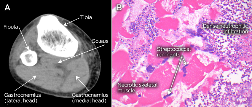
The patient’s condition further deteriorated during his first night in the ICU, necessitating aggressive fluid resuscitation, vasopressor support and haemodialysis. By the morning of the third day in hospital, 12 hours after ICU admission, an isolated, small, tender area of discolouration was noted over the distal posteromedial aspect of the right leg, with no other clinically apparent lesions, but persisting myalgia in all four limbs. This, in conjunction with the confirmation of gram-positive cocci grown from the admission blood and throat swab cultures, prompted the initial consideration of necrotising fasciitis. Subsequent imaging of the lower limbs with ultrasound and non-contrast computed tomography (CT) scans excluded venous thrombosis and fascial thickening, but both tests showed subtle swelling of the calf muscles, suggesting myositis (Figure, A). An urgent plastic surgery consultation mandated surgical exploration of the right calf, and the diagnosis of necrotising myositis (NM) (Figure, B) was subsequently obtained.

Due to the ongoing requirement for frequent soft tissue debridement, the patient was ventilated and transferred to the nearest quaternary hospital. Here, he underwent further imaging of all four limbs and successive debridement of his right leg and both arms for NM on Days 4, 5, 7, 9 and 18 of admission. After receiving confirmation of susceptibility, the ceftriaxone was changed to benzylpenicillin, while clindamycin was retained and intravenous immunoglobulin (IVIG) commenced. Microbiological serotyping confirmed Streptococcus pyogenes with type emm 89.0 strain; exotoxin assays were not conducted. The patient’s total ICU stay lasted 17 days, with liberation from haemodialysis after 7 days and the ventilator after 9 days, resolution of his multiple organ failure, and all four limbs preserved without amputation. After 33 days in hospital, he was discharged to a rehabilitation centre.

Necrotising myositis is a rare but potentially fatal form of infection, predominantly characterised by muscle necrosis capable of rapidly progressing to multiple organ failure in healthy young adults. Published literature attributes group A streptococcus as the most commonly implicated pathogen, but NM has also been associated with groups C and G streptococci, Bacteroides subtilis, Staphylococcus aureus and Peptostreptococcus.1,2

Our case highlights three important clinical lessons. First, NM typically involves a single limb or area. Multiple limb involvement in the initial presentation has only been reported in two previous cases.3,4 Second, despite the widespread limb involvement in our patient, skin discolouration was a subtle, late sign. It presented in only one limb 72 hours after symptom onset, at a stage when the toxic shock syndrome was already apparent. Of 14 previously reported cases, only five describe skin discolouration and two describe local erythema.1,38 Third, and most crucially, NM, like necrotising fasciitis, remains a clinical diagnosis, with most investigations being indeterminate.

Increased serum CK level has previously been lauded as a potential early warning sign,46 but the initial CK result at our patient’s hospital admission (already more than 48 hours after the onset of symptoms) was only marginally raised (380 U/L). We found two other previously reported cases of NM where the CK level remained below 500 U/L at 48–72 hours after symptom onset.6,7 These findings suggest that excluding NM on the basis of small rises in serum CK level (< 500 U/L) is unreliable. Similarly, a reliance on imaging to provide a diagnosis can result in non-specific or negative findings, delaying a definitive surgical diagnosis and treatment.8 While modern imaging can be performed rapidly, the CT and ultrasound scan findings in our patient were subtle, non-specific and ultimately delayed surgery by 3–4 hours.

Once a diagnosis of NM is suspected, aggressive surgical debridement, appropriate antibiotic therapy and supportive care are mandated for survival. Early surgical intervention has reduced mortality from 100% to 37%,1 but with the consequence of significant long-term morbidity for many survivors. Aggressive group A streptococcal infections respond less well to penicillin and continue to be associated with high mortality and extensive morbidity, leading to the use of adjunctive therapies.9 In a recent observational Australian study of 84 patients with severe invasive group A streptococcal infection, the addition of clindamycin resulted in a significant reduction in mortality, which was further enhanced by the inclusion of IVIG.10 Clindamycin inhibits bacterial protein synthesis at the level of the 50S ribosome, resulting in decreased exotoxin production and increased microbial opsonisation and phagocytosis, while IVIG increases the ability of plasma to neutralise superantigens.9,10 Finally, conclusive evidence is lacking for the use of hyperbaric oxygenation, aimed at reducing hypoxic leucocyte dysfunction, and it was not used for this patient.

Lessons from practice

  • Necrotising myositis is a rare but potentially fatal condition. It is a diagnostic conundrum, often presenting as systemic toxicity and widespread myalgia without focal features.

  • Improved survival is underpinned by early clinical diagnosis, appropriate antibiotic therapy including clindamycin to reduce exotoxin load, and urgent surgical referral. Adjunctive therapies such as intravenous immunoglobulin and hyperbaric oxygenation should be considered based on individual circumstances.

  • Previously suggested diagnostic investigations such as serum creatine kinase levels, ultrasound and computed tomography scans are unreliable, mandating a high index of clinical suspicion to make a diagnosis.

Figure


A: Computed tomography scan (transverse plane) of the right leg, showing subtle hypointense and mildly expanded gastrocnemius and soleus muscles with intact fascia, suggestive of myositis. B: Haematoxylin and eosin stained paraffin section of the right gastrocnemius muscle, showing necrotic skeletal muscle, inflammatory infiltrate with disintegrating neutrophils and colonies of streptococcal bacteria.

Perceptions of Australasian emergency department staff of the impact of alcohol-related presentations

Alcohol-related presentations are common in emergency departments (EDs) throughout Australia and New Zealand. Two point prevalence surveys indicate that one in eight presentations to EDs are alcohol-related.1,2

ED clinicians are at the forefront of responding to the consequences of alcohol-related harm. Verbal and physical violence and aggression are common in EDs, with adverse effects on staff wellbeing and job satisfaction.3 As little is known about this problem in the local context, our study surveyed perceptions of clinical staff of alcohol-related presentations to Australasian EDs. The study had two main objectives: to quantify the scale of the problem of alcohol-related violence experienced by ED staff, and to assess their perceptions of the effects of alcohol-related presentations on the functioning of the ED.

Methods

A mixed methods, cross-sectional online survey was developed after undertaking a literature search, and refined by the consensus of an expert steering committee. Definitions for verbal and physical aggression were taken from the Medicine in Australia: Balancing Employment and Life (MABEL) Longitudinal Survey (http://mabel.org.au/) (Appendix 1). Free-text items were included for qualitative analysis. The survey was piloted, leading to minor modifications of its wording.

The survey was conducted from 30 May to 7 July 2014. Participation was anonymous, voluntary, and consent implied by completion of the survey. The survey link was distributed by email to 156 directors of emergency medicine at EDs accredited by the Australasian College for Emergency Medicine (ACEM). Directors were asked to forward the survey link to all clinical staff in their ED to encourage participation. The ACEM e-bulletin and social media channels were also used to promote the survey. The College for Emergency Nursing Australasia (CENA) also distributed the survey. These distribution channels ensured the survey was targeted at clinicians working in Australasian EDs. At the time of its distribution, 1575 emergency registrars and 1270 emergency physicians were working in ACEM-accredited EDs, together with an average of eight nurses per physician.

The survey distribution methodology meant that a response rate could not be determined. A small number of responses were received from ED staff who were not doctors or nurses; these were excluded from analysis.

Statistical analysis

Quantitative data was analysed using SPSS Statistics for Windows 22.0 (IBM). Proportions with 95% confidence intervals (CIs) were calculated, cross-tabulated by clinician role, and compared in χ2 tests; P < 0.05 was defined as statistically significant. When analysing Likert scale data, “positive” and “very positive” responses were combined, as were “negative” and “very negative” responses. When assessing how frequently alcohol-related aggression was experienced, “frequently” and “often” responses were pooled, as were the responses “occasionally”’ and “infrequently”. Qualitative data were categorised according to thematic keywords derived from the free-text responses, and then analysed by the frequency distribution method.

Ethics approval

Ethics approval was provided by the Monash Health and Monash University Human Research and Ethics Committees (reference, MUHREC-CF14/1691-2014000782).

Results

Responses to the survey were received from 2002 clinicians (emergency physicians, ED registrars, resident medical officers, interns, and ED nurses) working in EDs in Australia and New Zealand (Box 1).

Alcohol-related verbal aggression from a patient had been experienced by 97.9% of respondents (1899 of 1940) in the past year, and physical aggression by 92.2% (1784 of 1935) (Box 2). Appendix 2 breaks downs the frequency of alcohol-related verbal or physical aggression experienced during the past year according to clinician type. Eighty-seven per cent of respondents (1682 of 1929) had felt unsafe in the presence of an alcohol-affected patient. Nursing staff were more likely than other ED staff to have felt unsafe (Box 3).

Sixty-eight per cent of respondents (1311 of 1940) reported having experienced verbal aggression often (a few times per month) or frequently (one or more times a week); 42% (807 of 1935) had often or frequently experienced physical aggression from alcohol-affected patients in the past year. Third party aggression (from patients’ relatives and carers) was also common. Although most staff had experienced alcohol-related verbal and physical aggression from patients and verbal aggression from a relative or carer in the past 12 months, nursing staff were more likely to have experienced this problem than non-nursing staff (χ2 test, P < 0.001) (Appendix 2).

Forty-eight per cent of respondents (931 of 1931) reported routine screening for alcohol consumption of patients presenting to their ED, and 44% (850 of 1928) reported screening, brief intervention and referral to treatment for patients at risk of alcohol harm.

Thematic analysis of qualitative responses (selected examples: Box 4) showed that alcohol-related aggression was a daily occurrence, as reflected in 24% of free-text comments on this theme (44 of 186). Respondents also commented that such behaviour should not be acceptable in the workplace.

Men and women reported similar frequencies of verbal and physical aggression from patients, but women were more likely to report verbal or physical aggression from relatives or carers (χ2 test, P = 0.01) (Appendix 3).

Alcohol-related presentations were perceived to have a negative impact on waiting times, other patients in the waiting room, and the care of other patients (summary: Box 5; full results: Appendix 4). Alcohol-related presentations were also widely viewed as having a negative or very negative impact on the workload, wellbeing and job satisfaction of ED staff (summary: Box 5; full results: Appendix 5).

Free-text responses about the impact of these presentations on ED functioning confirmed this. Sixty per cent of respondents (1191 of 2002) provided a comment about the effect of alcohol-related presentations on other patients attending the ED. Most described negative effects, with 48% (569 of 1191) commenting that other patients experienced distress and felt unsafe if an alcohol-affected patient displayed loud, aggressive, violent or antisocial behaviour. Seventeen per cent (201 of 1191) commented that this distress was heightened in vulnerable patients, including children, the elderly, and in those who were mentally unwell.

Twenty-three per cent of these respondents (272 of 1191) also commented that alcohol-affected patients caused increased waiting times for other patients because they often were treated as a priority. Alcohol-affected patients were perceived to affect other patients by diverting resources (17% of respondents; 205 of 1191) and clinicians (15%; 182 of 1191), and by generally compromising the quality of care that other ED patients received (16%; 188 of 1191).

Discussion

Our study found that more than 90% of ED clinicians had in the past year experienced physical aggression from a patient affected by alcohol, with 42% experiencing this aggression weekly or monthly. This frequency of physical aggression from a single cause is disturbing, particularly compared with a large survey of Australian general practitioners and hospital doctors in which 32.3% reported experiencing physical aggression in the past year.4

Verbal aggression from patients affected by alcohol was an ever-present part of clinical life for ED staff. This compares with 70.6% of a more general cohort of doctors reporting that they had experienced verbal or physical aggression.4 While all ED clinician types experience violence and aggression, it is more frequently experienced by ED nurses,5 and it has been suggested that nurses see violence and aggression as an inescapable part of their job.6,7

We also found that ED clinicians frequently experience both physical and verbal violence and aggression from alcohol-affected patients’ relatives and carers. While this third party aggression has been reported before, comments made by respondents suggest that accompanying persons were often also affected by alcohol, and this may explain the high rate of aggression. Although there was no difference in their experience of violence and aggression from the patients themselves, female staff were more likely to experience violence and aggression from the carers of alcohol-affected patients. Previous research did not find this gender difference for either doctors or nurses.8,9

Adverse impacts of alcohol-affected patients on other patients and the effective operation of the ED is concerning. The need to divert resources disrupts or delays care for other patients. Effects on the welfare of and care for other patients, particularly vulnerable groups, are further exacerbated by the disruptive and antisocial behaviours of alcohol-affected people in EDs.

Violence and aggression had a negative effect on respondents’ perceptions of their wellbeing and job satisfaction. This has been previously reported,8 and it has been suggested that this affects the quality of care beyond its obvious effects on workload. Patient aggression and violence has a profound impact on patients, clinicians and the therapeutic relationship.10 It can also affect staff retention and recruitment, and this highlights the importance of community education about alcohol-related harms and of changing the culture of unacceptable behaviour.9

The MABEL study found that medical practitioners were less likely to experience aggression in workplaces where strategies to reduce aggression had been implemented.11 Environmental and human factors should be taken into account to reduce the risk of workplace violence, while resources that enable appropriate medical care and access to safe sobering-up facilities will assist EDs to manage alcohol-affected patients.

Study limitations

Selection and non-responder bias inevitably affects voluntary surveys. ED clinicians who have recently experienced aggression and violence from alcohol-affected patients may be more likely to respond. Further, as the survey was anonymous, it was difficult to ensure that respondents did not complete the survey several times. However, our review of respondents’ IP addresses and their demographic data suggests that this was unlikely. More than half the respondents worked in major referral hospitals, suggesting that this group was over-represented. Recall bias was minimised by asking respondents only about the past 12 months. Definitions of violence and aggression were provided in the survey to limit misclassification of events by respondents. Misclassification may, however, have resulted in some respondents confounding alcohol-related presentations with those related to other drug use, or to a combination of alcohol and drug use.

Conclusions

Alcohol-related verbal aggression was commonplace for the clinicians who responded to this survey. Physical violence was experienced by a large majority. This violence and aggression has a negative impact on the care of other patients and on the wellbeing of clinicians. Managers of health services must ensure a safe working environment for staff. More importantly, however, a comprehensive public health approach to changing the prevailing culture that tolerates alcohol-induced unacceptable behaviour is required.

Box 1 –
Workplace characteristics of the 2002 emergency department staff who responded to the survey

Number

Percentage


Gender

Men

700

35.0%

Women

1285

64.2%

Not stated

17

0.8%

Staff role

ED nurse

904

45.2%

EM physician

507

25.3%

EM registrar

373

18.6%

Medical officer

136

6.8%

Other

67

3.3%

Not stated

15

0.7%

Location

Victoria

408

20.4%

Queensland

367

18.3%

Western Australia

300

15.0%

New South Wales

298

14.9%

South Australia

211

10.5%

Tasmania

46

2.3%

Northern Territory

29

1.4%

Australian Capital Territory

11

0.5%

Australia (total)

1670

83.4%

New Zealand

313

15.6%

Not stated

19

0.9%

Role delineation of ED

Major referral

1035

51.7%

Urban district

566

28.3%

Regional/rural

379

18.9%

Not stated

22

1.1%


ED = emergency department; EM = emergency medicine.

Box 2 –
Frequency of alcohol-related verbal or physical aggression experienced by respondents within the past 12 months (summary)*

Number of responses

Often/frequently

Occasionally/infrequently

Not at all


Verbal aggression from a patient

1940

67.6% (65.5–69.7%)

30.3% (28.3–32.3%)

2.1% (1.6–2.9%)

Physical aggression from a patient

1935

41.7% (39.5–43.9%)

50.5% (48.3–52.7%)

7.8% (6.7–9.1%)

Verbal aggression from a relative or carer

1923

30.8% (28.8–32.9%)

58.7% (56.5–60.9%)

10.5% (9.2–11.9%)

Physical aggression from a relative or carer

1930

18.9% (17.2–20.7%)

58.0% (55.8–60.2%)

23.1% (21.3–25.0%)


Data are presented as the percentage of received responses, with the 95% confidence intervals in parentheses. * For full results and breakdown by clinician group, see Appendix 2.

Box 3 –
Emergency department staff reporting that they have felt unsafe because of the presence of an alcohol-affected patient in their emergency department*

Staff role

Number

% (95% CI)


Emergency department nurses

863

91.8% (89.8–93.4%)

Emergency department registrars

360

88.6% (84.9–91.5%)

Emergency department physicians

499

82.6% (79.0–85.0%)

Emergency department medical officers

133

72.2% (64.0–79.1%)

Other/unknown

74

85.1% (75.3–91.5%)


* P < 0.001 (χ2 test).

Box 4 –
Selected qualitative responses to the survey

Alcohol-related aggression and violence in the emergency department (ED)

  • Verbal abuse is an hourly occurrence. One or two people removed for physical aggression each shift, a staff member injured severely enough to have days off every few months, patients restrained by security/code black every two to four hours. Serious property damage (window/wall broken) every one or two months. [ED doctor, female]
  • Staff are regularly faced with physical/verbal aggression due to alcohol presentations; several members of staff have had chairs thrown at them, one underwent a shoulder reconstruction after sustaining a dislocation from a patient and we are constantly abused. [ED nurse, male]
  • If I am out of uniform I do not have to tolerate these behaviours and have a course of action; if I am in uniform, I am fair game! [ED nurse, female]
  • I was assaulted and knocked unconscious by a patient. I was put in my own resus[citation] room with concussion and vomiting. I had residual effects for several weeks following. The patient was arrested in the ED. When asked why he hit the doctor, his reply was: “because no-one brought me a **** sandwich.” [ED doctor, male]
  • I was obviously pregnant and was threatened by a patient (in front of his kids and wife) that he was going to punch me in the stomach. [ED nurse, female]

The impact of alcohol-related presentations on patient care

  • They can feel threatened and intimidated in an environment where they should feel safe. They are often shocked and offended by what they see or hear. [ED nurse, female]
  • I always feel terrible when there is a parent with a sick kid and they are exposed to behaviour and language and sometimes violence or even police presence; it’s very unfair towards them. [ED doctor, female]
  • A woman and her 4-year-old daughter in a cubicle had a drunk man open the curtain and urinate over the bed thinking he was in the toilet. Distressing for mum and child. [ED nurse, female]
  • Other patients in the ED often have delayed care because of intoxicated patients, and sometimes even important tests or observations are missed or forgotten because the intoxicated patient is taking up so much of our time — either vomiting, abusing staff or other patients, creating a scene, or generally being unsafe. [ED nurse, female]
  • They use resources that may otherwise go to another case. If you have one remaining bed and two patients, and one of them is highly intoxicated, they will get preference over the patient who may be quite ill but is able to sit in the waiting room. [ED nurse, female]

Box 5 –
The impact of alcohol-related presentations on emergency department function and care of other patients* and on emergency department staff (summary)

Number of responses

Positive/very positive

Neutral

Negative/very negative

Don’t know


On patients

On waiting times

1980

0.5% (0.3–0.9%)

13.7% (12.3–15.3%)

85.5% (83.8–86.9%)

0.4% (0.2–0.7%)

On other patients in the waiting room

1980

0.4% (0.2–0.8%)

4.4% (3.6–5.4%)

94.4% (93.3–95.3%)

0.8% (0.5–1.3%)

On the care of other patients

1982

0.6% (0.4–1.1%)

10.9% (9.6–12.4%)

88.3% (86.8–89.6%)

0.2% (0.1–0.5%)

On emergency department staff

On staff workload

1991

0.8% (0.5–1.3%)

4.7% (3.8–5.7%)

94.2% (93.1–95.2%)

0.3% (0.1–0.7%)

On staff wellness

1981

0.7% (0.4–1.2%)

24.6% (22.7–26.5%)

74.1% (72.1–76.0%)

0.6% (0.4–1.1%)

On staff job satisfaction

1983

1.3% (0.9–1.9%)

17.4% (15.8–19.1%)

80.9% (79.2–82.6%)

0.4% (0.2–0.8%)


Data are presented as the percentage of received responses, with the 95% confidence intervals in parentheses. * For full results and breakdown by clinician group, see Appendix 4. † For full results and breakdown by clinician group, see Appendix 5.

ADHD and psychostimulants — overdiagnosis and overprescription

Careful assessment and universal precautions are necessary

Attention deficit hyperactivity disorder (ADHD) is the most widely studied child and adolescent mental health disorder, yet it remains the subject of ongoing debate, both about the validity of the diagnosis and its treatment. Increasing rates of psychostimulant prescription highlight the possibility of overprescription and overdiagnosis with the implication that disorders of children in particular are being “medicalised”. There are risks for children that the use of stimulant medication is a simplistic attempt to find solutions to more complex problems underlying behavioural and emotional difficulties1, and risks in adolescents and adults prescribed or exposed to stimulants, including poisonings, as identified in this issue of the MJA.2

Several factors appear to contribute to the increasing diagnosis of ADHD since the 1970s, before which the diagnosis was relatively rare. On the positive side, there is increasing awareness of the associated developmental morbidity and implications of early attentional disorders and related neurodevelopmental problems; increasing scientific understanding of the risk factors for neurodevelopmental difficulties such as ADHD, which are very broad and include in utero, peripartum and postpartum factors, with genetic and environmental components; and increasing recognition of the coordinated educational and family support needs for children with this spectrum of difficulties and evidence from a range of randomised control trails about the importance of comprehensive intervention. Despite this, the controversies around ADHD persist without consensus as to whether increases in diagnosis and treatment result in symptom reduction and improved long-term outcomes.3

The controversy around ADHD and its treatment has contributed to emotive and highly polarised discussions, with proponents of both over- and underdiagnosis positions. As is often the case, the complexity of this situation means that there are many developmental pathways to the condition commonly diagnosed as ADHD, and a major issue remains in the need for comprehensive assessment, which excludes other conditions. ADHD may be confused with other conditions, such as trauma-related neurodevelopmental difficulties, autistic spectrum disorder, and fetal alcohol spectrum disorders. In these instances, stimulants may be of benefit. The use of medication in various neurodevelopmental conditions may be quite appropriate but should not be seen as the sole treatment approach. It is therefore important that diagnosis includes a clear differential approach and that it is not made in a perfunctory fashion. It is also crucial that attention is paid to the needs of families, including parenting interventions and other strategies to support the development of positive emotional relationships and security of attachment in children who have major challenges in both behavioural and emotional regulation.

The issues related to the prescription of stimulant medications are also complex. There are increasing rates of prescription, especially of methylphenidate and an associated increase in poisonings.2 The proportion of deliberate overdoses and associated suicidal behaviour is of particular concern. Given concerns about the use of stimulant medication across the community in general, it is in some ways unsurprising that psychostimulants that may be appropriately prescribed can be misused. This pattern of greater rates of prescribing of psychoactive medications being associated with greater rates of misuse has also been dramatically and tragically seen with opioids.4

In the absence of national guidelines, the Royal Australian and New Zealand College of Psychiatrists5 supports the use of Canadian6 or United Kingdom 7 guidelines for ADHD treatment. Both highlight the need for comprehensive assessment of ADHD and substance use disorders. An approach supported by specialist Australian medical colleges that has been suggested for opioid prescription could be adapted for stimulant prescribing for ADHD.8 The concept of “universal precautions” implies routinely assessing all patients for risk of diversion, misuse or overdose, both before and on an ongoing basis while prescribing psychoactive drugs.

With regard to prescribing stimulants for ADHD, this approach could include ensuring comprehensive assessment with alternative diagnoses considered. These include multimodal non-pharmacological approaches to ADHD treatment; assessing all patients (including parents of children) for current and past history of substance use disorders; clinical assessment and drug toxicology to assess medication adherence and exclude substance use disorders; treatment agreements including informed consent; and addressing assessment of non-medical use. If substance use disorders are identified, they warrant concurrent specialist treatment.

In addition, prescription monitoring programs may have a role;9 however, with the exception of Tasmania, this has not occurred to date in Australia. While careful assessment and universal precautions will not stop all non-medical use of prescription stimulants, including poisonings, they remain practical and feasible approaches to limit misuse.

6 procedures and tests that should be questioned: physiotherapists

The Choosing Wisely campaign will reach the next stage in mid-March with the release of their next wave of recommendations from Australian medical colleges, societies and associations.

The campaign kicked off in 2015, with the Australasian College for Emergency Medicine, the Australasian Society of Clinical Immunology and Allergy, The Royal Australian College of General Practitioners, The Royal Australian and New Zealand College of Radiologists and The Royal College of Pathologists of Australasia all releasing their recommendations of tests and treatments to question.

Related: MJA – Choosing wisely: the message, messenger and method

In the next few weeks, the next wave of colleges will release their recommendations, including a second list from the RACGP.

In anticipation of the announcement, the Australian Physiotherapy Association has developed a list of 6 recommendations that clinicians and consumers should question. It is the only allied health profession among twelve medical colleges and societies taking part in Choosing Wisely.

They say their recommendations are not prescriptive and should merely help start a conversation about what is appropriate and necessary in each individual situation.

Related: Richard King: The right choice

Their list is:

  1. Don’t request imaging for patients with non-specific low back pain and no indicators of a serious cause for low back pain.
  2. Don’t request imaging of the cervical spine in trauma patients, unless indicated by a validated decision rule.
  3. Don’t request imaging for acute ankle trauma unless indicated by the Ottawa Ankle Rules, (localized bone tenderness or inability to weight-bear as defined in the Rules).
  4. Don’t routinely use incentive spirometry after upper abdominal and cardiac surgery.
  5. Avoid using electrotherapy modalities in the management of patients with low back pain.
  6. Don’t provide ongoing manual therapy for patients with adhesive capsulitis of the shoulder.

The six recommendations were collated from a member survey with around 2800 responses, which was then examined by an expert panel.

Latest news:

Hand hygiene initiative successful but not economical

A study has found the Australian National Hand Hygiene initiative run in hospitals was a successful program, protecting many hospital patients from dangerous bugs.

However at $2.9 million a year, it was found to be an expensive program to run.

The NHMRC-funded evaluation of the program that ran in 50 hospitals across Australian from 2009 – 2012 was published in PLOS One.

The aim of the program was to reduce healthcare associated Staphylococcus aureus bacteraemia and one of the key messages from the initiative was the ‘five moments of hand-hygiene’ which highlighted critical times for health workers to wash their hands to control infection.

Related: Doctors drag chain on hand hygiene

Professor Nicholas Graves from QUT’s Institute of Health and Biomedical Innovation (IHBI) said the program didn’t stack up against other health care programmes that might need to be funded.

“Health economists use the concept of ‘life years gained’ to assess the health benefits of competing programs,” Professor Graves said.

“We look for programs that provide extra years of life at the lowest cost, and we should pick the bargains first if we want to get the biggest bang for our health buck.

Related: Doctors reject hand hygiene reminders

“The extra $2.9 million bought us only 96 years of life for the whole country, this is about $29,700 per life year gained.”

The value for money varied across the states. In Queensland, the program got better value per month however in Western Australia the infection risk was very low and there were no new cases prevented, meaning almost $600,000 got spent for nothing.

As a comparison, Professor Graves said other research that looked at interventions for prevention, screening, diagnosis, and treatment had shown life years could be gained for $18,720, and a large number of programs cost under $10,000 per life year.

Latest news:

How much time have I got, doc? The problems with predicting survival at end of life

If you work in healthcare and have a blog topic you would like to write for doctorportal, please get in touch.

Predicting how long a patient will survive is critically important for them and their families to guide future planning, yet notoriously difficult for doctors to predict accurately. While many patients request this information, others do not wish to know, or are incapable of knowing due to disease progression.

Fuelling this complexity are families who prefer the patient not to be told for fear of torpedoing hope and reducing the quality of time remaining. Conversely, patients may want to know themselves, but do not want to distress their loved ones with this knowledge.

We can’t ever be sure

Central to these scenarios is whether accurate prognostication at end of life is actually possible. Providing a meaningful survival time to patients is often genuinely challenging for doctors. Accuracy declines further the longer the patient is expected to live.

A number of studies indicate clinicians tend to be over-optimistic in predicting survival times. Research from 2011 indicated surgeons’ prognosis for survival time for patients with abdominal malignancies was accurate in 27% of cases, too optimistic in 42% and too pessimistic in 31% of cases.

This is one of the reasons some doctors are reluctant to attempt to predict survival time at end of life. This has traditionally been seen as part of the doctor’s special domain of knowledge to be communicated at the doctor’s discretion (if and when it is the right time to tell the patient, so it is not going to cause harm).

This archaic and paternalistic view melds conveniently with popular positive thinking, replete with militaristic vocabulary such as “fighting to the end”, which some doctors share.

This mindset views meaningful discussions of prognosis as harmful, as it may cause the patient to lose hope and give up the fight. It underpins those not uncommon cases when a family requests the clinician not to disclose a prognosis or a diagnosis to their dying relative. Unfortunately, it may also shut down meaningful end-of-life discussion and planning and result in harm, including to the grieving who remain.

When patients do not want to know their prognosis, this should be respected. For others who ask and the outlook is poor, a relationship built on trust is important.

Author Bill, an emergency and palliative care physician, is asked to give a prognosis every day. A discussion about prognosis includes the caveat that the accuracy the patient usually seeks is elusive, if not impossible to nail down.

Much can be said, however, including an explanation of why there is no firm prognosis. If it is possible to estimate survival time (derived from a mixture of medical details about the clinical history, prior response to treatment, imaging results, pathology results, functional status of the patient and experience), this is best communicated in terms of a short number of months, (long months is very difficult), long weeks or short weeks, a week or a few days or a few hours.

The accuracy of survival time can become more obvious as time progresses, just as the doctor-patient relationship develops, enabling more explicit discussions about survival time. In emergency medicine, when there is no time for these relationships to develop and time is short, patients frequently seek honesty and are extremely good at telling if the doctor is hiding something. This may then lead to them imagining something worse than the reality.

If the doctor does get the prognosis wrong, there is surprisingly little Australian authority as to whether a doctor will be liable. Considering the general principles of medical negligence is useful here. It suggests that if a doctor provides a prognosis that is widely accepted as competent professional practice, shared by other respected clinical peers, then that prognosis is not negligent.

Even if the doctor provided a prognosis that was not widely accepted as competent professional practice, provided the incorrect prognosis did not cause additional damage to the patient, then no liability will follow.

How long have I got, doc?

Most of us are going to have to ask this one day – presuming we have not confronted it personally or through close relationships already. Despite the understandable imperative for those who want to know, the answer is rarely as crisp or accurate as the original diagnosis.

Breaking bad news to a patient is much more a process than an event, unfolding as symptoms develop and viable treatments recede. Best medical practice aims consistently for open, honest communication that is delivered sensitively.

Most doctors try to provide accurate information if able, despite clinical uncertainty. The aim is to maximise the good and minimise harm. When a prognosis appears wildly inaccurate, is not supported by a group of peer doctors and causes significant harm, Australians may be able to pursue the matter through legal action.The Conversation

Sarah Winch, Health Care Ethicist and Sociologist, The University of Queensland; Bill Lukin, Consultant Emergency Physician and Palliative Medicine Trainee Physician, School of Medicine, The University of Queensland, and John Devereux, Professor of Law, The University of Queensland. Photo: Christine Gleason/Flickr, CC BY-SA

This article was originally published on The Conversation. Read the original article.

Other doctorportal blogs

MBS Review progress

Last week the AMA convened its second meeting of medical profession leaders to discuss the progress of, and concerns about, the MBS Review.

AMA President Professor Brian Owler said all in attendance wanted to achieve an MBS that reflected modern medical practice, and which benefited patients, but warned delays in listing new MBS items to replace scrapped items could create an incomplete Schedule, with serious implications for patient care.

Professor Owler said it was clear that the Government was aiming to make savings from the MBS Review. He said that where savings could be made without adversely affecting patient access to services or clinical practice, they would have AMA support, but it was vital that any savings be reinvested in health.

Professor Bruce Robinson, Chair of the MBS Review Taskforce, told the forum that the Taskforce did not have a savings target, and its aim was to align the MBS with current clinical practice.

“My task is not to save money. The Government may make savings, but I hope that the money is reinvested in health,” Professor Robinson said.

Read the AMA’s exclusive report on the MBS Review forum

Assessing and treating functional disorders

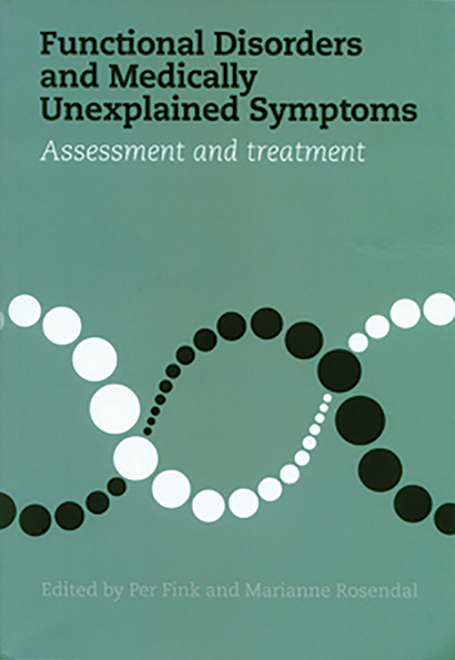
Functional disorders and medically unexplained symptoms: assessment and treatment. Per Fink and Marianne Rosendal, editors. Aarhus, Denmark: Aarhus University Press, 2015 (258 pages). ISBN 9788771248517.

The Research Clinic for Functional Disorders and Psychosomatics at Aarhus University Hospital in Denmark is the background for this book and its authors. This setting differs from the workplace of most Australian doctors, but many of the challenges are the same. Functional disorders are defined as “physical symptoms that cannot be attributed to any known, well defined, physical or psychiatric disorder”. “Medically unexplained symptoms, “bodily distress disorder or syndrome and health anxiety” are also useful descriptions for some patients.

The book covers the historical and cultural background, prevalence, diagnosis and classification of functional disorders. Some of this is rather Eurocentric. Theories of causation and contributing factors, including the doctor’s role, will be familiar to many readers. The chapter on patients’ symptom perception and illness belief is short but helpful.

Of most interest, but perhaps of less practical use, is the treatment plan used in this clinic. There is a general outline of familiar treatment options, including cognitive behavioural therapy and a supportive relationship with the general practitioner. The book describes the concept of stepped care, moving from the first step in primary care of normalisation and empowerment for mild symptoms, escalating to more specialised and multidisciplinary treatment for patients with severe, disabling symptoms. The second half of the book describes a primary assessment and treatment program, TERM, which includes understanding (taking a full history of symptoms and background), acknowledgement and feedback, negotiating a new or modified model of understanding, summarising, and planning follow-up.

If your practice has a significant number of patients who have medically unexplained symptoms, you will find this a useful guide to a more structured approach to helping them. Whether you have the time to carry through the suggestions in a busy general practice or a hospital setting may be more challenging.

Emergency department presentations with mammalian bite injuries: risk factors for admission and surgery

Animal bites, particularly by mammals, are common in Australia,1,2 and their treatment is a substantial public health burden.3 Clinical assessment and the subsequent decision to transfer patients to surgical centres may be challenging, especially for primary health care providers, paramedics and rural emergency departments. There have been few investigations into predictors of hospital admission and surgery for bite injury patients.2,4 We retrospectively analysed the characteristics of all mammalian bite injuries with which patients presented to seven major hospital emergency departments in Victoria during a 2-year period.

Methods

Study design

A retrospective review of all patients presenting with mammalian bite injuries to seven Victorian emergency departments (at the Alfred Hospital, Austin Hospital, Royal Melbourne Hospital, Frankston Hospital, Monash Medical Centre, St Vincent’s Hospital and Western Hospital) during the 2-year period 1 January 2012 – 31 December 2013 was undertaken. Patients were identified using International Statistical Classification of Diseases and Related Health Problems, tenth revision, Australian modification (ICD-10-AM) codes for animal-related injury, and by searching patient record systems for the terms “bite” and “animal-related injuries”. Injuries not involving mammalian bites were excluded.

Descriptive and univariate analysis

All statistical analysis was performed with SPSS Statistics 22 (IBM). Graphs were created in Excel 2013 (Microsoft) and Prism 5 (GraphPad). P < 0.05 (two-tailed) was defined as statistically significant.

The associations between each predictor and outcome of interest were analysed with univariate methods, χ2 tests, analyses of variance (ANOVAs) and Kruskal–Wallis tests as appropriate. Post hoc Bonferroni corrections were performed when appropriate. The choice of potential predictors was based on reports in the literature; age, sex, smoking status, diabetes mellitus, immunosuppression, time to presentation, type of animal and site of injury were assessed. The measured outcomes were hospital admission, surgery, readmission, reoperation, and positive microbiological culture.

Multiple regression analysis

We conducted multiple logistic regression analyses, with stepwise backward elimination by likelihood ratio tests, to further clarify the associations between predictors and outcomes. The probability for stepwise elimination was set at 0.10. This method allowed us to examine the effects of multiple predictors on an outcome. For each predictor, the category with the lowest rate of the outcome of interest was designated as the baseline or reference category. Each regression model was assessed with the Hosmer-Lemeshow test, the Nagelkerke R2, percentage of correct predictions, and area under the receiving operating characteristic curve.

Ethics approval

The investigation was approved by all hospitals involved in this research (Alfred Health Human Research Ethics Committee, reference QA535/13; St Vincent’s Hospital Human Research Ethics Committee, reference QA004/15; Western Health Human Research Ethics Committee, reference QA2014.02; Melbourne Health Office for Research, reference QA2013161; Monash Health Human Research Ethics Committee, reference 14386Q; Frankston Hospital Human Research Ethics Committee, reference QA13PH36; Austin Health Human Research Ethics Committee, reference LNR/15/Austin/525).

Results

Epidemiology of mammalian bites

We identified a total of 717 patients who presented with mammalian bite injuries to the seven Melbourne emergency departments during the study period. Their mean age was 36.5 years, with an equal number of males and females (sex unspecified in one case). Almost all cases (96.1%) involved bites to only one anatomical region; 60.9% involved the upper limbs, 18.7% the head and neck, 14.5% the lower limbs, and 2.0% another part of the body (trunk, back or perineum). Most patients had presented to an emergency department (84.5%) within 24 hours of the injury. The overall rate of hospital admission was 50.8%, and the mean length of stay was 2.7 days. Intravenous antibiotics were administered in 46% of cases; surgery was undertaken in 43.1% of cases. The reoperation rate was 4.5%, the readmission rate was 3%.

A comparison of the demographic and other data for patients presenting with bites by different mammals is shown in Box 1. Almost all bites sustained by patients aged under 15 years were dog bites (92%). Further, 63.1% of patients aged 0–15 years with dog bites were bitten on the head and neck (compared with 13.3% of older patients with dog bites). Dog and human bites were significantly more likely to be seen in male than in female patients (54% and 75%, respectively were sustained by males; P < 0.05); the reverse was true for cat bites (72% were sustained by females).

Patients presenting with cat bites were on average older (mean age, 46.8 ± 19.3 years) than those presenting with bites by other mammals (mean age, 35.7 ± 20.5 years; P < 0.0001). Cat bites comprised 24.0% of all bites in patients aged 60 years or over, compared with 15.9% in other age groups. Cat bites were seen significantly more frequently in female than in male patients (72.1% v 27.9%; P < 0.05). There was no seasonal trend in the frequency of presentation of mammalian bites according to type of bites (data not shown).

Box 2 summarises the rates of hospital admission and the management outcomes for patients presenting with the different bite types. Patients with dog bites usually presented to a hospital on the day of the injury, while presentation with bites by cats and other mammals was often delayed for up to 2 days. Hospital admission rates were significantly higher for cat bites (64% v 48% for all other bites; P < 0.05), and surgery rates were significantly higher for patients with dog bites (48% v 30% for all other bites; P < 0.05). Patients with cat and dog bites were more likely to receive intravenous antibiotics than were those with bites from other mammals (P < 0.05).

Predictive factors for admission to hospital

Patient age, type of animal, the site of injury, and time to presentation of 2 days or more were all significantly associated with admission to hospital (P < 0.01 for all tests). Children under 15 years of age and adults over 60 years of age were more likely to be admitted; the probability also increased with age from the age of 30 years. Admission was more frequent for patients with cat and dog bites than for bites by other mammals. Patients with isolated bites to the head and neck or an upper limb were more likely to be admitted than those with bites to the trunk, back or perineum. However, bites to multiple sites were associated with the greatest risk for admission (Box 3). Finally, smoking was identified by multiple regression analysis as a significant risk factor for admission (adjusted odds ratio [aOR], 1.99 v non-smokers; 95% CI, 1.21–3.28). Sex, immunosuppression and diabetes were not significant risk factors for admission.

Predictive factors for surgery, re-admission and re-operation

Surgery was significantly more frequent for patients with bites by dogs (P < 0.05) and bites to the head and neck, upper limb or multiple sites, and was more frequent in patients who smoked. Patients aged 15–29 years were more likely to undergo surgery as a result of their bite, but this difference was not statistically significant (P < 0.10). Sex, diabetes and immunosuppression were not statistically significantly associated with surgery (Box 4).

After pooling data for all mammalian bites, time to presentation of greater than 2 days was associated with an increased risk of re-operation (OR, 4.41; 95% CI, 1.39–13.95; P = 0.019).

Discussion

Our findings show that presentations by patients to emergency departments with animal bites are frequent, and that a substantial proportion of these patients are hospitalised or undergo surgery. Our data identified certain trends that are consistent with other findings in the literature. Males were more likely to sustain bite injuries, especially by dogs,59 and cat bites were more common in females, as in previous reports.1 In children under 15 years of age, dog bites were more common than other bites (92% of all mammalian bites in this age group were dog bites); further, 20.2% of all dog bites were presented by children under 15 years of age, similar to other reported findings.1,57,10

We also confirmed that the average age of patients presenting with cat bites was higher than for patients presenting with other animal bites.10 The most common site of injury for animal bites of any type was an upper limb, consistent with previous studies,1 although some authors found that the lower limbs were the predominant site of injury for dog bites.8,9 In our study, dog bites more frequently caused head and neck injuries in younger patients than in adults. It has been proposed that children are at particular risk because of their shorter stature, lower capacity for self-defence, and poorer risk awareness with regard to potentially provocative behaviour.7,9

Patients with dog bite injuries usually presented to hospital on the day of the injury, while presentations with bites by cats and other mammals were often delayed. This could be explained by the smaller wound sizes of cat bites, so that patients do not seek medical attention until after infections have developed.11,12 That patients with dog bites had the highest rate of surgery is reasonable, given the depth and complexity of dog bite wounds.5,13 Higher hospital admissions of patients with cat bites may be related to the need for prophylactic intravenous antibiotics, as is currently recommended.2,14

Our study found a relatively high admission rate of 50.8% for mammalian bites. This is at the high end of a broad range of admission rates reported in the literature (4.7–51%).69,15 The variability of these estimates may be explained by differences in the sources of the collected data; some studies analysed surveillance data based on presentations to general practice clinics,1,15 while the emergency department presentations in our study may include a larger proportion of more serious injuries. We also found that delayed presentation for treatment increased the risk of hospitalisation, surgery and reoperation. This is consistent with most studies,4 with the exception of one which found that smoking, an immunocompromised state, and location of the bite over a joint or tendon sheath were associated with hospitalisation.14 Our study confirmed the previously reported association between higher age and the risk of hospitalisation for bite injuries.4 Some traditional risk factors, such as diabetes and immunosuppression, were not significantly associated with hospitalisation, surgery or complications in our study, perhaps because only 3% of our sample were affected by these factors.

A limitation of this study was the retrospective nature of the data collection. Further, the outcomes we analysed were limited to reported hospitalisation and surgery; there was no long term patient follow-up, so that there were no recorded data about any subsequent disabilities.

Our study identified risk factors associated with hospitalisation and surgery. Further analysis of surgical findings in patients who have sustained bite injuries is needed, as this could allow the derivation of risk-stratifying scoring systems from regression models that predict whether a patient will require hospitalisation or surgery. A scoring system could prove beneficial for guiding primary health care providers and emergency physicians in identifying low-risk and high-risk patients who can be managed conservatively or in the outpatient setting, as well as who require timely interhospital transfer or assessment by a surgical unit.

Box 1 –
Demographic data for patients presenting with bites from different mammals

Variable

Source of bite


P

Dog

Cat

Human

Other*


Number of patients

509

122

36

50

Mean age (SD), years

34.8 (21.0)

43.9 (19.3)

37.4 (16.5)

34.9 (17.1)

0.0002

Age group

< 0.0001

< 15 years

103 (20.2%)

4 (3.3%)

1 (3%)

4 (8%)

15–29 years

127 (25.0%)

27 (22.1%)

12 (33%)

19 (38%)

30–44 years

114 (22.4%)

38 (31.1%)

12 (33%)

12 (24%)

45–59 years

97 (19.1%)

29 (23.8%)

8 (22%)

10 (20%)

≥ 60 years

68 (13.4%)

24 (19.7%)

3 (8%)

5 (10%)

Sex

< 0.0001

Female

232 (45.6%)

88 (72.1%)

9 (25%)

29 (58%)

Male

276 (54.2%)

34 (27.9%)

27 (75%)

21 (42%)

Unknown

1 (0.2%)

0 (0%)

0 (0%)

0 (0%)

Site of bite

49

< 0.0001

Head and neck

119 (23.4%)

4 (3.3%)

10 (28%)

1 (2%)

Upper limb

272 (53.4%)

105 (86.1%)

20 (56%)

39 (80%)

Lower limb

84 (16.5%)

12 (9.8%)

1 (3%)

7 (14%)

Trunk, back, perineum

11 (2.2%)

0 (0%)

2 (6%)

1 (2%)

Multiple sites

23 (4.5%)

1 (0.8%)

3 (8%)

1 (2%)

Diabetes mellitus

14 (3.5%)

9 (8.7%)

1 (3.8%)

1 (2%)

0.12

Current smoker

71 (18.0%)

19 (18.6%)

10 (38.5%)

14 (35%)

0.007

Immunosuppression

12 (3.0%)

3 (2.9%)

2 (7.4%)

1 (2%)

0.62


All percentages are column percentages. * Monkey, rat, possum and bat bites. † Data on diabetes, smoking and immunosuppression status were not recorded for all patients.

Box 2 –
Outcomes for patients presenting with mammalian bites

Clinical outcome

Source of bite


P

Dog

Cat

Human

Other*


Number of patients

509

122

36

50

Median time to presentation (IQR), days

0 (0–0)

0.5 (0–1.75)

0 (0–1)

0 (0–4)

< 0.0001

Time to presentation of 2 days or more

54 (11.4%)

29 (24.2%)

5 (14.7%)

17 (34.0%)

< 0.0001

Admission

262 (51.5%)

78 (63.9%)

12 (33.3%)

12 (24.0%)

< 0.0001

Surgery

246 (48.3%)

41 (33.6%)

11 (30.6%)

11 (22.0%)

< 0.0001

Median length of stay (IQR), days

1 (0–2)

2 (1–3.75)

2 (0–3)

2 (1–2.75)

< 0.0001

Positive wound or blood culture

32 (6.3%)

28 (23.0%)

4 (11.1%)

3 (6.0%)

< 0.0001

Administration of intravenous antibiotics

227 (44.6%)

79 (64.8%)

13 (36.1%)

11 (22.0%)

< 0.0001

Re-admissions (percentage of prior admissions)

7 (2.7%)

1 (1.3%)

1 (8.3%)

2 (16.7%)

0.021

Re-operation (percentage of prior operations)

8 (3.3%)

3 (7.3%)

1 (9.1%)

2 (18.2%)

0.074


IQR = interquartile range. All percentages are column percentages. * Monkey, rat, possum and bat bites.

Box 3 –
Univariate and multivariable predictors and prediction score for hospital admission following a mammalian bite injury

Risk factors for admission

Admission rate

Univariate tests (n = 717)


Multiple logistic regression model (n = 545)


Unadjusted OR (95% CI)

Adjusted OR (95% CI)

P


Animal type

< 0.0001

Other

24%

1

Human

33%

1.58 (0.61–4.09)

1.42 (0.44–4.60)

0.555

Dog

52%

3.36 (1.72–6.58)

3.54 (1.60–7.85)

0.002

Cat

64%

5.61 (2.66–11.85)

5.55 (2.32–13.25)

< 0.0001

Age group

0.001

15–29 years

37%

1

< 15 years

55%

2.13 (1.32–3.44)

2.16 (1.17–4.01)

0.014

30–44 years

48%

1.61 (1.06–2.45)

1.84 (1.09–3.12)

0.023

45–59 years

56%

2.21 (1.42–3.45)

2.01 (1.17–3.45)

0.011

≥ 60 years

68%

1.85 (1.28–2.69)

3.84 (2.04–7.21)

< 0.0001

Sex

Female

49%

1

Male

52%

1.12 (0.83–1.50)

NA

NA

Site of bite

0.003

Trunk, back or perineum

7%

1

Lower limb

37%

7.48 (0.94–59.48)

5.52 (0.61–50.0)

0.129

Upper limb

53%

14.51 (1.88–112)

10.94 (1.26–95.3)

0.03

Head and neck

59%

18.67 (2.37–147)

14.72 (1.63–133)

0.017

Multiple sites

57%

17.33 (1.98–151)

22.02 (2.12–229)

0.01

Diabetes mellitus

No

53%

1

Yes

72%

2.26 (0.93–5.49)

NA

NA

Current smoker

No

53%

1

Yes

61%

1.35 (0.89–2.05)

1.99 (1.21–3.28)

0.007

Immunosuppression

No

54%

1

Yes

61%

1.36 (0.52–3.55)

NA

NA

Time to presentation

< 2 days

49%

1

≥ 2 days

65%

1.90 (1.23–2.93)

2.39 (1.30–4.36)

0.005


NA = not applicable (not included in multivariate model because not significant in univariate model); OR = odds ratio.

Box 4 –
Univariate and multivariable predictors and prediction score for surgery following a mammalian bite injury

Risk factors for surgery

Surgery rate

Univariate tests (n = 717)


Multiple logistic regression model (n = 545)


Unadjusted OR (95% CI)

Adjusted OR (95% CI)

P


Animal type

< 0.0001

Other

22%

1

Human

31%

1.56 (0.59–4.14)

1.39 (0.44–4.44)

0.576

Dog

34%

1.79 (0.83–3.87)

1.92 (0.82–4.42)

0.135

Cat

48%

3.32 (1.66–6.62)

4.47 (2.03–9.81)

< 0.0001

Age group

0.074

15–29 years

35%

1

< 15 years

55%

2.29 (1.42–3.70)

1.77 (0.95–3.32)

0.074

30–44 years

38%

1.11 (0.72–1.70)

1.63 (0.97–2.77)

0.067

45–59 years

50%

1.85 (1.18–2.88)

2.13 (1.24–3.66)

0.006

≥ 60 years

44%

1.45 (0.88–2.38)

1.61 (0.89–2.89)

0.113

Sex

Female

41%

1

Male

45%

1.15 (0.85–1.54)

NA

NA

Site of bite

0.002

Trunk, back or perineum

7%

1

Lower limb

33%

9.14 (1.19–70.50)

7.886 (0.90–69.0)

0.062

Upper limb

41%

6.31 (0.79–50.28)

12.80 (1.52–108)

0.019

Head and neck

50%

13 (1.49–113)

21.13 (2.08–214)

0.01

Multiple sites

60%

19.26 (2.45–152)

23.89 (2.71–211)

0.004

Diabetes mellitus

No

53%

1

Yes

52%

0.96 (0.43–2.15)

NA

NA

Current smoker

No

52%

1

Yes

58%

1.29 (0.85–1.95)

1.96 (1.20–3.18)

0.007

Immunosuppression

No

53%

1

Yes

56%

1.13 (0.44–2.90)

NA

NA

Time to presentation

< 2 days

47%

1

≥ 2 days

36%

0.63 (0.41–0.97)

NA

NA


NA = not applicable (not included in multivariate model because not significant in univariate model); OR = odds ratio.