×

Factors associated with quality of care for patients with pancreatic cancer in Australia

The known Treating patients with pancreatic cancer is challenging, and socio-demographic factors influence whether patients receive specific treatment forms, such as surgery and chemotherapy. 

The new Our composite quality of care score was lower for patients from rural or socially disadvantaged areas; it was higher for patients who first presented to a hospital with a high pancreatic case volume. A higher score was significantly associated with improved survival. 

The implications Strategies should be developed which ensure that all patients with pancreatic cancer have the opportunity to receive optimal care from or in conjunction with high pancreatic case volume centres. 

In Australia, pancreatic cancer is the tenth most common cancer, and the fourth leading cause of cancer-related death.1 One-year survival is 20%, 5-year survival 6%.2 Treating pancreatic cancer presents distinctive challenges, and requires highly specialised care to achieve optimal outcomes.3 Studies in Australia and overseas have shown that fewer patients receive the recommended treatment than expected,4,5 that receiving recommended care is inconsistent,6,7 and that socio-demographic factors influence the treatment of patients with pancreatic cancer.7,8 Treating patients in non-specialised centres appears to at least partly explain these findings.9,10

Previous studies have tended to focus on individual types of treatment, such as surgery or chemotherapy. We took a more holistic approach and calculated an overall quality of care score for Australian patients diagnosed with pancreatic cancer. We examined variations in the score associated with patient and health service-related factors, and analysed the relationship between quality of care and survival.

Methods

This analysis was nested within a population-based study of patterns of care for patients in Australia with pancreatic cancer. Eligible patients were residents of Queensland and New South Wales diagnosed with pancreatic cancer between July 2009 and June 2011. Patients with histological confirmation of pancreatic adenocarcinoma were included, as were patients with presumed pancreatic cancer but without histological or cytological confirmation. Trained research nurses collected information about patient treatment from medical records in public and private facilities.4 Patients were excluded from this analysis if they died within one month of diagnosis or clinical staging data were unavailable.

We calculated a quality of care score based on the results of our previously reported Delphi process.11 Briefly, clinicians from a range of specialties involved in care for patients with pancreatic cancer were asked “What is important in the care of patients with pancreatic cancer?” A list of statements was prepared on the basis of a thematic analysis of the responses. The clinicians were asked to score each statement on a scale of 0 (“disagree”, “not important”) to 10 (“strongly agree”, “very important”). The mean score and the coefficient of variation (CV) were calculated for each statement.

Calculating the quality of care score

We calculated quality of care scores on the basis of the mean Delphi process scores, selecting statements about which there had been reasonable consensus in the Delphi process (CV ≤ 0.4) and when information for assessing whether the item of care had been delivered was available in our database. Eighteen items were included in the analysis (Box 1).

For each patient, we calculated a potential score by identifying the items that applied to their clinical situation and summing the mean scores from the Delphi survey for these items. For example, items related to surgical procedures were included only for patients who underwent attempted resection. We then identified items for which there was evidence that the specified care had been delivered and summed their mean Delphi scores as a score for care delivered. The proportional care score was calculated by dividing the care delivered score by the potential score, yielding a value between 0 and 1. The clinical information that determined eligibility and whether or not care specified by an item was delivered are shown in Box 1.

Measurement of potential determinants of care

Patient characteristics assessed included age, sex, Eastern Cooperative Oncology Group (ECOG) performance status, and Charlson comorbidity index.12 Based on their area of residence at diagnosis, each person was allocated a socio-economic index for areas (SEIFA)13 score and Accessibility/Remoteness Index of Australia (ARIA+)14 category. We grouped the SEIFA scores into quintiles, and collapsed the ARIA into three levels: major city, inner regional, and rural (which included the outer regional, remote and very remote categories).

Tumour-related factors included the stage of the tumour, categorised as potentially resectable or not, and as confined to the pancreas, locally advanced, or metastatic.

Health service-related factors included the type of specialist first seen, and the number of pancreatic cancer presentations (volume) for the facility to which the patient first presented.

Statistical analysis

The proportions of eligible patients who received each item of care were calculated; the statistical significance of differences between proportions according to socio-economic status and place of residence categories was assessed in χ2 tests.

We used linear regression analyses, with the proportional score as the outcome, to examine variation in the score attributable to patient-, tumour- and health service-related factors. Mean proportional scores for levels of each exposure variable were calculated and β coefficients reported (with 95% confidence intervals [CIs]). The β coefficients were interpreted as the difference between the mean score for patients in a particular category and that of patients in the reference category. Multivariable models included age, ECOG performance status, and comorbidity score as factors.

Survival time was calculated from the date of diagnosis until the death of the patient or the date of the final follow-up (February 2014). Patients were grouped in quartiles according to their proportional care scores; Kaplan–Meier graphs were generated and log-rank tests assessed differences in survival according to score quartile. We also performed the analysis with the proportional care score as a continuous variable; we report changes in survival associated with each 10 percentage point increase in score, using Cox proportional hazard models to adjust for patient-related factors and clinical stage. The association between the score and survival was further investigated by calculating adjusted hazard ratios for each care score item separately. Analyses were performed for the entire patient group and separately for patients with or without metastases identified at clinical staging. We used Stata 14 (StataCorp) for all analyses. P < 0.05 (two-sided) was deemed statistically significant.

Ethics approval

Access to medical records was approved under the Queensland Public Health Act and the NSW Privacy Act. Ethics approval was obtained from the QIMR Berghofer Medical Research Institute (reference, P1292), the Royal Brisbane and Women’s Hospital (on behalf of all public hospitals in Queensland; reference, HREC/10/QRBW/16), and the NSW Population and Health Services Research Ethics Committee (reference, HREC/10/CIPHS/45).

Results

A total of 1896 patients were eligible for inclusion in the patterns of care study. We were unable to locate medical records for 33 patients; 259 had died within one month of diagnosis, and staging information was not available for 33, so that 1571 patients (83%) were included in our analysis, including 867 men (55%). At clinical staging, 781 patients (49.7%) had non-metastatic disease and 790 (50.3%) metastatic disease. Most patients lived in major cities (1076, 68%); 338 (22%) lived in inner regional areas and 157 (10%) in rural areas. Almost three-quarters of patients (1151, 73%) died within one year of diagnosis. The median survival time was 6 months (11 months for patients without metastases; 4 months for those with metastases).

Younger patients and those with better ECOG performance status had higher care scores than older and less active patients with pancreatic cancer (Appendix 2). ARIA+ category, area level socio-economic status, age, ECOG performance status, institutional pancreatic cancer case volume, and specialist first seen were all factors that significantly influenced the care score (Box 2; Appendix 3). After adjusting for these factors, the care scores for patients living in rural areas were 11% lower (95% CI, 8–13%) than for those living in major cities. The care scores for patients living in more disadvantaged areas were up to 8% lower (95% CI, 6–11%) than for patients living in the least disadvantaged areas. Care score estimates for patients presenting to a low pancreatic cancer case volume hospital (fewer than ten presentations per year) were 13% lower (95% CI, 11–15%) than for those presenting to hospitals with more than 30 presentations annually. They were higher for patients for whom a hepatobiliary surgeon was the first specialist seen; scores for patients initially seeing a general surgeon were 10% lower (95% CI, 8–13%) (Box 2).

To further investigate the association between ARIA+ category and care score, models were then also adjusted for the pancreatic cancer case volume of the first hospital and specialist seen. The differences in the adjusted mean scores for major cities and rural areas (5% lower for rural patients; 95% CI, 3–8%) and between least and most disadvantaged areas (6% lower for most disadvantaged patients; 95% CI, 3–8%) were lower in this model.

For patients who had been clinically staged with non-metastatic disease, the factors most strongly associated with lower care scores were being seen initially by a general rather than a hepatobiliary surgeon (17% lower; 95% CI, 13–21%), living in a rural area rather than a major city (11% lower; 95% CI, 8–15%), and being at least 80 years of age (v aged less than 60 years: 16% lower; 95% CI, 13–20%). For patients diagnosed with metastatic disease, being seen at a lower volume facility (15% lower; 95% CI, 12–17%) and having a poorer ECOG performance status (11% lower; 95% CI, 7–15%) were the factors most strongly associated with quality of care.

Individual items of care were also examined. Less than one-third of patients received some items: 31% were presented to multidisciplinary teams (MDTs), received psychosocial support (19%), participated in clinical trials (7%), or were first seen by a hepatobiliary surgeon (19%). Most eligible patients were offered resection or received a valid reason why they were not (98%), had a tissue diagnosis (80%), saw a medical oncologist (86%), and were referred to palliative care (82%) (Box 1). There were significant differences for patients according to their ARIA+ category and area level socio-economic status; for example, 32 patients living in rural areas (41%) were referred to a hepatobiliary surgeon, compared with 53% of patients (290 of 548) in metropolitan areas (Appendix 4, Appendix 5).

Patients with scores in the highest quartile of proportional care scores had an estimated median survival time of 8 months, double that for those with scores in the lowest quartile. Median survival time for patients with non-metastatic disease in the highest and lowest score quartiles was 14 and 7 months respectively; for those with metastatic disease, it was 5 and 3 months (Box 3).

After adjusting for age, ECOG performance status, comorbidities, and clinical stage of pancreatic disease, each 10 percentage point increase in proportional care score was associated with a statistically significant 6% reduction in the risk of dying (hazard ratio [HR], 0.94; 95% CI, 0.91–0.97; Box 4). The reduction was greater for patients who were diagnosed with non-metastatic disease (adjusted HR, 0.91; 95% CI, 0.87–0.95) than for those with metastatic disease (adjusted HR, 0.95; 95% CI, 0.91–0.99).

Individual care score items that were statistically significantly associated with survival included having a diagnostic tissue sample collected (HR, 0.66; 95% CI, 0.57–0.77), being offered adjuvant chemotherapy (HR, 0.43; 95% CI, 0.33–0.56), being referred to a hepatobiliary surgeon if potentially resectable (HR, 0.82; 95% CI, 0.69–0.96), being presented to an MDT (HR, 0.86; 95% CI, 0.77–0.96), being offered psychosocial support (HR, 1.24; 95% CI, 1.09–1.12), pancreatic enzyme replacement therapy (HR, 0.83; HR, 95% CI, 0.73–0.94), and, if diagnosed with metastatic disease, referral to palliative care (HR, 1.42; 95% CI, 1.17–1.74) (Appendix 6).

Discussion

We found that the quality of care for patients with pancreatic cancer varied according to their age, where they live, and the pancreatic cancer case volume of the hospital to which they first presented. We also found that higher quality of care was associated with improved survival. This association was strongest for patients clinically staged with non-metastatic pancreatic cancer, for whom there is more scope for treatment that can increase survival.

Earlier studies found that receiving surgery, chemotherapy and palliative care was influenced by the age, education, place of residence, ethnic background, and marital status of patients.5,7,15 By applying a composite measure of care that included a broad range of factors, we found that age and ECOG performance status influenced its overall quality. While this is unsurprising, it is important to recognise that age alone is not a barrier to high quality care. Our more worrying finding is that quality of care varied according to the geographic classification and the area level socio-economic status of the patient’s place of residence. This is at least partly explained by differences in access to specialists and care in high case volume centres, suggesting that interventions which ensure that all patients are managed by high volume teams could improve the quality of care.

Our analysis of individual care items found that the proportion of people receiving care from specialist teams, as recommended, was particularly small: fewer than one-third of patients had been referred to an MDT, only half of potentially resectable patients had been referred to a hepatobiliary surgeon, and referral to a clinical trial was only rarely considered, even though these factors have consistently been found to influence the quality of care.9,16,17 These aspects of care were particularly poorly delivered to patients living in more rural areas. Distance causes particular challenges in Australia,1820 but they should not be insurmountable; it has been reported, for example, that a multi-level approach (such as telemedicine MDTs and formalising referral relationships between regional and metropolitan centres) can improve outcomes.21

Survival for patients with lower care scores was poorer, consistent with previous reports.2224 This association was stronger for patients diagnosed with non-metastatic disease, for whom there is more scope for influencing survival by ensuring that staging is adequate, that surgery is undertaken in high case volume centres, and that patients have access to adjuvant chemotherapy. For patients with metastatic disease, a focus on quality-of-life indicators is arguably more important; this could be explored in further investigations of care quality.

Some care items were associated with a greater hazard of dying when the care was received, including statements that patients should be “offered psychosocial support”, that “patients with metastatic disease should be referred to palliative care”, and that “patients with technically resectable disease should be offered resection or a valid reason for no surgery”. Receiving psychosocial and palliative care is more likely as the expected survival time shortens, and this probably explains these findings (reverse causation). The care item regarding resection was classed as having been delivered if a valid reason for the resection not being offered had been recorded. This applied to 28% of patients eligible for resection; the reasons for not attempting surgery included older age, comorbidity, and poor ECOG performance status, each of which were associated with poor survival. When these three care items were all omitted from the care score, the risk of death was 2% lower for each 10 percentage point increase in care score (data not shown).

Our study was comprehensive, reasonably large, and population-based, and was also the first Australian investigation to assess the overall quality of care with a single score. Nevertheless, it had some limitations. Firstly, different weights for the care items may have been obtained if another mix of specialists had participated in the Delphi process. Secondly, the Delphi study highlighted the importance of communication between patients and clinicians. This factor cannot be adequately captured in a medical record review and could therefore not be incorporated into our score, but may have influenced decisions regarding care. Thirdly, some patients may have been incorrectly classified as having resectable tumours, which would have affected their eligibility for certain care items and thereby the delivery of appropriate care. Finally, although we controlled for age, ECOG performance status and comorbidities, we may not have completely accounted for confounding patient-related factors.

In conclusion, our population-based study provides evidence that the geographical location of their place of residence, among other factors, influences the quality of care received by Australian patients with pancreatic cancer, and that survival can be improved by delivering optimal care. Systems of care need to be implemented which ensure that equitable treatment is provided for all Australian patients with pancreatic cancer.

Box 1 –
Statements about care for patients with pancreatic cancer deemed to be most important in our Delphi process, patient eligibility criteria, and definition of care received

Care statement

Weight*

Eligible patients

Number eligible

Number who received care

Care received


All patients with potentially resectable disease should be referred to a hepatobiliary surgeon§

9.3

Non-metastatic

781

401 (51%)

Any referral or consultation with hepatobiliary surgeon

All patients with technically resectable disease should be offered resection or valid reason for not doing so

9.2

Potentially resectable

519

509 (98%)

Surgery attempted or valid reason for not doing so

Surgery should be performed by surgeons who perform more than five pancreatic resections per year

9.0

Resection attempted

366

158 (43%)

Surgeon performed more than five resections per year

Tumour resectability should be assessed by an MDT at a tertiary hospital

9.0

Non-metastatic

781

229 (29%)

MDT prior to attempted surgery, or within 40 days of diagnosis if no surgery

All patients should have a triple phase/pancreas protocol CT scan for staging

8.9

All patients

1571

674 (43%)

Evidence of pancreas protocol CT

Entry into a clinical trial should be considered for all patients

8.8

All patients

1571

103 (7%)

Clinical trial discussed, considered, offered or participated in a trial

Surgery should take place in tertiary institutions where more than 15 resections are performed annually**

8.6

Resection attempted

366

152 (42%)

Attempted resection performed at hospital with more than 11 resections each year**

Each patient should be assigned a care coordinator and an individualised treatment/clinical plan

8.5

All patients

1571

345 (22%)

Evidence of a navigator, care plan or nursing referral

Tissue diagnosis should be obtained where possible

8.3

All patients

1571

1251 (80%)

Histology or cytology analysis completed

All patients should be presented to an MDT

8.3

All patients

1571

494 (31%)

Evidence of presentation to an MDT

Biliary obstruction should routinely be managed endoscopically in non-resectable patients

8.2

Non-resectable with biliary obstruction

416

346 (83%)

Evidence of endoscopic biliary stent, not bypass surgery

All patients should be offered adjuvant therapy after surgery, assuming performance status is adequate

8.1

Resection attempted

366

244 (67%)

Evidence of any adjuvant chemo- or radiation therapy

All patients should be offered psychosocial support

8.0

All patients

1571

301 (19%)

Evidence of referral to or consultation with psychological services

Pancreatic enzyme replacement therapy should be considered for all patients

7.9

All patients

1571

345 (22%)

Evidence of pancreatic enzyme replacement

All patients should see a medical oncologist

7.9

All patients

1571

1353 (86%)

Seen by a medical oncologist or valid reason why not

A specialist hepatobiliary surgeon should be the initial/primary specialist unless the patient has obvious metastases

7.3

Non-metastatic

781

146 (19%)

Hepatobiliary surgeon was the first specialist seen

All patients should be referred to a dietitian soon after diagnosis

7.3

All patients

1571

1000 (64%)

Evidence of referral to or consultation with dietitian

Patients with confirmed metastatic disease should be referred to palliative care

6.0

Metastatic

790

646 (82%)

Any evidence of palliative care consultation or referral


CT = computerized tomography; MDT = multidisciplinary team meeting. * Final mean average score of importance from Delphi process. † Patients eligible for care according to classification by clinical staging. ‡ Number and percentage of eligible patients who received the item of care. § Hepatobiliary surgeon: defined as a surgeon who had undergone recognised specialist hepatobiliary surgery training or who was recognised by peers as an experienced hepatobiliary surgeon. ¶ Includes all inpatient records and consultations. ** Only three hospitals from the patterns of care study performed 15 resections each year; this high volume classification was therefore amended, on the basis of Australian data and literature reports, to hospitals where 11 or more resections were performed each year.

Box 2 –
Associations between patient, tumour and health service-related characteristics and proportional care scores for all patients, and for patients with or without evidence of metastases at clinical staging

Adjusted β coefficient (95% confidence interval)*


All patients

Patients without metastases

Patients with metastases


Number of patients

1571

781

790

Age group

< 60 years

Reference

Reference

Reference

60–69 years

0.01 (−0.01 to 0.03)

0.01 (−0.02 to 0.04)

0.00 (−0.03 to 0.04)

70–79 years

−0.05 (−0.08 to −0.03)

−0.05 (−0.08 to −0.02)

−0.06 (−0.09 to −0.03)

≥ 80 years

−0.13 (−0.15 to −0.10)

−0.16 (−0.20 to −0.13)

−0.10 (−0.13 to 0.06)

P (overall; trend)

< 0.001; < 0.001

< 0.001; < 0.001

< 0.001; < 0.001

Sex

Women

Reference

Reference

Reference

Men

−0.01 (−0.02 to 0.01)

0.01 (−0.01 to 0.03)

−0.03 (−0.05 to −0.00)

P (overall)

0.34

0.40

0.03

Charlson comorbidity score

0

Reference

Reference

Reference

1

−0.01 (−0.03 to 0.01)

−0.00 (−0.03 to 0.02)

−0.01 (−0.03 to 0.02)

2

−0.01 (−0.03 to 0.01)

−0.01 (−0.04 to 0.02)

−0.01 (−0.04 to 0.02)

P (overall; trend)

0.64; 0.38

0.88; 0.63

0.89; 0.66

ECOG performance status

0

Reference

Reference

Reference

1

−0.01 (−0.03 to 0.01)

−0.01 (−0.04 to 0.02)

−0.01 (−0.04 to 0.02)

≥ 2

−0.06 (−0.08 to −0.03)

−0.06 (−0.09 to −0.03)

−0.05 (−0.08 to −0.02)

Not stated

−0.09 (−0.12 to −0.06)

−0.07 (−0.11 to −0.03)

−0.11 (−0.15 to −0.07)

P (overall; trend)

< 0.001; < 0.001

< 0.001; < 0.001

< 0.001; < 0.001

Residence (ARIA+ classification)

Major city

Reference

Reference

Reference

Inner regional

−0.06 (−0.08 to −0.04)

−0.03 (−0.06 to −0.00)

−0.08 (−0.11 to −0.05)

Rural

−0.11 (−0.13 to −0.08)

−0.11 (−0.15 to −0.08)

−0.09 (−0.13 to −0.06)

P (overall; trend)

< 0.001; < 0.001

< 0.001; < 0.001

< 0.001; < 0.001

Socio-economic status (quintiles)

1 (least disadvantaged)

Reference

Reference

Reference

2

−0.03 (−0.06 to −0.01)

−0.04 (−0.07 to −0.00)

−0.03 (−0.07 to 0.01)

3

−0.07 (−0.10 to −0.04)

−0.08 (−0.12 to −0.05)

−0.06 (−0.10 to −0.02)

4

−0.08 (−0.11 to −0.05)

−0.08 (−0.12 to −0.05)

−0.08 (−0.12 to −0.04)

5 (most disadvantaged)

−0.08 (−0.11 to −0.06)

−0.07 (−0.10 to −0.03)

−0.10 (−0.13 to −0.06)

P (overall; trend)

< 0.001; < 0.001

< 0.001; < 0.001

< 0.001; < 0.001

Clinical stage of disease

Confined to pancreas

Reference

NA

NA

Locally advanced

−0.02 (−0.04 to 0.01)

Metastatic

−0.02 (−0.04 to 0.00)

P (overall; trend)

0.26; 0.14

Pancreatic cancer case volume of first facility seen

> 30 per year

Reference

Reference

Reference

10–29 per year

−0.06 (−0.08 to −0.04)

−0.07 (−0.10 to −0.05)

−0.04 (−0.07 to −0.02)

< 10 per year

−0.13 (−0.15 to −0.11)

−0.10 (−0.13 to −0.07)

−0.15 (−0.17 to −0.12)

P (overall; trend)

< 0.001; < 0.001

< 0.001; < 0.001

< 0.001; < 0.001

First specialist seen

Hepatobiliary surgeon

Reference

Reference

Reference

Gastroenterologist

−0.09 (−0.11 to −0.06)

−0.12 (−0.15 to −0.09)

−0.03 (−0.07 to 0.01)

General surgeon

−0.10 (−0.13 to −0.08)

−0.13 (−0.16 to −0.10)

−0.05 (−0.09 to −0.01)

Other

−0.14 (−0.16 to −0.11)

−0.17 (−0.21 to −0.13)

−0.10 (−0.14 to −0.06)

P (overall)

< 0.001

< 0.001

< 0.001


ECOG = Eastern Cooperative Oncology Group; NA = not applicable. * Adjusted for age group at diagnosis (< 60, 60–69, 70–79, ≥ 80 years), ECOG performance status (0, 1, ≥ 2, not stated), and Charlson comorbidity index score (0, 1, ≥ 2). † Includes patients in outer regional, remote and very remote areas.

Box 3 –
Kaplan–Meier survival curves for all patients, patients with non-metastatic disease and patients with metastatic disease on clinical staging, by proportional care score (quartiles)


* Log-rank test of equality of survivor functions across proportional care score quartiles.

Box 4 –
Association between total care score and survival according to stage of pancreatic cancer at diagnosis

Number of patients

Hazard ratio (95% CI)*


Unadjusted

P

Adjusted

P


All patients

1571

0.90 (0.87–0.93)

< 0.001

0.94 (0.91–0.97)

< 0.001

Non-metastatic disease

781

0.87 (0.83–0.91)

< 0.001

0.91 (0.87–0.95)

< 0.001

Metastatic disease

790

0.95 (0.91–0.98)

0.006

0.95 (0.91–0.99)

0.013


* Reduction in the risk of dying associated with a 10 percentage point increase in care score. † Adjusted for age group, Eastern Cooperative Oncology Group performance status, Charlson comorbidity score, and clinical stage.

Major trauma mortality in rural and metropolitan NSW, 2009–2014: a retrospective analysis of trauma registry data

The known Trauma systems facilitate the timely treatment of major trauma patients at specialised centres, and this approach has reduced trauma-related mortality in Australia and overseas. Trauma in rural areas, however, has been associated with higher mortality. 

The new Trauma system changes introduced to New South Wales since 2009 may be associated with a decline in crude and adjusted inpatient mortality after major trauma in rural and regional locations. 

The implications Better transfer of rural patients, retrieval networks, and improved trauma care at major trauma centres may be benefiting severely injured patients from rural and regional locations in NSW. 

Injury causes significant physical and psychological disability around the world,1,2 and cost-effective systems of care are important for optimising patient outcomes and recovery. Trauma systems facilitate the timely treatment of severely injured patients, and this approach has reduced mortality among trauma patients in several Australian states3,4 and overseas.5,6 A system for trauma care was introduced in New South Wales in 1991, and the first analyses of major trauma outcomes in NSW were conducted in 2012.7,8 It was found that a survival benefit was associated with definitive care at designated major trauma centres located in NSW metropolitan areas.8

A number of factors may influence outcomes for trauma patients in rural and regional areas, particularly the sparse population density and the vast transport distances for many rural and regional trauma patients. Studies from other parts of Australia have indicated that rural trauma patients have poorer outcomes than metropolitan trauma patients, and it is recognised that road accidents in rural and remote locations are associated with higher risks of death and severe injury.9,10 The NSW Trauma Minimum Dataset now includes additional details on injuries, including those for a number of regional centres,11 allowing more detailed analyses of outcomes according to the geographic location of injuries incurred after 2009.

This change was particularly relevant because the revised NSW State Trauma Plan was implemented in 2009, formalising rural and regional referral networks for each of the seven adult major trauma centres.12 The aim of these networks was to facilitate the timely transfer of severely injured patients from sparsely populated rural and remote areas of NSW to major trauma centres in metropolitan areas along the east coast. We assessed and compared trends in crude and risk-adjusted mortality between 2009 and 2014 in the context of these changes, designed to improve trauma care and patient outcomes in NSW.

Methods

NSW is the most populous Australian state, with a population of 7.5 million in 2014 and an area of 809 000 km2. Around 70% of the population live in metropolitan areas on the eastern seaboard.13

We undertook a retrospective analysis of statewide trauma registry data. The statewide trauma registry was established and is maintained by the NSW Institute of Trauma and Injury Management; it receives data from seven adult major trauma centres and ten regional trauma centres.11 According to the standards of the American College of Surgeons,14 major trauma centres in NSW are equivalent to level 1 designated trauma centres (the top level), and regional trauma centres are equivalent to level 2 or 3 centres.

Adult patients (aged 16 years or more) were included in our analysis if they presented to a NSW hospital between 1 January 2009 and 31 December 2014, and their Injury Severity Score (ISS) was greater than 15.

Trauma centres that did not submit data for the entire study period were excluded. Patients were also excluded if the postcode of the site where they were injured was unknown or outside NSW, as were those who were dead on arrival at hospital. Duplicate records for patients who had been transferred between hospitals were identified, and the second and subsequent records excluded if the referral hospital provided trauma data to the NSW Trauma Registry.

Basic demographic characteristics, the mechanism of injury, vital signs on arrival at hospital, length of stay in hospital, and in-hospital mortality were analysed. Injuries were classified according to the Abbreviated Injury Scale (AIS). The AIS codes injuries according to their anatomic location (head, neck, chest, abdomen, lower limb, upper limb, external) and assigns a severity score, ranging from 1 to 6, according to the likelihood of death and disability. Severe injuries in this study were defined by AIS scores of 3 or more.15 The ISS, used to assess the overall severity of injury, was calculated by summing the squares of the AIS severity scores for the three most severely injured body regions. Major trauma was defined as an ISS greater than 15. Measures of hospital resource use included inter-hospital transfer, and the first major trauma procedures recorded by the referring hospital, ambulance or retrieval services, or trauma centre. The geographic location where the patient sustained their injury was identified by postcode, and categorised as metropolitan, inner regional, or outer regional/remote according to the Australian Statistical Geography Standard Remoteness Structure.16 As the number of deaths in outer regional/remote regions was small, all non-metropolitan postcodes were merged as rural/regional for multivariable analyses.

The primary outcome for our study was in-hospital mortality after major trauma, analysed according to the geographic location of the injury.

Statistical analyses

Descriptive statistics for baseline characteristics and crude in-hospital mortality are reported. Year-by-year differences and trends in crude in-hospital mortality were compared in χ2 and Cochran–Armitage linear trend tests. Risk-adjusted mortality trends were assessed in logistic regression models stratified by geographic location of the injury and adjusted for age, injury severity, intensive care unit admission, and year of admission. Variables were included in models according to the results of preceding univariate analyses (P < 0.1), or if they had been identified as influencing mortality risk in adult injury patients by previous studies.8 Adjusted odds ratios (ORs) for inpatient mortality in separate rural and metropolitan location regression models were plotted on a line chart to identify trend in ORs relative to the reference year, 2009. Multivariable adjusted trends were determined by changing the year predictor variable to a linear continuous variable for logistic regression modelling and non-parametric generalised additive models (GAM). Analyses were performed in SAS Enterprise Guide 6.1 (SAS Institute).

Ethics approval

Approval was obtained from the NSW Population Health Services and Research Ethics Committee (reference, 2015/04/036).

Results

Study population

A total of 18 652 patients were identified in the trauma registry data for the period 2009–2014. After the exclusions described in the Methods, data for 11 423 adult patients with an ISS greater than 15 from seven adult major trauma centres and three regional trauma centres were analysed (Appendix).

Box 1 compares the demographic characteristics of rural/regional (combined inner, outer regional and remote injury locations) and metropolitan major trauma patients, and the clinical characteristics of their injuries. With respect to location of injury, 8878 patients (77.7%) were in metropolitan locations, 1855 (16.2%) in inner regional locations, 601 (5.3%) in outer regional locations, and 89 (0.8%) in remote locations. The mean age of the patients was 53.5 years (SD, 23.1); 71.9% of the patients were men. The most common mechanisms of injury were falls (44.3%), road trauma (37.6%) and blunt assault (5.9%); penetrating injuries accounted for 3.6% of all cases.

The distribution of ages of rural/regional trauma patients, compared with metropolitan patients, was shifted to younger age groups. The proportions of head, chest, spinal, and lower limb severe injuries were higher for rural/regional trauma patients, consistent with the higher proportion of road trauma cases in this patient population (Box 1).

Inpatient major trauma mortality

Almost half of major trauma inpatient deaths (46.6%) occurred within 24 hours of admission, a further 30% occurred during the first week of admission, and 23.4% occurred after the second week of admission. The overall inpatient mortality rate for major trauma was 14.1% in 2009 and 14.5% in 2014, with no significant trend (Cochran–Armitage test, P = 0.66). There was no difference in overall inpatient mortality between admissions to major trauma and non-major trauma centres (14.0% v 13.0%; P = 0.47). For those injured in metropolitan locations, inpatient mortality was 14.7% in 2009 and 16.1% in 2014 (Cochran–Armitage test, P = 0.45). Major trauma in rural/regional locations was associated with a statistically significant decrease in in-hospital mortality over this period, from 12.1% in 2009 to 8.7% in 2014 (Cochran–Armitage test, P = 0.004). When rural/regional location was further stratified, major trauma mortality in inner regional locations decreased from 12.7% in 2009 to 10.4% in 2014 (P = 0.07); for outer regional/remote locations, it decreased from 10.3% in 2009 to 4.0% in 2014 (P = 0.005).

Box 2 shows the trend in risk-adjusted mortality for rural/regional and metropolitan injuries, compared with the reference year 2009. The adjusted OR for in-hospital mortality associated with rural/regional injuries was lower in 2013 (OR, 0.5; 95% CI, 0.3–0.8) and 2014 (OR, 0.6; 95% CI, 0.4–1.0) than in 2009. Mortality among rural patients declined by an average of 12% per year when year was analysed as a linear predictor (OR, 0.88; 95% CI, 0.81–0.96; P = 0.004); non-parametric GAM analysis indicated that this decline was statistically significant (multivariable GAM, P = 0.007). In contrast, there was no trend in risk-adjusted mortality for injuries in metropolitan locations (multivariable GAM, P = 0.25).

Mode of arrival and inter-hospital transfer

For the 2292 major trauma patients injured in a rural location and admitted to a major trauma centre, there was no change between 2009 and 2014 in the proportion who arrived directly (not transferred from another hospital) at a major trauma centre from a rural location (38.7% v 36.7%; Cochran–Armitage test, P = 0.96). There was an increase in the proportion who arrived at a major trauma centre by ambulance from a rural location (26.2% v 60.6%; P < 0.001), and a decline in the median time from injury to first trauma procedure from 276 min (interquartile range [IQR], 104–1420) in 2010 to 95 min (IQR, 48–495) in 2014 (P < 0.001).

Discussion

We found that overall inpatient mortality after major trauma remained steady between 2009 and 2014, but crude mortality declined among patients severely injured in rural and regional NSW. There was also a reduction in risk-adjusted mortality associated with rural location of injury. Comparison of data on crude inpatient mortality identified that this trend was most marked for injuries incurred in outer regional and remote locations.

These findings could be explained by a number of factors. Firstly, the establishment of trauma referral networks may have resulted in more efficient transfers between rural facilities and major trauma centres. We found that the time to first trauma procedure for patients from rural areas had declined since 2009. The NSW Ambulance Service trauma bypass protocol (protocol T1), revised in 2008, allowed for an increase in acceptable transfer time between the scene of the injury and arrival at the nearest designated trauma centre from 30 minutes to one hour.17 In addition, the introduction of the Rapid Launch Trauma Coordinator function in the NSW Ambulance Aeromedical Control Centre aimed to improve the early identification of major trauma incidents by monitoring emergency 000 calls to ensure that specialist pre-hospital resources are activated as early as possible. Both factors may have contributed to the steady increase in direct ambulance arrivals at major trauma centres from rural locations between 2009 and 2014. This has occurred although the proportion of inter-hospital transfers from rural and regional hospitals to major trauma centres was unchanged. It is unclear whether direct or secondary transfer from a rural hospital or direct transport to a major trauma centre is associated with improved survival. A 2011 systematic review of 36 observational studies found no difference, although most investigations were subject to potential referral bias, in that they excluded deaths from the data for referring hospitals.18

Secondly, improved outcomes for severely injured rural patients may have resulted from improved clinical care in regional trauma centres and rural referral centres. A core mission of the NSW Institute for Trauma and Injury Management over the past decade has been to coordinate and improve access to clinical expertise and education resources in these centres.12 This approach included employing regional trauma nurse coordinators, which achieved improved education, case management, data collection and audit capabilities. An American study of 18 rural level 3 and 4 trauma centres found that a rural trauma education course alone reduced the time to transfer of severely injured patients.19

Thirdly, the reduction in major trauma mortality in outer regional and remote locations underscores the importance of road safety initiatives for preventing deaths and critical injuries. The reductions reported here mirror the 11% drop in the rate of road accident deaths (per 100 000 population) in very remote locations between 2008 and 2012, compared with reductions of 0.7% in inner regional and 0.9% in metropolitan areas.20 The National Road Safety Strategy highlights the need to prioritise improving high risk rural and urban roads, vehicle safety standards, and safety for vulnerable road users.20 Nevertheless, rural road deaths continue to occur at two to three times the national average because of the longer travel distances, higher vehicle speed zones, and the greater likelihood of head-on collisions and vehicle rollovers than in more urban locations.21 This problem requires further attention and investment from all levels of government.

The lack of overall improvement in major trauma mortality in metropolitan NSW since the 2012 study by Curtis and colleagues8 is concerning. These authors had reported that overall mortality for NSW patients (rural and metropolitan) with an ISS greater than 15 had declined from 15.0% in 2003 to 12.9% in 2007. Mortality among trauma patients appears to have returned to around 15%, highlighting the ongoing need for quality improvements in the major trauma system in NSW, including regionalisation of trauma centres22 and models of care that sustainably manage the growing proportion of older major trauma patients.23 We found that the overall mortality of patients with an ISS greater than 12 during 2013–14 (all centres submitted data during this period for patients with an ISS over 12) was 11.1%, similar to the 11% reported by the Victorian population-based trauma registry for 2013–14.24

It remains to be seen whether risk-adjusted mortality associated with trauma in rural/regional areas continues to fall. Despite the downward trend in Box 2, the decrease was statistically significant only for 2013. The relatively small sample of patients in the rural/regional cohort may explain the lack of statistical significance for other years, as confidence limits for estimates were narrower when rural and regional case numbers were higher. Further analyses that include all years with complete data, including physiological data from rural/regional trauma centres, are needed to assess these trends, as is further risk adjustment modelling, consistent with analyses by other trauma registries.24

We also acknowledge other limitations of our study. Firstly, seven regional trauma centres did not submit data to the statewide trauma registry until after 2012, and were therefore not included in our study. Data for people who died at the scene of an injury were also excluded, and this may have biased our assessment of rural trauma mortality. Secondly, formal risk adjustment of inpatient mortality according to physiologic parameters was not possible, as vital signs data were not recorded for almost 30% of all cases in the trauma registry. Risk adjustment is important in order to correct for possible differences between locations and facilities in patient characteristics, as well as in mechanisms of injury, age, and physiological parameters. We utilised admission to an intensive care unit as a proxy marker for physiological abnormality, but the risk adjustment models reported here are not comparable with those of the Australian Trauma Registry or with similar studies used to benchmark performance of trauma systems across different locations and facilities.25 Finally, the period 2009–2012 saw the transition from individual hospital-based trauma registries to a uniform statewide registry designed to streamline data collection, auditing and research across all designated trauma centres. Some of the problems of incomplete data (ie, trauma centres unable to submit data for the full period) may be explained by this transition period.

As the purpose of our study was to evaluate in-hospital mortality and overall trauma system performance and trauma centre care, we analysed only patients who survived to hospital presentation and excluded those who died at the scene of the injury or were declared dead on arrival. These deaths, however, would be reflected in the overall road toll statistics, which have also documented a decline in mortality in NSW.20 Taken together, the results of our analysis and the declining rural road toll favour our interpretation that overall trauma mortality has declined markedly in rural areas since 2009.

In conclusion, crude and risk-adjusted inpatient mortality associated with major trauma has remained stable for people injured in metropolitan areas of NSW since 2009. The reduction in risk-adjusted mortality in rural and regional NSW is encouraging, but further analyses will be appropriate when data from other NSW regional trauma centres become available.

Box 1 –
Baseline demographic data, clinical characteristics, and in-hospital mortality for 11 423 major trauma patients (Injury Severity Score > 15), New South Wales Trauma Registry, 2009–2014, by geographic location of injury

Metropolitan NSW

Rural/regional NSW

P


Total number of patients

8878

2545

Age group

< 0.001

16–24 years

1191

13.4%

473

18.6%

25–44 years

2108

23.7%

735

28.9%

45–64 years

2091

23.6%

678

26.7%

64–84 years

2441

27.5%

565

22.2%

> 84 years

1047

11.8%

94

3.7%

Sex

Men

6294

70.9%

1920

75.4%


Mechanism of injury

< 0.001

Road trauma

2994

33.7%

1298

51.0%

Falls

4276

48.2%

713

28.0%

Penetrating trauma

347

3.9%

72

2.8%

Blunt assaults

527

5.9%

150

5.9%

Burns

180

2.0%

41

1.6%

Other

554

6.3%

269

10.6%

Injury Severity Score (ISS)

0.23

15–24

5119

57.7%

1428

56.1%

25–49

3518

39.6%

1036

40.7%

≥ 50

241

2.7%

81

3.2%

Severe injury site (AIS > 2)

Head

5128

57.8%

1266

49.7%

< 0.001

Chest

3028

34.1%

1080

42.4%

< 0.001

Abdomen

855

9.6%

275

10.8%

0.08

Spine/vertebral column

997

11.2%

413

16.2%

< 0.001

Upper limb

104

1.2%

48

1.9%

0.005

Lower limb

1342

15.1%

496

19.5%

< 0.001

External

143

1.6%

32

1.2%

0.20

Mode of arrival to initial hospital

< 0.001

Ambulance

7310

87.3%

1234

58.4%

Helicopter

547

6.5%

692

32.8%

Fixed wing aircraft

6

0.1%

47

2.2%

Private vehicle

470

5.6%

133

6.3%

Other

42

0.5%

7

0.3%


Inpatient deaths

2009 (N = 1790)

209

14.7%

45

12.1%

0.19

2010 (N = 1749)

207

15.4%

49

12.1%

0.10

2011 (N = 1808)

216

15.4%

39

9.6%

0.003

2012 (N = 1895)

206

14.5%

41

8.7%

0.002

2013 (N = 2062)

250

15.2%

26

6.2%

< 0.001

2014 (N = 2119)

266

16.1%

41

8.7%

< 0.001


AIS = Abbreviated Injury Scale score. N = Number of major trauma cases for year.

Box 2 –
Risk-adjusted odds ratios for mortality following major trauma, New South Wales, 2009–2014, stratified by geographic location of injury*


* The reference year is 2009. Estimates and 95% confidence intervals can be compared with the reference year, but not between rural/regional and metropolitan locations.

Health care homes: lessons from the Diabetes Care Project

Better care coordination, e-health tools and funding systems are essential for chronic disease management

One of the biggest health care challenges in Australia is ensuring that people with chronic diseases receive the care they need in a high quality and sustainable way. Today, one-third of the population — about 7 million people — have one or more chronic conditions, accounting for 85% of the total burden of disease, 90% of all deaths, 40% of general practitioner visits and 60% of disease-allocated health expenditure.1,2 As the National Health and Hospital Reform Commission noted in 2009, these patients often have great difficulty accessing appropriate care and “end up literally ricocheting between multiple specialists and hospitals, not getting access to community support services, and having endless diagnostic tests as each health professional works on a particular ‘body part,’ rather than treating the whole person”.3

In response to this challenge, and drawing on local and international experience,46 the commission recommended the concept of a health care home. The proposal was that people with chronic and complex health problems who chose to enrol with a single primary health care service as their health care home would be supported through a package of funding to strengthen continuity and coordinated, multidisciplinary care and health outcomes.3 The Diabetes Care Project (DCP) was a pilot of the health care home concept, conducted and evaluated from 2011 to 2014.7,8

In 2015, the Australian Government established the Primary Health Care Advisory Group (PHCAG) to re-examine this problem, and it recently announced that, from 1 July 2017, it would begin implementing a trial of health care homes in seven primary health network regions across the country.9,10 The health care home concept, as defined by PHCAG, aims to “provide holistic support and coordinated care for patients [and] support enhanced team based care … [while being] underpinned by shared information … [and] supported by new payment models”.9 Under the proposed model, eligible people with chronic diseases will be able to enrol with a GP practice or Aboriginal medical service, which will “co-ordinate all of the medical, allied health and out-of-hospital services required as part of a patient’s tailored care plan”.10 This will involve significant changes for both Medicare and the wider health care system. Moreover, funding to support people enrolled in health care homes will be bundled together into regular quarterly payments, signalling a move away from the current fee-for-service payment system for this population (except where a health problem does not relate to their chronic disease).

There have been various definitions of medical homes and health care homes described in the literature.1114 The concept of the health care home proposed by the government is similar to the approach tested in the DCP, and it is timely to reflect on how lessons learned during that trial could inform current efforts to introduce a health care home model in Australia.7,8

The DCP was one of the largest randomised controlled trials of coordinated care for people with a chronic disease ever conducted. It involved 184 general practices and 7781 people with diabetes in South Australia, Victoria and Queensland from 2011 to 2014. Practices were randomised into a control group or one of two intervention groups. Group 1 received a new information technology system and regular updates on their performance, and group 2 received the same interventions as group 1 plus a new funding model similar to that being proposed by PHCAG for the new health care homes. After 18 months, participants in group 2 showed an improvement in the mean glycated haemoglobin (HbA1c) level (the primary endpoint of the trial), while group 1 showed no benefits (Box).

How can these findings help us design and implement an effective health care home model for Australia?

First, the DCP highlighted that modifying current funding mechanisms is important if we are to create a health care system more suited to the needs of people with chronic and complex conditions. Better information systems and quality improvement processes alone were not sufficient to improve health outcomes in the trial. However, combining these changes with a new funding model that made it easier for providers to coordinate a patient’s care and that rewarded quality care made a significant difference. Although designing and implementing changes to funding systems is never easy (the status quo will always have a strong pull), this finding demonstrates that such changes can have a considerable impact on health outcomes for people with chronic diseases.

Second, the results from the DCP showed the challenge of implementing e-health tools and better information systems without sufficient focus on support to encourage their adoption. One of the most surprising findings from the DCP was that group 1 did not show any improvement in health outcomes. A closer look at the data suggests that this may, in part, reflect this group’s limited use of cdmNet — an online service that allows clinicians to access a shared electronic health record, automatically send referrals, generate pre-populated electronic care plans and display aggregated information about the health of their enrolled patients. In group 2, GPs used cdmNet twice as often, practice nurses used it three times as often, and allied health providers used it six times as often as their counterparts in group 1. Care facilitators in group 2 also relied heavily on cdmNet to prioritise tasks and identify the problems they could help with. Both intervention groups received the same training and technical support, but it is likely that cdmNet was used more in group 2 because the tool automated payments to practices and allied health providers (which made it much easier for them to get paid) and care facilitators reinforced its use in practices. As these results suggest, it is not sufficient to simply give people new health tools. Instead, these tools must be incorporated into the day-to-day model of care and people must be provided with compelling reasons for using them to have a meaningful impact on care delivery and health outcomes.

Last, the data gathered during the DCP highlight the importance of coordination between primary and secondary care. In the year before the trial, hospital costs accounted for almost half of total health care expenditure in the enrolled population.8 These costs were unevenly distributed, with 5% of participants accounting for about 50% of hospital costs, and 20% of participants accounting for over 80% of hospital costs. Despite this, people who were hospitalised more frequently did not receive a significantly greater allocation of chronic disease management and allied health funding than people in better health. In future programs, improved information sharing between primary and secondary care may help identify those most at risk of repeated hospitalisations and allow better targeting of resources to keep people well and reduce avoidable hospitalisations.

Shifting our health system towards a health care home model is a challenging task, and it is unlikely that initial attempts will be perfect. For this reason, it is important that implementation is accompanied by thorough and ongoing evaluations of the impact of this model on health outcomes, patient experience and value for money. The resulting data can then be used to inform refinements where necessary. In the longer term, the findings can be used to answer broader questions about the health care home model, such as: which people benefit most from the program? what is the clinician experience and how is clinical practice impacted? what is the ideal mix of fee-for-service, population-based funding and payment for outcomes? how do providers manage switching between the health care home model for some people and normal fee-for-service visits for others? and is the health care home model reducing hospital costs in the long term?

The government has indicated that a review of the health care home model will be considered in 2018 to determine whether it will be implemented in other parts of the country.15 Establishing the evaluation framework from the outset will strengthen the implementation and the value of the results, paving the way towards better-coordinated and more appropriate care for those with the greatest health needs.

Box –
Diabetes Care Project interventions and results8

Group

Interventions

Results


Group 1

cdmNet: an online care planning and shared health record tool for clinicians and patients.Regular reporting to practices on their clinical performance compared with peers.

No change in HbA1c level (the primary endpoint).

Group 2

cdmNet: an online care planning and shared health record tool for clinicians and patients.Regular reporting to practices on their clinical performance compared with peers.Flexible payments of $130–$350 to practices, and $140–$666 for allied health care per year (which replaced funding for GP management plans and team care arrangements).Incentive payments of up to $150 per patient per year tied to quality of care, improvements in HbA1c and patient experience.Funding for a salaried care facilitator, shared between several practices.

Improvement in HbA1c level of 0.2 percentage points across the whole population (the primary endpoint).Larger improvements for people with starting HbA1c above target range (eg, 0.6 percentage point improvement for people with HbA1c above 9%).Statistically significant improvements in blood pressure, blood lipids, waist circumference, depression, diabetes-related stress, care plan take-up, completion of recommended annual cycles of care and allied health visits.


Barrett’s oesophagus: epidemiology, diagnosis and clinical management

In most industrialised countries, including Australia, the incidence of oesophageal adenocarcinoma has increased fivefold in the past 40 years.1 Almost all of these cancers arise from underlying Barrett’s oesophagus,2 a condition described by Australian-born Norman Barrett in 19573 in which the normal oesophageal squamous epithelium is partially replaced by an intestinal metaplastic columnar epithelium. This narrative review discusses the epidemiology of Barrett’s oesophagus and its relationship to cancer, considers recent developments around screening and surveillance, and briefly reviews the management of dysplasia and early adenocarcinoma arising in Barrett’s oesophagus. It is based on comprehensive Australian guidelines recently published by Cancer Council Australia (http://wiki.cancer.org.au/australia/Guidelines: Barrett%27s).4

Definition

In the Australian guidelines (as in most other international guidelines), a diagnosis of Barrett’s oesophagus requires two components: first, endoscopic evidence of a salmon-pink coloured columnar epithelium extending above the gastro-oesophageal junction and partially replacing the normal tubular oesophageal squamous epithelium; and second, biopsies from the oesophageal columnar epithelium showing evidence of intestinal metaplasia, with the presence of mucin-containing goblet cells (Box 1).46 Under Australian guidelines, patients with a columnar-lined oesophagus on endoscopy but no evidence of intestinal metaplasia on biopsy do not meet the definition for Barrett’s oesophagus; the significance of this finding is uncertain, and we discuss the management of such patients below.

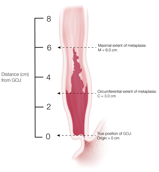
The length of the columnar epithelium at endoscopy is described using the Prague C (circumferential length) and M (maximal length) criteria (Box 2).7 Barrett’s oesophagus is defined as long segment when maximal segment length is ≥ 3 cm and as short segment when maximal length is < 3 cm.

Prevalence

Because Barrett’s oesophagus is asymptomatic and requires endoscopic examination and histological confirmation to establish the diagnosis, estimates of prevalence in unselected populations are scarce. The arguably best data were derived from a sample of 1000 Swedish residents recruited at random from the community who underwent upper gastrointestinal endoscopy, of whom 16 were identified with Barrett’s oesophagus (5 long segment, 11 short segment).8 Well conducted surveys in comparable populations (the United States and Europe) suggest community prevalence < 5%, with estimates converging around 2%; Australian data are limited to studies of patients referred for endoscopic investigation of symptoms.911 There is evidence that Australian detection rates have increased recently, with higher proportions of patients who undergo upper gastrointestinal endoscopy being diagnosed with Barrett’s oesophagus in consecutive surveys (rising from 0.3% in 1990 to 1.9% in 2002).10

Risk factors

Pooled analyses and meta-analyses of high quality epidemiological studies have consistently identified age, male sex, gastro-oesophageal reflux, central obesity and smoking as risk factors for Barrett’s oesophagus. In most populations, Barrett’s oesophagus is twice as common in men as in women,12 and prevalence rises with age.13

The longstanding clinical association between Barrett’s oesophagus and acid regurgitation or heartburn has been confirmed in research studies; a recent meta-analysis concluded that symptoms of gastro-oesophageal reflux increased the risks of long segment Barrett’s oesophagus more than fivefold.14 In addition, factors that promote reflux, such as hiatal hernia, are also observed more frequently in patients with Barrett’s oesophagus than among endoscopy controls with non-erosive reflux disease.15

While obesity is an established risk factor for oesophageal adenocarcinoma, epidemiologic studies have reported inconsistent associations between body mass index and Barrett’s oesophagus.16 However, studies measuring abdominal obesity (eg, waist circumference, waist–hip ratio) have identified two-to-threefold higher risks for high versus low waist circumference.16 Strong evidence of a likely causal association between obesity and Barrett’s oesophagus came from a Mendelian randomisation analysis, in which investigators demonstrated that people with a strong genetic propensity to develop obesity have significantly higher risks of Barrett’s oesophagus than those with a weak genetic propensity to obesity.17 The mechanisms remain speculative, but include mechanical (increased pressure on the lower oesophageal sphincter promoting reflux), metabolic and hormonal pathways. Importantly, the association between abdominal obesity and Barrett’s oesophagus is observed in people with and without reflux symptoms, indicating that mechanical reflux does not explain the whole effect.18 Metabolic factors are strongly implicated, with recent investigations reporting positive associations with markers of the metabolic syndrome, including insulin resistance19 and high serum concentrations of leptin.2022 Increasingly, it seems likely that male–female differences in fat deposition and metabolism may account for some of the observed sex-specific differences in the prevalence of Barrett’s oesophagus.23

Many other lifestyle exposures have been assessed as possible risk factors for Barrett’s oesophagus, two of which have been consistently implicated. Smoking increases the risk of the condition by about 50%24,25, whereas past infection with Helicobacter pylori reduces risk by about 50%.26,27 Previously, it was hypothesised that H. pylori infection inhibits gastric acid production and thus reduces acid-associated damage.27 Recent studies suggest that, in Western populations, H. pylori infection occurs predominantly in the antrum and likely reduces the risk of Barrett’s oesophagus by disrupting ghrelin and leptin pathways.28,29

Aside from the factors described above, no others have been consistently associated with the disease. Thus, despite considerable investigation, there is no evidence that alcohol is a risk factor.3033 Similarly, several well conducted case–control studies34,35 have investigated the role of aspirin and other non-steroidal anti-inflammatory drugs, based on strong and consistent inverse associations with oesophageal adenocarcinoma in observational and experimental studies. However, there is no evidence that this class of drugs alters a person’s risk of Barrett’s oesophagus. Relatively few studies have examined dietary factors and no conclusions can be drawn.

In the past 5 years, several large scale genome-wide association studies have identified a number of single nucleotide polymorphisms significantly associated with Barrett’s oesophagus.36 Moreover, there appears to be considerable genetic overlap between patients with Barrett’s oesophagus and patients with oesophageal adenocarcinoma, lending weight to the notion that these two conditions share similar causal origins.37,38 This is a rapidly moving field, but the clinical utility of these findings remains unknown.

Progression to cancer

Clinical interest in Barrett’s oesophagus stems largely from the concern that the condition is a precursor or risk marker for adenocarcinoma of the oesophagus. Most cases of oesophageal adenocarcinoma arise from underlying Barrett’s metaplasia in which there is a histological progression over time from low grade dysplasia (LGD) to high grade dysplasia (HGD) and subsequent intramucosal and invasive carcinoma (metaplasia–dysplasia–carcinoma sequence). Early oesophageal adenocarcinoma refers to invasion of the carcinoma beyond the basement membrane into the lamina propria (T1a on the current tumour–node–metastasis staging system) or superficial submucosa (T1b), but no deeper. Early oesophageal adenocarcinoma represents 6–12% of patients presenting with oesophageal cancer.39,40

The key question relates to the rate at which patients diagnosed with Barrett’s oesophagus progress to cancer. Early studies, largely conducted in tertiary referral centres, suggested rates as high as 1–2 per 100 patients per year. Since the year 2000, a number of large, population-based, record linkage studies have observed considerably lower progression rates for patients with uncomplicated Barrett’s oesophagus, converging at around 1–3 per 1000 patients per year (an order of magnitude lower than earlier reports).4145 The risk of progression is greater in those with dysplasia46 and those with long segment Barrett’s oesophagus.47

Considerable uncertainty remains about progression rates among Barrett’s oesophagus patients with LGD. In the community, LGD patients progress to HGD or cancer at a rate of about 1.5% per annum, whereas a recent European academic centre-based study reported much higher progression rates to HGD or cancer (about 13% per annum).42,48 The explanation appears to be that patients attending academic centres are reviewed by multiple expert gastrointestinal pathologists, and up to 75% of community referral LGD patients are downstaged to non-dysplastic Barrett’s oesophagus following expert review. Among the downstaged patients, progression rates to HGD or cancer of about 0.5% per year have been observed,48 similar to those reported from community-based studies.42 Studies from other academic centres of progression rates of patients with expert-confirmed LGD are awaited.

Little is known about lifestyle factors that increase or decrease the rate of progression to cancer. The literature underpinning this area is limited in scope and challenged by methodological issues such as small sample sizes, losses to follow-up, possible selection bias and confounding. Notwithstanding these limitations, it would seem that men with Barrett’s oesophagus progress to cancer at about twice the rate of women,46,49 and smokers progress at twice the rate of non-smokers.50,51 While prospective observational studies suggest that non-steroidal anti-inflammatory drugs,52,53 proton pump inhibitors54 and statins52,55 might retard progression to cancer, to date there are no randomised trials to support such conclusions and caution is warranted. Clinical factors associated with high rates of progression include longer segment length,45,46,50,52 and the presence of nodules,56 ulceration57 and strictures57 on endoscopy.

Screening

Screening is the process of identifying new cases of disease in an unselected population. Endoscopic screening for Barrett’s oesophagus in an unselected population with gastro-oesophageal reflux symptoms is not recommended, as it is not cost-effective. Focused endoscopic screening programs in those at greatest risk for Barrett’s oesophagus improve cost-effectiveness.58 Guidelines published by the British Society of Gastroenterology in 2014 recommend that endoscopic screening be considered in patients with chronic gastro-oesophageal reflux symptoms and multiple risk factors for Barrett’s oesophagus (at least three of aged 50 years or older, Caucasian background, male sex, and obesity). They suggest that the threshold of multiple risk factors should be lowered in the presence of a family history including at least one first-degree relative with Barrett’s oesophagus or oesophageal adenocarcinoma.59

For more widespread Barrett’s oesophagus screening to be considered, the costs of detection need to be reduced substantially with no compromise in accuracy. Studies of less costly screening methods (eg, ultrathin endoscopes, cytosponges) have yielded promising results but it is too early for these to be recommended at a population level.60,61

Endoscopic surveillance

Surveillance is the strategy of systematically following up patients with a known precursor condition to reduce (or prevent) the harms of cancer progression. The aim is to detect progression early, so that disease can be treated with the least invasive method, thereby reducing morbidity and mortality from cancer. The decision to commence endoscopic surveillance should be individualised for each patient, after considering factors such as age, comorbidities and the patient’s wishes and ability to participate in a long term surveillance program.

Endoscopic surveillance in Barrett’s oesophagus involves careful and meticulous examination of the Barrett’s segment with a high resolution white light endoscope, followed by biopsies from the segment. It is recommended that biopsies be taken according to the Seattle protocol, with biopsies of any mucosal irregularity (labelled separately) and quadrantic biopsies every 2 cm, unless there is known or suspected dysplasia, in which case quadrantic biopsies should be taken every centimetre. In the presence of erosive oesophagitis, it is recommended that acid suppression be maximised and surveillance endoscopy repeated in 2–3 months. This allows the oesophagitis to heal, thereby permitting underlying (masked) lesions to be identified and further biopsies to be taken.

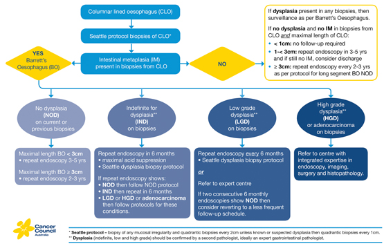
The interval between surveillance endoscopies depends on segment length and the presence of dysplasia (Box 3). In patients with Barrett’s oesophagus with no current or past dysplasia, follow-up endoscopy is recommended every 2–3 years in those with long segment disease, and every 3–5 years in those with short segment disease. Follow-up and management of patients with dysplasia is discussed below.

In some patients, a columnar-lined oesophagus is found at endoscopy but no intestinal metaplasia or dysplasia is seen histologically. The biological implications of this finding remain uncertain. The Australian guidelines recommend follow-up intervals based on segment length: < 1 cm, no endoscopic follow-up; 1–< 3 cm, 3–5 years; and ≥ 3 cm, 2–3 years.4

Although endoscopic surveillance in Barrett’s oesophagus is the current recommended practice, there is no direct evidence from randomised trials for its effectiveness or cost-effectiveness. Economic modelling studies suggest that current surveillance practices are unlikely to be cost-effective, and that identifying patients at high risk of progression to oesophageal adenocarcinoma substantially improves cost-effectiveness.39,62,63 The future hope is that a combination of clinical, endoscopic, blood or tissue markers might be used to develop risk stratification tools for identifying high risk patients most likely to benefit from surveillance and early intervention.64

Management of gastro-oesophageal reflux disease

In patients with Barrett’s oesophagus and gastro-oesophageal reflux symptoms, proton pump inhibitor treatment is recommended at a dose titrated to control symptoms and heal reflux oesophagitis. If proton pump inhibitors fail to control gastro-oesophageal reflux symptoms or heal reflux oesophagitis, surgical fundoplication can be considered.5 There is no strong evidence to suggest that medical or surgical therapy of gastro-oesophageal reflux disease leads to any substantial regression in segment length or influences progression to cancer.65

Management of low grade dysplasia

Management of LGD patients is currently uncertain, as new data suggest cancer progression rates are higher in patients whose LGD has been confirmed by an expert pathologist.48 A recent multicentre European randomised study of radiofrequency ablation in patients with expert-confirmed LGD found that control patients undergoing intensive endoscopic surveillance had a progression rate to cancer of 8.8%, while patients in the intervention arm had a progression rate to cancer of 1.5%.66 The Australian guidelines recommend that those with LGD be either closely monitored with frequent endoscopic assessment and biopsies every 6 months or referred to an expert centre for ongoing follow-up and consideration of ablative therapy of the Barrett’s segment.4 The decision regarding management of patients with LGD needs to take into account the features of the Barrett’s segment and histology as well as patient age, fitness and preference.

Management of indefinite for dysplasia

Indefinite for dysplasia is reported when biopsies from the Barrett’s segment show some histological features of true dysplasia but other processes (eg, inflammation) cannot be excluded as a cause for the changes. As with LGD and HGD, such biopsies should be reviewed by a second pathologist, ideally an expert gastrointestinal pathologist. If indefinite for dysplasia remains the diagnosis, then Australian guidelines recommend that the patient be placed on maximal acid suppression and undergo repeat endoscopy with dysplasia protocol biopsies in 6 months.4

Management of high grade dysplasia and early oesophageal adenocarcinoma

Patients with HGD or early oesophageal adenocarcinoma should be referred to an expert centre that has integrated expertise in endoscopy, imaging, surgery and histopathology. This allows the initial diagnosis to be confirmed by a second pathologist (ideally an expert gastrointestinal pathologist) and allows assessment and management by a multidisciplinary team.

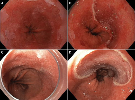
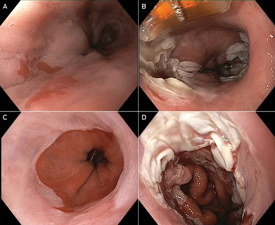
Until a decade ago, the only definitive management option for patients with HGD or early oesophageal adenocarcinoma was oesophagectomy. Because of the low risk of metastatic disease in cancers confined to the mucosa (1–2% for T1a lesions),67 the past decade has seen a number of endoscopic techniques developed to manage these conditions. These techniques can be divided in to two groups: resection (endoscopic mucosal resection [EMR] and endoscopic submucosal dissection); and ablation (radiofrequency ablation [RFA], argon plasma coagulation, photodynamic therapy and cryotherapy). In Australia, EMR and RFA are the most commonly used resection and ablation techniques, respectively (Box 4 and Box 5). In EMR, the oesophageal mucosa is aspirated into a cap on the end of the endoscope, a band applied and the captured mucosa and submucosa resected and retrieved endoscopically. In RFA, thermal injury delivered by an endoscopically placed device is used to destroy the oesophageal mucosa. Although these endoscopic methods do carry a small risk of complications (pain, bleeding, perforation and stricture formation), they are substantially less morbid, less expensive and more organ-preserving than surgery.68

Initial management of patients with histologically confirmed HGD and early oesophageal adenocarcinoma involves detailed endoscopic assessment and staging of the Barrett’s segment, with EMR of any visible lesions and biopsies of the Barrett’s segment according to the Seattle protocol. EMR of visible lesions enables accurate histological staging of the depth of invasion; studies have shown that EMR can change staging assessments in 48% of patients.69

Endoscopic treatment of high grade dysplasia

In patients with HGD without adenocarcinoma, further endoscopic treatment of the remaining Barrett’s segment is advised because of the risk of metachronous lesions. Treatment options for the residual flat segment vary from patient to patient, depending on factors such as segment length, the presence of a circumferential segment or the presence of an oesophageal stricture and involve EMR and/or endoscopic ablation. Follow-up studies of endoscopic therapy in HGD have shown promising long term results, with complete eradication of dysplasia and metaplasia in 89% of patients at 2 years.70 Longer term outcome studies are awaited. Post-treatment oesophagitis may be associated with decreased success rates of endoscopic therapy. It is therefore recommended that endoscopically treated patients receive ongoing medical therapy with a proton pump inhibitor to control gastro-oesophageal reflux symptoms and to prevent and heal oesophagitis.5 If medical therapy is unable to achieve these goals, surgical fundoplication may be considered. Long term, frequent endoscopic surveillance following treatment is recommended because of the risk of recurrence and metachronous lesions.

Endoscopic treatment of early oesophageal adenocarcinoma

In patients with early oesophageal adenocarcinoma and favourable histology (T1a; size, < 2 cm; well differentiated grade; no lymphovascular invasion; clear resection margins), further endoscopic treatment of the remaining Barrett’s segment can be planned.71 Treatment of the residual segment is advised because of the risk of future metachronous lesions within the segment. The endoscopic method for treating the residual flat dysplastic and non-dysplastic mucosa varies depending on patient factors, but will typically involve EMR and/or RFA. Australian guidelines recommend that ablation should only be used to treat flat dysplastic and non-dysplastic mucosa, and not as primary endoscopic therapy for early oesophageal adenocarcinoma.4

Because of the higher risks of lymph node metastases in T1b lesions (12–50%), surgically fit patients with T1b lesions should be offered oesophagectomy as a potentially curative treatment.40,72 In those patients who are unfit or unwilling to have surgery, endoscopic treatment with or without adjuvant therapy can be offered, but recognising the significant risk of lymph node metastasis that will remain undiminished by endoscopic therapy.73,74

Metachronous lesions or recurrent oesophageal adenocarcinoma have been described in up to 15% of patients undergoing endoscopic therapy for T1a lesions; therefore, long term, frequent post-treatment endoscopic surveillance is recommended. In most cases, lesions found on surveillance can be successfully managed endoscopically, with an overall 94% long term complete remission rate.75 In patients for whom endoscopic therapy is unsuccessful or not appropriate, oesophagectomy should be considered. Surgery in patients with HGD or early oesophageal adenocarcinoma carries a lower perioperative mortality rate (1.6%) than surgery for more advanced oesophageal adenocarcinoma.76

Conclusion

Barrett’s oesophagus describes a metaplastic change to the epithelium of the lower oesophagus that predisposes the person affected to oesophageal adenocarcinoma. While risks of progression are not as high as previously assumed, they are not insignificant, posing a challenge for clinical management. Australian guidelines have been developed to assist practitioners in this area.4 New endoscopic techniques for treating dysplasia and early adenocarcinoma are now available that have markedly lower morbidity than older approaches. In the future, it is possible that new screening and surveillance technologies may prove cost-effective for identifying and managing patients with Barrett’s oesophagus in the community.
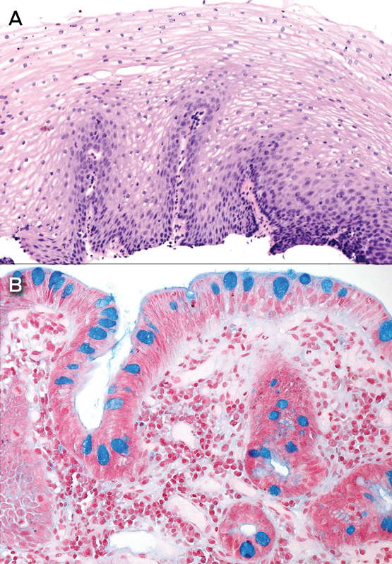
Box 1 –
Biopsies from normal oesophagus and Barrett’s oesophagus


A: Normal oesophageal squamous mucosa. B: A segment of columnar-lined oesophagus showing intestinal metaplasia with goblet cells highlighted by Alcian blue staining.

Source: A Clouston, with permission from Cancer Council Australia.

Box 2 –
Endoscopic classification of Barrett’s oesophagus using the Prague criteria7


Prague classification of Barrett’s oesophagus showing the circumferential extent of metaplasia (C) and maximal extent of metaplasia (M) above the true position of the gastro-oesophageal junction (GOJ). This example is classified as C3 M6, with 3 cm of circumferential metaplasia and 6 cm of maximal extent of metaplasia above the GOJ.

Box 3 –
Algorithm for recommended endoscopic surveillance schedule for Barrett’s oesophagus


Source: This graphic is licensed under the Creative Commons Attribution-ShareAlike 3.0 Australia license.

Box 4 –
Endoscopic mucosal resection (EMR)


A: C3 M4 Barrett’s oesophagus; after careful inspection, a focal abnormality was noted at 2 o’clock. B: Focal EMR was performed for staging, confirming high grade dysplasia. C: C7 M8 Barrett’s oesophagus; using a distal attachment cap for improved visualisation, a nodular lesion with slight depression was noted at 12–2 o’clock. D: This area is completely excised by EMR; histology confirmed Barrett’s oesophagus with high grade dysplasia and focal area of intramucosal adenocarcinoma (T1a).

Source: Reproduced with permission from Whiteman et al.4

Box 5 –
Radiofrequency ablation (RFA)


A: C5 M7 Barrett’s oesophagus with high grade dysplasia previously treated by endoscopic mucosal resection and RFA, showing residual disease remaining at 7 o’clock proximally and 12–4 o’clock distally. B: Focal RFA to sites of residual Barrett’s oesophagus. C: C2 M4 Barrett’s oesophagus previously treated by RFA for flat high grade dysplasia. D: Residual Barrett’s oesophagus is treated by focal RFA.

Source: Reproduced with permission from Whiteman et al.4

Central retinal venous pulsations

Diagnosing raised intracranial pressure through ophthalmoscopic examination

The ophthalmoscope is one of the most useful and underutilised tools and it rewards the practitioner with a wealth of clinical information. Through illumination and a number of lenses for magnification, the direct ophthalmoscope allows the physician to visualise the interior of the eye. Ophthalmoscopic examination is an essential component of the evaluation of patients with a range of medical conditions, including diabetes mellitus, systemic hypertension and conditions associated with raised intracranial pressure (ICP). The fundus has exceptional clinical significance because it is the only location where blood vessels can be directly observed as part of a physical examination.

Optic disc swelling and central retinal venous pulsations are useful signs in cases where raised ICP is suspected. Both signs can be obtained rapidly by clinicians who know how to recognise them. Although optic disc swelling supports the diagnosis of raised ICP, the presence of central retinal venous pulsations may indicate the contrary.

In the standard technique for direct ophthalmoscopy, the patient is positioned in a seated posture and asked to fix their gaze on a stationary point directly ahead. Pupillary dilation, removal of the patient’s spectacles and dim room illumination usually aid the examination. To start examining the patient, set the ophthalmoscope dioptres to zero — alternatively, a suitable setting would be the sum of the refractive errors of the patient and the examiner. Use the right eye to examine the patient’s right eye and vice versa. Using a slight temporal approach facilitates the identification of the optic disc, which also minimises awkward direct facial contact with the patient. Examine the red reflex at just under arm’s length. A pale or absent red reflex may suggest media opacity, such as a cataract. Next, on approaching the patient and obtaining a clear view of a retinal vessel, follow its course toward the optic disc. The presence or absence of venous pulsations should be appreciable (see the video at www.mja.com.au; pulsations of the central vein are clearly visible at the inferior margin of the optic disc). These pulsations, usually of the proximal portion of the central retinal vein, are most readily identified at the optic disc. The examination of the fundus should be concluded by visualisation of the four quadrants of the retina and examination of the macula.

Central retinal venous pulsations are traditionally attributed to fluctuations in intraocular pressure with systole, although this is may be an incomplete explanation.1 Patients with central retinal venous pulsations generally have cerebrospinal fluid pressures below 190 mmHg.2 Based on the results of Wong and White,3 the positive predictive value for retinal venous pulsations predicting normal ICP was 0.88 (0.87–0.9) and the negative predictive value was 0.17 (0.05–0.4).

This is important when considering lumbar puncture and when neuroimaging is not available. A limitation of this sign is that about 10% of the normal population4 do not have central retinal venous pulsations visible on direct ophthalmoscopy.4 The absence of central retinal venous pulsations does not, by itself, represent evidence of raised ICP; some patients with elevated ICP may still have visible retinal venous pulsations.

Papilloedema (optic disc swelling caused by increased ICP) may develop after the loss of retinal venous pulsations. This change in the appearance of the optic disc and its surrounding structures may be due to the transfer of elevated intracranial pressure to the optic nerve sheath. This interferes with normal axonal function causing oedema and leakage of fluid into the surrounding tissues. Progressive changes include the presence of splinter haemorrhages at the optic disc, elevation of the disc with loss of cupping, blurring of the disc margins, and haemorrhage. In later stages, there is progressive pallor of the disc due to axonal loss. A staging scale, such as that of Frisén,5 can be used to reliably identify the extent of papilloedema (Box).

Box –
Stage 4–5 papilloedema (5) showing disc and nerve fibre swelling, haemorrhage, loss of the optic cup and obscuration of the vessels at the disc margin


Source: Bruce AS, O’Day J, McKay D, Swann PG. Posterior eye disease and glaucoma A–Z. London: Elsevier Health Sciences, 2008.

Teaching approaches in medicine made easier

Teaching professional attitudes and basic clinical skills to medical students: a practical guide. Jochanan Benbassat. Springer, 2015 (143 pp, €51.99). ISBN 9783319200880.

Training, learning styles, role modelling and behaviours vary among doctors. As a result, students experience different approaches to teaching, which later shape their own approaches to patient interviewing, data collection and problem solving as doctors. Some doctors encourage patients’ narratives by using open-ended questions; others favour closed questions. This can leave medical students confused by the different techniques.

Teaching professional attitudes and basic clinical skills to medical students: a practical guide aims to help tutors, clinicians and teachers by providing an approach to teaching patient interviews, physical examination and clinical reasoning and by bringing the reader closer to the behavioural and social sciences.

The book is easy to read and provides a thorough description of the paradigm shift seen in the teaching of medicine in recent decades. It includes analyses of the difficulties in teaching patient interview and communication techniques, physical examination, data recording and clinical reasoning, and gives insightful and well researched pedagogical suggestions for different approaches to teaching aimed at improving students’ learning. The tables included help readers to quickly summarise the differences in teaching approaches, suggested priorities and important learning outcomes.

The book was written by Jochanan Benbassat, Professor of Medicine and Chair of Sociology of Health at the Ben-Gurion University of the Negev, Israel, from 1992 to 1997 and currently a research associate at the Myers-JDC-Brookdale Institute in Jerusalem.

Eliciting and responding to patient histories of abuse and trauma: challenges for medical education

Toward trauma-informed medical education

Traumatic experiences such as childhood abuse, family violence, elder abuse and combat exposure influence both physical and mental health, health-related behaviour, and the ways in which patients interact with medical practitioners.1,2 Despite greater knowledge of the pervasive sequelae of psychological trauma, the implications for medical practice and for medical education are not well articulated. Many doctors lack confidence and remain ill-informed or avoidant when dealing with patients’ psychological trauma.3,4 The consequences of this include non-recognition of somatisation and of psychiatric disorders, delay in instituting proper treatment, and costs to the patient and health care system of unnecessary investigations and treatments.5,6 Here, we discuss why and how we should better train doctors to elicit and respond to patient histories of trauma.

High prevalence of trauma and its clinical sequelae

The lifetime prevalence of exposure to traumatic events is high (74.9% in Australian adults).7 Most people who experience trauma do not develop mental illness; however, trauma and abuse are substantial contributors to the burden of mental and physical ill health. The risk of post-traumatic stress disorder after trauma is about 10%,8 but childhood abuse and neglect in combination with later life stress contribute to the development of mental illnesses as diverse as psychoses, depression, eating disorders and addictions, as well as a range of physical illnesses.2,9 There are also clear associations between past trauma and abnormal illness behaviour10 and, related to this, increased health care use.6 These sequelae of patient trauma pervade all medical specialties and also dentistry.1

Incorporating teaching about trauma in medical curricula: key issues

Although there are several studies that describe training interventions for specific forms of trauma,3,11,12 there is little in the literature on current practices in medical education, either in Australia or elsewhere. The diversity in general structure, content and methods of medical curricula as outlined by the Australian Medical Council (AMC) probably extends to trauma-relevant components.13 Despite this diversity, it is possible to offer initial considerations for trauma-informed education. We focus on six interrelated aspects: communication skills; knowledge of the health effects of trauma and abuse; knowledge about the effects of trauma and abuse disclosures on doctors and other health professionals; specific knowledge relevant to different medical specialities and settings; teaching formats and methods; and the need for a staged, incremental, integrated program, structured to achieve continuity between undergraduate, prevocational and specialist phases.

Communication skills curricula13 in pre-clinical and clinical phases afford opportunities for trauma-specific education, but are also relevant to junior hospital and specialty training. Common issues that need to be addressed include the personal discomfort many doctors experience asking about trauma and abuse; when not to screen for or discuss trauma; and when to seek advice from senior colleagues, as overconfidence can be harmful, leading to patient distress and even re-traumatisation. Communication skills education should extend to discussion of services relevant to different forms of trauma, such as social work, refuges, police and the courts. Here, it would be valuable for medical students to visit these settings or meet with workers from them.

Training needs to be realistic in that doctors often work in settings that are not conducive to asking about trauma, such as busy emergency departments, hospital wards that lack privacy, and overloaded outpatient clinics, often with a lack of psychiatric support. However, these realities should not be a pretext for avoiding clinically competent trauma-informed practice. We do not propose that doctors become trauma therapists; rather that they become competent in empathically recognising and eliciting information about trauma, and at effective referral of patients to relevant services, including psychology and psychiatry.

Exposure to patients who have experienced trauma or abuse evokes a range of psychological reactions in students and trainees, from normal discomfort and distress through to vicarious traumatisation.3,4,14 In addition, there may be unhelpful, if not harmful, responses: doctors may adopt an avoidant “don’t ask, don’t tell” style; over-investigate; refer patients to other clinicians; exhibit stigmatising attitudes toward patients; or become over-involved.4 Course content on these issues could be introduced early and developed further during clinical and postgraduate phases.

There are many opportunities pre-clinically to learn about the effects of abuse and trauma on human development, including the short and long term effects on the brain and behaviour. Relevant knowledge can be taught within sections of the curriculum devoted to neuroscience, cognitive science and population health. In addition, medical humanities have a powerful capacity to expand our knowledge and understanding of diverse human experiences, including trauma, and to foster empathy. Every clinical specialty that medical students and trainees encounter in hospital training brings opportunities to learn specialty-specific trauma knowledge and skills. For example, rotations in paediatrics and in obstetrics and gynaecology are opportunities for teaching about child abuse and about family violence.

Trauma is an everyday part of clinical discourse within psychiatry and a key dimension of academic and clinical learning in psychiatry rotations. However, trauma-informed education is relevant to all clinical specialties. Relevant specific knowledge encompasses common clinical presentations of trauma in those specialties; how trauma-relevant inquiry can be embedded within the specialty-specific clinical interview; clinical, social and legal services relevant to various forms of abuse; and legal requirements regarding mandatory reporting.

Some medical students and trainees have their own experiences of trauma, including childhood trauma and abuse, but also vicarious trauma stemming from clinical encounters, such as witnessing horrific physical injury or disfigurement. Post-traumatic stress disorder in medical practitioners is often unrecognised.14 Such experiences may increase or may impair empathic capacity to engage with traumatised patients.

Although the AMC standards discuss the stressful and traumatic nature of medical work and provide recommendations about availability of counselling, peer support and other measures, they construe the reality of trauma as external to the core business of medical education.13 Instead, we propose that learning about the emotional impacts of clinical work should be core medical education, to be dealt with in lectures, tutorials, simulations — that is, a range of appropriate, complementary educational methods, as is done for other topics. In addition, curricula should include safe, confidential, non-coercive opportunities for experiential learning in small groups, allowing participants to reflect on and share their own emotional reactions to patients and understand how these reactions can shape their clinical practice. Ideally, some teaching should be conducted jointly with students from other disciplines, notably nursing.

The AMC standards stress the centrality of clinical clerkships in the development of clinical competence and judgement.13 We agree, but when it comes to trauma-informed clinical teaching, there are several entailments. Teaching about trauma has to become a routine, everyday feature of clinical teaching in wards and clinics, not something outsourced by referral to psychiatry or social work. Clinician teachers in all specialties need to acquire the skills to do such teaching and to act as role models. Given the limited evidence base, it is premature to recommend a mix or staging of methods, and this should be a focus of future research and curriculum innovation.

The medical education literature is marked by separate discourses on differing forms of trauma. For example, education regarding intimate partner violence has been extensively explored and excellent curricula have been implemented.3,13,14 However, patients have often experienced several concurrent or sequential traumas; and the clinical sequelae of different traumas have many similarities, demand similar clinical skills (albeit allied with different bodies of specific knowledge), and thus present similar challenges for medical education. Valuable educational synergies are likely if the currently disparate, unconnected trauma-relevant elements in medical curricula are integrated.

These considerations point to the need for the creative design and evaluation of staged, incremental, integrated programs, structured to achieve continuity between undergraduate, pre-vocational and specialist phases of medical education. We do not propose removing the various trauma-specific educational components from medical curricula and replacing them with some form of generic trauma education. We do propose, however, examining creatively how they may be better integrated to become mutually reinforcing.

Conclusion

Trauma-informed health care is an invaluable concept which we propose should extend to trauma-informed medical education.15 Although the arguments for trauma-informed medical education are compelling, new lines of educational research will be needed to guide curriculum design and build on the small body of work already available.3,4,11,12,16 It is likely that if doctors of all kinds have the knowledge, skills and attitudes to deal competently with abuse and trauma, we can expect improvements in patient care and health service costs, and in the health and wellbeing of medical practitioners. These possibilities deserve empirical study. As well as becoming better clinicians, medical students and trainees will also become better teachers and role models and, as they move into more senior and leadership roles, advocates for competent trauma-informed medical care.

Vocational training of general practitioners in rural locations is critical for the Australian rural medical workforce

The known In efforts to reduce the longstanding geographically inequitable distribution of Australian GPs, current policy requires that 50% of GP vocational training (registrar) positions are located in rural or remote areas. 

The new We identified a strong association between rural training pathways and subsequent rural practice, and it is intensified by a rural origin effect. Despite some attenuation over time, these associations remained strong up to 5 years after vocational registration. 

The implications Ongoing support for rural GP vocational training opportunities and the selection of rural origin medical students are critical components of GP workforce policy. 

The geographically inequitable distribution of the Australian medical workforce continues, and rural and remote general practitioner positions are largely filled by international medical graduates (IMGs).1 This dependency persists despite substantial government efforts to stimulate recruitment and retention of Australian-trained GPs in rural areas. Recent government initiatives have included a large increase in the number of federally supported medical school places for students, and supporting medical education and training in rural communities through the Rural Clinical Training and Support (RCTS) program.1,2 A quota for the proportion of domestic students with a rural background selected by medical schools (at least 25%) has also been introduced, and rural clinical exposure during undergraduate and pre-vocational medical training programs has increased. In addition, Australian policy now requires that 50% of GP vocational (registrar) training occurs outside metropolitan areas.1 This policy is based chiefly on research that has indicated that a positive educational experience in rural settings, targeted training of GP registrars for rural practice, and clear pathways to rural practice are the most effective incentives for interesting a GP in a rural career.3,4 Doctors accepted into GP training are selected into either the Rural Pathway or the General (mostly metropolitan) Pathway, with about 50% of candidates allocated to each.5

Evidence for the effectiveness of these interventions for increasing rural recruitment and retaining Australian medical graduates in rural areas has accumulated. Ranmuthugala and colleagues6 reported that evidence for the effectiveness of increased rural exposure during undergraduate medical training on the uptake of rural practice was inconclusive, but Wilkinson and colleagues7 found that postgraduate rural GP training had a stronger association with rural practice uptake than rural exposure during undergraduate training (although the availability of rural GP postgraduate training was low at the time of this study because the number of rural training positions was limited). More recent empirical data810 and data on intentions collected at training completion11,12 suggest moderate improvement in the uptake of rural practice by students who have participated in RCTS programs. However, as reported in three literature reviews on the recruitment and retention of medical practitioners in rural areas3,13,14 and as lamented in a recent letter to the Medical Journal of Australia,15 there remains a large evidence gap as to the effectiveness of rural exposure during vocational training programs. A review of the outcomes of the regionalised Australian General Practice Training Program16 found that only 27% of former Rural Pathway registrars remained in rural practice after 7 years. In addition, several North American studies have produced limited quantitative evidence of associations between vocational training in a rural primary care setting and subsequent rural practice.1720

The geographic origin of doctors also has an impact on their commencing rural practice, with convincing evidence about a strong link between an individual’s rural upbringing and their subsequent decisions about a rural career.21,22 The consistency of the reported association between GPs having a rural background and their choosing a rural career suggests that their origin is a critical factor in making this decision, regardless of vocational training location. Our study therefore aimed to investigate the association between vocational training location and the subsequent choice of practice location for newly registered GPs, including the effect of a rural background.

Methods

This study was based on data from the Medicine in Australia: Balancing Employment and Life (MABEL) study, conducted by the Centre for Research Excellence in Medical Workforce Dynamics (https://mabel.org.au/). MABEL is a national longitudinal survey that collects annual data from a panel of doctors, with a regular small participation top-up. The first wave of the MABEL study (2008) invited the entire medical workforce to participate, and 10 498 doctors (19.4% of the medical population) completed the initial survey, including 17.7% of GPs. There has subsequently been an annual 70–80% study retention rate. Further participants (generally recently graduated, non-specialist hospital doctors or IMGs newly registered in Australia) are added to the MABEL pool each year.

Our study analysed data from MABEL waves 1 to 7 (2008–2014), and was restricted to respondents who had completed their GP vocational training and were transitioning to independent practice. The transition year for a GP was identified from MABEL data on the basis of their participation in GP registrar training and details of newly completed medical qualifications. Data for IMG GPs — defined as those who had completed their initial medical training outside Australia and New Zealand — were analysed separately.

Rural origin and work location

Rural origin was defined for doctors trained in Australia or New Zealand as their having resided for at least 6 years in a rural area before the age of 18 years. Each doctor’s work location was geocoded in each MABEL wave to a specific town or suburb, then classified as metropolitan or rural. Rural location was defined as including Australian Standard Geographic Classification Remoteness Areas (ASGC-RA) 2 to 5;23 it was self-defined for New Zealand-trained doctors. Vocational training location was defined in two ways: as rural or metropolitan by work location in the year the doctor completed their training (final training location), and as an aggregate of work locations in the 2 to 3 years preceding their completion of training.

Statistical analysis

Four cohorts were defined by a combination of origin type and final training location: rural origin/rural training, metropolitan origin/rural training, rural origin/metropolitan training, and metropolitan origin/metropolitan training. For comparison purposes, IMGs were separately divided into two cohorts: rural training and metropolitan training.

A secondary (sensitivity) analysis defined four cohorts by multiple training locations: rural training only; completed training in a rural area, but also had some metropolitan training; completed training in a metropolitan area, but also had some rural training; metropolitan training only.

For each cohort, the proportions of GPs working in rural and metropolitan locations were calculated for each of the first 5 years after they had completed their vocational training. Rurally trained GPs were further classified according to whether they were working in the same or a different rural community from that in which they completed their vocational training; a buffer of 20 kilometres was allowed.

Separate generalised estimating equation (GEE) models with a logit link function and exchangeable correlation structure were used to test associations between vocational training pathways and subsequent work location for the four primary cohorts (non-IMGs only) for each of the 5 years after completing vocational training. Adjustments were made for four additional demographic variables during each particular year: sex, age, living with a partner, and having dependent children. A further variable — whether the GP was rurally bonded (contracted to work for part of their early career in rural locations) in a particular year — was included in each regression model. These models were repeated for the four secondary cohorts, with rural origin as an additional covariate; its multi-year cohort definitions limited analysis to 4 outcome years. All calculations were performed in StataSE 12 (StataCorp).

Ethics approval

The MABEL study was approved by the University of Melbourne Faculty of Business and Economics Human Ethics Advisory Group (reference, 0709559) and the Monash University Standing Committee on Ethics in Research Involving Humans (reference, CF07/1102 – 2007000291).

Results

During the 7-year study period, 610 doctors completed their GP vocational training and commenced in at least one subsequent work location. The demographic characteristics of these GPs are summarised in Box 1. Just under half of the local graduates (ie, those who graduated in Australia) trained in the Rural Pathway, and about one quarter were of rural origin (consistent with current policy requirements for GP training posts and medical student intakes); fewer than 10% were rurally bonded. Most local medical graduates were women, most lived with a partner, and almost 40% had dependent children. The proportions of IMGs who trained in the Rural Pathway, were men, were aged 35 years or more, lived with a partner, or had dependent children were higher than for local medical graduates (Box 1).

Box 2 summarises the practice location as an independent GP for the four primary cohorts of local medical graduates for each of the 5 years following their completion of vocational training. There were very strong and sustained associations between final vocational training location type and subsequent practice location for the rural origin/rural training and metropolitan origin/metropolitan training cohorts; 74–91% and 87–95% respectively remained in their origin/training type during their first 5 post-training years. Moreover, 61–70% of the rural origin/rural training cohort practised in the same rural community in which they trained during the first 4 years after completing their vocational training. Outcomes for GPs from cohorts 2 and 3 also showed a clear pattern: initially, these GPs generally remained in their final vocational training location type, but there was subsequently a gradual move in work location toward their origin type. The career patterns of rurally trained IMGs was similar to those of metropolitan origin/rural trained local graduate GPs, with a gradual move in work location toward metropolitan areas during the 5 years after vocational registration (Box 3).

The rural training pathway, regardless of childhood location, was highly significantly associated with subsequent rural practice. The odds of rural practice for each of the rural training cohorts of GPs decreased with time, but a strong and highly significant association was nevertheless retained across the 5 years. Unsurprisingly, rural bonding and rural origin were positively associated with rural practice. Higher age was also associated with rural practice, while there were no consistent statistically significant associations between practising in a rural location and sex, or with having a partner or dependent children (Box 4).

Secondary analysis, using the multiple year training location definition, confirmed the importance of rural training, particularly that of the final GP training year (Box 5).

Discussion

We have provided empirical evidence for the contribution of rural vocational training, in combination with the selection of rural origin students, to the Australian rural GP workforce. This is highly significant for rural workforce policy, as the Australian government requires that more than half of Australian GP vocational training positions be located in rural areas; our study allows an opportunity to assess the effect on the workforce of these policies.1

We found that training in the rural training pathway and the trainee having a rural background were each strongly associated with early career rural practice. The strength of the association between vocational training location and choosing rural practice remained strong and statistically significant up to 5 years after completing GP training for doctors of either rural or metropolitan origin (primary cohorts 1 and 2). Sustained rural practice was very strongly linked with the combination of a rural origin and rural training, but this cohort alone is unlikely to provide a sustainable rural GP workforce while only 25% of Australian-trained doctors are of rural origin, as about 30% of the Australian population live in rural or remote areas.

Most mixed rural/metropolitan origin/training GPs (cohorts 2 and 3) subsequently practised in a same location type as that in which they trained, although some gradually returned to their origin type. Diminution of the pathway effect over time is perhaps expected, as 50% of GP registrar training positions are in rural areas but about 75% of young doctors are of metropolitan origin. Other research has found that work location changes are most likely during early career stages,24 when personal circumstances, including relationships with spouses and dependents, are more fluid. The secondary analysis confirmed the strong influence of rural training on subsequent rural practice, especially location during the final year of vocational training. Together, these findings suggest that the periods leading up to and immediately following vocational training are critically important windows of opportunity for ensuring that appropriate policies optimise recruitment of GPs for rural practice and their subsequent retention.25,26

The largest cohort, metropolitan origin doctors undertaking GP training in metropolitan areas (cohort 4) largely remained in metropolitan practice. Further, there was no evidence that rural origin Australian doctors were more likely than metropolitan origin doctors to choose general practice as their specialty (unpublished MABEL data). Consequently, metropolitan origin doctors continue to remain the major source of non-IMG rural GPs, making cohort 2 (metropolitan origin/rural training) critical for the rural GP workforce. This cohort is nearly twice the size of cohort 1, and the association with rural practice was much stronger than for those in the metropolitan pathway (cohort 4). However, more than 50% of cohort 2 had moved to metropolitan practice after 5 years, further highlighting the importance of targeted retention initiatives focused on this cohort.

The odds of members of the smallest cohort (cohort 3: local medical graduates with a rural background who undertook their training in metropolitan areas) practising in rural areas was three times that for metropolitan origin/metropolitan training GPs, although the association was statistically significant only from 3 years after completing vocational training. However, the odds were much lower than for the rural origin/rural training cohort 1, highlighting the importance of the rural training pathway.

A key limitation of this study is that it cannot establish cause and effect. There is probably a strong self-selection bias, in that the rural training pathway attracts those who are interested in a rural career. Further limitations include the use of a self-selected cohort, the participants of the MABEL survey, who represent 15–18% of all Australian GPs. While the panel design of our study enabled individual tracking of doctors over a 7-year period and application of GEE (logit) modelling, the observed patterns, particularly in the mixed origin/training cohorts, suggest that these doctors have not yet decided on their long term preferred work location, and it is therefore difficult to accurately predict outcomes at, for example, 10 or 20 years. Additionally, vocational training location was primarily defined for the purposes of this study as the location of the trainee in the year they completed their training, as this was considered to be the most influential year for subsequent practice location. Our secondary analysis partially examined this aspect by separately analysing GPs who had undertaken vocational training in a mix of rural and metropolitan locations. Further, our key focus was on the joint effects of rural origin with rural/metropolitan training pathways. This necessitated a focus on GPs who had completed their medical degrees in Australia or New Zealand, despite IMGs comprising a considerable proportion of the rural GP workforce in Australia (more than 50% in some regions). Finally, this study used a binary measure of rurality (metropolitan v non-metropolitan) that may not adequately adjust for the substantial heterogeneity in the attractiveness to GPs of different rural and remote Australian locations. It is possible that more nuanced measures of rurality, including multiple levels of remoteness and population size, might have identified different associations for the four cohorts.27

Conclusion

Our study analysed the best available Australian longitudinal data about individual GPs to provide new quantitative evidence of a strongly positive association between rural GP vocational training location and subsequent rural practice, even after adjusting for the influence of rural origin. This evidence supports the objectives of existing policies that require at least 50% of GP training to occur in rural locations, and that at least 25% of medical students should be of rural origin. While Australia strives to reduce its reliance on IMG GPs for the rural workforce, this aim requires long term improvements in the rural recruitment and retention of Australian-trained GPs. Ongoing support for rural GP vocational training opportunities is a critical component of rural GP workforce policy in Australia.

Box 1 –
Demographic characteristics of participating doctors at the time they completed general practitioner vocational training

Local medical graduates

International medical graduates


Number

467

143

Rural Pathway (year of training completion)

221 (47.3%)

101 (70.6%)

Rural origin

118 (25.3%)

NA

Sex (women)

322 (69.0%)

74 (51.8%)

Age, median

32 years

41 years

Age, ≥ 35 years

153 (32.9%)

125 (89.9%)

Living with a partner

335 (72.7%)

119 (83.2%)

Has dependent children

179 (39.4%)

119 (83.8%)

Rurally bonded

35 (7.5%)

NA


NA = not applicable. Percentages exclude missing data for local medical graduates (age, 2; living with partner, 6; dependent children, 13) and international medical graduates (age, 4; dependent children, 1).

Box 2 –
Final vocational training location and general practice location for local medical graduates during the first 5 years after completing general practitioner vocational training

Time since completion of training

Location of practice

(1) Rural origin/rural training

(2) Metropolitan origin/rural training

(3) Rural origin/metropolitan training

(4) Metropolitan origin/metropolitan training


Number of GPs

78 (17%)

143 (31%)

42 (9%)

204 (44%)

1 year

Same rural

70%

54%

Other rural

20%

22%

18%

5%

Metropolitan

10%

25%

82%

95%

2 years

Same rural

62%

42%

Other rural

24%

31%

30%

13%

Metropolitan

14%

27%

70%

87%

3 years

Same rural

68%

24%

Other rural

15%

42%

35%*

11%

Metropolitan

18%

34%

65%*

89%

4 years

Same rural

61%

25%

Other rural

30%

29%

46%*

9%

Metropolitan

9%

45%

54%*

91%

5 years

Same rural

42%*

15%

Other rural

32%*

33%

33%*

9%

Metropolitan

26%*

52%

67%*

91%


* Groups with fewer than 20 participants.

Box 3 –
Final vocational training location and general practice location for international medical graduates during the first 5 years after completing general practitioner vocational training

Time since completion of training

Location of practice

Rural training

Metropolitan training


Number of GPs

101 (71%)

42 (29%)

1 year

Same rural

81%

Other rural

6%

4%

Metropolitan

13%

96%

2 years

Same rural

57%

Other rural

17%

8%

Metropolitan

26%

92%

3 years

Same rural

49%

Other rural

10%

0*

Metropolitan

41%

100%*

4 years

Same rural

45%

Other rural

21%

18%*

Metropolitan

34%

82%*

5 years

Same rural

53%*

Other rural

7%*

20%*

Metropolitan

40%*

80%*


* Groups with fewer than 20 participants.

Box 4 –
Odds of local medical graduates practising in a rural location during the first 5 years after completing general practitioner vocational training

Odds ratio (95% confidence interval)


1 year post-GP training

2 years post-GP training

3 years post-GP training

4 years post-GP training

5 years post-GP training


Primary cohorts

(1) Rural origin/rural training

159 (45–558)

65 (27–158)

48 (22–102)

50 (24–106)

52 (24–111)

(2) Metropolitan origin/rural training

68 (26–175)

32 (16–60)

28 (16–51)

23 (13–41)

24 (13–43)

(3) Rural origin/metropolitan training

2.8 (0.7–11)

2.4 (0.9–6.2)

2.9 (1.2–6.7)*

3.3 (1.5–7.4)

3.5 (1.5–7.9)

(4) Metropolitan origin/metropolitan training

1.00

1.00

1.00

1.00

1.00

Age (for each 1-year increase in age)

1.06 (1.00–1.13)*

1.04 (0.99–1.08)

1.04 (1.00–1.08)*

1.05 (1.01–1.08)*

1.04 (1.01–1.08)*

Sex (reference: men)

1.00 (0.48–2.1)

0.9 (0.5–1.6)

1.03 (0.6–1.7)

0.8 (0.5–1.4)

0.8 (0.5–1.4)

Living with a partner

0.8 (0.3–1.9)

0.9 (0.5–1.7)

0.9 (0.5–1.7)

0.98 (0.6–1.7)

0.9 (0.6–1.5)

Has dependent children

1.8 (0.8–4.1)

1.9 (1.06–3.3)*

1.4 (0.9–2.3)

1.3 (0.9–2.0)

1.3 (0.9–1.9)

Rurally bonded

5.1 (1.2–22)*

3.5 (1.1–11)*

3.8 (1.4–11)*

3.7 (1.4–10)*

3.6 (1.3–10)*


Odds ratios from generalised estimating equation (logit) model: * P < 0.05; † P < 0.01.

Box 5 –
Odds of practising in a rural location for each of the 4 years after completing general practitioner training for local medical graduates

Odds ratio (95% confidence interval)


1 year post-GP training

2 years post-GP training

3 years post-GP training

4 years post-GP training


Secondary cohorts

(1) Rural training only

92 (27–312)

49 (21–115)

41 (19–88)

29 (14–59)

(2) End training rural, with some metropolitan training

17 (5–58)

11.6 (4.6–29)

11.5 (4.9–26)

9.9 (4.3–23)

(3) End training metropolitan, with some rural training

0.94 (0.09–9.4)

2.8 (0.8–9.4)

2.9 (1.00–81)

2.7 (0.96–7.9)

(4) Metropolitan training only

1.00

1.00

1.00

1.00

Rural origin

4.1 (1.3–13)*

2.0 (0.9–4.3)

2.1 (1.02–4.1)*

2.5 (1.3–4.9)

Age (for each 1-year increase in age)

1.2 (1.04–1.3)

1.08 (1.01–1.16)*

1.07 (1.01–1.14)*

1.05 (1.00–1.12)

Sex (reference: men)

0.9 (0.3–2.4)

0.8 (0.4–1.7)

0.9 (0.5–1.9)

0.8 (0.4–1.5)

Living with a partner

0.6 (0.2–2.1)

1.1 (0.5–2.6)

1.1 (0.5–2.4)

1.07 (0.5–2.1)

Has dependent children

0.6 (0.2–2.0)

1.3 (0.6–2.7)

1.09 (0.6–2.1)

1.02 (0.6–1.8)

Rurally bonded

2.0 (0.4–10)

2.21 (0.6–7.8)

3.8 (1.2–13)*

3.6 (1.1–11)*


Odds ratios from generalised estimating equation (logit) model: * P < 0.05, † P < 0.01.

Rural recruitment and training promotes rural practice by GPs, but is it enough to retain them?

Challenges to keeping general practitioners in the bush remain

The findings reported by McGrail and colleagues in this issue of the MJA support the effectiveness of Australian government incentives for recruiting and training general practitioners in rural areas as a strategy for reducing rural medical workforce shortages.1 The study found that rural origin of trainees and rural vocational training of GPs were each strongly associated with their practising in rural areas in the early years after completing vocational training. However, their findings also suggest that these effects had started to diminish by 4 years post-training.1 This finding is consistent with another recent Australian study, which found that the effects of rural recruiting and training diminished over time.2

As evidence emerged in the early 1990s that a rural background and a positive rural training experience promoted the subsequent uptake of rural practice by trainees, the Australian government introduced several initiatives for recruiting and training medical students in rural areas. The Rural Undergraduate Support and Coordination Program (RUSC) was in 1993 among the first of these initiatives, followed by the Rural Clinical School (RCS) and the Rural Clinical Training and Support Program (RCTS). These initiatives required that 25% of the intake of students by federally funded medical schools be from a rural background; that all federally supported medical students undertake a 4-week structured rural placement; and that 25% of students undertake at least 12 months’ clinical training in a rural location.3 Initiatives such as the Australian General Practice Training Program followed, ensuring that at least 50% of general practice vocational training placements are in rural or remote areas.4 These training initiatives have contributed to the success achieved in increasing the number of GPs who adopt rural practice: it was recently reported that the rural and remote GP workforce increased by 23% between 2010 and 2014, compared with a 3.5% increase in the rural and remote community population, and a 10% increase in the metropolitan GP workforce over the same period.5

It is now timely to consider whether an increase in the number of rural and remote GPs necessarily translates into a sustained and well supported workforce which can deliver quality health care that meets the needs of rural communities. Factors that motivate practitioners to remain in rural areas include access to training, professional development and career development opportunities.3 While I focus in this article on the role of training and education in rural retention, other factors known to be important include peer and professional support, assistance with heavy workloads and on-call requirements, locum relief,3 access to infrastructure (such as information and communication technology and electronic health data systems), housing, and family support.6

In addition, being a principal of the medical practice has been identified as significantly increasing the likelihood of a doctor remaining in a rural location (by 72%), while being a salaried or contracted employee significantly reduces the likelihood (by 20–30%).7 GPs in rural and remote locations work longer hours than their metropolitan counterparts, increasing steadily from an average of 38 hours per week in metropolitan locations to 45.8 hours in very remote locations.5 Such demands, and the need to travel, make it more difficult for rural or remotely located practitioners to participate in professional development and to take up training opportunities. Innovative business and work model solutions are needed to support the rural GP workforce.

It should also be noted that the proportion of GPs practising procedural skills increases with remoteness (from 8.0% in inner regional areas to 13.8% in outer regional and 20.9% in remote and very remote locations).5 Recognising that rural and remote practitioners must have procedural skills in general surgery, obstetrics, anaesthesia, radiology and endoscopy, the Royal Australian College of General Practitioners has incorporated procedural skills training into their curriculum.8 Additional training is provided through the General Practitioner Procedural Training Support Program. Nevertheless, the period 2010–2013 saw a drop in the proportion of GPs practising procedural skills;5 the decline was greatest in outer regional areas (4.1%), followed by remote (3.9%), inner regional (1.9%) and very remote locations (0.6%). Reasons for this decline are not clear and need further exploration, especially given a recent finding that undertaking hospital work significantly increases the likelihood that rural and remote GPs remain in rural locations (by up to 40%).7 As exercising one’s skills contributes to increased job satisfaction, motivation, commitment and retention,9 there is a need to provide the infrastructure and opportunity for these practitioners to enhance and practise the procedural skills that have been identified as an important aspect of rural practice.

The early training initiatives are having positive effects on recruitment, but they must be reviewed and updated as new evidence emerges. Accordingly, in light of consistent support for the influence of longer term rural clinical placements on the likelihood of choosing rural practice, the initial requirement that all federally supported medical students undertake a 4-week rural placement has been reduced to 50% of students, but with no change to the proportion required to undertake a year-long rural clinical placement.10 It will be another 5–10 years before the effect of these revised funding parameters on the recruitment and retention of the rural medical workforce will be apparent.

New plan to find poor performing doctors

Screening for at-risk doctors and strengthening CPD are two key points highlighted in a new expert report released by the Medical Board of Australia.

The consultation and discussion paper were released on Wednesday, proposing a new approach to revalidation of doctors in Australia.

Board Chair, Dr Joanna Flynn AM said in a statement: “Regulation is about keeping the public safe and managing risk to patients. Part of this involves making sure that medical practitioners keep their skills and knowledge up to date.”

The proposal has two main parts.

    1. Strengthened CPD – a ‘smarter not harder’ approach that will be evidence based to drive practice improvement and better patients outcomes.
    2. Identify and assess at risk and poorly performing practitioners – an accurate and reliable way to screen practictioners at risk of poor performance will be developed. The report identifies that doctors more at risk include age (from 35 years, increasing into middle and older age), the male gender, number of previous complaints and time since last complaint. Other risk factors include getting qualification in some countries of origin, certain specialties, those who don’t respond to feedback, those who have an unrecognised cognitive impairment, doctors practising in isolated areas, doctors who do low levels of high-quality CPD activities and who have had a change in scope of practice.

According to Dr Flynn: “Most of the practitioners in the at-risk groups will be able to demonstrate that they are performing satisfactorily, just as most people who are screened in a public health intervention do not have the disease for which the screening program is testing.

Related: Blaming individual doctors for medical errors doesn’t help anyone

Those who have been found to be under-performing would then go through a ‘tiered, multi-faceted assessment strategy’ which would be scaled to match the perceived level of risk. There could be peer review and feedback processes or a more thorough evaluation for those considered to be seriously under performing.

Remediation would also depend on the nature and level of risk, although according to the report, “there is little information about long-term outcomes of remediation on doctors’ subsequent performance.” There will be continued research to confirm the efficacy of remediation interventions.

Related: Blocking overseas practitioners won’t solve rural doctor shortage

The Medical Board is now consulting about the proposed changes and want to hear from the medical profession about their thoughts.

“We want a system in Australia that is practical, effective and evidence-based, and we want to hear what the community and the medical profession think about the approaches proposed by the expert advisory group,” Dr Flynn said.

Options on the revalidation page include:

      • have your say in the online discussion
      • take a short survey to provide your views on the approach
      • send your written submission by email or mail
      • read submissions made by others

The consultation closes on 30 November 2016 and the final report is expected by mid 2017.

Latest news: