×

Point-of-care testing for coeliac disease antibodies — what is the evidence?

To the Editor: The recent introduction of rapid point-of-care testing (PoCT) in Australian pharmacies to screen for coeliac disease has attracted controversy1 and provides an important opportunity to review the current literature.

PoCT provides a rapid (within 10 minutes) assessment of the presence or absence of coeliac disease-specific antibodies using a skin-prick blood sample. Based on lateral flow immunochromatography, circulating IgG and IgA antibodies to deamidated gliadin peptides, if present, bind to a membrane, which generates a coloured line of varying intensity.2 Total IgA antibodies are also assessed to detect the 3% of patients with coeliac disease who are IgA-deficient.

Coeliac Australia’s Medical Advisory Committee has developed a position statement, supported by the Royal College of Pathologists of Australasia, that reviews the evidence base for PoCT in coeliac disease and provides a detailed explanation of the technology used in currently available PoCT kits.3

The diagnostic accuracy of current assays to perform PoCT for coeliac antibodies is inferior to laboratory-based testing, particularly in the context of average-risk populations, where coeliac disease prevalence is relatively low.4,5 A positive PoCT result does not confer a definitive diagnosis of coeliac disease; nor does a negative test sufficiently exclude it. Diagnosis of coeliac disease still requires demonstrating the characteristic enteropathy in a small intestinal biopsy specimen.6

Interpretation of PoCT results requires a suitably trained practitioner, as it is inherently subjective and greater reader experience is associated with improved accuracy. Interpretation of results where antibody binding generates a “faint positive” line is challenging.4 Validated standards for reporting results, sound clinical governance, and protocols that establish regular control procedures will be important to ensure robust performance of PoCT.

Although it is an attractive technology, the accuracy and clinical utility of PoCT by community clinics, general practitioners and pharmacies have not been studied, and prospective data are required. Given the clinical implications of a positive or negative screen for coeliac disease and the multitude of differential diagnoses in patients presenting with a range of symptoms, professional medical review remains a crucial factor in the diagnostic work-up of coeliac disease.

Acute pancreatitis: update on management

Acute pancreatitis is a common acute surgical condition. The annual incidence worldwide is 4.9–73.4 cases per 100 000 people,1,2 with the incidence in Australia on the higher end of the spectrum.3 The mortality rate for pancreatitis is between 1.5% and 4.2% in large epidemiological studies,46 but varies according to the severity of pancreatitis, increasing to 30% in those with infected pancreatic necrosis.7 A recent Australian study has reported a low mortality rate of 0.08%,8 thought to be due to earlier recognition of severe pancreatitis and the appropriate use of intensive care support.8

As acute pancreatitis is common, costly and potentially life-threatening it is important that management is guided by an evidence-based approach. The American College of Gastroenterology (ACG) and the International Association of Pancreatology (IAP) in collaboration with the American Pancreatic Association (APA) have recently published new guidelines for the management of acute pancreatitis.9,10 These guidelines reviewed all of the evidence of a wide range of areas within the management of acute pancreatitis and made recommendations of various strengths based on the level of available evidence.

Here, we provide an update on diagnosing and managing acute pancreatitis based on the strong recommendations in those guidelines. These recommendations are summarised in Box 1.

There are two aspects of management that have changed in the newer 2013 guidelines. The most significant change is the recommendation that patients with biliary pancreatitis and concurrent cholangitis or common bile duct obstruction should have early endoscopic retrograde cholangiopancreatography (ERCP), whereas the old guidelines recommended ERCP for all patients with severe pancreatitis. This is based on new evidence from a meta-analysis showing no benefit of ERCP in severe pancreatitis. Second, the definition of severe pancreatitis has been revised in the new guidelines to include moderately severe acute pancreatitis, distinct from severe pancreatitis, in accordance with the revision of the Atlanta classification published in 2013.7

Aetiology of pancreatitis

Gallstones and alcohol together make up to 80% of all causes of pancreatitis,11 with gallstones leading at a ratio of 2 : 1 in a recent Australian study.12 The incidence of idiopathic pancreatitis is increasing,13 which may be explained by increasing rates of morbid obesity in our communities. One explanation may be an increase in biliary microlithiasis given that most cases of idiopathic pancreatitis are actually biliary pancreatitis due to microlithiasis.14

Diagnosis

Diagnosis is established by the presence of two of the three criteria:

  • acute upper abdominal pain;
  • serum amylase and/or lipase levels greater than three times the upper limit of normal; and/or
  • characteristic findings from abdominal imaging; either computed tomography (CT), magnetic resonance imaging (MRI) or ultrasound.7,9,10

CT should only be used for initial assessment if there is doubt about the clinical diagnosis or a normal level of lipase and/or amylase because of a delayed presentation.9,10 The doubt in clinical diagnosis may be because the pain is not typical or an alternative diagnosis needs to be excluded (eg, if there is a suspicion of bowel perforation or ischaemia).

Routine use of CT scanning is not recommended in patients with a clear diagnosis of pancreatitis established by the presence of the first two criteria. CT scanning should be reserved for those whose condition fails to improve clinically after 5 days to assess the severity and extent of necrosis, as it is after this time interval that the presence and extent of pancreatic necrosis are most obvious (Box 2).9,10 The reason for not performing routine early CT scans is based on strong evidence that they do not improve clinical outcomes, increase the risk of contrast allergy and nephrotoxicity, and increase the duration of hospital stay.15,16

Assessment of aetiology

Determining the aetiology of pancreatitis is important for guiding management and preventing recurrence. The following should be performed in all patients presenting with acute pancreatitis:

  • a detailed history, including alcohol intake, medications, hyperlipidaemia, trauma, recent ERCP, and family history of pancreatitis;
  • abdominal ultrasound to evaluate for cholelithiasis; and
  • laboratory tests for liver enzyme, serum triglyceride and calcium levels.9,10

Findings on liver function tests, particularly the level of alanine aminotransferase (ALT), can be used as a surrogate marker for gallstone pancreatitis. ALT levels greater than or equal to 150 IU/L have a positive predictive value of 95% in diagnosing gallstone pancreatitis.17

The patient is considered to have “idiopathic pancreatitis” if the above fails to identify a cause. Endoscopic ultrasound is recommended as a first-line investigation in those with idiopathic pancreatitis to assess for occult microlithiasis, neoplasms and chronic pancreatitis.10 If findings on endoscopic ultrasound are negative, magnetic resonance cholangiopancreatography (MRCP) should be performed as a second-line investigation.10 A recent study has shown that endoscopic ultrasound and MRCP can identify a possible biliary cause in 57% of cases of “idiopathic” pancreatitis.14 However, it should be noted that there is a low level of evidence to support the use of endoscopic ultrasound as a first-line investigation, and the guidelines advise that the relative risks and benefits of endoscopic investigation in these patients are unclear.

Assessment of severity

Severity is an important indicator of mortality and facilitates management decisions about the need for a critical care bed and nutritional support. The revised Atlanta classification7 is used to define severity of pancreatitis as follows.

Mild acute pancreatitis

  • No organ failure or local or systemic complications.
  • Most episodes of pancreatitis are mild and self-limiting, lasting less than 7 days.

Moderately severe acute pancreatitis

  • Transient organ failure of less than 48 hours or local complications (peripancreatic fluid collection, pancreatic necrosis) or systemic complications (exacerbation of pre-existing disease).

Severe acute pancreatitis

  • Persistent organ failure of greater than 48 hours.
  • High mortality rate of 20%–30%.

There are several scoring systems available to predict which patients will develop severe disease, including the Acute Physiology and Chronic Health Evaluation II (APACHE II),18 Ranson criteria19 and modified Glasgow scores,20 which rely on clinical and laboratory values, and the Balthazar score, which is based on CT findings.21 A newer prognostic scoring system, the Bedside Index for Severity in Acute Pancreatitis (BISAP), relies on the blood urea nitrogen level, impaired mental status, systemic inflammatory response syndrome (SIRS), age over 60 years and pleural effusions to stratify patients, and has a prognostic accuracy similar to the other scoring systems.22 There is no definitive consensus as to which scoring system should be used, and recent guidelines suggest that existing scoring systems have limited value as they all have a good negative predictive value but low positive predictive value.9,23 However, it is recommended that risk assessment be performed for all patients with pancreatitis to stratify them into higher and lower risk categories.9 Most scoring systems require 48 hours for accurate scoring, and CT-based systems are also inaccurate in the early course of disease as necrosis only becomes evident after 48 hours.9 Both the ACG and IAP/APA guidelines recommend a clinical approach to predicting the outcome of acute pancreatitis, combining patient factors (age, body mass index, comorbid disease), the presence of SIRS and laboratory findings (elevated creatinine level, rising haematocrit).9,10

Management of pancreatitis

Mild pancreatitis

Early aggressive intravenous hydration (with isotonic crystalloid solution, such as Hartmann’s solution) in the first 12–24 hours of presentation is essential, with frequent reassessment of fluid status.9,10 Early aggressive fluid resuscitation reduces the risk and the extent of pancreatic necrosis, leading to improved clinical outcomes. The success of this therapy is thought to be related to improved pancreatic perfusion. In addition, patients should be kept nil by mouth until abdominal pain and nausea and vomiting are resolving.9,10 In mild pancreatitis, nutritional support is rarely needed as oral feeding is usually commenced early.

In patients with mild biliary pancreatitis, laparoscopic cholecystectomy should be performed during the index admission.9,10 A delay of more than 4 weeks is associated with recurrent gallstone pancreatitis or other complications of gallstone disease.24

Severe pancreatitis

In addition to aggressive intravenous fluid resuscitation and fasting, patients with severe pancreatitis may require an intensive care bed and nutritional support. All patients with severe acute pancreatitis as defined by the revised Atlanta classification7 (persistent organ failure) should be treated in an intensive care setting.9,10

Enteral feeding is indicated in patients with predicted severe acute pancreatitis and should be commenced early, ideally after 48 hours of fasting.9,10 Early nutritional support with nasoenteric or nasogastric feeding has been shown to improve clinical outcomes in patients with severe pancreatitis, resulting in reduced infective complications, a reduced length of stay and a tendency toward improved mortality.25,26 Parenteral nutrition should be avoided because of the increased risk of infectious complications.10,11

Previous guidelines from 2005 recommended that all patients with severe biliary pancreatitis should have ERCP performed within 72 hours of presentation.27 However, a recent meta-analysis has shown that early ERCP does not affect mortality or complications in severe gallstone pancreatitis, and a benefit is only seen in patients with concurrent cholangitis or biliary obstruction.28 The more recent guidelines reflect this finding, with both the IAP/APA and ACG guidelines recommending that ERCP should be performed at less than 24 hours after presentation in patients with concurrent acute cholangitis rather than in all patients with severe biliary pancreatitis.9,10

Routine antibiotic therapy is not indicated in severe acute pancreatitis.9,10 Antibiotics should only be given for extrapancreatic infection (eg, cholangitis) or to patients with infected pancreatic necrosis. Meta-analyses have not shown improvement in outcomes with prophylactic antibiotic therapy,29,30 and some studies show an increased risk of pancreatic fungal infection with routine antibiotic use.31,32

Transfer to specialist centres

Referral to a specialist centre is recommended for patients with severe acute pancreatitis and those who may require surgical or endoscopic intervention.10 A specialist centre is defined as a high-volume centre with intensive care facilities and daily access to interventional radiology, endoscopy and surgical services. There is evidence that management of severe pancreatitis in high-volume centres results in shorter length of stay and lower mortality rates.33

1 Summary of recommendations

Recommendations

Strength and level of evidence*


Diagnosis


The diagnosis of acute pancreatitis is made if two of the following three criteria are met:
(i) upper abdominal pain;
(ii) serum amylase and/or lipase levels > 3 times the upper limit of normal;
(iii) characteristic findings on abdominal imaging.

1B

Use of computed tomography (CT)


CT of the abdomen should be performed if either: (i) there is an unclear diagnosis; or
(ii) the patient’s condition fails to improve clinically after 3–5 days.

1B

Aetiology


For all patients with acute pancreatitis, aetiology should be determined by features of the history, results of laboratory tests (liver function tests, serum calcium triglyceride levels) and findings on transabdominal ultrasound.

1B

Patients with idiopathic pancreatitis should have endoscopic ultrasound ± magnetic resonance cholangiopancreatography to assess for microlithiasis, neoplasms and chronic pancreatitis.

2C

Severity


Acute pancreatitis should be predicted on the basis of patient characteristics (age, comorbid conditions, body mass index, presence of systemic inflammatory response syndrome, and laboratory findings. There is no evidence that a particular severity scoring system is more accurate than any other.

Management of mild pancreatitis


Early aggressive intravenous hydration with isotonic crystalloid solution within the first 12–24 hours.

1B

Oral feeding can be commenced once pain is resolving and there is no nausea or vomiting.

2B

Cholecystectomy in mild biliary pancreatitis should be performed during the index admission.

1B

Management of severe pancreatitis


Early aggressive intravenous hydration with isotonic crystalloid solution within the first 12–24 hours.

1B

Patients with severe acute pancreatitis as defined by the revised Atlanta classification (persistent organ failure)7 should be managed in the intensive care setting.

1C

In severe acute pancreatitis, enteral feeding should be commenced early in patients requiring nutritional support.

1B

Endoscopic retrograde cholangiopancreatography (ERCP)


ERCP should be performed within 24 hours of presentation in patients with concurrent acute cholangitis.

1B

Antibiotics


Routine, prophylactic intravenously delivered antibiotics are not recommended in acute pancreatitis.

1B

Intravenously delivered antibiotics are recommended for extrapancreatic infection.

1A


1A = strong recommendation from both guidelines, high quality of evidence. 1B = strong recommendation from both guidelines, moderate quality of evidence. 1C = strong recommendation from both guidelines, low quality of evidence. 2B = Weak recommendation from both guidelines, moderate quality of evidence. 2C = Weak recommendation from both guidelines, low quality of evidence.

* Quality of evidence was adapted from the assessment of evidence in the guidelines of the American College of Gastroenterology and the International Association of Pancreatology/American Pancreatic Association using the Grading of Recommendations Assessment, Development and Evaluation (GRADE) system.9,10 † Weak recommendation with no consensus between guidelines, low quality of evidence.

2 Arterial phase computed tomography (CT) images (Day 0,* Day 4 and Day 7) of a 65-year-old man with severe idiopathic pancreatitis who developed pancreatic necrosis


* CTperformed on presentation to the emergency department for diagnostic purposes showing pancreatitis with significant peripancreatic stranding and free fluid. † Progress CT performed on Day 4 after presentation because of ongoing systemic inflammatory response syndrome and respiratory failure; this shows no significant change and did not aid the management. ‡ Another progress CT, performed on Day 7 after presentation because of worsening sepsis; this shows pancreatic necrosis.
Arrows indicate the pancreas in each image.

Hep C cure comes with $3 billion price tag

Thousands of Australians living with hepatitis C are a step closer to a cure after the Commonwealth’s chief medicines adviser recommended they be given subsidised access to a hugely expensive but effective drug credited with eliminating the disease in the majority of patients.

But the Pharmaceutical Benefits Advisory Committee (PBAC) has warned that, at its current price tag of around $110,000 for a 12-week course, subsidising the drug for around 62,000 chronic hepatitis C patients would cost the country more than $3 billion over five years.

Nonetheless, in recommending that sofosbuvir (marketed under the name Sovaldi) be listed on the Pharmaceutical Benefits Scheme for the treatment of chronic hepatitis C, the PBAC said there was a “high clinical need” for such a treatment to be available on the PBS.

The Therapeutic Goods Administration approved the use of Sovaldi as part of a combination antiviral treatment for chronic hepatitis C last year, raising hopes of improved outcomes for the estimated 233,000 people living with the disease.

But the medicine’s huge price tag means it will have to be subsidised through the Pharmaceutical Benefits Scheme if it is to be put within financial reach for many patients.

In its initial assessment of the drug in late 2014, the PBAC recommended against listing on the PBS, cautioning that doing so would have “a high financial impact on the health budget”, warning that estimates of its cost to taxpayers were probably understated given the likelihood of a jump in demand.

But in its latest assessment, the PBAC took a more expansive view.

It said “it was appropriate for the new all-oral treatment to be listed in the General Schedule, rather than Section 100 Highly Specialised Drug Program, to facilitate the longer term objectives for access to treatment, increase treatment rates and better outcomes with a view to treat all patients with CHC [chronic hepatitis C] over time”.

However, the Committee said the drug was not cost-effective at the price proposed by the manufacturer, and warned that the expense of providing subsidised access through the PBS would come at “a large opportunity cost to the health care system”.

While viral hepatitis has become increasingly common – the Kirby Institute estimates more than half a million Australians now live with either hepatitis B or C – treatment rates are low.

Fewer than 5 per cent of those with hepatitis B receive treatment, and only around 1 per cent of those with chronic hepatitis C.

Unsurprisingly, in this environment, Sovaldi is regarded as something of a wonder drug.

Manufacturer Gilead Sciences said hepatitis C patients can be cured of the disease in as little as 12 weeks, eliminating the lifetime burden of an otherwise chronic infection.

Director of gastroenterology at Melbourne’s St Vincent’s Hospital, Professor Alex Thompson, told the Herald Sun last year that Sovaldi was a major advance on current hepatitis C treatments.

“This is a game-changing medicine,” Professor Thompson said. “This disease could become rare or non-existent, you could be talking about eradication.”

Hepatitis Australia has warned that hepatitis C could become a major health burden for the country unless urgent action is taken.

Viral hepatitis damages the liver and, without effective treatment, it can lead to liver cirrhosis, cancer and failure – currently around 1000 a year die from hepatitis-related liver cancer, according to the Institute.

“Without urgent investment in rigorous treatment programs, Australia will continue to fail in its efforts to halt escalating rates of serious liver disease due to chronic hepatitis B or C,” Hepatitis Australia said.

It said hepatitis B and C infections had continued to spiral despite national strategies aimed at curbing their growth, showing that “Australia needs to redouble its efforts and investment in prevention”.

“We know what works – educating the community on the risks of infection and improving access to hepatitis B vaccinations and needle and syringe programs for vulnerable populations,” the group said. “It’s now time for the investment to make it happen.”

Clinical trials of Sovaldi evaluated by the TGA demonstrated that the hepatitis C virus was undetectable in up to 90 per cent of patients 12 weeks after completing therapy.

Professor Gregory Dore, Head of the Kirby Institute’s Viral Hepatitis Clinical Research Program, hailed the drug as “a major advance” in the treatment of hepatitis C because it was able to achieve results more quickly than existing treatments, and with fewer side effects.

But humanitarian organisation Medicins Sans Frontieres has complained that the high cost of the medicine puts it out of the reach of most of the world’s poor.

The medical charity said drugs such as Sovaldi had the potential to revolutionise treatment of hepatitis C, but not at current prices.

Sovaldi, is Gilead’s trade name for sofosbuvir, which in the United States costs $US84,000 ($A90,000) for a 12-week course of treatment – roughly $US1000 a pill. Even in Thailand, its costs $US5000 for a course.

“The price Gilead says it will charge for sofosbuvir in developing countries is still far too high for people to afford,” said MSF Director of Policy and Advocacy Rohit Malpani. “When you’re starting from such an exorbitant price in the US, the price Gilead will offer middle-income countries like Thailand and Indonesia may seem like a good discount, but it will still be too expensive for many of these countries to scale up treatment.”

Adrian Rollins


Domestically acquired hepatitis E successfully treated with ribavirin in an Australian liver transplant recipient

We describe a rare case of domestically acquired hepatitis E in Australia and the first in an Australian liver transplant recipient. The infection was successfully treated with ribavirin.

Clinical record

A 48-year-old Australian man of European ancestry received his third liver transplant in February 2013 for hepatic failure precipitated by ischaemic cholangiopathy and secondary biliary cirrhosis. His first liver transplant was performed 10 years earlier for complications of cirrhosis arising from autoimmune hepatitis – primary sclerosing cholangitis overlap syndrome, but required retransplantation after 3 months due to hepatic vein thrombosis and hepatic infarction. The second liver transplant was complicated by hepatic artery thrombosis, resulting in ischaemic cholangiopathy.

For his third transplant, from 13 days before to 13 days after transplantation, the patient received blood products from 22 individual donors. His initial immunosuppressive regimen comprised cyclosporin 150 mg twice daily, prednisolone 20 mg once daily and mycophenolate mofetil (MMF) 500 mg twice daily. Subsequently, moderate renal impairment (creatinine, 170 µmol/L; reference interval [RI], 60–110 µmol/L) and cytopaenias prompted gradual cyclosporin and MMF dose reductions. Blood products were not required, and results of liver function tests (LFTs) remained normal.

Ten weeks after transplantation, an elevation in LFT levels occurred (alanine aminotransferase [ALT], 131 U/L [RI, < 40 U/L]; aspartate aminotransferase [AST], 53 U/L [RI, < 45 U/L]; γ-glutamyltransferase [GGT], 76 U/L [RI, < 60 U/L]). Abdominal ultrasound was unremarkable, and the raised LFT results gradually settled without adjustment of the immunosuppressive regimen.

Twenty-two weeks after the patient’s transplantation, he developed significant transaminitis (ALT, 338 U/L; AST, 215 U/L; GGT, 183 U/L; alkaline phosphatase, 126 U/L [RI, 35–135 U/L]; total bilirubin, 13 µmol/L [RI, < 20 µmol/L]) (Box 1).

The patient adhered to his immunosuppressive regimen and had not commenced other medications. Physical examination was unremarkable and abdominal ultrasound did not show biliary or hepatic vascular abnormalities. A liver biopsy was performed and reported as consistent with moderately active acute rejection. This prompted methylprednisolone therapy (500 mg once daily for 3 days) followed by prednisolone (50 mg once daily) and replacement of cyclosporin with tacrolimus (3 mg twice daily). MMF was continued unchanged. A second liver biopsy was performed 1 week later due to a further rise in the ALT level (456 U/L), which demonstrated non-specific hepatitis without definite features of rejection or an autoimmune aetiology (Box 2). Subsequent comparison of these biopsies by a specialist histopathologist confirmed acute hepatitis in both samples, without significant features of rejection on either biopsy.

Investigation for infectious causes excluded hepatitis A, hepatitis B, hepatitis C, Epstein–Barr virus, cytomegalovirus, and human herpesvirus 6. Anti-hepatitis E IgG antibody (HEV ELISA, MP Biomedicals Asia Pacific) was not detected, but an in-house hepatitis E virus (HEV) reverse transcription polymerase chain reaction (RT-PCR) assay (Appendix) detected HEV RNA in the patient’s blood. HEV RNA was also detected at the Victorian Infectious Diseases Reference Laboratory (VIDRL), and nucleotide sequencing demonstrated genotype 3 HEV. Paraffin-embedded liver tissue from the biopsy collected 22 weeks after transplantation was HEV RNA positive, as were blood samples collected 22 and 23 weeks after transplantation. Retrospective testing of the patient’s and liver donor’s blood at the time of transplantation was negative for anti-HEV IgG antibody and HEV RNA. A biopsy of the donor liver, collected at the time of transplantation, was also HEV RNA negative.

Stored blood from all donors of the blood products were tested for anti-HEV IgG antibody by two assays (HEV IgG ELISA, Genelabs Diagnostics; HEV-IgG ELISA, Beijing Wantai), anti-HEV IgM antibody (HEV-IgM ELISA, Beijing Wantai) and by in-house (VIDRL) and commercial (RealStar HEV RT-PCR 1.0, Altona Diagnostics) HEV RT-PCR. All 22 donors’ samples were negative for anti-HEV IgM antibody and HEV RT-PCR. Three donors were found to have detectable anti-HEV IgG antibody levels. Two of these donors were found to have detectable IgG anti-HEV antibody levels in both assays; one was born in South Africa but had not left Australia in 6 years, and the second had travelled frequently to India in the past 5 years, most recently 8 months before donation. The third donor had never travelled outside of Australia, but had worked in a piggery. Repeat samples collected 9 months after donation from these donors and 18 of the 19 seronegative donors produced the same serological results.

The patient was born in Australia, had not recently travelled overseas, and worked in a city office. He had no contact with overseas travellers, and had not visited rural areas, farms or had any livestock exposure. He consumed pork regularly, which was sourced from local supermarkets.

Despite reducing the patient’s immunosuppression, the hepatitis continued to worsen over the next 5 weeks (ALT, 669 U/L) and HEV RNA remained detectable (Box 1). Ribavirin at a dose adjusted for his renal impairment (200 mg once daily) was commenced with an immediate improvement of his liver function. HEV RNA was last detected 17 days after commencement, and HEV RT-PCR was negative after 24 days. Pegylated interferon alfa was not used due to pre-existing cytopaenia and the risk of precipitating acute rejection. The patient received a 12-week course of ribavirin and remained HEV RNA negative 15 weeks after cessation. The patient had not developed anti-HEV IgG antibody 7 months after onset of hepatitis.

Discussion

HEV is a non-enveloped RNA virus identified in 1980 as the cause of “epidemic, non-A, non-B hepatitis”, a waterborne illness similar to hepatitis A.1 After an incubation period of 2–9 weeks2 the illness is usually self-limiting, but can progress to severe disease, particularly during advanced pregnancy, and among very young children and those with pre-existing chronic liver disease.3

There are four human HEV genotypes. Genotypes 1 and 2 cause large outbreaks in Asia, Africa and Central America via contaminated water. Genotypes 3 and 4 are predominantly swine viruses causing sporadic zoonotic disease in Europe, the United States and Eastern Asia.4

Acute hepatitis E is most reliably diagnosed either serologically by IgG anti-HEV antibody seroconversion,1,3 or by detection of HEV RNA in blood or faeces. HEV RNA is detectable in blood samples from up to 2 weeks before and 1 week after the onset of jaundice, and in stool it is detectable for up to 3 weeks after the onset of jaundice.

In developed countries, genotype 3 HEV is mostly transmitted by the consumption of undercooked pork or raw offal,3 and occasionally from animal contact or blood transfusion.1,4 The seroprevalence in Europe and the US is lower than for hepatitis A, but higher than for hepatitis B and hepatitis C.1,3 However, the incidence of acute hepatitis E in developed countries is unknown, with only five US cases of domestically acquired acute hepatitis E reported from 1997 to 2006.3

The source for HEV infection in our patient was unknown, but the donor liver, blood products, or contaminated food or water could have been responsible. Solid organ donors are not routinely screened for HEV infection in Australia, although this is recommended where HEV is endemic.4 Donor liver transmission of HEV, presenting 5 months after transplantation from an anti-HEV antibody negative but HEV RNA positive donor has occurred,5 but in our case both donor blood and liver tested negative for anti-HEV IgG antibody and HEV RNA. The blood donor who had travelled to India is a possible source as this donor’s anti-HEV IgG antibody sample-to-cut-off ratio was high in both IgG assays, which has been correlated with a recent illness compatible with hepatitis E and overseas travel.2 A contaminated food source cannot be excluded, as anti-HEV seropositive pigs have been found in Australian piggeries6 and, although processed, up to 80% of the ham, bacon and smallgoods sold in Australia is made from imported pig meat, mostly from the US, Canada and the European Union.7

Chronic hepatitis E can develop in solid organ transplant recipients, patients receiving cancer chemotherapy, and people with HIV. About two-thirds of solid organ transplant recipients infected with HEV develop chronic disease,8 which can be severe and cause significant inflammation and fibrosis.1 In our patient, the undetectable anti-HEV IgG antibody is likely a reflection of his immunosuppression.9

Management options for hepatitis E in solid organ transplant recipients include reducing immune suppression, pegylated interferon alfa or ribavirin therapy, or a combination of these. Reduction of immunosuppression alone can clear HEV in a minority of solid organ transplant recipients and pegylated interferon alfa has been used effectively to treat chronic hepatitis E after transplantation, but may precipitate donor organ rejection.4,8 Although not approved for this use in Australia, ribavirin for at least 3 months has been shown to produce sustained virological responses in at least two-thirds of patients with chronic hepatitis E,8 and is recommended as first-line treatment in solid organ transplant recipients who do not clear the virus despite reducing the immunosuppression.4

Domestically acquired hepatitis E has been reported rarely in Australia since the mid 1990s.1012 To our knowledge, this is the first case of an Australian organ transplant recipient with hepatitis E successfully managed with antiviral therapy. Domestically acquired cases in Australia may be missed due to infrequent HEV serological testing in the absence of a travel history and the relative unavailability of HEV RNA testing. Hepatitis E should be considered in patients with unexplained hepatitis, and solid organ transplant recipients or those with compromised immune systems with hepatitis should be tested for HEV RNA because anti-HEV antibody tests may be negative in these patients.

1 Timeline of laboratory test results and treatment


HEV = hepatitis E virus. PCR = polymerase chain reaction. RI = reference interval, < 40 U/L.

2 Liver biopsy findings 5 months after transplant


A: Mild portal tract inflammation with interface and lobular hepatitis (haematoxylin and eosin stain [H & E], x20). B: Lobular disarray with spotty necrosis (H & E, x40). C: Portal tract expansion by early fibrosis with extension into the lobule (Masson trichrome, x10).

Cytomegalovirus disease in immunocompetent adults

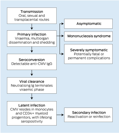
Cytomegalovirus (CMV) is an internationally ubiquitous human herpes virus with a worldwide seroprevalence ranging from 45% to 100%.1 A national serosurvey in 2006 estimated that 57% of Australians between the ages of 1 and 59 years were seropositive.2 While primary CMV infection is common in the general community, it is usually asymptomatic or causes a mild mononucleosis-like syndrome.3 The viraemic phase is generally self-limiting in healthy adults, and is followed by a lifelong bloodborne latent phase within peripheral monocytes and CD34+ myeloid progenitor cells4 (Box 1).

However, in certain circumstances CMV infection is capable of producing severe, life-threatening disease, including a wide range of potential clinical manifestations, owing to systemic haematogenous dissemination and a very broad tissue tropism6 (Box 2). Typically, severe CMV disease occurs in the context of an immature, suppressed or compromised immune system, and can lead to death or permanent major sequelae.7 As such, severe CMV infection is a well recognised cause of morbidity and mortality in neonates and immunocompromised adults, such as pharmacologically immunosuppressed transplant recipients and patients with AIDS.7

CMV disease in immunocompetent adults

While CMV is a well recognised pathogen in neonates and immunocompromised adults, the burden of CMV disease in immunocompetent adults is less well understood. This is because severe CMV disease is of considerably lower incidence in this population. However, it is far from non-existent; over 380 published case reports document instances of severe tissue-invasive CMV infection in immunocompetent adults. Similarly to CMV disease in immunocompromised individuals, these cases show a wide range of manifestations, including colitis,9 vascular thrombosis,10 pneumonia11 and myocarditis.12

The most comprehensive evaluation of severe CMV infection in immunocompetent adults to date included a systematic meta-analysis of case reports and reviews documenting 290 instances, across all manifestations.8 This study found that CMV infection most commonly involved the gastrointestinal tract (primarily colitis), followed by the central nervous system (including meningitis, encephalitis and myelitis) and then haematological abnormalities (including haemolytic anaemia and thrombocytopenia). CMV disease of the eye, liver, lung and vasculature were also documented, among other conditions. The authors ultimately concluded that the incidence of severe manifestations of CMV infection in immunocompetent individuals appeared to be significantly more common than previously appreciated.8

It should be noted that the definition of immunocompetency in this analysis, like most published reviews and case reports, excluded only individuals with profound loss of immune function, including patients who had AIDS, pharmacologically immunosuppressed transplant recipients and chemotherapy recipients. However, many case reports of CMV disease in “immunocompetent” adults document comorbidities that may be associated with a degree of immune dysfunction, such as diabetes mellitus or renal failure. Indeed, studies have also shown an increased risk of CMV-related morbidity and mortality in “immunocompetent” critically ill patients.13 It is therefore highly feasible that partial immune dysfunction may represent a currently overlooked risk factor for severe CMV disease. As such, further studies are needed to evaluate the risk of CMV disease in these populations. Nonetheless, this possibility reinforces the importance of considering CMV as a potential infectious agent even in patients with a low degree of immune dysfunction.

Diagnostic challenge of CMV disease

Although uncommon, severe CMV infection in immunocompetent adults often poses a significant diagnostic challenge, and a number of case reports have documented considerable delay to diagnosis.1416 Some patients with CMV colitis, in particular, have had protracted hospitalisations and have undergone surgery to investigate persistent and undiagnosed disease.

The diagnostic difficulty in CMV disease arises from three factors. First, the low incidence of severe CMV disease in immunocompetent individuals warrants a lower index of clinical suspicion for CMV infection at initial presentation. Second, CMV disease can present with a wide array of potential clinical manifestations, owing to broad tissue tropism. Third, certain presentations of CMV disease strongly mimic other diseases, potentially causing diagnostic confusion and delay in diagnosis. Indeed, case studies have documented initial misdiagnoses of colon carcinoma,14 ischaemic colitis,15 inflammatory bowel disease,16 dengue fever17 and lung cancer,18 among others.

Notable case studies

Siegal and colleagues documented the case of an 82-year-old man presenting with a 2-day history of diarrhoea and epigastric pain.15 Non-contrast computed tomography (CT) showed faecal impaction and thickening of the wall of the distal colon, with generalised large-bowel dilatation. Despite repeated negative results from assays for Clostridium difficile toxin, antibiotics were administered. There was no clinical improvement. Sigmoidoscopy and biopsy during the second week of admission showed acute inflammation and lymphoid aggregates, but did not lead to a diagnosis. The patient was treated with mesalamine for suspected ulcerative colitis, with minimal effect. A positive faecal occult blood test result raised the suspicion of ischaemic colitis. However, repeat sigmoidoscopy and biopsy at 1 month after admission showed mucosal ulceration with viral inclusions diagnostic of CMV infection, and treatment with intravenous (IV) ganciclovir was commenced. The authors concluded that CMV should be considered as a potential aetiological agent of severe colitis in immunocompetent individuals when other differentials have been excluded.15

A case study by Falagas and colleagues highlighted the diagnostic difficulty posed by CMV disease in non-immunocompromised adults.14 In this instance, a 57-year-old, HIV-negative man with chronic renal failure presented with acute abdominal pain, diarrhoea, and per rectal bleeding. Colonoscopy showed a large polypoid mass in the hepatic flexure, suspicious for colorectal carcinoma, although a colonic biopsy specimen did not show neoplastic changes. The results of subsequent CT scanning, stool examination and a C. difficile toxin assay were normal. Recurrent symptoms prompted an exploratory laparotomy and right hemicolectomy on Day 9 of admission, at which time histological analysis showed intranuclear inclusions diagnostic of CMV disease. The patient commenced a 2-week course of IV ganciclovir, and his symptoms subsequently abated. The authors concluded that the endoscopic findings of CMV colitis may resemble colon carcinoma and should be considered as a differential diagnosis, even in patients without severe immunosuppression.14

Yu and colleagues published a case report of a 39-year-old woman with a 6-week history of fever of unknown origin who was referred to a tertiary hospital.19 Her liver enzyme levels were elevated, and an abdominal CT scan was consistent with acute hepatitis. Results of serological screening for hepatitis A, B and C viruses, Epstein–Barr virus and HIV were negative. The result of a CMV-polymerase chain reaction (PCR) analysis was positive, and other causes (including drug-induced and autoimmune) were excluded. The severity and progressive nature of the disease necessitated urgent living-donor liver transplantation. A biopsy specimen from her explant liver showed widespread hepatic necrosis and stained positive for CMV protein. Initially, no antiviral therapy was commenced. The patient’s postoperative course was characterised by a rising serum bilirubin level and CMV antigenaemia. A liver biopsy specimen taken on Day 14 after the operation showed moderate degenerative changes and stained positive for CMV protein, at which point IV ganciclovir therapy was initiated, leading to improvement in her clinical condition and liver function. The authors concluded that CMV should be investigated as a potential cause of severe hepatitis, regardless of the patient’s immune status, after more common aetiologies have been excluded.19

Clinical implications

Delayed diagnosis of CMV disease in immunocompetent adults creates the potential for numerous adverse outcomes. Delay in initiation of targeted therapy leads to increasing morbidity and mortality as a result of disease progression. Prolonged hospitalisation is associated with health risks such as nosocomial infection and venous thromboembolism. Patients may also receive unnecessary radiation exposure from repeated CT imaging and be exposed to risks associated with surgical interventions. In addition, financial costs associated with extended hospitalisation and the potential for numerous investigations, surgery and intensive care unit admission are substantial. These consequences of diagnostic delay are of particular note, given the availability of non-invasive diagnostic testing for CMV infection, including serological tests, CMV-PCR and viral culture.20

Treatment of CMV disease in immunocompetent adults

While ganciclovir or valganciclovir are currently recommended as first-line treatment for severe CMV disease in immunocompromised adults, few studies have appropriately evaluated the use of these antiviral agents for the treatment of severe CMV disease in immunocompetent adults. These agents may have major side effects, including myelosuppression and potential carcinogenicity. However, untreated CMV disease is associated with considerable morbidity and mortality, and published case studies and reviews provide consistent case-based evidence of rapid clinical improvement after commencement of therapy in this clinical setting.3,11,17,2123 Furthermore, a recent study found ganciclovir to be a safe and effective treatment for CMV-associated pneumonia in immunocompetent children.24 Therefore, the continued use of antivirals for the treatment of very likely or proven CMV disease in immunocompetent adults appears justified at present. While formal studies evaluating the efficacy and utility of these therapies in the context of immunocompetency would be beneficial, such studies would be difficult to pursue, given the low incidence of severe CMV disease in this population.3

Conclusion

Although severe CMV disease primarily occurs in neonates or severely immunocompromised adults, the burden of disease in immunocompetent adults appears to be greater than previously understood. This may be partly owing to underrecognised risk from immune dysfunction associated with comorbidities such as renal failure or diabetes mellitus. It also appears that diagnostic delay is more likely in this clinical setting, especially for instances of CMV colitis, creating the potential for a range of adverse outcomes. Severe CMV disease in immunocompetent adults is likely to remain a diagnostic challenge in many circumstances. However, earlier consideration of CMV as a potential aetiological agent in individuals with atypical or refractory disease, regardless of immune status, may facilitate early non-invasive diagnosis and the initiation of appropriate directed antiviral therapy.

1 Overview of primary and secondary cytomegalovirus (CMV) infection and disease4,5

2 Non-exhaustive list of recognised potential manifestations of cytomegalovirus disease7,8

Direct effects


Gastrointestinal

Cardiovascular

Colitis

Myocarditis

Enteritis

Venous thrombosis

Gastritis

Neurological

Hepatitis

Meningitis

Pancreatitis

Encephalitis

Cholangitis

Myelitis

Respiratory

Retinitis

Pneumonitis

Uveitis

Haematological

Urological

Thrombocytopenia

Nephritis

Leukopenia

Prostatitis

Anaemia


Disseminated intravascular coagulation


Myelodysplastic change


Indirect effects


Atherosclerosis acceleration

Accelerated AIDS progression

Graft dysfunction and rejection

Increased opportunistic infections

Colorectal cancer screening and subsequent incidence of colorectal cancer: results from the 45 and Up Study

With a mean of 43.8 new cases diagnosed per 100 000 individuals in 2008, Australia has one of the highest age-standardised colorectal cancer (CRC) incidence rates worldwide, accounting for 12.7% of total cancers and 10% of all cancer deaths nationally.1

Survival from CRC highly depends on stage at diagnosis, and clinical trials have demonstrated that screening using faecal occult blood testing (FOBT) increases the detection rate of early-stage disease and reduces CRC mortality.2 Further, the efficacy of endoscopic polypectomy in preventing adenomas from progressing to CRC has led to a decrease in the incidence of CRC in screening trials.3 As a result, population-based screening programs to reduce mortality from CRC have been implemented in many nations in recent years.46

In Australia, national guidelines for CRC screening were introduced in 1999, recommending asymptomatic persons aged 50 years and over be screened using FOBT at least every 2 years.7 However, CRC screening tests were not freely available until the National Bowel Cancer Screening Program (NBCSP) was launched in 2006 with a one-off faecal occult blood test mailed to all people turning 55 and 65, and, from 2008, additionally to people turning 50.4 Although NBCSP-detected cancers are being diagnosed at an earlier stage than symptomatic cancers,8,9 the effects of opportunistic screening that occurred before the NBCSP on CRC incidence remain largely unexplored. Observational data on CRC incidence after CRC screening do not directly indicate screening effectiveness in reducing incidence, as cancers diagnosed after screening constitute those either missed at screening or arising de novo during the interval between screens (interval cancer), and a reduced incidence compared with non-screened individuals is to be expected. However, such data are useful in providing a real-world indication of the likely outcomes and health services use after CRC screening in the general population and giving indirect insights into the impact of screening.10,11

In this study, we present data on the relationship between reported CRC screening history and CRC incidence in a large population-based Australian cohort study, the 45 and Up Study.

Methods

Study population

The 45 and Up Study is a population-based Australian cohort study designed to investigate healthy ageing.12 Briefly, eligible participants were randomly selected from the Australian universal health insurance records (Medicare Australia). A total of 267 113 individuals (123 906 men and 143 207 women) aged ≥ 45 years from the general population in New South Wales joined the study by completing a postal questionnaire (distributed from January 2006 to December 2008) and giving written consent. Ethics approval for the study was provided by the University of New South Wales Human Research Ethics Committee and the NSW Population and Health Services Research Ethics Committee.

We excluded participants with pre-existing cancer (other than non-melanoma skin cancer), missing date of study entry, body mass index (BMI) outside the range of 15–50 kg/m2, and with invalid or most likely implausible values for physical activity and diet as previously defined.13 We excluded 55 777 participants with pre-existing cancer (other than non-melanoma skin cancer), 11 with missing date of study entry, 2113 with BMI outside the range of 15–50 kg/m2 and 12 759 with invalid or most likely implausible values for physical activity and diet. Exclusions left 196 464 participants for analysis.

Exposure assessment and definitions of variables

Information on all variables was derived from the self-administered questionnaire.14 The questionnaire included information on sociodemographic characteristics, medical history, body weight and height, smoking, alcohol, diet and physical activity.

For screening history, participants were asked whether they had ever been screened for CRC, and if so which test they had undergone (FOBT, sigmoidoscopy or colonoscopy). Year of most recent test was also recorded. The following exposure groups were defined: ever versus never screened, time since last screening (never, screened ≤ 3 years ago, screened > 3 years ago), screening modality (ever FOBT v never screened; ever endoscopy v never screened) and time since last screening according to single screening procedures. Initially, no distinction was made between primary screening by endoscopy or endoscopy after another screening procedure, most likely FOBT. However, we separately evaluated primary screening by endoscopy in additional analyses.

Ascertainment of colorectal cancer

Information on cancer incidence was obtained through record linkage with the NSW Central Cancer Registry. For our analysis, the specific censoring date at which the cancer registry was considered complete was 31 December 2008. Registry information was complemented with record data from the NSW Admitted Patient Data Collection (APDC) for the period from 1 January 2009 to 31 December 2011. The APDC is a complete census of all hospital admissions and discharges in NSW and contains, among other details, the principal reason for admission. As the primary treatment of CRC is surgical, the APDC is considered to provide reliable independent data on such diagnoses. Recent studies on CRC and breast cancer have shown that cancer diagnosis can be accurately identified using hospital data.15,16

We only considered first primary incident cases of CRC and participants were followed up from study entry to cancer diagnosis, death or follow-up termination (31 December 2011), whichever came first. Incidence data were coded using the International Classification of Diseases for Oncology, 3rd edition (ICD-O-3), with CRC comprising C18–C20 (excluding C18.1, cancers of the appendix). Proximal colon tumours included the caecum, ascending colon, hepatic flexure, and transverse colon (C18.0, 18.2–18.4). Distal colon tumours included the splenic flexure (C18.5), and descending (C18.6) and sigmoid (C18.7) colon. Overlapping lesions (C18.8) and unspecified colon (C18.9) were grouped among all colon cancers only (C18.0, C18.2–C18.9). Cancer of the rectum included tumours occurring at the rectosigmoid junction (C19) and rectum (C20). Anal canal tumours were excluded.

Statistical analysis

Associations between history of CRC screening and incidence of CRC were investigated by calculating hazard ratios (HRs) using proportional hazards regression stratified by age. Age was taken as the underlying time metric, with entry and exit time defined as the participant’s age at recruitment and age at cancer diagnosis or censoring, respectively. HRs are presented in relation to never screened individuals and are adjusted for sex; BMI (per kg/m2); highest qualification (no school, intermediate/high school, apprenticeship, university), household income (< $20 000, $20 000–$49 999, $50 000–$69 999, and ≥ $70 000); remoteness based on the Accessibility/Remoteness Index of Australia (city, regional, remote); a diagnosis of diabetes; family history of CRC; aspirin use; smoking (never, former, current); alcohol use (drinks/day); vigorous physical activity (none, 0 to 1 hour/week, > 1 to 3.5 hours/week, > 3.5 hours/week); and intake of red meat, processed meat, cereals, fruits, vegetables and wholemeal bread.

To appropriately handle missing data (up to 13% missing data for some variables), we used multiple imputation techniques (PROC MI and PROC MIANALYZE in SAS).17 All variables included in the multivariate model were included in the imputation procedure, and five imputation cycles were performed.

In sensitivity analyses, we excluded cases occurring during the first 2 years of follow-up and restricted the analysis to individuals with complete information on all variables to compare results with those obtained using multiple imputation.

All analyses were performed using SAS, version 9.3 (SAS Institute) and two-sided P values were considered.

Results

During a mean follow-up of 3.78 years (SD, 0.92 years) — 741 829 person-years — a total of 1096 cases of incident CRC accrued (454 proximal colon, 240 distal colon, 349 rectal and 53 unspecified cancers).

Screened individuals were more likely to have a family history of CRC and less likely to be current smokers than unscreened individuals (Box 1).

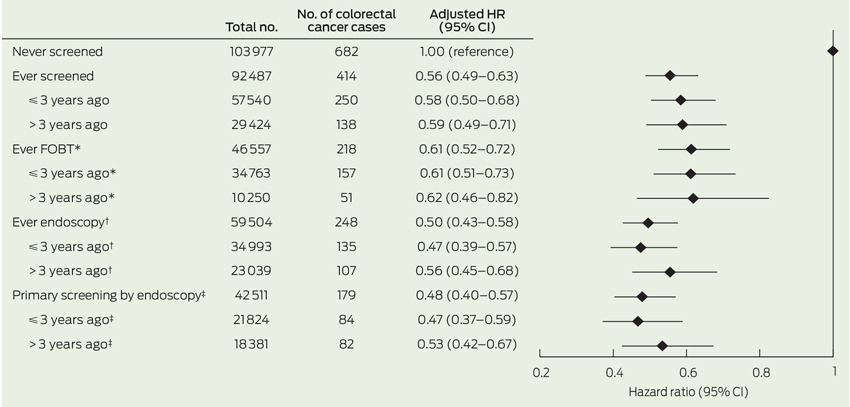
Having ever been screened before baseline was associated with a 44% reduced risk of developing CRC during follow-up in comparison to never having been screened (HR, 0.56; 95% CI, 0.49–0.63) (Box 2). HRs of CRC were 0.61 (95% CI, 0.52–0.72) and 0.50 (95% CI, 0.43–0.58), respectively, among those reporting ever having had FOBT and those reporting endoscopy versus no screening. We observed negligible differences in the HRs for those screened more than 3 years versus 3 years or less before baseline.

In relation to CRC subtypes, the inverse association between all screening exposure variables was strongest for rectal (HR, 0.35; 95% CI, 0.27–0.45) followed by distal cancer (HR, 0.60; 95% CI, 0.46–0.78), while the relationship was weaker for proximal colon cancers (HR, 0.76; 95% CI, 0.62–0.92) (Box 3).

Similar HRs were observed in subgroup analyses; for example, men and women and across smoking status (data not shown). The risk of developing CRC was particularly low for individuals with a family history of CRC (HR, 0.38; 95% CI, 0.27–0.54) compared with individuals without (HR, 0.59; 95% CI, 0.52–0.68) (P for interaction, 0.02).

Restricting the analysis to individuals with complete information on all variables (626 CRC cases) did not materially alter the results (data not shown). Exclusion of patients diagnosed during the first 2 years of follow-up slightly attenuated results for FOBT (HR, 0.72; 95% CI, 0.58–0.90), while estimates remained virtually unchanged for ever having had an endoscopy (HR, 0.52; 95% CI, 0.42–0.65).

Discussion

In this large population-based prospective study, a history of CRC screening was related to a 44% lower risk of subsequent CRC compared with never having undergone screening. Reductions in CRC risk were about 40% for ever having had FOBT and 50% for endoscopic procedures, compared with never having undergone CRC screening. The reduction in CRC incidence was observed up to 4 years after baseline and applied most strongly to rectal cancer.

Although our data cannot be used to directly evaluate CRC screening effectiveness, they are largely consistent with trial-based evidence on the efficacy of CRC screening in reducing CRC incidence and mortality. A meta-analysis of four randomised controlled trials (RCTs) indicated that those allocated to FOBT had a 16% reduction in CRC mortality compared with those not randomised to screening,18 while risk reduction may be even greater among those who adhere to a screening program.19 Based on data from five RCTs, sigmoidoscopy-based screening reduced CRC incidence by 18%.3 Colonoscopy is considered the “gold standard” for examination of the entire colon and rectum; however, its efficacy in relation to CRC incidence and mortality has never been investigated in RCTs and its magnitude of effect is currently unknown.20

Our observation of a 50% lower CRC risk after screening endoscopy is in line with two observational prospective studies from the United States.10,11 Interestingly, the reduction in CRC risk after endoscopy was greater for rectal and distal colon cancers than tumours located in the proximal colon. In our study, we reported combined estimates for sigmoidoscopy and colonoscopy. Because flexible sigmoidoscopy only allows inspection of (and polyp removal from) the distal but not proximal colon, our finding of stronger risk associations with distal CRC might be due to the lack of association of sigmoidoscopy with proximal colon cancer. However, only a small proportion of screened individuals reported having undergone sigmoidoscopy (< 6% v 64% colonoscopy and 52% FOBT), with 70% of them additionally having had colonoscopy, presumably as a follow-up of abnormal sigmoidoscopy. Our risk estimates are thus likely to be driven by the effect of colonoscopy. Differences in the strength of association with colonoscopy for proximal and distal colon cancers have been noted previously.11,21,22 A recent long-term prospective study from the US found reductions in incidence of 30% and 75% for proximal and distal CRC after colonoscopy, respectively, which compares well with our finding.11 Possible explanations for the difficulties in detecting precancerous lesions in the proximal colon may include incomplete colonoscopies or poor quality of bowel preparation resulting in missed lesions; flat lesions which are difficult to detect and remove; and the occurrence of rapidly growing cancers.21,23 Large, long-term RCTs on the effectiveness and the magnitude of effect of colonoscopy on the incidence of CRC and its subtypes in the general population are needed and underway, but results are unlikely to be available within the next 10 years.24,25

The association of FOBT with reduced CRC incidence appeared to be driven primarily by the strong relation of FOBT with rectal cancers in our study. The ability of FOBT to detect proximal colon cancers has been debated.26 FOBT exploits the tendency of CRC and large adenomas to bleed; however, haemoglobin from proximal neoplasia may degrade on passage to the anus, which may affect the accuracy of the test. In a systematic review of seven prospective screening studies, most of the studies indicated lower sensitivity for detecting proximal advanced neoplasia than for distal advanced neoplasia.26 In general, sensitivity for detecting precancerous lesions is low for guaiac faecal occult blood tests (16%–31%) and moderate for the newer faecal immunochemical tests (27%–67%).27

The lack of differences in the association of screening with CRC incidence across the two periods may be due to the relatively short time frame of our study. Case–control studies have suggested that the effect of endoscopy sustains for more than 10 years with little attenuation.28,29 For FOBT, the effect attenuation we observed after excluding cases occurring during the first 2 years of follow-up may reflect its ability to detect early-stage disease and its rather low to moderate sensitivity for detecting precancerous lesions.27

The results of our study need to be interpreted against the backdrop of its limitations. First, as has been noted, the results cannot be directly translated into effectiveness of population screening. Greater emphasis should be placed on the data as providing insights into the likely experiences of consumers and on health services use after CRC screening. Second, assignment of screening status was based on self-report and may be subject to misreport. A recent meta-analysis, however, indicates self-reports of CRC screening to be reasonably reliable.30 Although the questionnaire explicitly asked for screening, we cannot rule out that some participants may have reported having had endoscopy as a diagnostic test (due to symptoms) rather than as true screening (as part of a routine health check-up), and our risk estimates may not entirely represent the effect of true screening. This potential mixture of effects, however, will most likely have biased our estimates towards the null, and the protective effect of true screening by endoscopy might be even stronger. Third, we were not able to take into account information about the number of previous screens and a potential diagnosis or removal of polyps. Removal of polyps would have altered individuals’ CRC risk. Further, people having polyps removed would be more likely to participate in screening regularly. People screened more than once will have a lower risk of CRC irrespective of whether screening is beneficial, as they must not have had CRC detected on a previous screen and will later be diagnosed only if the last screen produced a false-negative result or cancer developed afterwards. Similarly, because we excluded pre-existing CRC at baseline, all incident cases must have had either a false-negative screen or developed cancer since last screening. Fourth, we lacked information on the type of FOBT and sigmoidoscopy. Finally, we had no information on screening during follow-up; however, the likelihood of bias due to repeated screening appears low, given the relatively short follow-up in our study.

In conclusion, this population-based prospective study illustrates a lower CRC risk among individuals with a history of CRC screening, compared with individuals who have never had CRC screening, through either FOBT or endoscopy, lasting for at least 4 years after screening.

1 General characteristics of the 196 464 included participants from the 45 and Up Study, by screening history


Never screened (n = 103 977)

Ever screened (n = 92 487)

P for comparison


Age, mean (SD)

59.7 (11.1)

62.8 (10.1)

< 0.001

Body mass index, mean (SD)

27.0 (5.0)

26.9 (4.8)

< 0.001

Men

41.1%

44.6%

< 0.001

Diabetes

8.0%

8.1%

0.95

University education

23.8%

25.3%

< 0.001

Yearly household income ≥ $70 000

27.1%

26.8%

0.06

Remote

2.3%

1.8%

< 0.001

First-degree relative with CRC

7.2%

20.0%

< 0.001

Regular aspirin use

17.1%

22.5%

< 0.001

Former smokers

32.7%

36.0%

< 0.001

Current smokers

9.7%

5.4%

< 0.001

Alcoholic drinks per day*

1.47

1.50

< 0.001

Vigorous activity > 3.5 hours/week

8.8%

8.6%

0.18

Red meat ≥ 5 times/week

20.9%

22.4%

< 0.001

Processed meat ≥ 5 times/week

5.4%

5.0%

< 0.001

Fruits ≥ 2 serves/day

55.9%

60.7%

< 0.001

Vegetables ≥ 5 serves/day

32.8%

36.0%

< 0.001

Breakfast cereals > 1 times/week

76.5%

82.9%

< 0.001

Wholegrain bread ≥ 5 serves/week

47.1%

51.6%

< 0.001


CRC = colorectal cancer. * Among drinkers only.

2 Adjusted hazard ratios for the associations between different screening procedures and incidence of colorectal cancer in the 45 and Up Study


HR = hazard ratio. FOBT = faecal occult blood testing. Numbers for analyses on time since screening do not add up to number of cases used for overall analyses because individuals without information on time since last screening were excluded from these analyses. HRs were obtained from proportional hazards regression, and were stratified by age and adjusted for demographic and health-related variables as described in the text, in addition to footnoted adjustments. * Adjusted for having undergone primary endoscopy. † Adjusted for having undergone FOBT only. ‡ Adjusted for having undergone FOBT or FOBT and endoscopy.

3 Adjusted hazard ratios for the associations between different colorectal cancer (CRC) screening procedures to the incidence of different subtypes of CRC in the 45 and Up Study (n = 196 464)


Proximal colon (n = 454)


Distal colon (n = 240)


Rectum (n = 349)


Screening variables

Total no.

No.

Adjusted HR (95% CI)

No.

Adjusted HR (95% CI)

No.

Adjusted HR (95% CI)


Never screened (reference)

130 977

245

1.00 (reference)

149

1.00 (reference)

253

1.00 (reference)

Ever screened

92 487

209

0.76 (0.62–0.92)

91

0.60 (0.46–0.78)

96

0.35 (0.27–0.45)

≤ 3 years ago

57 540

128

0.82 (0.66–1.03)

56

0.63 (0.46–0.87)

57

0.33 (0.25–0.45)

> 3 years ago

29 424

68

0.76 (0.58–1.00)

30

0.62 (0.42–0.92)

34

0.38 (0.27–0.54)

Ever FOBT*

56 557

106

0.87 (0.69–1.10)

53

0.72 (0.52–1.00)

52

0.36 (0.26–0.49)

≤ 3 years ago*

34 763

76

0.83 (0.64–1.09)

36

0.69 (0.47–1.01)

39

0.36 (0.25–0.51)

> 3 years ago*

10 250

24

0.79 (0.52–1.21)

15

0.88 (0.51–1.50)

12

0.35 (0.20–0.64)

Ever endoscopy

59 504

133

0.73 (0.58–0.90)

46

0.46 (0.33–0.65)

58

0.33 (0.25–0.45)

≤ 3 years ago

34 993

73

0.68 (0.52–0.89)

26

0.47 (0.30–0.72)

31

0.30 (0.20–0.43)

> 3 years ago

23 039

56

0.74 (0.55–1.00)

20

0.52 (0.32–0.84)

26

0.38 (0.25–0.57)

Primary screening by endoscopy

42 511

92

0.71 (0.55–0.90)

30

0.43 (0.29–0.64)

38

0.32 (0.23–0.45)

≤ 3 years ago

21 824

49

0.72 (0.52–0.98)

16

0.45 (0.27–0.76)

16

0.26 (0.16–0.43)

> 3 years ago

18 381

42

0.70 (0.50–0.97)

14

0.45 (0.26–0.79)

21

0.39 (0.25–0.61)


HR = hazard ratio. FOBT = faecal occult blood testing. HRs were obtained from proportional hazards regression, and were stratified by age and adjusted for demographic and health-related variables as described in the text, in addition to footnoted adjustments.* Adjusted for having undergone primary endoscopy. † Adjusted for having undergone FOBT only. ‡ Adjusted for having undergone FOBT, or FOBT and endoscopy.

Splenic rupture: a rare complication of infectious mononucleosis

A 28-year-old man presented to the emergency department with acute left upper quadrant tenderness and postural hypotension. He reported having had fever and cervical tenderness for 1 week before his presentation.

Blood tests showed an elevated white cell count with reactive lymphocytosis. A test for infectious mononucleosis heterophile antibody was positive, consistent with recent infection.

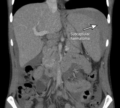
A contrast scan of the abdomen showed splenomegaly with subcapsular haematoma.

Splenic rupture after infectious mononucleosis is rare (incidence, 0.1%–0.5%), but can have disastrous consequences if overlooked.1,2

Feeding a hungry microbiome: large bowel fermentation and human health

Dietary fibre largely comprises indigestible plant carbohydrates and was thought to act primarily through its faecal bulking action — as “roughage”. Emerging evidence now shows that many of the actions of fibre are mediated through short chain fatty acids (SCFAs), principally acetate, propionate and butyrate, produced by the fermentation of its constituents by beneficial large-bowel bacteria (Physiol Rev 2001; 81: 1031-1064). Evidence is growing that SCFAs are important for general health as well as for normal gut function. Advances in genomics are enabling detailed identification and characterisation of the hundreds of different microbes that inhabit the large bowel, including fibre fermenters and SCFA generators (Nature 2012; 486: 207-214). Unfortunately, these dazzling technical advances have tended to divert attention away from the substrates, on which the microorganisms depend.

Questions about supply of colonic substrate are increasingly significant for public health. Australians have taken the advice to eat more fibre very seriously and consume fibre at high levels, largely as cereal fibres. This has not led to the anticipated fall in colorectal cancer (CRC) rates, which remain high (Int J Mol Sci 2013; 14: 13525-13541). In comparison, populations at low risk of CRC who consume wholegrains as staples do not have the high cereal fibre intake of Australians. However, the culinary practice of eating foods cold after cooking favours the generation of starch that is resistant to digestion by the small intestine. Resistant starch is fermented extensively in the large bowel, producing SCFAs, which promote a normal colonocyte phenotype — possibly lowering CRC risk (http://tv.csiro.au/?v=xbz189as9jwd). Levels of resistant starch in most Australian processed consumer foods are low, suggesting a general deficiency of fermentable substrate — and thus a collectively “hungry microbiome”. We suggest that filling this gap by increasing the availability of new fermentable fibre-rich foods to feed the colonic microbiota and raise large-bowel SCFA levels has considerable potential to improve the health of the population.

A bowel cancer screening plan at last

More lives will be saved by fully implementing the National Bowel Cancer Screening Program in 2020

The 2014–15 federal Budget included an announcement of $95.9 million for the long-awaited full implementation of the National Bowel Cancer Screening Program (NBCSP) by 1 July 2020.1 From that date, all Australians aged 50 to 74 years will finally be invited to screen for bowel cancer every 2 years with a faecal occult blood test (FOBT).

The announcement included a plan to incrementally expand the program, currently offered to people aged 50, 55, 60 and 65 years. The program will include 70-year-olds (through a previous funding commitment in 2012) and 74-year-olds from July 2015; people turning 64 and 72 years from 2016; and those aged 54, 58 and 68 years from 2017. The four remaining age groups (52, 56, 62 and 66 years) will be included from 2018 to 2020.1

The rationale is consistent with results from a study by Cenin and colleagues published in this issue of the Journal, which prioritised age groups according to the mortality-reduction benefit that can be expected from FOBT screening.2 Benefit is derived from prioritising screening according to age-based risk and closing gaps in the existing age cohort to shift from 5-yearly to biennial screening.3

A final implementation plan for the NBCSP has been a long time coming. The program was introduced in August 2006 with the mail-out of FOBT kits to people turning 55 and 65 years. While sporadic funding increases in the interim have been welcomed, there have also been unacceptable delays and ongoing concerns. For example, the 2012–13 Budget provided a much-needed $50 million to expand the NBCSP; however, the final implementation date was set as 2034.4 Cancer Council Australia therefore believed it was critical to provide evidence of the enormous potential benefits of completing the program by an acceptable date of 2020.

Cenin and colleagues used the MISCAN (microsimulation screening analysis)-Colon model to examine mortality gains with full implementation of the NBCSP by 2035 compared with full implementation by 20202 — the year recommended by Cancer Council Australia in its 2013 election priorities. The model estimated that full implementation by 2020 would prevent 35 000 (100% extra) bowel cancer deaths over the following four decades.

Now that the future of the NBCSP is assured, it is essential to engage with general practitioners and other health care professionals to improve participation and facilitate continuous improvement in service delivery.

The most recent data available show that, of people invited to participate in the NBCSP between July 2012 and June 2013, only 33.5% did so.5 This is an unacceptably low rate. However, it was not unexpected, given the low awareness among Australians about bowel cancer,6 the novelty of population screening for men and the lack of targeted communication about the NBCSP. A large-scale communications campaign, during program expansion and after full implementation, will be needed to improve participation rates if the NBCSP is to fulfil its potential to reduce bowel cancer mortality.

Since the inception of the NBCSP, GPs have been identified as critical partners. The government has sought to promote GP involvement in the NBCSP through GP representation on relevant committees, and through engagement with the Royal Australian College of General Practitioners (RACGP). While general practice resources such as the RACGP “red book” recommend FOBT screening for 50–74-year-olds, it will become increasingly important to consult closely with the primary care sector and provide support to GPs to facilitate their role in the expanded NBCSP.

GPs are well placed to promote the use of FOBT as the recommended screening tool for average-risk people currently outside the NBCSP. This would help reduce the strain on colonoscopy services. More than 500 000 colonoscopies are conducted annually in Australia.7 While there is no national dataset on how many of these are performed on asymptomatic, average-risk patients, it is thought that a significant number are done as first-line screening.

Currently, 7.5% of FOBT tests completed through the NBCSP return a positive result.5 Of those patients, around 70% present for colonoscopy. Of these, one in 32 are diagnosed with a confirmed or suspected cancer and one in 17 are diagnosed with advanced adenoma.5 FOBT is therefore a valuable tool for prioritising the use of colonoscopy for patients who are at higher than average bowel cancer risk or are symptomatic.

Throughout the NBCSP’s expansion, there has also been discussion about virtual colonoscopy, flexible sigmoidoscopy and plasma DNA testing as alternative screening tools. There is no evidence to suggest virtual colonoscopy would be a feasible alternative to FOBT.8 Although flexible sigmoidoscopy has been shown to be effective in randomised trials,9 it is significantly more expensive than FOBT and questions remain about its acceptability. DNA biomarker tests using plasma and faecal stool samples are also available; however, they are unsuitable for screening, as they have significantly lower sensitivity than FOBT for advanced adenoma and for stage A cancer.1012

Importantly, South Australian data have shown that twice the number of stage A cancers were diagnosed in people invited to participate in the NBCSP compared with people who were not and who had presented with a symptom.13

The NBCSP’s potential to prevent a total of 70 000 Australian bowel cancer deaths over the next four decades is compelling.

Dysphagia, regurgitation and weight loss in an elderly man

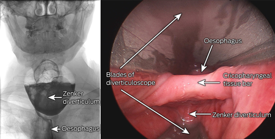
An 84-year-old man reported unintentional weight loss of 15 kg over 18 months and dysphagia and regurgitation of undigested food over 3 months. Barium swallow showed a large pharyngo-oesophageal (Zenker) diverticulum. These are caused by elevated intraluminal pressure at the Killian dehiscence, an area of hypopharyngeal weakness between the cricopharyngeus and inferior pharyngeal constrictors.

Zenker diverticuli are rare, but can cause significant morbidity and mortality. A barium swallow should precede gastroscopy, as there is a risk of perforation and mediastinitis. We performed endoscopic stapling, as it is less invasive than open repair.