×

The Value of Audio Devices in the Endoscopy Room (VADER) study: a randomised controlled trial

Music has been used in medical therapy and healing since Ancient Greek times.1 Its use in the operating theatre has been longstanding, with reports of its potential therapeutic effects dating back to the 1940s.2 Music therapy before and during an operation has been shown to reduce anaesthetic requirement and patient anxiety,3 and improve overall satisfaction.4 There is also evidence that proceduralist-selected music improves surgical skill acquisition.5

In gastrointestinal endoscopy, music has shown benefits for patients;614 however, there are no data on the effects of music on improving the performance of endoscopists and on whether a specific style of music would lead to optimal performance. This is particularly relevant in an era where high volumes of endoscopic procedures are performed in an often stressful clinical environment, and where procedural outcomes, such as adenoma detection rate (ADR) in colonoscopy, are reported as validated benchmarks of quality endoscopy.

The appropriate style of music to use and avoid in the operating theatre has been previously proposed,15 but music from epic movies was absent from this list. The soundtracks from such movies often contain uplifting musical tracks associated with glory, success and large-scale victory. Given we are avid fans of the Star Wars movies (Lucasfilm Ltd), and with the imminent release of the latest instalment, Star Wars: The Force Awakens, we felt it would be important and timely to evaluate the effect of music from the Star Wars soundtrack in colonoscopy. Therefore, we hypothesised that Star Wars music (SWM) would be superior to endoscopist-selected popular music (PM) when measuring quality outcomes in colonoscopy.

Materials and methods

In a hospital far, far away (Melbourne, Australia), patients were recruited from endoscopy lists from a single quaternary-centre gastroenterology unit between June and August 2015. All human subjects undergoing colonoscopy for any indication were included in the study. Ewoks and Wookies were excluded from the study owing to their inability to provide informed consent, but fortunately none were identified on the colonoscopy lists. The Austin Health Human Research Ethics Committee approved the project. The Jedi Council could not be contacted despite numerous attempts — clouded their contact details were.

Bowel preparation was performed using three sachets of sodium picosulfate for healthy patients and a volume-reduced regimen for patients with substantial comorbidities. A split-dose bowel preparation was used for afternoon procedures. All colonoscopies were performed under anaesthetist-administered monitored sedation with intravenous propofol. An Olympus 190-Series colonoscope (Olympus Medical Systems) was used in all cases. ProVation MD (ProVation Medical) was used for procedure documentation and coding.

Randomisation was performed using a coin toss by the endoscopist before colonoscopy to determine which music would be played: heads for SWM; tails for endoscopist-selected PM. SWM included the soundtrack from Star Wars: Episode III Revenge of the Sith, of which “Battle of the Heroes” (composed by John Williams) was the most commonly played track. The music was played through a personal computer in the endoscopy suite at a volume of between about 40 and 70 decibels, depending on the tolerance of the other endoscopy staff, and began before commencement of the colonoscopy. Patients were blinded to the music type being used.

Five endoscopists participated in the study: one consultant with 10 years’ experience (Jedi Master) and four specialist registrars with 1–2 years’ experience (Padawans). An independent member of the endoscopy team kept timings of procedures, and endoscopists were blinded to these timings. Timing commenced when the scope entered the anus (outer rim) and continued until the scope was withdrawn from the colon. The Padawans were supervised by a Jedi Master endoscopist for each procedure and advised to use the Force, and not force, during the colonoscopy.

Patient demographics and the presence of diverticular disease were recorded. The quality of bowel preparation was assessed by the endoscopist using qualitative descriptors (“excellent”, “good”, “fair” or “poor”). Primary end points were the procedure time, polyp detection rate (PDR) and adenoma detection rate (ADR). The PDR was defined as the number of colonoscopies in which one or more polyps were removed, divided by the total number of colonoscopies performed. The ADR was defined as the number of colonoscopies in which one or more adenomas were removed, divided by the total number of colonoscopies. A secondary end point was the recently proposed quality measure, adenomas per colonoscopy (APC), defined as the total number of adenomas detected divided by the number of colonoscopies.16

Data were analysed using the statistical software GraphPad Prism, version 6 (GraphPad Software, Inc). Non-parametric data comparisons between the two groups used Mann–Whitney tests to determine differences in procedure time and PDR. P ≤ 0.05 was used as the statistical significance level.

Results

Box 1 details the patient characteristics and colonoscopy outcomes. One hundred and three colonoscopies were included in the analysis: 58 in the SWM group and 45 in the PM group. A similar number of colonoscopies were performed by the Jedi Master in both groups (P = 0.45). Mean ages were 64 and 66 years (P = 0.13); 62% and 51% of patients in the SWM and PM groups, respectively, were men (P = 0.055). The most common indications for colonoscopy were positive faecal occult blood test, anaemia for investigation, and a personal history of polyps. Colonoscopies for bowel cancer screening and in patients with a history of polyps were evenly distributed between the two groups (P = 0.18).

Bowel preparation was assessed as good or excellent in 57% of the SWM group compared with 69% in the PM group (P < 0.01). Caecal intubation rates were high in both groups (98% and 97%, SWM and PM groups, respectively). The procedure duration was similar in both groups, with total procedure times of 20 minutes and 22 minutes and withdrawal times of 14 minutes in both groups (P = 0.87) in the SWM and PM groups, respectively.

The PDR was significantly higher in the SWM group than in the PM group (60% v 35%; P = 0.006). Similarly, the ADR was significantly higher in the SWM group than the PM group (48% v 27%; P = 0.01). The APC in the SWM group was 84% compared with 35% in the PM group (P = 0.01).

Discussion

Colonoscopy is a key intervention to prevent bowel cancer.17 With an increasing uptake of bowel cancer screening, demand on colonoscopy services is high. Of note, individuals from the baby boomer generation, many of whom were fans of the original Star Wars trilogy, are approaching the appropriate age to undergo bowel cancer screening. It is therefore essential that strategies to enhance quality outcomes in colonoscopy are evaluated. Environmental considerations are important, and music has been shown to improve a proceduralist’s skills. Our prospective, single-centre randomised controlled trial study supports the positive effects of music in procedures, and demonstrated a higher PDR and ADR with SWM compared with PM during colonoscopy.

Higher PDRs and ADRs were achieved in the SWM group compared with the PM group, despite a similar procedure time and a lower rate of optimal bowel preparation. It would be expected that a poorer quality of bowel preparation would lead to a lower PDR and ADR and prolonged procedure duration. Poor preparation leads to missed lesions. Missed lesions lead to interval cancers. Interval cancers may have serious consequences for the affected patients. SWM may break this cycle. Our finding that the PDR and ADR were higher in the SWM group may relate to the heightened observation skills of the endoscopist in a conducive musical environment. Proceduralist-selected music, which was used in both groups of our study, has been found to induce neurohormonal and immune system changes that lead to improved procedural performance;18 however, this does not explain the differences observed between the groups. As Star Wars fans, we must suspect that the Force was also involved, enhancing our Force-sensitive abilities to detect polyps, despite an overall more hostile luminal environment. Akin to Luke Skywalker’s destruction of the Death Star in Star Wars: Episode IV A New Hope, despite being pursued by Darth Vader and TIE fighters, we trusted the Force to guide us through the murky colonic waters to locate and destroy polyps.

There are some limitations in our study. Despite being randomised, there was a non-significant trend towards a greater proportion of men in the SWM group compared with the PM group. Also, there were disproportionate numbers in the two groups. Both may be explained by flaws in randomisation; however, other key parameters known to influence PDR and ADR, such as being referred for colonoscopy after receiving a positive faecal occult blood test result from participation in the National Bowel Cancer Screening Program, were distributed evenly. Differences by sex between the groups were unlikely to account for the almost twofold increase in ADR in the SWM group compared with the PM group. Given that the Star Wars fan base is predominantly male and there were more males randomised to the SWM group, it is again plausible that the Force was involved — influence the coin toss it may have.

Without concealment allocation, it could be argued that the higher PDRs and ADRs in the SWM group were due to performance bias. However, the ADRs in the PM and SWM groups were at the expected level for the study population,1922 so it is unlikely that deliberate underperformance in the PM group led to the observed difference in PDRs and ADRs between the groups. Additionally, the attainment of at least a 90% caecal intubation rate and an ADR of 20% was achieved in both groups and is in keeping with the current recommended guidelines for quality colonoscopy.23

It is unclear from this study if the improved endoscopic outcomes are specific to SWM or can be generalised to other epic movie soundtracks. A future study could, for example, assess the impact of music from the Lord of the Rings on endoscopic parameters. Of course, other non-Star Wars soundtracks would not be able to harness the power of the Force. If the Force is responsible for the superior endoscopic outcomes demonstrated in this, the addition of other Star Wars paraphernalia to the endoscopy suite may be of additive value. The wearing of Star Wars costumes may become a standard of practice in the future (Box 2).

In conclusion, SWM improves PDR and ADR in colonoscopy despite poorer quality bowel preparation when compared with PM. We recommend the widespread use of SWM in endoscopy rooms to improve quality outcomes. May the Force be with you.

Box 1 –
Patient characteristics and colonoscopy outcomes

Variable

Popular music (n = 45)

Star Wars music (n = 58)

P


Sex, men

23

36

0.055

Mean age, years (range)

63.8 (22–88)

65.5 (23–93)

0.13

Indications

Anaemia

6

18

National bowel colon cancer screening

19

21

Follow-up

Polyp

5

5

Cancer

1

0

Constipation

1

4

Diarrhoea

2

3

Pain

3

2

Perirectal bleeding

3

0

Weight loss

0

1

Inflammatory bowel disease surveillance

2

2

Other

3

2

Preparation quality

Excellent

1

1

Good

30

32

Fair

12

20

Poor

2

5

Good or excellent quality (%)

31/45 (69%)

33/58 (57%)

< 0.01

Procedures carried out by:

Consultant (Jedi Master)

22

28

0.45

Registrar (Padawan)

23

30

Extent

Caecum

13

22

0.016

Terminal ileum

31

35

0.23

Insertion time, min

Mean (SD)

8.0 (7.6)

6.8 (5.8)

0.056

Median

6

6

Range

2–42

1–35

Withdrawal time, min

Mean (SD)

13.7 (8.6)

14.0 (8.0)

0.87

Median

12

12

Range

6–50

6–52

Polyp detection rate (%)

16/45 (36%)

35/58 (60%)

0.006

Second polyp detection rate (%)

7/16 (43%)

20/35 (57%)

0.19

Polyp retrieval rate (%)

15/16 (93%)

32/35 (91%)

Adenoma detection rate (%)

12/45 (27%)

28/58 (48%)

0.01

Adenoma per colonoscopy (%)

16/45 (35%)

49/58 (84%)

0.01

Total adenomas

16

49

Diverticulosis (%)

23/45 (51%)

16/58 (27%)

0.007


∗Data are the number of patients unless otherwise indicated. †There was one aborted procedure in each group due to poor bowel preparation.

Box 2 –
Artist’s impression of the future standard practice in endoscopy

Food avoidance: some answers, more questions

The growing number of consumer foods and ingredients branded as “gluten-free” and/or “lactose-free” suggests that avoidance of specific dietary factors is common. CSIRO (Commonwealth Scientific and Industrial Research Organisation) survey data indicate that this behaviour is a real phenomenon, with one in seven adult Australian respondents not diagnosed with coeliac disease reporting avoidance of wheat or dairy foods. The behaviour is largely self-initiated, occurring predominantly without a formal medical diagnosis and is driven principally by adverse reactions attributed to those foods. Mostly, these comprise gastrointestinal symptoms that include bloating and cramps, commonly associated with irritable bowel syndrome. Wheat contains gluten, which provokes an immune reaction in susceptible individuals. However, the pattern of survey responses regarding symptoms does not suggest an allergenic or autoimmune cause; wheat avoiders do not seem to be undiagnosed coeliacs (Public Health Nutr 2014; 18: 490-499).

Our results raise several causes for concern. Translation of the data to the general population suggests that a significant proportion of adult Australians are at risk of nutritional imbalance. Wheat- and dairy-based foods are important sources of essential nutrients, so their exclusion could lead to dietary deficiencies (eg, of fibre, calcium) or, of equal concern, dietary imbalances caused by consumer overcompensation for perceived deficiencies. Clinically, self-diagnosis of symptoms of intolerance also carries the risk of delays in the identification and treatment of potentially serious medical conditions (BMJ 1988; 297: 719-720). For the majority of avoiders, the actual causes of symptoms and their mechanisms need to be established to improve condition diagnosis and management. Complicating matters is the issue of poly-avoidance, with more than 50% of wheat avoiders also avoiding dairy foods, and we need to establish whether the behaviours share a common aetiology. We also need to clarify whether consumers are making other self-prescribed dietary changes, either in response to their symptoms or in the belief that they are improving their personal health.

Bad times for good bacteria: how modern life has damaged our internal ecosystems

Human actions damage ecosystems on a global scale. Our influence is so great we’ve triggered a new geological epoch, called the Anthropocene, simply because of the changes we’ve brought about. But it’s not just the outside environment we’ve changed, we’ve also damaged the ecosystems inside us.

Our activities alter natural processes, such as weather patterns, and the way nutrients, such as nitrogen and phosphorus, move within ecosystems. We cause declines in species diversity, trigger extinctions and introduce weeds and pests.

All this comes with costs, caused by the increasing unpredictability of both physical and biological systems. Our infrastructure and agriculture rely on a consistent climate, but that’s now becoming increasingly unreliable. And it’s not just the outside world that’s unpredictable; it may come as a surprise to some that we have internal ecosystems, and that these have also been damaged.

Shrinking population

Every adult is made up of 100 million, million human cells (that’s a one followed by 14 zeroes). But the human body is also home to ten times this number of bacterial cells, which, collectively, are called the microbiota. Biologists have only been exploring this internal ecosystem for a decade or so, but surprising and important results are already emerging.

Bad times for good bacteria: how modern life has damaged our internal ecosystems - Featured Image

Humans damage ecosystems on an epic scale. Global Water Forum/Flickr, CC BY

Because the laboratory where I work is interested in how humans affect evolutionary processes, it was natural for us to ask how much humans might affect microbial ecosystems. The answer turns out to be quite a lot.

Possibly the most direct and personal effects are on our own microbiota. And these changes come with consequences for health and well-being. Exactly the same processes we see in external ecosystems – loss of diversity, extinction, and introduction of invasive species – are happening to our own microbiota. And damaged ecosystems don’t function as well as they should.

Scientists have tried to “go back in time” and ask what the original human microbiota might have looked like. There are three ways of doing this: biologists can look at the microbiota of our nearest relatives, the great apes; we can examine DNA from fossils; or we can look at the microbiota of modern-day humans who still have a hunter-gatherer lifestyle.

All these approaches tell the same story. Modern humans have a lower diversity of microbiota than our ancestors, and there’s been a consistent decline in this diversity across ancient and recent human history.

There are a number of reasons for the decline. The widespread use of fire from 350,000 years ago increased the calories we could obtain from food. This probably decreased our need for a big gut, and a smaller gut means less room for microbes.

The invention of agriculture between 8,000 and 10,000 years ago changed our diet, and with it, our microbiota. The end result was the extinction of some components of the microbiota in farming populations. Even today, hunter-gatherers and subsistence societies have many bacterial species in their gut that are never found in the guts of people from westernised societies.

Bad times for good bacteria: how modern life has damaged our internal ecosystems - Featured Image

One of the last hunter-gatherer societies in the world, the Yanomami people of South America, have a highly diverse and stable microbiota, and don’t suffer from diseases common in the developed world. christian caron/Flickr, CC BY-ND

Modern onslaught

Changes in microbiota have been tracked using bacteria preserved on the teeth of skeletons, and this showed falls in diversity linked to dietary changes, as well as a shift to microbial species associated with disease.

The changes are particularly apparent after the Industrial Revolution, when processed flour and sugar became widely available. And diet continues to have a major influence on our microbiota.

But the greatest disruption probably happened after the 1950s. This time period corresponds to a number of changes that directly affect the composition of the human microbiota. One involves the opportunity for microbiota to colonise newborns and infants. Normally, babies obtain some microbiota from their mother during childbirth, but caesarean births interrupt this opportunity. Bottle feeding, increased sanitation, and eating processed, sterile foods also limit opportunities to acquire microbiota.

Modern medicine has been very successful at controlling bacterial diseases with antibiotics. Unfortunately, antibiotics cause considerable collateral damage to innocent and beneficial bacteria. After antibiotic therapy, the microbiota may never return to their original abundance, and genetic diversity is reduced in those bacteria that remain.

Bad times for good bacteria: how modern life has damaged our internal ecosystems - Featured Image


Antibiotics can also damage beneficial bacteria. Photo: Shutterstock

Collectively, these changes mean that our microbial ecosystems have become degraded, much like natural ecosystems globally. The microbiota are less functional and resilient than they should be. And it turns out they have essential roles in developing our immune systems, and in regulating metabolism. So it shouldn’t be surprising that altered microbiota are now being associated with many diseases of the modern world.

These diseases include obesity, allergic reactions, chronic inflammatory conditions and autoimmune disorders. More recently, it’s also been suggested that psychological conditions, such as depression and anxiety, are linked to the bacteria that live inside us.

In some cases, the parallels with more conventional ecosystems are clear. Clostridium difficile is a bacterium that can grow out of control in our gut, like an invasive weed. And, like a weed invading degraded land, it often spreads rapidly after other bacteria have been eliminated from the gut by antibiotics. The most effective cure is similar to bush regeneration; donating microbiota from healthy volunteers (a “poo transplant”) helps restore a healthy ecosystem.

But, for many diseases associated with our microbiota, there are no immediate cures. Like most ecosystems, our gut bacteria are complex and dynamic. The challenge now is to understand this system and how to acquire and maintain a healthy microbiota, so that in the future, a microbiota check-up might be a routine part of a visit to the doctor.

In such a future, hunter-gatherers such as the Yanomami of the Amazon may turn out to be the custodians of valuable species that are extinct in the microbiota of the developed world.


This article was originally published on The Conversation. Read the original article.

Renowned clinician next MJA Editor in Chief

Influential medical clinician and researcher, Laureate Professor Nicholas Talley, has been appointed as the new Editor in Chief of the Medical Journal of Australia.

AMPCo Board Chair Richard Allely said Professor Talley, who is currently Pro Vice Chancellor, Global Research, at the University of Newcastle and a part-time staff specialist gastroenterologist at the John Hunter Hospital, came to the position with a wealth of local and international experience in medical research, practice and publishing.

“Professor Nick Talley is a clinician, educator, writer, author, researcher, and editor, with a strong track record in medical practice, medical education, and medical publishing, in Australia and overseas,” Mr Allely said.

As well as having authored 800 original and review articles in peer-reviewed academic journals, Professor Talley is currently Co-Editor in Chief of the international journal Alimentary Pharmacology and Therapeutics (a position he will relinquish soon after he takes up the MJA post on 1 December), and served for six years as Co-Editor in Chief of the American Journal of Gastroenterology.

“He brings significant experience, knowledge and expertise to the MJA, and is perfectly suited to guiding Australia’s leading medical journal at a time of rapid change, innovation and technological revolution in media and publishing,” Mr Allely said.

In addition to his ongoing academic, clinical and publishing work, Professor Talley is President of the Royal Australasian College of Physicians and Chair-elect of the College of Presidents of Medical Colleges.

He also holds several international adjunct appointments, including Professor of Medicine and Professor of Epidemiology at the Mayo Clinic, and Foreign Guest Professor at Stockholm’s Karolinska Institute.

Professor Talley’s appointment was announced soon after it was revealed that AMA Federal Councillor and former Australian Medical Students’ Association President Jessica Dean had been recruited to the Board of mental health organisation beyondblue.

beyondblue Chairman Jeff Kennett said Ms Dean’s experience as a young doctor would be “invaluable” for his organisation as it sought to work with medical students and practitioners at risk of experiencing depression and anxiety.

Ms Dean has been a member of beyondblue’s Victorian Doctors’ Mental Health Advisory Group, and earlier this year addressed a meeting of senior Victorian doctors, health officials and administrators about the mental health of medical practitioners and the culture in which they work.

Adrian Rollins


Klebsiella pneumoniae liver abscess complicated by endogenous endophthalmitis: the importance of early diagnosis and intervention

Clinical record

A 51-year-old man presented to the Royal Victorian Eye and Ear Hospital with 3 days of progressive visual loss and pain in the left eye, and a 6-hour history of painless visual loss in the right eye. He reported a 1-week history of fever, night sweats, sore throat and a non-productive cough. The man was systemically well, with no features of sepsis or abdominal pain.

His only medical history was for hypercholesterolaemia. He was Malaysian, but had lived in Australia for 10 years. He had recently travelled to Malaysia and Vietnam.

Visual acuity (VA) was hand movements in the left eye and 6/12 in the right. He had a left relative afferent pupillary defect and bilateral hypopyon. Vitritis limited posterior segment examination. The left eye had lid swelling, conjunctival chemosis, proptosis, and computed tomography (CT) showed evidence of scleritis (Figure, A).

White cell count (26 × 109/L; reference interval [RI], 4.0–11.0 × 10⁹/L) and C-reactive protein levels (199 mg/L; RI <5 mg/L) were both elevated; liver function tests were deranged, with evidence of cholestasis. Liver ultrasonography revealed a 5.3 cm abscess in segment VII. A CT scan showed two abscesses: a 5.0 × 5.7 cm abscess in segments V/VIII and a 3.3 × 5.1 cm abscess in segment VII, with cholelithiasis and segmental thrombosis of the right hepatic vein (Figure, B). The results of blood cultures were negative.

VA in the right eye deteriorated to counting fingers over a 24-hour period, and endogenous Klebsiella pneumoniae endophthalmitis was suspected. The patient received systemic ceftriaxone; bilateral intravitreal vancomycin, ceftazidime and dexamethasone; and oral and topical corticosteroid therapy. K. pneumoniae was cultured from ultrasound-guided drainage of one liver abscess and from urine. Repeated vitreous samples included polymorphonuclear leukocytes, but no bacteria could be cultured. Hepatic vein thrombosis was treated with therapeutic enoxaparin and, later, rivaroxaban.

Pars plana vitrectomy (PPV) was performed in the right eye 2 days after presentation, and a superotemporal subretinal abscess was noted. Vision initially improved to 6/24, but deteriorated to hand movements 2 days after the operation, presumably following rupture of the abscess. A second PPV was performed, and the abscess was drained. A rhegmatogenous retinal detachment was managed intraoperatively with endolaser and silicone oil (Figure, C). The right eye received four doses and the left eye five doses of intravitreal vancomycin, ceftazidime and dexamethasone.

The left eye underwent a two-stage PPV, with the first surgery 4 days after presentation. An inferior subretinal abscess was drained during the second PPV (Figure, C and D). An associated rhegmatogenous retinal detachment was treated with endolaser and silicone oil.

Both eyes settled well (Figure, E and F). The retinae remained attached bilaterally under oil, and best-corrected VA had improved to 6/12 in the right eye and 6/24 in the left eye at the 1-month follow-up.

Endophthalmitis refers to inflammation of the intraocular space, and is predominantly of infectious aetiology. It is usually exogenous and may complicate intraocular surgery, penetrating trauma or corneal ulceration. Around 2%–8% of cases occur via endogenous spread, often in the context of immunosuppression, diabetes or injecting drug use.1

In Australia, fungal organisms cause 65.9% of endogenous endophthalmitis, while gram-negative organisms cause 19.5%.1 In East Asia, there is an increasing incidence of disease caused by gram-negative organisms, with Klebsiella pneumoniae now the most common cause of endogenous endophthalmitis.24 K. pneumoniae is a known cause of pyogenic liver abscess in southeast Asia, with metastatic spread of infection occurring in 3.5%–20% of cases and endogenous K. pneumoniae endophthalmitis (EKPE) in 3%–7.8% of cases.3

EKPE has an extremely poor prognosis.25 Retinal damage occurs rapidly following irreversible necrosis of the retinal photoreceptor layer and the associated subretinal abscess formation.17 Final visual outcomes of hand movements or worse are reported in 66%–78% of cases,3,7 no perception of light in 57.8%–62%,7,8 and evisceration or enucleation in 26.8%–75% of patients;1,8 in one report, three cases of EKPE required enucleation despite appropriate intensive antibiotic treatment.6

The poor prognosis is exacerbated by delayed diagnosis, particularly in patients who, because of overwhelming systemic illness, are unable to describe visual symptoms.2 Further significant predictors of a worse prognosis include poor initial visual acuity3 and the presence of hypopyon at presentation.8

In rare cases, EKPE may be the first manifestation of K. pneumoniae infection.3 EKPE is usually unilateral, but bilateral ocular involvement is reported in up to 26% of patients.1 Diabetes is a risk factor for EKPE.2,5 There is no general consensus about the most appropriate treatment for EKPE.

It has been suggested that pars plana vitrectomy (PPV) reduces intraocular bacterial and inflammatory load in EKPE and aids the penetration of intravitreal antibiotics.2,6 The best visual outcomes are seen in patients who undergo surgery early.4 In a Korean report about seven patients (10 eyes) with EKPE, early PPV was performed on eight eyes and delayed PPV on two. Operative findings included extensive retinal necrosis with subretinal abscess formation and dense vitritis. Five eyes maintained a final VA of counting fingers or better, VA in two eyes improved to 6/19 and 6/38; no enucleations or eviscerations were required.2 Another group described a patient who had PPV within 8 hours of the onset of EKPE symptoms; VA had improved from 6/120 to 6/6 at the 12-month follow-up.9 These results support the role of early PPV in the management of EKPE.

A series of five patients had previously been treated for EKPE at the Royal Victorian Eye and Ear Hospital. All were treated with intravenous and intravitreal antibiotics. Four of the five patients required enucleation or evisceration, and the fifth became phthisical. Our patient, the sixth case, presented early in the course of disease, and was treated with systemic and intravitreal antibiotics, as well as with early PPV. In contrast to the preceding cases, the visual outcome was excellent.

The incidence of K. pneumoniae liver abscess in the Asia-Pacific region is increasing. Early ophthalmology referral for patients with suspected or proven K. pneumoniae liver abscess is recommended. In patients with known EKPE, early PPV should be performed in conjunction with intensive systemic and repeated intravitreal antibiotic and steroid therapy to reduce intraocular bacterial and inflammatory load, and to aid the penetration of intravitreal antibiotics to the subretinal focus of infection. This may increase the chances for rescuing the eye and improving the visual outcome.

Lessons from practice

  • Endogenous Klebsiella pneumoniae endophthalmitis (EKPE) carries an extremely poor visual prognosis.

  • In patients admitted to general hospitals with K. pneumoniae liver abscess, a high index of suspicion of EKPE is recommended, and early referral to ophthalmology services advised.

  • Early surgical intervention in EKPE can salvage vision in this otherwise devastatingly blinding disease.

Figure


A, B: Intraoperative photographs of the posterior segment of the eye, showing retinal detachment with a subretinal abscess in the right eye (A) and a subretinal abscess in the left eye (B); the white arrows indicate the optic nerves. C: Computed tomography (CT) image of the orbits at presentation, showing left-sided proptosis with associated inflammation of the sclera (white arrow) and periorbital soft tissues. D: Abdominal CT scan, showing a 5.0 × 5.7 cm abscess (white arrow) in segments V/VIII of the liver. E, F: Slit lamp photos of the right (E) and left (F) eyes 13 days after initial presentation.

Splenic abscess complicating gastroenteritis due to Salmonella Virchow in an immunocompetent host

Clinical record

A 20-year-old man was admitted to a regional hospital with fevers, rigors, anorexia and left upper quadrant pain. It was his fourth presentation to the emergency department in the preceding 10 days. On the first two presentations, he had been sent home with a provisional diagnosis of renal colic. After review by his general practitioner, he had undergone outpatient imaging that identified filling defects in the pulmonary arteries of his left lower lobe, which were reported as being consistent with pulmonary emboli. In addition, two hypodense splenic lesions were identified, as well as collapse and possible consolidation of the left lower lobe. His GP had referred him to the emergency department for further review (his third presentation), after which he had commenced therapeutic anticoagulation for a presumed diagnosis of pulmonary emboli.

The patient’s history was notable for a self-limiting episode of gastroenteritis 6 weeks before his initial presentation, with sick family contacts. On his fourth presentation, he described progressive left upper quadrant and flank pain over the preceding 10 days, with intermittent fevers and rigors. He had no other focal infective symptoms on review.

On examination, he was found to have a fever (temperature, 39.3°C), sinus tachycardia (heart rate, 154 beats/min), tachypnoea (respiratory rate, 28 breaths/min), hypotension (blood pressure, 97/66 mmHg), decreased breath sounds at the left base of his lung fields and mild left upper quadrant tenderness. Investigations showed a white cell count of 16 × 109/L (reference interval [RI], 4.0–11.0 × 10⁹/L), with a predominant neutrophilia (neutrophils, 14 × 109/L [RI, 2.0–7.0 × 10⁹/L]). Results of his liver function tests and electrolyte, urea and creatinine levels were all within reference intervals.

A computed tomography scan of the chest and upper abdomen again showed two low-density lesions of unclear aetiology in the spleen, as well as a left-sided pleural effusion and collapse of the left lower lobe. Given the possibility that the hypodense splenic lesions represented septic emboli from a cardiac source, the patient was treated empirically with benzylpenicillin, flucloxacillin and gentamicin for a provisional diagnosis of endocarditis. However, a transthoracic echocardiogram performed the next day did not support this diagnosis, with no abnormalities detected. Beyond the radiological findings described, there were no other clinical grounds to support a diagnosis of endocarditis.

Blood cultures taken on Day 1 of admission were positive for gram-negative bacilli, with confirmation of a non-typhoidal Salmonella species (later confirmed as Salmonella Virchow) the following day. This allowed targeted antibiotic therapy, once susceptibilities were known, with ampicillin (2 g every 6 hours). Cultures of stool samples taken at admission were positive for the same isolate, consistent with the patient’s self-limiting episode of gastroenteritis 6 weeks before his first presentation.

Magnetic resonance imaging of the abdomen suggested that the two splenic lesions were likely to represent abscesses in this clinical context (Figure). Given our patient’s ongoing sepsis, a decision was made to perform a laparoscopic splenectomy for source control on Day 5 of admission. Surgical specimens tested positive for Salmonella Virchow. Histopathological testing identified cystic lymphangiomas of the spleen. Despite problems with postoperative pain and a prolonged ileus, the patient made a full recovery. He received appropriate post-splenectomy vaccinations, along with a total of 2 weeks’ intravenous ampicillin, followed by a 2-week course of oral amoxicillin.

Non-typhoidal salmonellae are common foodborne pathogens. In Australia, they are the second most frequent bacterial isolates identified in cases of acute gastroenteritis, after Campylobacter jejuni. In 2010, OzFoodNet sites reported 11 992 cases of Salmonella infection, a rate of 53.7 cases per 100 000.1 Salmonella Virchow was the third most common isolate, after Salmonella Typhimurium and Salmonella Enteritidis. Non-typhoidal Salmonella infection outbreaks are most commonly associated with consumption of poultry and eggs, but have also been linked to fresh produce and, increasingly, contact with pet reptiles.2

Up to 8% of patients with gastroenteritis secondary to non-typhoidal Salmonella infection develop bacteraemia.3 Risk factors for invasive infection include extremes of age, immunosuppressed states, malignancy, HIV infection and use of tumour necrosis factor-blocking medication.4 Our case is unusual in that bacteraemia occurred in an otherwise immunocompetent host.

Extraintestinal focal infections have been reported to occur in 5% to 10% of patients with non-typhoidal Salmonella bacteraemia.3 The best recognised complications are endovascular infections, most commonly involving the aorta, that result from seeding of atherosclerotic plaques and aneurysms.5 However, focal infections of almost all organ systems have been reported.

Splenic abscesses are most commonly seen as a complication of infective endocarditis, occurring in about 5% of patients.6,7 They are also found as a rare complication of non-typhoidal Salmonella infections. In one case series of 49 patients from southern Taiwan, Salmonella species were the third most common pathogens isolated from splenic abscesses, accounting for 11% of cases.8 The most common presentations among the 49 patients with splenic abscesses were fever (47 patients), abdominal pain confined to the left upper quadrant (33 patients), left pleural effusion and splenomegaly (both 27 patients), all of which were present in our patient.

About 50% of patients presenting with splenic abscesses have pre-existing anatomical abnormalities.9 The cystic lymphangiomas identified in our patient almost certainly predisposed him to developing splenic abscesses.

According to the literature, the mainstay of treatment for splenic abscesses is splenectomy. Data from 287 cases published between 1987 and 1995 suggested that non-operative management, which included invasive treatment with percutaneous aspiration and catheter drainage, had a success rate of less than 65%.10 The same series suggested that antimicrobial therapy alone had a success rate of less than 50%. Salvage splenectomy, however, was not shown to result in increased mortality. Another retrospective study of 51 patients in a tertiary hospital between 1998 and 2003 reported survival rates of 48% with antimicrobial therapy alone, 45% with pigtail catheter insertion and drainage in addition to antimicrobial therapy, and 100% with splenectomy and antimicrobial therapy.11 These results may be influenced by selection bias but do suggest improved outcomes with splenectomy over less invasive strategies.

Lessons from practice

  • Splenic abscesses are a rare but potentially life-threatening complication of non-typhoidal Salmonella bacteraemia.
  • Splenic abscesses should be considered as a possible source of infection in patients presenting with unexplained fevers and left upper quadrant or left flank pain.
  • Splenectomy plus appropriate antimicrobial therapy remains the mainstay of treatment for splenic abscesses.
  • Interventional radiological techniques should be considered as a spleen-preserving strategy on a case-by-case basis and where experienced radiologists are available.

Splenic abscesses are a rare but serious complication of non-typhoidal Salmonella bacteraemia that may occur in otherwise immunocompetent individuals. Splenic abscesses should be suspected in patients with unexplained fevers and left upper quadrant pain. The mainstay of treatment is splenectomy with appropriate antimicrobial therapy.


A: Axial T2-weighted magnetic resonance image (MRI) of the abdomen, without contrast, showing an abscess in the inferior pole of the spleen (circle).


B: Saggital T2-weighted MRI of the abdomen, without contrast, showing two splenic abscesses (circles).


John Lachlan Gollan MB BS, MD, PhD, FRACP, FRCP, FACP

John Gollan was a world expert in liver diseases. He was born in Launceston, Tasmania, on 5 March 1942, the son of highly respected physician Neil Golan. After schooling in Launceston, John studied medicine at the University of Adelaide, graduating in 1966.

John undertook advanced clinical training in liver diseases in the United Kingdom and gained a doctorate in biochemistry from the University of London. He then relocated to the United States to work at the Liver Center at the University of California, San Francisco. In 1981, he moved to Harvard Medical School, where he established the study of liver diseases and was director of the gastroenterology division at Brigham and Women’s Hospital.

From 1999 to 2001, John was professor of medicine at the University of Adelaide, after which he returned to the US to chair the department of internal medicine at the University of Nebraska. In 2003, he became dean of the College of Medicine, a position he held until he retired in 2010 with severe Parkinson disease.

In Nebraska, he helped to raise millions of dollars for the Michael F Sorrell Center for Health Science Education, which opened in 2008 and is one of the world’s most technologically advanced education facilities. He also worked with Nebraska Medicine to establish the Biocontainment Unit, which gained international prominence recently for its care of Ebola patients.

John established a fund to provide the annual Neil Gollan Memorial Lecture at Launceston General Hospital in memory of his father and, in 2003, he returned to present the inaugural lecture.

John died on 7 January 2015 in Omaha, Nebraska. The University of Nebraska has now established a memorial fund. Comments from his many associates emphasise his warm personality and ready and welcome smile. He was the consummate gentleman, exercising gentle leadership and displaying integrity, wisdom, compassion and a sense of humour.

John’s wife Roseanne trained as a nurse in Launceston and was a great companion and support to him throughout his august career. Their three children Jackie, Tim and Jenny all live in the US.

Imported gluten-free foods: free of gluten?

To the Editor: The recent hepatitis A outbreak associated with imported berries has brought the problem of imported food quality acutely into the public spotlight. By contrast, the serious adverse effects for many people with coeliac disease of non-compliant imported foods being labelled “gluten-free” (GF) is more insidious and less easily assessed.

Concern has previously been expressed about proposals to raise the amount of gluten permitted in GF foods.1 In Australia, the current standard for claiming that a food is “gluten-free” is that it contains “no detectable gluten”;2 on the basis of the limits of current laboratory test sensitivity, this equates to less than 3 parts per million (ppm).

Closely aligned with this concern is the fact that imported food labelled “GF” may comply with standards in the country of manufacture but not with tighter Australian standards. For example, “GF” in Europe and North America indicates gluten levels of less than 20 ppm; accordingly, GF-labelled foods imported from these regions may contain detectable gluten. Further, gluten-level testing of GF-labelled foods is not mandatory in the United States;3 in one report, 20% of US foods labelled “GF” did not comply with the Food and Drug Administration standard.4

Governance of food regulation in Australia is unfortunately complex. Food Standards Australia New Zealand set food standards federally; individual states set laws based on the federal standards; local government health officials implement state laws and monitor compliance. The Australian Competition and Consumer Commission, responsible for consumer law, has also contributed to food regulation and compliance. Further, the federal Department of Agriculture has responsibility for regulating imported foods. Local importers and retailers should also facilitate food safety.

Testing of imported foods labelled “GF” is ad hoc, lacking coordination across multiple jurisdictions, and is hampered by financial constraints. There is a tendency for organisations to suggest that the responsibility for compliance lies elsewhere. Enhanced transparency of laboratory food testing outcomes is required, for there are scant published data that assure the consumer about food code compliance for foods labelled “GF”. It is to be hoped that some good will come of the hepatitis A food contamination incident, by providing the impetus for significant change in the governance of Australian food safety.

HCV-infected patients need access now to new direct-acting antiviral agents to avert liver-related deaths

To the Editor: We recently used a modelling approach to describe how the burden of infection with hepatitis C virus (HCV) and the associated health care costs in Australia will increase as the infected population ages.1 We showed that increasing the efficacy of antiviral therapy and the number of patients treated could avert the expected increase in deaths from HCV-related liver disease and in the number of patients with end stage HCV-related liver disease. We did not include the specific costs of new direct-acting antiviral (DAA) regimens, as these are yet to be determined in Australia. We know that the cost of the new regimens has elicited discussion internationally about the ability of payers to meet those costs.

Importantly, compared with previous regimens, DAA therapies offer higher cure rates, simplified dosing, shorter treatment duration and are better tolerated — albeit at a substantial price.

Given the difficult decisions that will need to be made by the Australian Government, we examined the impact of delayed access to DAA treatment by modelling 1-year and 2-year delays. Currently, an estimated 2550 patients are treated with interferons and/or first-generation protease inhibitors.1 In the DAA scenario, we assumed cure rates of over 90%, drug availability in 2015 and an increase in the number of patients treated to 3550 in 2015, 7100 in 2016 and 14 000 after 2018. To provide the chance of cure of HCV infection to those at greatest risk, we limited treatment to advanced liver fibrosis (stage F3 or F4) from 2015 to 2017, with no restriction on the stage of fibrosis beginning in 2018. Based on those assumptions, we then compared the cumulative incident cases of liver-related deaths, decompensated cirrhosis, and hepatocellular carcinoma from 2014 to 2030. Under no circumstance did we consider that DAAs would never be available, thus the 1-year and 2-year delay scenarios were only compared with the DAA scenario (Box).

While other scenarios could be considered, we believe that it is important to remember that an estimated 230 000 people in Australia are chronically infected with HCV,1 and those with advanced liver disease remain at greatest risk of liver-related death. We believe that it is critical to provide patients with access to highly effective therapeutic regimens to cure HCV infection without delay to diminish future HCV-related morbidity and mortality.

Projected cumulative new cases of hepatitis C-related advanced liver disease and death with current treatments compared with direct-acting antiviral (DAA) agents from 2014 to 2030, inclusive, plus projected new cases if access to DAA agents is delayed for 1 or 2 years


Cumulative new cases


Treatment

Liver-related deaths

Decompensated cirrhosis

Hepatocellular carcinoma


Current treatments

22 200

19 100

13 500

DAAs

13 500

11 000

8 200

DAAs, 1-year delay

14 400

11 800

8 700

DAAs, 2-year delay

15 300

12 600

9 200

Impact of delay


1-year

900

800

500

2-year

1 800

1 600

1 000

Pancreatic cancer: gradual rise, increasing relevance

Pancreatic cancer is likely to become one of the leading causes of cancer death in Australia

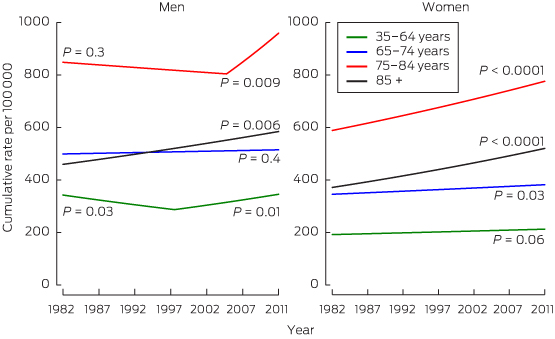
Pancreatic cancer is the 10th most frequently occurring cancer but the fifth most common cause of cancer death in Australia, as is also seen in other developed regions of the world.1 A gradual increase in incidence has been observed since the 1980s in almost all age groups in both sexes in Australia (Box 1).3 Increases have been attributed only to trends in smoking, which is considered causal,4 with local published data suggesting a lag of about 30 years between smoking trends and incidence.5 However, overweight and obesity may also have contributed, in part, to incidence trends.6

In Australia (and other developed countries), only about 50%–70% of cases of pancreatic cancer are histologically confirmed based on review of the primary tumour,1 because pancreatic biopsy procedures have been associated with significant risks7 and are often avoided. But improvements in imaging modalities, particularly endoscopic ultrasound and pancreas-specific computed tomography, and magnetic resonance imaging protocols, together with endoscopically guided biopsy procedures, are likely to have led to some of the increase in incidence through improved detection.

Survival and mortality

In 2011, the latest year for which results are available, 5-year survival from pancreatic cancer was 5.2% in Australia and 7.3% in the United States (among patients on selected Surveillance, Epidemiology and End Results Program registers) with only modest improvements observed over the past several decades. Five-year survival from pancreatic cancer was about 3% in the mid 1980s in both places.8,9 Between 1987 and 2007 in Australia there was only a 6% drop in mortality from pancreatic cancer in both sexes (in those aged less than 75 years), compared with decreases in mortality of 34% from lung cancer, 47% from bowel cancer and 28% from all cancers overall.10 Current projections suggest that within 10 years, pancreatic cancer will be the second-highest cause of cancer death in the US as mortality and survival from the other four leading causes of cancer death (lung, bowel, prostate, and breast cancers) improves.11 If these trends are reflected in Australia, we would anticipate that pancreatic cancer will become one of the leading causes of cancer mortality.

Complete resection of the primary tumour currently offers the only hope of cure. While 5-year survival in those who have local or regional involvement remains low, at 15%–20%, it is significantly better than the 5-year survival of 3% in people with metastatic disease. Thus, increasing the proportion of patients diagnosed with resectable disease from the currently dismal 15% seen in Queensland and New South Wales12 by diagnosing the disease earlier may be beneficial. However, beyond the setting of high-risk families, screening to identify precursor or early invasive lesions is not feasible for two main reasons. First, endoscopic ultrasound is invasive and can only be used in specialised settings, so does not meet criteria for a population screening test. Molecular-based screening may become available in the future, at least partially overcoming this problem. Second, the positive predictive value of screening is limited by the low population prevalence of pancreatic cancer. Attempts to stratify the population using known risk factors, including several known single nucleotide polymorphisms, have not yet identified population subgroups at sufficiently high risk to warrant screening.13

Optimising outcomes

An avenue to optimise outcomes for patients is to ensure that all receive high-quality care in the most appropriate setting. There is evidence from the US that not all patients with potentially resectable tumours are offered surgery.14 Detailed data are not currently available for Australia, but it appears that there is similar underutilisation of surgery here.12,14,15 It is thus important that all patients without metastatic disease are reviewed by a multidisciplinary team in a major centre to determine the resectability of their pancreatic tumours. In addition, resections should be performed in hospitals that carry out a large number of these procedures annually, as this has been shown to improve survival.16

Investment in research to identify cure, beyond surgery, has increased in the past decade. Sequencing of tumour tissue to identify tumour-specific genetic profiles offers the possibility of tailored treatment, with the hope that this will improve survival and avoid unnecessary side effects. In a whole-genome sequencing study of 100 patients with pancreatic cancer, four subtypes with potential clinical utility were identified, and in a subgroup of eight patients receiving platinum-based therapy, four out of five with the “on-genotypes” had exceptional or partial responses, while three patients who did not have these genotypes did not respond.17 Other promising approaches, such as targeting tumours with immunotherapy, are also being explored.18

In conclusion, while the rise in pancreatic cancer incidence in Australia is only slow, as the population ages, more people will be affected with this devastating disease. The burden of pancreatic cancer relative to other cancer types is likely to increase. A multilevel approach is needed to control pancreatic cancer, including reducing the prevalence of risk factors such as smoking and obesity, identifying effective biomarker screening tools and populations in whom screening or early detection might be feasible, discovering new treatment modalities and ensuring that all patients have access to optimal care.

Cumulative age-specific incidence per 100 000 population for cancer of the pancreas in men and women in Australia, 1982–2011*


P values are shown for the slope of trend, with joinpoint regression analysis used to identify points where this slope changed significantly.2
Data source: Australian cancer incidence and mortality (ACIM) books.3