×

The possible risks of proton pump inhibitors

These drugs have revolutionised the management of gastrointestinal diseases, but their long term use may have risks

Proton pump inhibitors (PPIs) suppress gastric acid secretion by selectively inhibiting the enzyme hydrogen–potassium adenosine triphosphatase, located in gastric parietal cells. These drugs superseded H2-receptor antagonists as first-line acid suppressants in the 1980s, and their potent effect has revolutionised the management of common upper gastrointestinal (GI) disorders, including gastro-oesophageal reflux disease, peptic ulcer disease, and functional dyspepsia. These drugs are also widely used as part of regimens designed to eradicate Helicobacter pylori infection, and as prophylaxis against the deleterious effects of non-steroidal anti-inflammatory drugs on the GI tract.

PPIs are among the most commonly administered medications worldwide. In recent years, there has been a marked increase in prescribing PPIs,1 concurrent with an overall reduction in their cost with the advent of inexpensive generic formulations. This reduction in cost is likely to have contributed to injudicious overprescribing of PPIs, with up to 60% of primary care physicians making no attempt to reduce patients’ doses over time, and almost 50% of patients receiving long term PPI therapy having no clear indication for its continuation.2 Despite their well documented benefits, the use of PPIs may be associated with an increased risk of certain GI and non-GI conditions.

Gastrointestinal risks of proton pump inhibitor use

Commensal bacteria are thought to influence and maintain metabolic and immune pathways in the gut. Manipulation of GI microbiota has been identified as a potential target for therapeutic intervention in the management of several GI disorders, including irritable bowel syndrome and inflammatory bowel disease, by means of faecal microbial transplantation or the use of prebiotics, probiotics, symbiotics or antibiotics. Changes in the faecal microbiome have also been identified in PPI users, with a reduction in the α-diversity of bacterial communities observed in the gut,3 presumably secondary to the alteration in pH of the intestinal luminal contents. This reduction in bacterial diversity may be associated with an increased risk of developing GI infections. A meta-analysis of observational studies reported increased odds of taking PPIs for patients with Clostridium difficile-associated diarrhoea (odds ratio [OR], 1.96; 95% CI, 1.28–3.00) and other enteric infections, including with Salmonella and Campylobacter species (OR, 3.33; 95% CI, 1.84–6.02).4

Aside from any potential association with enteric infection, diarrhoea is a common side effect of PPI use. The cause of this is uncertain, although a case–control study noted increased odds of microscopic colitis (OR, 3.37; 95% CI, 2.77–4.09),5 and, in a meta-analysis, small intestinal bacterial overgrowth (SIBO) was found to be more common in PPI users (OR, 2.28; 95% CI, 1.24–4.21).6 Given that both of these conditions present with diarrhoea, which is a side effect of PPIs, it may be that this association is the result of detection bias, as individuals using PPIs are more likely to be investigated to rule out microscopic colitis or SIBO.

The hypergastrinaemia documented in long term PPI users has led to concerns about the possibility of enterochromaffin-like cell hyperplasia being associated with PPIs, and therefore a theoretical increase in the risk of neuroendocrine tumour development, although this has not been substantiated.7 Observational studies also report an increase in the incidence of gastric fundic gland polyps (FGPs) in patients using PPIs long term. Although these polyps are benign, this has raised the possibility that long term PPI use could lead to an increased risk of gastric cancer. A recent systematic review and meta-analysis investigating the association between PPI use and the incidence of FGPs suggested a statistically significant increase in the odds of developing such polyps (OR, 2.45; 95% CI, 1.24–4.83), with a longer duration of exposure further increasing this risk.8 In the same meta-analysis, the risk of PPI-associated gastric cancer was examined in a small number of studies of heterogeneous design, and a small increase in the risk of gastric cancer was observed (risk ratio [RR], 1.43; 95% CI, 1.23–1.66). However, follow-up in these studies extended to a maximum of 36 months, a timeframe one would consider too short to develop gastric cancer de novo. This suggests that the association may be the result of protopathic bias, or reverse causation, with PPI therapy more likely to be initiated in individuals presenting with dyspepsia, which may herald early gastric cancer.

The anti-secretory properties of PPIs can also lead to the development of vitamin B12 and magnesium deficiencies. Gastric acid is required to facilitate the breakdown of digestible vitamin B12, and the risk of vitamin B12 deficiency may be increased in people with achlorhydria. In a retrospective case–control study investigating risk factors for the development of vitamin B12 deficiency, PPI exposure greater than 2 years was associated with a significant increase in the odds of vitamin B12 deficiency (OR, 1.65; 95% CI, 1.58–1.73), with a dose–response relationship observed.9 Similarly, the intestinal absorption of magnesium is adversely affected by an increase in luminal pH. A meta-analysis of studies examining this question also suggested increased odds of hypomagnesaemia with PPI use (OR, 1.78; 95% CI, 1.08–2.92).10 The United States Food and Drug Administration recommends monitoring of serum magnesium levels before commencing PPI therapy and periodically thereafter if long term use is likely to be required.

Non-gastrointestinal risks of proton pump inhibitor use

Conflicting evidence regarding the association between PPI use and the risk of bone fracture has been reported. Potential pathophysiological mechanisms include impaired intestinal calcium absorption and alterations in bone metabolism, mediated by inhibition of osteoclast proton pumps. A meta-analysis reported increased odds in patients taking PPIs of hip fracture (OR, 1.25; 95% CI, 1.14–1.37) or vertebral fracture (OR, 1.50; 95% CI, 1.32–1.72).11 However, no dose–response relationship was observed and significant heterogeneity was noted. Given the observational nature of the included studies, it is likely that these findings are secondary to unmeasured confounding, with PPI users being more likely to have a comorbid condition, such as obesity or tobacco smoking, that is associated with both PPI use and an increased risk of fracture.

The association between PPI use and community-acquired pneumonia (CAP) has been investigated, again with conflicting results from the numerous observational studies. A meta-analysis of these studies indicated an increase in the odds of developing CAP with short term PPI use (OR, 1.92; 95% CI, 1.40–2.63), but not with prolonged use (OR, 1.11; 95% CI, 0.90–1.38).12 This lack of a dose–response relationship, the absence of any plausible pathophysiological mechanism to explain the association, and the significant heterogeneity of the included studies cast doubt on this association, again suggesting prescription channelling may have influenced the study findings.

The most common side effect of antiplatelet medication is GI haemorrhage secondary to peptic ulcer disease, resulting in the prophylactic use of PPIs for gastro-protection in many patients with cardiovascular disease. A reduction in the in vitro antiplatelet effect of clopidogrel, when used in conjunction with omeprazole, led to concerns about a possible increase in cardiac event rates when these drugs were co-prescribed after percutaneous coronary intervention.13 However, a randomised controlled trial (RCT) investigating the incidence of GI complications and subsequent cardiac events in patients randomly allocated to receive either dual antiplatelet therapy and omeprazole, or dual antiplatelet therapy and placebo found that there was no difference in the cardiac event rate in patients receiving omeprazole or placebo (hazard ratio [HR], 0.99; 95% CI, 0.68–1.44), but a significant reduction in overt upper GI bleeding was noted with omeprazole (HR, 0.13; 95% CI, 0.03–0.56).14

The incidence of dementia is increasing. A recent cohort study suggested PPI use may be a risk factor for the onset of dementia in patients aged over 75 years, after adjusting for potential cardiovascular, cerebrovascular, and pharmacological confounders (HR, 1.44; 95% CI, 1.36–1.52).15 However, morbidity and mortality associated with adverse events, including those associated with GI bleeding, were not assessed, meaning that the effect of unmeasured confounding could not be accounted for and, in a case–control study, PPIs appeared to protect against dementia (OR, 0.93; 95% CI, 0.90–0.97).16 Despite this, it is plausible that PPIs may increase the risk of dementia, as there is evidence suggesting that PPIs cross the blood–brain barrier, and may promote the production, or impair the degradation, of amyloid,17 although another potential mechanism could be through the effects of PPIs on vitamin B12 absorption, as discussed above.

Conclusion

Although PPIs have revolutionised the management of several upper GI disorders, their use may be associated with intestinal and extra-intestinal complications. Most studies that have exposed these risks are observational and retrospective in nature. In the only RCT that has examined the impact of one of these associations on patients, no increase in the risk of cardiac events was observed with PPIs. Unlike RCTs, the inherent flaws of observational studies do not account for unmeasured bias and confounding, and only provide evidence for the presence or absence of an association, rather than a causal link between PPI use and the risk of such complications. Moreover, the magnitude of the increased associations observed in many of these studies, although statistically significant, is unlikely to be large enough to be clinically relevant, and is probably explained by a combination of residual confounding, prescription channelling, and protopathic bias. Patients should therefore be informed that the benefits of PPIs outweigh any potential deleterious effects. Nevertheless, just like any other drug, PPIs should be prescribed judiciously, with a clear indication and regular review of the appropriateness of continued long term use to minimise these theoretical risks. Currently, no routine monitoring of PPIs is recommended other than periodic measurement of serum magnesium levels if long term use is required.

Reversal of end-stage renal failure using direct-acting antiviral agents for chronic hepatitis C

Clinical record

A 58-year-old Eritrean woman presented to hospital in December 2014 with a vasculitic rash, peripheral oedema, acute renal impairment and microscopic haematuria with proteinuria. She had a history of genotype 4 hepatitis C virus (HCV) infection, which was unsuccessfully treated with pegylated interferon and ribavirin in 2010. Results of laboratory investigations showed a pattern consistent with mixed cryoglobulinaemia (Box 1). After a renal biopsy to assess progressive renal impairment, a diagnosis of membranoproliferative glomerulonephritis (MPGN) was made. The patient began taking an angiotensin-converting enzyme inhibitor and diuretic therapy.

The patient’s creatinine level continued to worsen, from 202 μmol/L (reference interval [RI], 45–90 μmol/L) (estimated glomerular filtration rate [eGFR], 27 mL/min/1.73 m2 [RI, > 60 mL/min/1.73 m2]) in January 2015 to 412 μmol/L (eGFR, 10 mL/min/1.73 m2) in July 2015. The renal impairment was associated with increasing serum cryoglobulin levels, worsening proteinuria and increased requirement for ascitic drainage. The ascitic fluid was a transudate, and a transjugular liver biopsy excluded cirrhosis. Infection and malignancy were also excluded. As the eGFR was below 15 mL/min/1.73 m2 and fluid overload had become refractory to medical management, preparations for dialysis were made and a Tenckhoff (peritoneal dialysis) catheter was inserted. At this stage, direct-acting antiviral medication was obtained through a compassionate access program to treat the HCV infection.

The patient began a 12-week course of HCV treatment with paritaprevir–ritonavir (150 mg/100 mg) and ombitasvir (25 mg) plus ribavirin (200 mg) daily on 2 July 2015. Ribavirin was discontinued at Week 5, as her haemoglobin level had fallen from a baseline of 102 g/L (RI, 115–160 g/L) to 76 g/L. Overall, the treatment was well tolerated. HCV viral load was undetectable at Week 2, and sustained viral response (SVR) 12 weeks after completion of treatment was achieved, indicating the HCV was cured. Creatinine levels improved markedly with treatment, associated with a rapid decline in serum cryoglobulin level and improvement in the urine albumin:creatinine ratio (Box 2). The ascites was last drained on 24 August 2015, and the Tenckhoff catheter was subsequently removed.

Mixed cryoglobulinaemia is a frequently encountered extrahepatic complication of chronic HCV infection and can be associated with considerable morbidity and mortality.1 The most common renal manifestation of mixed cryoglobulinaemia is MPGN, which is associated with a mixed cryoprecipitate, hypocomplementaemia, positive rheumatoid factor, microscopic haematuria and proteinuria.1 The clinical presentation ranges from asymptomatic proteinuria to nephritic or nephrotic syndrome.1 Progression to end-stage renal failure occurs in 10–33% of patients.1

Treatment of mixed cryoglobulinaemia due to HCV is reserved for symptomatic patients and centres on viral eradication.2 In a series of 86 patients with HCV infection and cryoglobulinaemic vasculitis treated with pegylated interferon and ribavirin, remission of symptoms occurred in 88% of patients who achieved SVR.2 However, management of patients with concurrent renal disease is complicated by the difficulties associated with using pegylated interferon and ribavirin in the setting of severe renal failure, and a lower response rate is reported in this cohort.2 In clinical practice, interferon and ribavirin therapy is therefore rarely pursued in patients receiving dialysis. In addition, the reversibility of renal impairment in the setting of HCV-induced cryoglobulinaemia has traditionally been thought to be poor, particularly once end-stage renal disease has been reached.

There are limited data available on the outcomes of interferon-free HCV treatment regimens in patients with mixed cryoglobulinaemia.3 The efficacy of sofosbuvir-based regimens has been studied in a series of 12 patients with HCV-associated mixed cryoglobulinaemia, with seven patients also having renal impairment related to MPGN.3 In this study, patients who achieved SVR had decreased proteinuria and improved renal function; however, the degree of baseline renal impairment was mild (eGFR, 30–60 mL/min/1.73 m2).3 Sofosbuvir is not currently recommended for use in patients with an eGFR < 30 mL/min/1.73 m2.3

The combination of ombitasvir and ritonavir-boosted paritaprevir is an interferon-free antiviral regimen for the treatment of genotype 4 HCV infection, with the addition of dasabuvir required for genotype 1 HCV infection.4 The efficacy and safety of this combination in patients with end-stage renal disease have been demonstrated in a pilot trial of 20 patients with genotype 1 HCV infection,5 although the impact of viral clearance on renal function has not been reported.

A literature review of the PubMed database on this topic did not identify other cases, so we believe this to be the first reported case of significant recovery of renal function in this clinical setting.

Lessons from practice

  • In patients with chronic hepatitis C virus (HCV) infection and renal failure from cryoglobulin-related glomerulonephritis, direct-acting antiviral medications may be safe and effective, and successful treatment may significantly improve renal function.

  • Chronic HCV infection can have numerous extrahepatic manifestations that may abate considerably after virus eradication.

  • Patients with chronic HCV infection who were previously considered difficult to treat due to medical comorbidities should be reassessed for HCV treatment with direct-acting antiviral medications.

Box 1 –
Results of diagnostic investigations

Laboratory test

Result

Reference interval


Urinary protein

+++

nd

Urinary blood

+++

nd

Albumin:creatinine ratio (mg/mmol)

121.1

< 3.5

Creatinine (μmol/L)

179

45–90

Cryoglobulins (g/L)

0.44

< 0.08

C3 (g/L)

0.80

0.88–1.98

C4 (g/L)

0.04

0.16–0.52

Rheumatoid factor (kU/L)

117

< 14

Hepatic venous pressure gradient (mmHg)

10

1–5


nd = not detectable.

Box 2 –
Results of investigations showing response to treatment with direct-acting antiviral medications

Laboratory test

Reference interval

Result


Before treatment (July 2015)

End of treatment (Sept 2015)

12 weeks after treatment (Dec 2015)


Haemoglobin (g/L)

115–160

102

125

113

Albumin (g/L)

35–50

33

35

40

Creatinine (μmol/L)

45–90

412

186

196

eGFR (mL/min/1.73 m2)

> 60

10

25

24

HCV viral load (IU/mL)

nd

2.00 × 10⁶

nd

nd

Cryoglobulins (g/L)

< 0.08

3.46

0.84

0.56

Albumin:creatinine ratio (mg/mmol)

< 3.5

394

nr

67.7

C3 (g/L)

0.88–1.98

0.67

0.94

0.87

C4 (g/L)

0.16–0.52

< 0.03

0.06

0.06


eGFR = estimated glomerular filtration rate. HCV = hepatitis C virus. nd = not detectable. nr = no result available.

Streamlining ethics review for multisite quality and safety initiatives: national bariatric surgery registry experience

The current ethics review process is inappropriate for clinical quality registries

Rigorous methods for assessing and improving the quality of health care have proven difficult to develop by traditional research approaches.1 Clinical quality registries (CQRs) systematically collect an agreed minimum dataset of data across multiple sites on clinically relevant outcome measures. Data are analysed, comparing procedures, providers and institutions.2 Feedback to practitioners has been shown to drive performance improvement, especially if the data are perceived to be high quality.3

Because CQRs collect and store health information, protocols require human research ethics committee (HREC) approval to ensure that they comply with the Australian Privacy Act 1988 (Cwlth). Principle 6 of this Act states that stored personal information must not be used or disclosed for a secondary purpose unless patient consent is obtained or there is a permitted health situation. Section 16B of the Act defines the permitted health situations, which include research relevant to public health or public safety. The use or disclosure of personal information must be conducted under guidelines approved under section 95A of the Act. Current National Health and Medical Research Council guidelines state that ethical review is required at each contributing site for CQRs except where multi-institutional approval is in operation.4

Bariatric surgery is burgeoning in Australia. In 2016, it is estimated that there will be over 15 000 such procedures performed in Australia at a direct cost of over $225 million. Yet there are no evidence-based guidelines directing who should be offered this surgery, nor are there any long-term community data documenting its safety and efficacy in Australia.

The Obesity Surgery Society of Australia and New Zealand (OSSANZ) partnered with Monash University Department of Epidemiology and Preventive Medicine (DEPM) to establish a national registry of all bariatric procedures with the aim of filling these knowledge gaps. The pilot commenced in 2012 and national rollout commenced in May 2014 (with federal government funding).

The Bariatric Surgery Registry (BSR) collects information on each procedure performed, the devices used, changes in patients’ weight and diabetes status, and adverse events. Data are collected primarily from surgeons and are validated against hospital International Statistical Classification of Disease and Related Health Problems, 10th Revision, Australian Modification (ICD-10-AM) discharge codes. Because the BSR is tracking and storing identifiable sensitive health information longitudinally as well as cross referencing data points to external data sources, HREC review is required at every site contributing to the BSR.4,5

We believe that there are 164 hospitals undertaking bariatric surgery in Australia. As of 30 April 2015, there were 52 hospitals (31.7%) for which HREC approval for participation in the BSR had been obtained. Private hospitals accounted for 67% of these sites, 29% were public hospitals and an additional approval was received from both the Royal Australasian College of Surgeons and Monash University. Applications for high risk projects were requested from 31.4% of private hospital and 33.3% of public hospital HRECs. Seven sites (13.5%) provided approval through an affiliated site. Fifty sites (96%) had additional governance requirements and, in 76% of cases, this was a separate process. The median time from the first application to final approval was 86 days (range, 17–414 days). The maximum numbers of queries from or changes requested by an HREC was 67.

The process of obtaining ethical approval at these initial hospital sites cost the BSR $180 698.58 in salaries and $3474.97 per application. In addition, the BSR has had to pay five sites a total of $3927.00 for ethics approval application fees.

The number of CQRs in Australia is growing rapidly in response to community demands for better monitoring of health care outcomes.6 HREC review of registry processes is one way of ensuring that the rights of individuals participating in registries are protected and complying with the Privacy Act. However, as highlighted by our experience in rolling out the BSR, the lack of a consistent process for obtaining HREC approval across multiple sites for these quality and safety initiatives creates cost and slows implementation.

HRECs are typically set up to review research projects rather than quality and safety initiatives. Unlike clinical trials, which are hypothesis driven and in which patients are given the option of participating, CQRs attempt to ensure quality through benchmarking, and so need to recruit all patients who undergo a given intervention to avoid the risk of bias. Many HRECs are not familiar with these basic and essential differences, and this can lead to confusion and delays, as the processes for clinical trials and CQRs invariably differ.7 Based on the cost and time involved in obtaining even 31.7% of the required approvals for the BSR, we call for a bespoke national process for HREC review of CQRs. This would streamline implementation and reduce costs while still protecting patient’s privacy. Examples of such a process could be having all sites apply to a single national ethics committee, as in New Zealand, or implementing specific federal legislation protecting the transfer of information to and from approved CQRs.

Detecting ascites

Most cases can be diagnosed by good clinical assessment at the bedside

The presence of ascites is a common physical finding and the detection of ascites is important for both diagnostic and prognostic reasons. Ascites is defined as the pathological accumulation of fluid in the peritoneal cavity.1 It may be due to a number of causes (Box 1). The most common is portal hypertension as a result of cirrhosis (> 75%) but malignancy (10%), heart failure (3%) and infection (2%) are other possibilities.1

Patients with ascites usually present to clinicians with increasing abdominal distension, weight gain and discomfort. However, ascites may be detected incidentally in patients developing other complications of their cirrhosis, such as variceal haemorrhage and encephalopathy. Moreover, a patient may present when their underlying heart failure or malignancy progresses.

Initial assessment of the patient will involve taking a history to determine the risk of liver disease. This will include questions about alcohol consumption and risk factors for chronic hepatitis, especially hepatitis C. Cardiac symptoms (shortness of breath and orthopnoea) can be assessed and patients should also be asked about symptoms that might indicate an underlying malignancy, such as weight loss and decreased appetite. Patients who do not complain of ankle swelling or abdominal distension are unlikely to have significant ascites.2

The examination of a patient with ascites includes two main components: inspection and palpation.

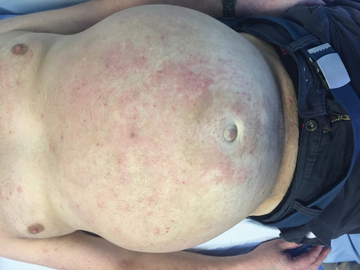
General inspection of the patient should include looking for signs of chronic liver disease, such as jaundice, spider naevi, palmar erythema, gynaecomastia and loss of body hair. Prominent collateral veins in the abdominal wall may also be present. Bulging flanks (Box 2) may be due to ascites or obesity, but the absence of this sign makes the presence of ascites unlikely.2

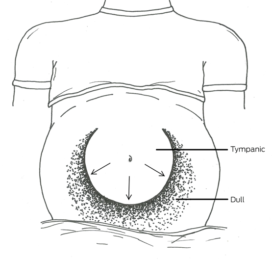
Palpation of significant hepatomegaly and splenomegaly may help the diagnostic process, but can be difficult to perform in a patient with a large volume of ascites or tenderness. With the patient lying supine with their head on a single pillow, there will be a tympanic area to percussion in the midline of the abdomen, which will normally be bordered by an area of dullness in either flank (Box 3). To assess this, the clinician should place their hand in the midline, parallel to the direction of the expected change in resonance, and percuss away from themselves until the percussion note changes from tympanic to dull. It is possible to repeat this manoeuvre in each direction. However, to demonstrate shifting dullness, the clinician will need to ask the patient to roll towards them, and on to the patient’s right side, while keeping their hand on the patient at the location of the dull percussion note. After waiting up to a minute for fluid to shift, the clinician can percuss once again from the tympanic area, which will now be in the flank, to the dull area, which will be in the midline. The sensitivity and specificity of this test for ascites is more than 70%.3

With a significant volume of ascites, it may be possible to elicit a fluid thrill or wave. This may require two people. The second person places the ulnar border of their forearm, or both hands, on the midline of the patient’s abdomen to prevent transmission of the thrill through the subcutaneous fat. The clinician then taps the flank firmly and feels for an impulse on the opposite side. This sign lacks sensitivity but is highly specific.3

Box 4, adapted from a review by Williams and Simel,3 summarises the significant symptoms and signs to consider when evaluating a patient with ascites, and their accuracy and precision. The absence of reported ankle swelling and abdominal distension, combined with no bulging flanks, flank or shifting dullness, are most helpful in excluding ascites. A diagnosis can be positively made when a patient has a fluid thrill together with shifting dullness and ankle swelling. Studies suggest high levels of agreement between clinicians, especially those with experience, on the detection of ascites using these techniques.2,4,5 Using ultrasound or computed tomography, the volume of ascites that can be detected is probably as small as 100 mL. However, it is unlikely that volumes of this magnitude will be detectable on clinical assessment. For flank dullness, more than 1 L of ascitic fluid needs to be present.

The gold standard for detecting ascites is aspiration of fluid after visualisation with imaging.3 However, with good clinical assessment, using the presence of a fluid thrill, shifting dullness and peripheral oedema as the positive indicators, the majority of cases can be diagnosed by a clinician at the bedside.

Box 1 –
Frequency of causes of ascites

Frequency

Cause


Very common

Cirrhosis

Common

Right-sided heart failure

Malignancy

Rare

Tuberculosis

Pancreatitis

Nephrotic syndrome

Very rare

Constrictive pericarditis

Budd–Chiari syndrome

Protein-losing enteropathy

Chylous ascites

Serositis (lupus, familial Mediterranean fever)


Box 2 –
Bulging flanks and abdominal distension secondary to ascites

Box 3 –
Tympanic area of the abdomen in the presence of ascites


Diagram courtesy of Rebecca Veysey.

Box 4 –
Significant pooled results for the accuracy of clinical history and physical examination in the detection of ascites*

Symptom or sign

Sensitivity

Specificity

Likelihood ratio


Positive

Negative


Abdominal distension

87%

77%

4.2

0.2

Ankle swelling

93%

66%

2.8

0.1

Bulging flanks

81%

59%

2.0

0.3

Flank dullness

84%

59%

2.0

0.3

Shifting dullness

77%

72%

2.7

0.3

Fluid thrill

62%

90%

6.0

0.4


* Adapted from Williams and Simel.3 † The likelihood that a symptom or sign would be expected if a patient had ascites: positive likelihood ratio indicates how much more likely the presence of ascites will be if the sign or symptom is present; negative likelihood ratio indicates how unlikely ascites would be if the symptom or sign were absent.

Refining the care of patients with pancreatic cancer: the AGITG Pancreatic Cancer Workshop consensus

Pancreatic adenocarcinoma is the 11th most frequent cancer in Australia.1 The disease has a poor prognosis: the overall 5-year survival rate is about 4%, and 5-year survival in those who are eligible for surgery is 20–21%,2 which is significantly better than the 5-year survival rate of 3% for people with metastatic disease. Internationally, about 20% of patients have potentially resectable disease after staging; there is currently no strategy for early detection. Recent Australian data indicate that about 15% of patients with pancreatic adenocarcinoma undergo surgery.3

Modern imaging techniques have improved diagnostic precision, especially in differentiating adenocarcinoma from non-malignant pancreatic masses (pseudo-cyst, lymphoma, chronic pancreatitis, intraductal papillary mucinous neoplasms, neuroendocrine tumours). Important prognostic indicators for long term patient survival include negative or tumour-free surgical margins, as well as tumour size, lymph node status, and the absence of metastases.46 Margins, number of lymph nodes identified (and examined) and accurate determination of tumour size are influenced by the quality of surgery and the surgical definitions used. The lack of widely accepted clear and precise definitions of surgical margins, surgical procedures and pre-operative resectability has contributed to significant variability in 5-year survival rates and local control, and hampers efforts to define the role of adjuvant therapy in treating pancreatic cancer.

Post-operative adjuvant therapy can improve survival,2 but the ability of patients to safely receive chemotherapy after pancreatic surgery is compromised by their slow recovery and the consequences of surgical morbidity. As many as 25% of patients never receive the indicated treatment, and, even with treatment, local recurrence at distant sites is frequent.2 A new approach to improving outcomes is the increasing use of pre-operative adjuvant therapy, which has advantages over post-operative treatment in terms of lower toxicity and improved tolerability, because the patients are usually fitter and less nutritionally compromised. These advantages have been observed in the treatment of selected breast, oesophageal, gastric and rectal cancers. Neo-adjuvant therapy may have three benefits: increasing the proportion of patients receiving systemic therapy; identifying early disease progression before a potentially debilitating surgical procedure without associated benefit; and possible tumour shrinkage (downstaging).

The Australasian Gastro-Intestinal Trials Group (AGITG), in partnership with the Avner Nahmani Pancreatic Cancer Foundation, convened the Australasian Pancreatic Cancer Workshop in July 2015.7 The primary aims of this expert panel of health professionals and consumers — including medical and radiation oncologists, surgical subspecialists, anatomical pathologists, gastroenterologists, and nursing and allied health professionals — were to develop a consensus statement on the operability of pancreatic cancer and the role of neo-adjuvant therapy in enhancing the benefits of surgery. The discussion below represents an agreed optimal approach to the key aspects of surgical management.

Staging

The accurate staging of pancreatic cancer prior to surgery guides appropriate and individualised treatment of patients. The detection of advanced disease at diagnosis may prevent unnecessary, morbid surgery. In addition, the ability to precisely describe the relationship of the tumour to the surrounding vasculature influences consideration of the appropriate procedure and the risk of a positive surgical margin, and accordingly influences decisions about pre-operative chemotherapy or considering a clinical trial.

Imaging

Modern computed tomography (CT) allows high definition imaging of the primary lesion and its relationship to the vascular anatomy of the pancreas,8 as well as assessment of the regional and non-regional lymph nodes and potential sites of metastases. The minimum requirement for radiological staging is consequently a pancreatic protocol CT scan of the chest, abdomen and pelvis. Standardised reporting templates have been shown to improve decision making and to facilitate clinical trials.9 The accuracy of CT is lower for small or cystic lesions of the pancreas, for which magnetic resonance imaging (MRI) may provide better delineation. MRI is also superior for assessing small liver lesions when metastases need to be excluded. Endoscopic ultrasound (EUS) is useful for diagnosing small lesions not visible on cross-sectional imaging, and is a good adjunct to CT in local staging. 18F-Fluorodeoxyglucose positron emission topography/computed tomography (FDG PET/CT) may significantly alter treatment intention in a small group of patients and may prevent unnecessary laparotomy.10

Biopsy

Endoscopic ultrasound is the preferred method for tissue and fluid acquisition (primary and lymph node), as there is a low sampling error and reduced risk of peritoneal soiling.11 EUS-guided fine needle and core biopsy, especially when an on-site cytopathologist is available, has greatly improved accuracy and allows for the molecular profiling of tumours.12 Biopsy is preferred but not mandatory for all patients, despite a 2% risk of pancreatitis. However, biopsy is mandatory prior to neo-adjuvant and palliative therapy (to exclude alternative diagnoses for which another systemic therapy may be appropriate, such as pancreatic neuroendocrine tumours), and is also required for most clinical trials and when the diagnosis of malignancy is unclear.

Tumour markers

The serum tumour marker carbohydrate antigen (CA) 19-9, which binds to the tumour surface marker sialyl-LewisA, is often used in pancreatic cancer management for assessing treatment benefit.13 It has limited specificity and sensitivity, and is not produced by patients lacking the blood Lewis antigen, so it has only a minor role in diagnosis. However, CA 19-9 levels greater than 130 U/mL may predict unresectable disease, and very high levels may indicate the presence of occult metastatic disease, even when disease is localised on imaging. In this situation, further investigations, such as PET/CT and staging laparoscopy, should be employed.

Surgical definitions

Standardisation of surgical definitions allows clearer comparison of surgical and oncological outcomes. International guidelines published in 2014 categorise the nomenclature for surgical procedures, vascular resection and lymph node resection.14,15 The pre-operative classification of resectability has significant impact on which patients undergo surgery, whether surgery should be performed at a unit experienced in vascular resection, and whether neo-adjuvant treatment should be employed. Similarly, standardisation of specimen handling and the definition of a clear surgical margin aids comparison of different treatments and outcomes, which is paramount when conducting clinical trials or assessing and benchmarking outcomes.

Pre-operative classification of resectability

The decision to perform surgery is based on all available pre-operative information and the assessment of whether clear margins can be achieved. Current guidelines do not discriminate between tumours that do or do not require portal vein resection to achieve a clear margin, despite differences in outcomes and morbidity.16,17 There is also no differentiation regarding the borderline resectability of the venous and arterial margins. The panel recommends classification of operability into five categories, as the most commonly used international guidelines differ and do not account for the need for vascular resection, the likelihood of obtaining a clear margin, or differences in survival for these patient groups:

  • clearly resectable tumours with a standard pancreatectomy;

  • clearly resectable tumours that may require portal venous resection;

  • borderline resectable tumours that require venous resection;

  • borderline resectable tumours that require arterial resection; and

  • locally advanced inoperable tumours or metastatic disease.

Detailed information about the criteria for each category is available in the full report of the Workshop.7

The multidisciplinary team

The increasing complexity of cancer care and the implementation of multimodality treatment of pancreatic adenocarcinoma highlight the value of a specialist multidisciplinary team (MDT) meeting. This should result in increased numbers of patients receiving optimal personalised management and appropriate surgical treatment, and more frequent recruitment to clinical trials. The MDT discussion should include an experienced lead clinician as well as broad surgical, medical and radiation oncology, palliative care, genetics, nursing and allied health expertise, especially in the area of nutrition. Each patient should be presented to the MDT by a senior clinician responsible for the patient, who represents the interests of the patient and ensures that decisions made by the MDT are implemented. Workshop panel members recommended that all patients with pancreatic cancer should be registered, that their demographic details and the extent of disease be documented, and that this should be formally presented at an MDT meeting; however, this may not always be practical. Patients with widespread metastatic disease should be registered to ensure later tracking of treatments and outcomes; their discussion and management may be triaged to an alternative multidisciplinary forum, such as a medical oncology review meeting.

The group of patients presented to an MDT should include:

  • all newly diagnosed patients who have potentially operable disease (including borderline resectable) and initially locally advanced (unresectable) disease;

  • patients for whom there is diagnostic uncertainty, or uncertainty about how to proceed with their management; and

  • patients with single organ metastatic disease or recurrence.

Network arrangements, such as virtual meetings, can ensure that patients from private hospitals and regional centres are not disadvantaged in terms of access and expertise. These networks add value to discussions about these patients, especially for borderline cases and clinical trials.

Surgical procedures

Pancreatic resection should be performed after adequate pre-operative staging and assessment of fitness for surgery. All pancreatic resections carry the risk of significant morbidity and mortality, and should only be undertaken in experienced pancreatic surgical units.18 The standard procedure for a pancreatic head or uncinate process tumour is a Whipple procedure (pancreaticoduodenectomy), including removal of associated lymph nodes.14,15 For tumours of the body or tail of the pancreas, a subtotal or distal pancreatectomy (with splenectomy) is the standard procedure. Extended pancreatectomy (when associated vascular resection is performed) can be completed with survival rates similar to those of standard resections, but should be limited to specialist units with experience in vascular reconstruction.19,20 The general consensus among panel members was that extended multivisceral and arterial resections should be performed only in exceptional circumstances, because of the associated increase in morbidity and mortality.16,17

Specimen handling and pathological reporting

At present, the definition of a clear tumour resection margin is not internationally standardised. Many centres in Australia and Europe define an R0 resection (no tumour cells within a defined distance from the margin) as one without evidence of tumour within 1 mm of the edges of the resection margin, but some centres still use a 0 mm definition (ie, no evidence of tumour cells at the margin). While meeting the first definition is associated with significantly improved survival, a clear margin by either definition is correlated with better outcomes than involved margins.21,22 A recent systematic review found that varying definitions of clear margins had a significant impact on R0 resection rates in pancreatic cancer surgery (49% for a 1 mm margin, compared with 72% for a 0 mm margin).23 This review also reported that inconsistent use of terminology, lack of agreement on structured reporting guidelines, and variations in pathological techniques (axial slicing v other slicing techniques) hampered comparative analysis of the outcomes of international studies and identifying optimal pathways of care.

The Workshop recommended adoption of the Royal College of Pathologists of Australasia guidelines, as they reflect best practice and are easily reproducible. A resection specimen is considered R0 if all seven surgical margins are clear from 1 mm. However, regardless of the R status, the microscopic clearance of critical margins (ie, superior mesenteric artery, pancreatic transection, and superior mesenteric vein/portal vein margins) must be recorded in the pathology report. The use of a structured pathology reporting protocol that standardises the reporting of surgical margin status and other relevant information, such as depth of portal vein invasion, is recommended.21

Neo-adjuvant therapy

In Australia (and internationally) some centres use pre-operative neo-adjuvant therapy in patients with borderline resectable disease,24 and there is evidence for improved survival outcomes for patients with resectable and borderline resectable pancreatic cancer.25 Interpretation of these data is complicated by inconsistent or imprecise definitions of resectability, the rapid evolution of chemotherapy agents, and their combined use with radiotherapy. These confounding factors have resulted in confusion when interpreting patient outcomes associated with neo-adjuvant therapy.

Neo-adjuvant therapy and clearly resectable disease

Active research is currently being conducted in this area. The Australian gemcitabine and nab-paclitaxel (GAP) study26 examined the feasibility of giving pre- and post-operative chemotherapy to patients with resectable pancreatic cancer. The results indicated that pre-operative treatment (gemcitabine–nab-paclitaxel) was safe, did not impair the ability to perform surgery, and improved the R0 resection rate for patients with resectable disease. The study also found that post-operative chemotherapy was given to only 60% of patients, whereas pre-operative treatment was administered to 93% of patients, reflecting experience elsewhere.26 Although it can be considered for patients with clearly resectable pancreatic adenocarcinoma, the ultimate survival benefit of this approach remains to be established, and patients should be referred for clinical trials whenever possible.

Neo-adjuvant therapy and borderline resectable disease

Although several overviews have suggested the benefits of neo-adjuvant chemotherapy,25,27 variability in definitions has caused substantial uncertainty about whether it has a significant impact on resectability or curability in the borderline setting. The choice of chemotherapy or chemoradiation remains unclear. It has been reported that FOLFIRINOX (folinic acid–fluorouracil–irinotecan–oxaliplatin) and gemcitabine–nab-paclitaxel achieve objective shrinkage of primary tumours in small numbers of patients. Similarly, the addition of radiotherapy may lead to downstaging to operability for some patients. In this setting, however, re-imaging of local disease is unreliable, and patients should be re-evaluated by the MDT with respect to surgery, even in the absence of metastatic disease.28 Given the lack of clarity about the benefits, patients with borderline tumours should be offered the opportunity to participate in clinical trials.

Neo-adjuvant therapy and locally advanced disease

Chemotherapy is recommended as the initial therapy for the management of patients with unresectable loco-regional pancreatic cancer. The value of adding radiation after initial chemotherapy remains uncertain. However, downstaging chemoradiation is an option, although its use in this population is controversial; some patients are medically unfit for such intensive treatment, and it should usually be offered in the context of a clinical trial.27

Conclusion

The treatment of operable pancreatic cancer is entering a new phase of multimodal therapy in an attempt to improve upon the limited benefit of surgery alone. The considerable difficulties encountered in comparing studies and evaluating the quality of care will continue unless we have clear and precise definitions of a complete resection margin (preferably 1 mm R0), and of the choice of surgical procedures and pre-operative resectability criteria, as well as structured pathology and radiology reporting and compulsory MDT registration. Implementing a structured approach, as outlined in this article, is an essential step toward improving the care and outcomes for patients with pancreatic cancer in Australasia.

Probiotics – do they benefit healthy people?

A systematic review has found that probiotics have little effect on the gut microbiota of healthy people.

The study was undertaken by researchers at the Novo Nordisk Foundation Center for Basic Metabolic Research at the University of Copenhagen and published in Genome Medicine.

It involved a review of seven randomised controlled trials to investigate the effect of probiotics on the faecal microbiota of healthy adults. The probiotics products administered were biscuits, milk-based drinks, sachets, or capsules for periods of 21 to 42 days.

The researchers looked at reported effects on the overall structure of faecal microbiota including the number of species present, the evenness and whether the healthy people taking probiotics had different changes of bacteria living in their guts compared to the placebo groups.

Related: Patrick Charles: Power of poo

The researchers investigated the trials for reported effects of probiotics on the overall structure of the fecal microbiota of healthy adults, including the number of species present, the evenness (distribution of species within the populations) and whether the probiotics groups of study participants as a result of the intervention had different changes in bacteria living in their gut than the placebo groups.

“According to our systematic review, no convincing evidence exists for consistent effects of examined probiotics on fecal microbiota composition in healthy adults, despite probiotic products being consumed to a large extent by the general population,” PhD student and junior author Nadja Buus Kristensen said.

Of the seven original trials, only one found observed changes in the bacterial composition of faecal microbiota.

Related: John Dwyer: Promoting wellness

The authors note that various limitations include small sample size, use of different probiotic strains, variations of diet and variation in susceptibility of probiotics between individuals could mask the true impact of probiotic intake.

Oluf Pedersen, professor at the University of Copenhagen and senior author of the paper said: “While there is some evidence from previous reviews that probiotic interventions may benefit those with disease-associated imbalances of the gut microbiota, there is little evidence of an effect in healthy individuals. To explore the potential of probiotics to contribute to disease prevention in healthy people there is a major need for much larger, carefully designed and carefully conducted clinical trials. These should include ideal composition and dosage of known and newly developed probiotics combined with specified dietary advice, optimal trial duration and relevant monitoring of host health status.”

Latest news:

[Comment] Gastroenterology and hepatology: a call for papers

Remarkable advances have been made in gastroenterology and hepatology in recent years. For instance, the development of direct-acting antiviral treatments for hepatitis C virus raises the prospect of eliminating such infections—cost and access notwithstanding.1 And exciting progress is being made in harnessing the immune system to tackle previously hard-to-treat gastrointestinal cancers.2,3 But other challenges remain, or are arising. For example, as a result of increases in obesity, non-alcoholic steatohepatitis (NASH) is predicted to replace infection as the leading cause of cirrhosis, hepatocellular carcinoma, and liver transplant in the USA in the next 10–20 years;4 similar transitions are likely to take place elsewhere if global trends in obesity continue.

Carbapenemase-producing Klebsiella pneumoniae: a major clinical challenge

Clinical record

A 59-year-old man from rural Victoria, with no hospital contact for 15 years or recent history of international travel, presented to his local hospital with severe acute pancreatitis secondary to gallstones. He was transferred to a metropolitan hospital for further management, including intermittent admissions to the intensive care unit (ICU) for haemodynamic support. On Day 4 of admission, empirical antibiotics were prescribed for severe pancreatitis and concurrent nosocomial pneumonia, according to hospital guidelines and advice from the infectious diseases team; initially ceftriaxone, later changed to piperacillin–tazobactam and then meropenem, due to clinical deterioration. Diagnostic microbiology did not reveal any significant pathogens.

Serial computed tomography demonstrated persistent peri-pancreatic fluid collections despite repeated percutaneous drainage and broad-spectrum antibiotics. One month into admission, vancomycin-resistant Enterococcus faecium, Candida albicans and Stenotrophomonas maltophilia were identified in peri-pancreatic fluid. Contact precautions were implemented, and an infectious diseases physician recommended piperacillin–tazobactam, fluconazole, co-trimoxazole and linezolid (later changed to teicoplanin) to cover these organisms. Teicoplanin, co-trimoxazole and fluconazole were ceased after 8 weeks of treatment.

Pancreatic debridement performed 2 months into admission due to persistent pancreatic infection identified carbapenem-resistant Klebsiella pneumoniae in the pancreatic tissue. Testing by polymerase chain reaction detected the blaKPC-2 gene. Antimicrobial-susceptibility results are shown in the Box. Surrounding patients were screened.

Owing to limited antibiotic options, gentamicin combined with dual carbapenems (high-dose prolonged meropenem infusion three times a day combined with daily ertapenem) was prescribed for the K. pneumoniae. Gentamicin was continued for 3 weeks in conjunction with repeated pancreatic debridements in an attempt to control infection. Oliguric renal failure and sepsis developed, requiring ICU transfer, renal replacement therapy and cessation of gentamicin.

Three months into admission, following further attempted pancreatic debridement, multiple blood cultures grew blaKPC-2-producing K. pneumoniae that now demonstrated intermediate gentamicin susceptibility (minimum inhibitory concentration, 8 μg/L). Renal replacement therapy continued, all intravenous lines were replaced, two doses of gentamicin were administered and intravenous doxycycline was added to meropenem, ertapenem and fluconazole. Repeat blood cultures were negative. Application for compassionate access to ceftazidime–avibactam was made (to which the isolate was susceptible) and it was supplied 1 week later.

Because of further deterioration and isolation of doxycycline-resistant K. pneumoniae from abdominal fluid, antibiotics were changed to ceftazidime–avibactam (adjusted for renal function), metronidazole and teicoplanin. Over the next 3 weeks while receiving these agents, the patient had resolution of fever, a decrease in serum inflammatory markers, reduction in vasopressor requirements and radiological improvement of the peri-pancreatic collections. No side effects were reported from ceftazidime–avibactam.

During the fifth month, a laparotomy was performed in a final attempt to control pancreatic infection, but was unsuccessful due to the compromised state of pancreatic and peri-pancreatic tissues. Intra-abdominal drain tube fluid continued to grow blaKPC-2-producing K. pneumoniae that was susceptible to ceftazidime–avibactam. Shortly after this, and following discussion with the patient, family and treating teams, the patient was discharged home for palliation and died soon after.

Klebsiella pneumoniae carbapenemase (KPC)-producing Enterobacteriaceae have been responsible for nosocomial outbreaks worldwide and have become endemic in several countries. These organisms provide immense challenges for healthcare systems, health care providers and patients. Reports of KPC-producing organisms in Australia have been uncommon, with most cases found to be imported from endemic countries.1 Genes responsible for KPC production (eg, blaKPC-2) are acquired via transferable plasmids and, when expressed, result in enzymatic hydrolysis of all β-lactams including carbapenems.2 Additional antimicrobial resistance genes frequently accompany carbapenem-resistance mechanisms, limiting the choice of effective antimicrobials.2

Multiple risk factors have been associated with carbapenem-resistant Enterobacteriaceae (CRE) acquisition. These include prolonged duration of hospital stay, receipt of broad-spectrum antibiotics, presence of invasive devices, use of mechanical ventilation, total parental nutrition or nasogastric feeds, and colonisation pressure.3

Such infections pose management challenges given their propensity for causing severe sepsis in patients with multiple comorbidities. Many remaining active antibiotics have limitations in terms of efficacy (eg, tigecycline is not ideal for bacteraemia or urinary tract infections) and toxicity (eg, colistin can have significant nephrotoxicity).

There is a paucity of evidence to guide management decisions, and optimal antibiotic treatment is unknown.4,5 Current expert recommendations are largely based on retrospective observational data. These suggest that combination therapy with two or three active agents should be used. Antibiotic classes including fluoroquinolones and sulphonamides are usually inactive against these organisms. Despite the inherent presence of carbapenemases, inclusion of meropenem (usually high-dose extended infusions) in treatment regimens is usually recommended.4,5 However, more recent studies have suggested that a benefit may be restricted to isolates with only low-level carbapenem resistance (minimum inhibitory concentration, ≤ 8 μg/mL).4 At the time of this case, a small number of reports used dual carbapenems as salvage therapy for pandrug-resistant K. pneumoniae, which informed the decision to use combination ertapenem and meropenem. However, the clinical value of this practice remains uncertain.6,7

Ceftazidime–avibactam plus metronidazole has been shown in Phase II studies to have similar efficacy in complicated intra-abdominal infections when compared with meropenem,8 and has been approved for this indication in the United States by the Food and Drug Administration. Avibactam is a new β-lactamase inhibitor in the diazabicyclooctane class and, in combination with ceftazidime, retains activity against some KPC-producing Enterobacteriaceae in vitro.9 There is a paucity of clinical data relating specifically to its efficacy in infections caused by KPC-producing Enterobacteriaceae. Our patient demonstrated a clinical, biochemical and radiological response after administration of ceftazidime–avibactam, metronidazole and teicoplanin, with no development of in vitro resistance after 6 weeks of treatment. However, microbiological clearance was not achieved. Given that early treatment may be effective in managing CRE infections, timely access to antibiotics such as ceftazidime–avibactam and associated antibiotic susceptibility testing in Australia is crucial.

CRE infections are an increasing problem that Australian hospitals are facing; now in both local residents and returned travellers.10 Combination strategies and newer agents under investigation, such as ceftazidime–avibactam, are potential treatment options.

Lessons from practice

  • Carbapenem-resistant Enterobacteriaceae (CRE) infections pose a clinical challenge for management with limited effective antibiotics available.

  • New strategies, and new antibiotics, will be required to manage the increasing threat of CRE.

  • Ceftazidime–avibactam, a novel antimicrobial combination with activity against many CRE, may be a future option for treating such infections.

Box –
Initial Klebsiella pneumoniae antimicrobial-susceptibility results*

Antibiotic

Resistance

MIC (μg/mL)


Amoxycillin-clavulanic acid

R

≥ 32

Piperacillin–tazobactam

R

≥ 128

Ceftriaxone

R

≥ 64

Cefepime

R

≥ 64

Cefoxitin

R

≥ 64

Ciprofloxacin

R

≥ 4

Meropenem

R

≥ 16

Amikacin

R

≥ 64

Tobramycin

R

≥ 16

Gentamicin

S

4

Co-trimoxazole

R

≥ 320

Nitrofurantoin

R

≥ 512

Colistin

R

4

Fosfomycin

R

≥ 1024

Tigecycline

R

4

Tetracycline–doxycycline

S

4


MIC = minimum inhibitory concentration. R = resistant. S = susceptible. * Using Vitek 2 gram-negative antibiotic susceptibility cards (bioMérieux) according to Clinical and Laboratory Standards Institute (CLSI) interpretative criteria, unless otherwise indicated. † Etest (bioMérieux), according to European Committee on Antimicrobial Susceptibility Testing interpretative criteria (CLSI interpretative criteria not available).

Unexpected cause of urticaria

A 30-year-old man presented with acute generalised urticaria (Figure, A) 6 hours after ingestion of raw fish. While most adult food allergies occur within 1–2 hours of ingestion, allergy to the live fish parasite Anisakis simplex which has penetrated the gastrointestinal mucosa manifests 1 hour to a few days after ingestion.1 Emergency upper endoscopy detected the live larva in the stomach (Figure, B) and disinfestation resolved his symptoms. Allergy to A. simplex can be confirmed by testing for allergen-specific IgE or skin prick testing with Anisakis extract. Gastrointestinal anisakidosis can be an unexpected cause of urticaria owing to delay between the ingestion of fish and the appearance of symptoms. While most cases of anisakidosis currently occur in Japan, this may become an increasing problem in Australia due to increasing consumption of sushi and sashimi.2

Figure

First reported outbreak of locally acquired hepatitis E virus infection in Australia

Hepatitis E virus (HEV) outbreaks have not previously been reported in Australia. HEV infection mostly occurs in developing countries where transmission occurs via the faecal–oral route and contaminated water, causing large outbreaks.1 HEV genotypes 1 and 2 predominate in these settings.2 Like other forms of acute viral hepatitis, symptoms of HEV include jaundice, malaise, anorexia, fever and abdominal pain.1 The incubation period is 15–64 days.3

Recently, HEV transmission has been reported in developed countries, where infection has occurred via HEV-contaminated food. Consumption of pork products, deer meat, wild boar and shellfish has been implicated, with HEV genotypes 3 and 4 being detected in infected persons.2,48

Pigs, in particular, may play a role in human HEV transmission.9 An increased risk of HEV infection associated with the consumption of processed pork products was found by a recent case–control study in the United Kingdom.10 Human and swine HEV strains exhibit a high degree of sequence homology.5,11,12 Occupational exposure may be important, as seroprevalence rates have been found to be higher in pig veterinarians, pig farmers and abattoir workers than in healthy controls.1315

In Australia, HEV infection is notifiable to state and territory public health authorities. Common laboratory practice has been to test for HEV infection only in those with a history of overseas travel. Each year, 30 to 40 infections in returned travellers from HEV-endemic regions are reported, including 10 to 20 in New South Wales.16

In October and November 2013, NSW Health was notified of two apparently unrelated cases of HEV infection within 2 weeks. Each person had been tested because of preceding overseas travel, albeit outside the incubation period for HEV infection. The HEV RNA isolated from these two people was genetically identical. A family member of one of the patients presented with symptoms of HEV infection 4 weeks later.

In May 2014, we received a further HEV notification, an infection in a man who reported that a work colleague from another state was also infected with HEV. Neither had travelled overseas during their incubation periods. The only common exposure was a meal shared with seven other colleagues at restaurant X, and the index patient reported that three of the seven were symptomatic. All co-diners were interviewed and tested, and HEV RNA was detected in the three symptomatic co-diners. HEV RNA from the five infected persons was genotypically identical, and also with that from two of the three 2013 cases. During routine interview of the three HEV-infected people in 2013, one had reported eating at restaurant X during their incubation period, while another had not. During a follow-up interview in 2014, the third person was specifically asked about this exposure, and reported eating at restaurant X during their incubation period.

In this article we report our epidemiological investigation of the source and extent of the apparent outbreak.

Methods

Epidemiological investigation

Case definition

We defined a case of HEV infection as a person who resided in NSW with laboratory-confirmed HEV, verified by IgG seroconversion or detection of HEV-specific IgM or HEV RNA, with an onset date (or specimen collection date, if onset date was unknown) between 1 January 2013 and 31 December 2014.

Case finding and data collected

We identified cases in three ways:

  • Routine notification: As part of routine surveillance, pathology laboratories are required by the NSW Public Health Act 2010 to notify public health units of HEV infections. Surveillance specialists interview infected persons, using a standardised questionnaire. The information collected includes symptoms of illness, occupation, travel history, and water and food sources (including restaurants) during the incubation period. When an infected person had eaten at restaurant X, the interviewer asked about details of the food consumed there.

  • Testing of co-diners from restaurant X: Co-diners of infected persons from restaurant X were interviewed and tested for HEV.

  • Retrospective serological surveys: We tested all sera stored at a large public laboratory, with specimen dates between 1 September 2013 and 31 May 2014, for which HEV testing had been requested but not conducted because laboratory protocols excluded testing in the absence of a relevant travel history (survey 1). We also tested sera stored at a major NSW private pathology laboratory, with specimen dates between 1 January and 31 May 2014, where the alanine transaminase (ALT) level was > 200 IU/L and hepatitis A, hepatitis B, hepatitis C, Epstein–Barr virus and cytomegalovirus infections had been excluded, but HEV testing was not performed (survey 2).

Laboratory investigation

Serology

Anti-HEV IgM and IgG were detected using HEV IgM ELISA 3.0 and HEV ELISA kits respectively (MP Diagnostics) according to the manufacturer’s instructions. Reactive sera were re-tested and reported as positive if again reactive.

Viral detection and sequencing

Serum samples from confirmed cases were analysed at the Victorian Infectious Diseases Reference Laboratory. HEV RNA was extracted from serum using the QIAamp Viral RNA Mini kit (QIAGEN) and initially tested using a commercial HEV RNA polymerase chain reaction (PCR) assay (RealStar HEV RT-PCR). Samples containing HEV RNA were re-assayed by an in-house PCR assay using primers designed to amplify a portion of open reading frame (ORF) 2. The resulting PCR product was directly sequenced with internal primers. Sequences were aligned and compared with sequences in GenBank.

Environmental investigation

Investigation and food testing linked to restaurant X

Food handling and safety procedures at restaurant X were reviewed on 15 May 2014. Preparation of pork liver pâté was observed in detail. The internal temperature of sliced pork livers was measured by inserting a thermometer into the thickest part after 3 and 4 minutes’ cooking.

Three lots of chorizo sausage, three batches of cooked pork liver pâté, one sample of raw pork shoulder and raw pork jowl, one batch of cooked pork liver and eight raw pork liver samples from restaurant X were collected on 15 and 22 May 2014.

After extraction and purification using the MagMax Total RNA Isolation Kit (Life Technologies), samples were tested for HEV by Advanced Analytical Australia with real-time PCR, using Hepatitis E@ceeram Tools (Ceeram).

Pork products were traced back to their source by identifying the supplier from restaurant records; through the supplier we identified the farms from which the products originated.

Testing of pork liver sausages linked with an HEV case not linked to restaurant X

One of the infected persons without a link to restaurant X reported eating pork liver sausages during their incubation period, and had stored frozen uncooked sausages in a domestic freezer. Multiple samples were collected from several sausages and analysed for HEV at the Virology Laboratory of the Elizabeth Macarthur Agriculture Institute. Nucleic acid was purified and tested by real-time quantitative reverse transcription PCR (qRT-PCR)17 using previously published primers and probe sequences.18

Data analysis

Responses to questionnaires administered to interviewees were transferred to a Microsoft Excel spreadsheet for analysis. Responses about food histories were analysed, and relative risks and confidence intervals calculated using Epi Info 7 (Centers for Disease Control). The Fisher exact test (two-tailed) was used to test for differences between groups; P < 0.05 was defined as statistically significant.

Ethics approval

These studies were conducted as part of a public health investigation under the NSW Public Health Act 2010 and review by a human research ethics committee was not required.

Results

Epidemiological investigation

Notified HEV cases

Between January 2013 and December 2014, 55 cases of HEV infection were notified (Box 1). The median age of the patients was 45 years (range, 4–77 years), 36 (65%) were male, and all but one (98%) lived in metropolitan Sydney. Twenty-four (44%) required hospitalisation, with a reported median length of stay (where known) of 7 days (range, 1–67 days). Three people (identified as co-diners of notified patients) were asymptomatic, and details about symptoms were unknown in one case. ALT levels were elevated in 33 of the 37 patients for whom they were recorded, with a median value of 1058 IU/L (range, 26–4868 IU/L; reference interval, 10–40 IU/L). None were pregnant.

Of the 55 patients, 30 (55%) reported a history of overseas travel during their incubation periods: to South Asia (17), East Asia (six), South-East Asia (two), Africa (two), Europe (two), or the Middle East (one). One patient could not be contacted; the remaining 24 (44%) did not report overseas travel.

Restaurant X outbreak

Restaurant X mainly served dishes suitable for sharing by a group. The menu included more than 28 meat, seafood and vegetarian options. Seventeen cases of HEV infection in nine separate groups who dined between October 2013 and May 2014 were linked to restaurant X. Of these 17, seven were identified by routine surveillance, eight by testing co-diners, and two by the retrospective serosurveys. Two people refused further interview; food histories were collected from the remaining 15 infected persons and from seven dining companions who tested HEV-negative by serology.

The demographic data for the diners is summarised in Box 2; the food items most commonly consumed are listed in Box 3. The highest attack rates were in those who consumed pork liver pâté, pork chorizo or roast pork. All 15 patients who provided a food history reported consuming pork liver pâté, compared with four of the seven uninfected co-diners (P < 0.05).

Locally acquired cases not linked to restaurant X

During interviews, the seven infected persons not linked to restaurant X reported eating a number of pork products during their incubation periods, including supermarket ham, prosciutto, pork liver, homemade pork liver sausage, pork chops and pork belly.

Retrospective serological surveys

Of 136 serosurvey samples (31 in survey 1, 105 in survey 2), nine (6.6%) were IgG-positive, four (2.9%) were IgM-positive, and four (2.9%) were both IgM- and IgG-positive for HEV. Of the eight people who were IgM-positive, HEV RNA was detected in four; sequencing confirmed infection with genotype 3. Two of these four people reported eating at restaurant X but not overseas travel, one reported travel to an HEV-endemic country, and one could not be contacted.

Laboratory investigation

HEV RNA was detected in samples from ten of the 17 restaurant X cases; of the others, five with mild or no symptoms were PCR-negative, one was PCR-negative but showed seroconversion, and a sample was unavailable in one case. Sequencing of the ORF2 region was successful for all ten samples, and the HEV isolate was classified as genotype 3. There was at least 99% between-sample sequence homology in the targeted portion of ORF2 among restaurant X isolates.

HEV RNA was also detected in six of the seven locally acquired infections not linked to restaurant X (the specimen supplied by one person was insufficient for testing): five were genotype 3, and one sample was insufficient for genotyping. The viral sequence of these samples was about 90% homologous with samples from the restaurant-linked cases.

Environmental investigation

Investigation of restaurant X

Restaurant X was found to be well managed; no breaches in food safety or handling were identified. Staff were trained in handwashing and general food safety, including understanding cross-contamination and temperature control. During the observed cooking process, the internal temperature of the pork livers reached 51°C at 3 minutes, and between 82°C and 97°C at 4 minutes.

The livers used for pâté preparation were traced to a single farm. The pork shoulder, jowl and chorizo products were all sourced from different suppliers to the pork livers. HEV was not detected in any of the food samples obtained from the restaurant.

Investigation of pork products of locally acquired cases not linked to restaurant X

Pork products eaten by the seven infected persons not linked to restaurant X were bought from four different butchers and three different supermarkets. Pork livers from two of these butcheries could be traced back to two abattoirs supplied by several farms; further tracing was not undertaken. Pork liver sausages still held by one patient were found to contain very low levels of HEV RNA; the levels were too low for sequencing.

Public health interventions

NSW Health convened an expert panel involving public health, clinical, laboratory, agricultural and industry experts to assess the risks and to guide the investigation. On 15 May 2014, restaurant X was informed about its possible link with a number of cases of HEV infection. The importance of thorough cooking of pork products, including of pork liver pâté, was stressed, and the restaurant voluntarily removed this item from its menu. No further cases of HEV infection were linked to restaurant X.

As part of case finding, NSW Health issued an alert to gastroenterologists and public and private pathology laboratories in May 2014. The information garnered was then used to inform general practitioners in an alert, issued in September 2014, which requested that they consider HEV infection in people with a compatible illness, regardless of overseas travel. A joint media release with the New South Wales Food Authority, also issued in September 2014, urged the public to cook pork products thoroughly and, in particular, to cook pork livers to 75°C at the thickest part for 2 minutes.19

Discussion

This is the first reported Australian outbreak of locally acquired HEV infection and one of the largest linked with a restaurant reported anywhere. Seventeen cases were linked to consuming pork liver pâté at restaurant X during a 9-month period, and seven cases were linked to eating pork products bought from four butchers and three supermarkets with at least two different suppliers.

Retrospective serological testing identified a further eight previously undiagnosed cases of HEV infection (anti-HEV IgM). In two of these cases, HEV RNA was detected in people who reported no overseas travel but who had dined at restaurant X during their incubation periods. A further six cases were notified after the restaurant outbreak, probably as a result of increased vigilance and testing by clinicians. Data from a large public health laboratory confirmed this, with more than triple the number of HEV tests requested and carried out from July to December 2014 (after the laboratory began testing for HEV in people without a travel history) than during the same period in 2013 (unpublished data).

Active case finding among co-diners of restaurant cases detected locally acquired HEV infections that were either asymptomatic or mildly symptomatic, suggesting under-recognition and under-diagnosis of infection. A recent HEV serosurvey of blood donors by the Australian Blood Service identified past HEV infection in 14 of 194 blood donors without a history of overseas travel (7%).20 A case report in the Northern Territory21 and a study in Victoria22 each described single cases of HEV infection in which overseas travel was not implicated and no other risk factors were identified.

Common source outbreaks of HEV infection in high-income countries are rare. However, our investigation concurs with previous French,5 English10 and Japanese11 studies that have linked HEV infection with consumption of undercooked pork products. In these countries, locally acquired HEV infections predominate, and in 2013 accounted for 99% of all cases in France23 and almost 70% of cases in the UK.24

HEV is inactivated by heating to 71°C.19 Review of pork liver pâté preparation at restaurant X found that it was adequately cooked at the time of inspection, and testing available pork samples did not detect HEV RNA. It is nevertheless possible that, at the time of the restaurant infections (some weeks earlier), pork livers contaminated with HEV could have been undercooked at the thickest part before blending into pâté. This may explain the relatively low proportion of patrons infected with HEV at this popular restaurant. While we did not have access to leftover pâté samples from meals served to people infected at restaurant X that could be tested for HEV RNA, it was detected in pork liver sausages retained by one of the non-restaurant X patients.

Most fresh pork products in Australia are locally produced. The presence of HEV in Australian pigs was first noted in 1999 by a study that reported seropositivity rates of 17% in wild-caught pigs and more than 90% in commercial pigs by 16 weeks of age.25 To our knowledge, no further studies investigating the epidemiology of HEV in Australian pigs have been conducted. Despite the link between HEV outbreaks and pork products overseas, this discovery of HEV in Australian pigs did not translate into clinical practice, perhaps because HEV was not widely recognised as being endemic to Australian pigs, and because of a lack of awareness among Australian clinicians of the veterinary literature.

A limitation to this investigation was the time lag between some infected persons and co-diners being exposed, interviewed and tested for HEV, particularly co-diners of symptomatic persons from restaurant X. A lag in interviewing some infected persons and co-diners, coupled with the long incubation period of HEV (15–64 days), may have led to a recall bias in responses to the questionnaires and providing food histories. The limited sample size made it difficult to achieve statistically significant results. However, our findings are biologically plausible, and important associations could be deduced.

This study adds to our current understanding of the potential for HEV to be a food-borne illness in developed countries. Clinicians should request HEV testing in patients with acute hepatitis, irrespective of travel history, particularly where no aetiology has been determined. Laboratories should test for HEV where indicated to prevent under-recognition of infection. Health departments must be aware of the possibility of underestimating the prevalence of hepatitis E when using surveillance data. Pork products, particularly pork livers, should be cooked until they reach 75°C at the thickest part for 2 minutes.

Increased awareness, ongoing research and collaboration between primary industries, animal and human health authorities should help detect and prevent this and other emerging infectious diseases in Australia.

Box 1 –
Notifications of hepatitis E virus infections in New South Wales with onset dates between January 2013 and December 2014, by likely source of acquisition*


* Excludes three asymptomatic cases and one case with unknown symptom history. † May 2014: restaurant X was inspected, and pork pâté identified as the possible source of infection; restaurant voluntarily removed pork pâté from their menu. An alert was issued to gastroenterologists and pathology laboratories. ‡ September 2014: alert issued to general practitioners and the general public. § July–December 2014: increased HEV testing reported by the main public pathology laboratory.

Box 2 –
Characteristics of infected diners and healthy co-diners at restaurant X, October 2013 – May 2014

Infected persons (cases)

Healthy co-diners


Number

17

7

Median age (range), years

48 (29–75)

45 (29–47)

≤ 39 years

5 (29%)

1 (14%)

40–59 years

6 (35%)

5 (71%)

≥ 60 years

6 (35%)

0

Unknown

0

1 (14%)

Sex: men

12 (71%)

4 (57%)


Box 3 –
Commonly reported food items consumed by infected diners and healthy co-diners at restaurant X between October 2013 and May 2014*

Number of people who ate the item


Number of people who did not eat the item


Risk ratio (95% CI)

P

Infected persons (cases)

Healthy co-diners

Attack rate (%)

Infected persons (cases)

Healthy co-diners

Attack rate (%)


Brussel sprouts

5

3

63%

8

4

67%

1 (0.5–1.8)

1.00

Calamari

3

2

60%

10

5

67%

1 (0.5–2.0)

1.00

Eggplant

7

5

58%

6

2

75%

0.8 (0.5–1.5)

0.66

Pork chorizo

7

2

78%

6

5

55%

1.5 (0.8–2.7)

0.36

Pork pâté

15

4

79%

0

3

0

Undefined

0.02

Roast pork

9

4

69%

4

3

57%

1.2 (0.6–2.6)

0.64


* Food histories were available for 15 of the 17 infected persons (13 were complete and two were incomplete) and for all seven well co-diners.