×

Managing thyroid disease in general practice

Thyroid disease can be broadly categorised as thyroid dysfunction (hypothyroidism, hyperthyroidism) and structural disease (goitre, nodules and cancer). Management is often straightforward, but there are pitfalls that may lead to misdiagnosis, overdiagnosis and inappropriate treatment. This article reviews the approach to common thyroid problems in general practice.

Epidemiology

Worldwide, iodine deficiency is the most common cause of thyroid disease. Iodine deficiency has long been known in Tasmania, and a survey published in 2006 found evidence of iodine deficiency in mainland Australia, particularly New South Wales and Victoria.1 In 2009, use of iodised salt in bread became mandatory in Australia and New Zealand (following a Tasmanian program started in 2001), and Australia is now considered iodine sufficient.2

Autoimmune thyroid disease is the commonest cause of thyroid dysfunction in Australia, with prevalence as shown in Box 1. Some 10–15% of the population have positive thyroid antibodies, most commonly to thyroid peroxidase (TPOAb), with a higher prevalence in women than men.3 Patients who are euthyroid with positive thyroid antibodies do not require treatment, but are at increased risk of thyroid dysfunction (particularly hypothyroidism),4,5 and should be followed with annual measurement of serum thyroid-stimulating hormone (TSH).

TSH testing is the most sensitive means of detecting thyroid dysfunction. Debate surrounding the laboratory reference interval for TSH has largely resolved, and a range of about 0.4–4.0 mU/L is generally accepted.6,7 TSH concentrations increase with age,7,8 and some laboratories have adopted age-related reference intervals, with upper limits of up to 7 mU/L in older patients.

Hypothyroidism

Diagnosis

Classic symptoms of hypothyroidism include fatigue, weight gain, cold intolerance, arthralgia, constipation, menorrhagia, and dry skin and hair. Physical signs include pallor, coarse skin and hair, bradycardia and goitre, but may be absent in mild hypothyroidism. These symptoms and signs are non-specific and common in people without thyroid disease,9 so laboratory diagnosis is required. Serum TSH should be measured; if this is in the reference range, then additional tests such as free thyroxine (T4), free triiodothyronine (T3) or thyroid antibodies are rarely helpful. Tests such as basal metabolic rate and reverse free T3 have no diagnostic value.

Overt hypothyroidism (high TSH, low free T4) is usually symptomatic, readily diagnosed and can be treated without further investigation. A more common presentation in general practice is an elevated level of serum TSH with normal free T4. This may indicate subclinical hypothyroidism caused by autoimmune thyroid disease, but can arise from non-thyroidal, systemic illness, particularly in the recovery phase. In patients with a mildly elevated TSH (up to 10 mU/L), TSH normalises without treatment in over 50% of cases,10 so treatment need not be offered immediately. Instead, serum TSH testing should be repeated 6–8 weeks later, together with free T4 and TPOAb, and treatment offered if the abnormality persists. Thyroid imaging (including ultrasound) is not indicated in the investigation of hypothyroidism.11

The importance of detecting and treating subclinical hypothyroidism remains uncertain.12,13 Randomised controlled trials of thyroxine have shown inconsistent results on symptoms and quality of life, particularly in older patients and when TSH levels are mildly elevated (up to 10 mU/L). Untreated subclinical hypothyroidism with TSH levels above 10 mU/L is associated with increased risks of cardiovascular disease.14,15 The benefits of treating subclinical hypothyroidism are probably greater in younger and middle-aged patients than in older people.13,16

Treatment

Thyroid hormone replacement is indicated for overt hypothyroidism and for subclinical hypothyroidism with TSH levels above 10 mU/L. Patients with persistent mild subclinical hypothyroidism (TSH, 4–10 mU/L) and minimal or no symptoms can be offered a choice between thyroxine treatment or observation with annual follow-up testing to detect progressive hypothyroidism. Progression is more likely in TPOAb-positive patients.4 Women with subclinical hypothyroidism who are planning pregnancy should be treated (see below). When it is uncertain whether non-specific symptoms are caused by, or merely coexist with, mild subclinical hypothyroidism, a 3-month trial of thyroxine is reasonable to assess symptomatic benefit. Thyroid replacement therapy is not indicated for individuals with symptoms suggestive of hypothyroidism if TSH levels are within the reference interval.17

Thyroxine is the standard treatment for hypothyroidism.18 The usual approach is an initial dose of 50–100 μg/day with subsequent titration based on thyroid function tests checked 6–8 weeks later. Smaller initial doses (25 μg/day) should be used in very frail or elderly patients and in those with symptomatic ischaemic heart disease. Ideally, thyroxine should be taken in a fasting state, 1 hour before breakfast, but this may be inconvenient and reduce adherence, and it is probably more important that daily dosing is consistent with regard to time of day and relationship to meals.18 The long half-life of thyroxine means that if a dose has been missed, a catch-up dose can be taken later in the day or the following day.

When treating hypothyroidism, the targets are relief of symptoms and return of TSH to within the reference interval. In thyroxine-treated patients, serum free T4 may be within the reference range or elevated; the latter is not an indication to reduce dosage if TSH is within the reference interval. Measurement of free T3 is unhelpful in monitoring thyroxine replacement.11,18 In some patients, TSH levels remain elevated despite apparently adequate thyroxine dosage. The most common cause is non-adherence; other causes and ways to address these are shown in Box 2.

Three thyroxine preparations are available in Australia. Two of these (Eutroxsig and Oroxine, Aspen Pharma) are identical and interchangeable. A third preparation, Eltroxin (Aspen Pharma), has recently been marketed. It has a different formulation, a wider range of tablet strengths and (unlike Eutroxsig/Oroxine) does not require refrigeration, so may be more convenient. Eltroxin product information states that it is not bioequivalent to Oroxine/Eutroxsig, but this is based on a study using single, large doses of thyroxine in healthy volunteers, which may not predict clinically relevant differences during clinical use.18 Because of this uncertainty, patients should not be switched between Eltroxin and Eutroxsig/Oroxine (or vice versa), except where explicitly intended by the prescribing doctor. Patients who do switch brands should have their serum TSH checked 6 weeks later, and dosage adjusted if necessary.

Dissatisfaction with thyroxine replacement

In some patients, symptoms of ill health persist despite adherence to treatment and normalisation of TSH.18,19 There are three likely explanations for this. First, the persistent symptoms may be unrelated to thyroid dysfunction. For example, a patient may present with fatigue and be found to have mild subclinical hypothyroidism, but without a causal relationship between the two. In such cases, comorbidities including coeliac disease (which is associated with autoimmune thyroid disease) and depression should be sought to account for the symptoms, but often, none can be identified.

Second, standard thyroid replacement therapy may in some way be suboptimal for some patients. Anecdotally, some patients feel better if thyroxine dosage is increased until serum TSH levels are in the lower part of the reference interval (0.4–2.0 mU/L) or below the interval (0.1–0.4 mU/L), although this has not been confirmed in clinical trials.20 This approach appears to be safe, particularly in younger, otherwise healthy patients, so long as TSH is not suppressed below 0.1 mU/L.21 The healthy thyroid secretes small amounts of T3 as well as T4, and randomised controlled trials have explored whether the addition of T3 to thyroxine treatment results in symptomatic benefit. Overall, no convincing superiority of combined thyroxine/T3 treatment has been shown,18,22,23 but it remains possible that there is a subgroup of patients who respond symptomatically. Dessicated thyroid extract (from porcine thyroid) is sometimes prescribed for hypothyroidism. In a clinical trial, its effects on hypothyroid symptoms and neurocognitive function were equivalent to those of thyroxine, but resulted in modest weight loss compared with thyroxine (1.3 kg over 4 months) and was preferred by some participants.24 It is not approved by the Therapeutic Goods Administration or listed on the Pharmaceutical Benefits Scheme, and is not considered standard therapy.

Third, autoimmune thyroiditis is an inflammatory disorder associated with increased cytokine production, and it is possible that this causes symptoms of ill health, independent of thyroid dysfunction. In some studies, selenium supplementation (100–200 μg/day, equivalent to ingestion of two to four Brazil nuts per day) reduces inflammatory markers and improves quality of life in patients with Hashimoto’s disease.25 Although not an established treatment, this can be considered for patients with persistent symptoms.

Central (secondary) hypothyroidism

In patients with pituitary disease, serum TSH is unreliable in diagnosing central hypothyroidism and in monitoring thyroxine replacement. Monitoring is based on free T4 measurements and clinical assessment,18 and should include specialist input.

Hyperthyroidism

Overt hyperthyroidism

Hyperthyroidism is less common than hypothyroidism. The clinical picture is often characteristic, with symptoms including weight loss, heat intolerance, palpitations, breathlessness, anxiety, diarrhoea, tremor and proximal muscle weakness. Physical signs include tremor, tachycardia, ophthalmopathy, goitre and difficulty rising from a squatting position. The diagnosis is confirmed by thyroid function tests showing suppressed TSH (usually undetectable) with elevated free T4 and/or free T3.26

Hyperthyroidism is most commonly caused by Graves’ disease, thyroiditis or toxic nodular goitre. It is important to establish the cause of hyperthyroidism before starting treatment: Box 3 shows the key clinical features and diagnostic tests. Positive TSH-receptor antibodies (TRAb) establish a diagnosis of Graves’ disease. Radionuclide thyroid scanning is often helpful (Box 4). Thyroid ultrasound is not routinely indicated; it does not distinguish reliably between Graves’ disease and thyroiditis, and frequently identifies nodules that are unrelated to the hyperthyroidism, resulting in diagnostic confusion and further unnecessary investigation.11,26

Thyroiditis characteristically has a triphasic course of hyperthyroidism followed by hypothyroidism, resolving to euthyroidism. In subacute (viral) thyroiditis, full recovery is the rule, whereas in autoimmune thyroiditis (with positive TPOAb), hypothyroidism may persist. Often no treatment is required during the thyrotoxic phase, although β-blockers may be helpful symptomatically. TSH, free T4 and free T3 should be checked every 6–8 weeks until resolution. If hypothyroidism persists, then thyroxine treatment is indicated.

Graves’ disease should be treated initially with carbimazole (15–20 mg daily for mild to moderate hyperthyroidism, 30–40 mg for severe hyperthyroidism). In patients who respond well, it can be continued for an 18-month course, aiming for long term remission. Other treatment options are radioactive iodine treatment and thyroidectomy.

Toxic nodular goitre can be treated with surgery or radioactive iodine. Antithyroid drugs can be used, but need to be continued lifelong as remission of hyperthyroidism is unlikely, and are not the preferred option.

Hyperthyroid patients with Graves’ disease, toxic nodular goitre and those in whom the diagnosis is unclear should generally be referred.

Subclinical hyperthyroidism

Mild subclinical hyperthyroidism, with TSH levels between 0.1 and 0.4 mU/L, may be caused by autonomous thyroid nodules but may also be found in healthy individuals (healthy outliers). It often resolves without treatment,10 so follow-up with repeat testing may be all that is required. Subclinical hyperthyroidism with TSH levels persistently below 0.1 mU/L is classified as mild hyperthyroidism and should be managed as above.

Thyroid nodules and cancer

Palpable thyroid nodules

Palpable thyroid nodules are present in about 5% of the population.27,28 Most are benign, commonly colloid nodules, cysts, nodular thyroiditis or benign neoplasm, whereas about 5% are malignant. Large nodular goitres can be symptomatic and require surgery for relief of pressure symptoms, but most thyroid nodules are asymptomatic, and the diagnostic work-up is aimed at assessing the risk of thyroid cancer. The diagnostic approach to palpable thyroid nodules is shown in Box 5. TSH levels should be measured but are usually normal, and the key investigation is ultrasound-guided fine needle aspiration (FNA) biopsy. Where clinical assessment, sonographic features and cytology are all consistent with a benign pathology, no further assessment is required. When cytology is suspicious of cancer or indeterminate, or if clinical suspicion persists, referral to an endocrine surgeon or head and neck surgeon is indicated. If there is uncertainty regarding the need for surgery, an opinion from an endocrinologist may be helpful.

When TSH is suppressed, radionuclide scanning may detect one or more autonomous (“hot”) nodules. These are rarely malignant, and do not routinely require biopsy. When TSH is normal or raised, radionuclide scanning is not indicated.

Thyroid nodules and cancer: the risk of overdiagnosis

Although 5% of people have a palpable thyroid nodule, the prevalence of nodules detectable by ultrasound is much higher, and increases with age up to 70% in the elderly.27 Clinically diagnosed thyroid cancer is uncommon, with a lifetime risk of less than 1%. In autopsy studies, however, small thyroid cancers are present in up to 36% of individuals.29 Most of these are small papillary cancers < 1 cm in size.

In Australia and many other countries, the incidence of thyroid cancer is increasing dramatically.3032 The increase is largely accounted for by small papillary cancers found using ultrasound, most of which would never have become clinically apparent, an example of overdiagnosis.33 The most extreme example is in Korea, where screening with thyroid ultrasound has resulted in a 15-fold increase in thyroid cancer incidence, with no reduction in mortality (which is very low).34

Australia can avoid an epidemic of thyroid cancer by judicious use of thyroid ultrasound as follows:

  • Thyroid ultrasound should be performed to assess clinically detected, visible or palpable thyroid nodules or goitre. It is not indicated for hypothyroidism or hyperthyroidism in the absence of goitre; nor is it indicated for globus, non-specific symptoms, or for screening.

  • FNA biopsy should be considered for nodules > 1 cm in size, based on sonographic appearances. Nodules < 1 cm in size should not be routinely biopsied.27,33

Thyroid disease in pregnancy and postpartum

Pregnancy requires a 30–50% increase in thyroid hormone secretion because of stimulatory effects of chorionic gonadotropin (hCG) on the thyroid, increased circulating levels of thyroxine-binding globulin and degradation of thyroid hormone by the placenta.35 Maternal T4 crosses the placenta and is important for fetal brain development until 18–20 weeks’ gestation, when the fetal thyroid is fully functional. Mild iodine deficiency during pregnancy may impair fetal brain development,36 and dietary sources of iodine may not be sufficient for increased requirements during pregnancy.37 Iodine supplementation (150 μg/day) is therefore recommended for women who are pregnant or trying to conceive.

It remains controversial whether universal screening of pregnant women for thyroid dysfunction is indicated.38,39 The main value of screening is probably the detection of rare cases of overt hypothyroidism and hyperthyroidism for which treatment is clearly indicated, rather than minor abnormalities of uncertain significance (which are more common).35 Where screening is performed, TSH should be measured during the first trimester.

General laboratory reference intervals for TSH and free T4 do not apply to pregnancy. American Thyroid Association guidelines recommend that laboratories should develop trimester- and method-specific reference ranges from local populations. Instead, many laboratories have simply adopted suggested TSH reference intervals from the guidelines as follows: first trimester, 0.1–2.5 mU/L; second trimester, 0.2–3.0 mU/L; and third trimester, 0.3–3.0 mU/L.38 These intervals were originally based on expert opinion and limited data. Subsequent reference interval studies from different countries have produced widely varying results; in most, the upper limit for TSH is higher than 2.5 or 3.0 mU/L,4043 suggesting that these cut-offs may not be appropriate.

Hypothyroidism

Overt hypothyroidism during pregnancy is associated with adverse outcomes, including miscarriage, pre-eclampsia, placental abruption, preterm birth, low birth weight and reduced IQ in offspring.38,39 The impact of subclinical hypothyroidism during pregnancy is uncertain. Although some observational studies have shown adverse outcomes,44 the data are inconsistent and some studies have found no association.45,46

There are few clinical trials of thyroxine in pregnancy. In one study, thyroxine treatment of TPOAb-positive, euthyroid pregnant women resulted in fewer miscarriages and preterm births.47 More recently, thyroxine treatment of pregnant women with elevated TSH or reduced free T4 concentrations at a mean gestation of 12 weeks had no effect on obstetric outcomes or on cognitive function in the offspring.48 Other trials are in progress.

Until better data are available, the following approach is recommended. Women with pre-existing hypothyroidism who are planning a pregnancy should have their thyroxine dose optimised, aiming for serum TSH in the lower reference range (0.4–2.5 mU/L). When pregnancy is confirmed, thyroxine dosage should be increased by about 30%, conveniently achieved by doubling the thyroxine dose on 2 days per week (with no change on other days). Serum TSH should be checked every 4–6 weeks until 20 weeks’ gestation, then once in the third trimester. After delivery, thyroxine dosage can be reduced to pre-pregnancy levels and TSH levels checked at 6–8 weeks postpartum.38,39

In women found to have elevated serum TSH during pregnancy, free T4 and TPOAb should be measured. If TSH is > 4 mU/L or free T4 is reduced, thyroxine treatment should be started, at an initial dose of 75–100 μg/day.40

It is uncertain whether pregnant women with TSH values in the 2.5–4.0 mU/L range benefit from thyroxine treatment. American Endocrine Society guidelines recommend thyroxine treatment for all such women,39 but this is increasingly challenged as overly simplistic, and is likely to result in overdiagnosis of subclinical hypothyroidism and unnecessary treatment.4143,49 American Thyroid Association guidelines recommend treating pregnant women with TSH levels > 2.5 mU/L if they are TPOAb-positive.38

Hyperthyroidism in pregnancy

Overt hyperthyroidism during pregnancy is uncommon. Gestational hyperthyroidism can occur in the first trimester because of the stimulatory effect of hCG on the thyroid, mediated by the TSH receptor in women with very high hCG levels, particularly in hyperemesis gravidarum or multiple pregnancy. It usually resolves rapidly without treatment. Persistent hyperthyroidism in pregnancy is usually caused by Graves’ disease. Radionuclide scanning is contraindicated in pregnancy, and the key diagnostic test is measurement of TRAb. Hyperthyroidism increases the risk of pregnancy loss and other adverse outcomes, and patients should be referred urgently.

Subclinical hyperthyroidism can be a normal variant in pregnancy, or can be caused by thyroid disease. It is not associated with adverse outcomes and may not require treatment,50 but should be monitored closely.

Postpartum thyroid dysfunction

Thyroid dysfunction occurs in up to 10% of women in the first year postpartum. Hyperthyroidism may be caused by postpartum (autoimmune) thyroiditis or Graves’ disease, which can be distinguished by measurement of serum TRAb or radionuclide scanning (although scanning is contraindicated in breastfeeding women). Postpartum thyroiditis can be managed as above; it results in long term hypothyroidism in 10–20% of affected women.38,39

Box 1 –
Prevalence of thyroid disease in Australia

Condition

Definition

Prevalence


Thyroid autoimmunity

Positive TPOAb or TgAb

12%

Subclinical hypothyroidism

Increased TSH, normal free T4

5%

Overt hypothyroidism

Increased TSH, low free T4

0.5%

Subclinical hyperthyroidism

Decreased TSH, normal free T4 and free T3

0.3%

Overt hyperthyroidism

Decreased TSH, elevated free T4 and/or free T3

0.3%

Palpable thyroid nodules

About 5%

Thyroid nodules at ultrasound

Increases with age, up to 70% of the elderly


T3 = triiodothyronine. T4 = thyroxine. TgAb = thyroglobulin antibodies. TPOAb = thyroid peroxidase antibodies. TSH = thyroid-stimulating hormone.

Box 2 –
Causes of persistent elevation of thyroid-stimulating hormone levels despite thyroxine replacement

Cause

Remedies


Inadequate dosage

Increase dose

Non-adherence

Monitor and encourage adherence (eg, Webster pack)Supervised dosing (including weekly administration of a full week’s dose)

Thyroxine expired, heat- or moisture-exposed

Replace tablets

Thyroxine absorption reduced by food

Take thyroxine with water in fasting state, 60 minutes before breakfast; or at bedtime, 3 hours after evening meal

Co-administration with other medications (calcium carbonate, ferrous sulphate, multivitamins, sucralfate, raloxifene)

Separate timing of thyroxine from other medications (eg, thyroxine in morning and other medications at night)

Malabsorption from bowel disease

Investigate for bowel disease (coeliac antibodies, upper gastrointestinal endoscopy)


Box 3 –
Common causes of hyperthyroidism and their management

Disease

Other names

Pathogenesis

Clinical pointers

Key investigations

Initial treatment


Graves’ disease

Stimulating antibodies to TSH receptor, causing increased thyroid hormone secretion

Diffuse goitreThyroid bruitOphthalmopathy

Radionuclide scanTSH-receptor antibodies

Antithyroid drugs

Lymphocytic thyroiditis

Hashitoxicosis, silent thyroiditis, painless thyroiditis; includes postpartum thyroiditis

Destructive, autoimmune thyroiditis causing release of stored thyroid hormoneTransient hyperthyroidism, may be followed by transient or permanent hypothyroidism

Often none

Radionuclide scanThyroid peroxidase antibodies

Usually none (consider β-blockers if symptomatic)

Subacute thyroiditis

De Quervain thyroiditis, viral thyroiditis

Destructive, viral thyroiditis causing release of stored thyroid hormoneTransient hyperthyroidism, may be followed by transient hypothyroidism

Preceding viral illnessPainful, tender thyroid

Radionuclide scanC-reactive protein

Usually none (consider β-blockers if symptomatic)For painful thyroiditis: analgesia, NSAIDs; rarely prednisolone

Toxic nodular goitre

Toxic adenoma

Single or multiple adenomas secreting thyroid hormone

Asymmetric, irregular goitre

Radionuclide scan

None or antithyroid drugs


NSAIDs = non-steroidal anti-inflammatory drugs. TSH = thyroid-stimulating hormone.

Box 4 –
Radionuclide scanning appearances of the thyroid in hyperthyroidism


A: Graves’ disease, with diffusely increased tracer uptake. B: Thyroiditis, with absent thyroidal uptake of tracer. C: Solitary autonomous nodule with focal tracer uptake in left lobe and reduced uptake in right lobe. D: Toxic multinodular goitre, with multiple areas of increased and reduced uptake.

Box 5 –
Algorithm for investigation of a palpable thyroid nodule


FNA = fine needle aspiration. T3 = triiodothyronine. T4 = thyroxine. TSH = thyroid-stimulating hormone.

Endocrine Society of Australia position statement on male hypogonadism (part 1): assessment and indications for testosterone therapy

The Endocrine Society of Australia formulated guidelines for testosterone prescribing in 2000, aiming to restrict inappropriate usage.1 Since then, prescriptions of testosterone have risen dramatically in Australia and elsewhere without any new proven indications, consistent with its use extending beyond the treatment of men with pathological hypogonadism due to pituitary or testicular disease.24 Controversy has arisen over the role of testosterone treatment in older men with medical comorbidities who have low levels of circulating testosterone, in the absence of hypothalamic, pituitary or testicular disease.5 There are gaps in the evidence base in relation to the potential benefits of testosterone treatment in men with obesity or type 2 diabetes and those receiving long term glucocorticoid or opioid therapy, who may exhibit low levels of circulating testosterone. There is also ongoing debate about the risk of cardiovascular adverse events related to testosterone treatment.6 In view of the rising rates of testosterone prescription, in 2015 the Australian Government tightened the criteria for which testosterone therapy would be subsidised in the absence of pathological hypogonadism.7 In this context, the Endocrine Society of Australia commissioned a position statement to update its 2000 guidelines and to inform the recommended management of men with androgen deficiency.

Methods

The Council of the Endocrine Society of Australia invited the authors of the 2000 guidelines and new authors with recognised expertise in this field to participate in a working group in 2014. A distinguished endocrinologist (H G B) was appointed to chair the working group. Extensive communication within the working group took place by email before and after a face-to-face meeting in Adelaide on 26 August 2015. All competing interests of participating authors were declared. Members of the group were asked to identify, consider and cite relevant evidence, sourced from their personal knowledge and searches of the literature, and to consider previous published guidelines. Controversies were resolved by discussion within the group. The draft statement was submitted to the Council of the Endocrine Society of Australia, who provided feedback. The working group responded to the feedback, and the final version of the position statement was approved and submitted for publication in April 2016. This article forms Part 1 of the position statement, focusing on assessment of male hypogonadism, including the indications for testosterone therapy. Part 2 will deal with treatment and therapeutic considerations.8

Recommendations

Pathological hypogonadism: clinical diagnosis

Hypogonadism refers to a pathological disorder of the hypothalamic–pituitary–testicular (HPT) axis that results in the testes being unable to produce both physiological levels of testosterone (androgen deficiency) and adequate numbers of functional sperm for paternity (male infertility).9,10 Thus, in its complete form, hypogonadism affects both virility and fertility. In many infertile men, virility may be relatively preserved, as the germinal epithelium is more vulnerable to both congenital and acquired defects.

Most often, hypogonadism is due to congenital or acquired defects of the testes (primary testicular failure), in which case levels of gonadotropins (luteinising hormone [LH] and follicle-stimulating hormone [FSH]) are elevated (hypergonadotropic hypogonadism). A failure of hypothalamic–pituitary stimulation with onset before or after puberty (hypogonadotropic hypogonadism) is less common.11 Rarely, there can be combined defects at both levels, presenting special diagnostic and therapeutic challenges (Box 1).

Although inaccurate, the term “hypogonadism” is often used interchangeably with “androgen deficiency”, as most often the problems concern whether testosterone treatment is required or justified for androgen deficiency. Accordingly, this position statement focuses primarily on the management of androgen deficiency, while fertility is mentioned only when relevant.

The diagnosis of androgen deficiency requires an initial careful clinical assessment to determine whether a compatible presenting symptom complex exists (Box 2). A comprehensive medical history, including information on smoking and the use of medication, alcohol and other recreational drugs, in particular androgens, is required. In addition, a focused review of the reproductive system should include any developmental history of undescended testes or other genital abnormalities, pubertal development, prior fertility, erectile function, sexual desire and any history of pelvic surgery, genital trauma or infection. The physical examination must include height and weight (and, if obese, waist circumference), a check for gynaecomastia, the adequacy of age-appropriate virilisation and especially scrotal palpation, using an orchidometer to assess testicular volume (usually 15–35 mL in men aged 21–35 years with normal reproductive function12).

Initial hormonal assessment

Having identified the possibility of pathologically based androgen deficiency on clinical grounds, laboratory testing is undertaken. Measurement of serum testosterone levels is not otherwise warranted (eg, for population screening). A serum total testosterone level, together with serum LH and FSH levels, should be measured early in the morning, between 08:00 and 10:00, as testosterone levels may be lower during the remainder of the day.13 Fasting results in higher serum testosterone levels.14 This time frame should be adjusted for shiftworkers or men who are sleep-deprived for any reason, in whom measurement shortly after waking may be preferred.15

Accurate testosterone assays are required. As most laboratories offer immunoassays that exhibit non-specificity and method-dependent bias, mass spectrometry is preferred where available.16 Serum testosterone level peaks at about the age of 20 years17 and gradually declines thereafter as age-related comorbidities accumulate.18 In men aged 21–35 years with normal reproductive function (ie, proven normal testes and semen analysis), the reference interval for total testosterone measured using mass spectrometry is 10.4–30.1 nmol/L.12 The reference interval using mass spectrometry in unselected young men is 7.4–28.0 nmol/L.19 In very healthy men aged 70–89 years, the reference interval using mass spectrometry is 6.4–25.7 nmol/L.20 The differences between the reference intervals for younger and older men may represent unresolved confounding by underlying reproductive and other health disorders that accumulate with age. At high and low ends of the reference interval, serum testosterone level must be interpreted in relation to serum sex hormone-binding globulin (SHBG) level. Furthermore, reference intervals of testosterone immunoassays vary between methods, as these direct immunoassays do not correspond accurately to mass spectrometry-based measurements. Even lower thresholds defined by mass spectrometry are not 100% sensitive or specific for the detection of androgen deficiency due to pathological hypogonadism. Clearly normal serum testosterone and LH levels (using clinical judgement to assess assay results for individual men) exclude androgen deficiency.

Confirmatory blood testing

An initial low serum testosterone level, especially when accompanied by a serum LH level that is within the reference interval, should be confirmed, as up to 30% of men (of any age) who have an isolated initial low serum testosterone level will have a normal serum testosterone level on repeat measurement.21,22 Unless one measurement of serum testosterone, LH and FSH levels is clearly diagnostic of pathological hypogonadism, at least two measurements of serum testosterone, LH and FSH are recommended to diagnose androgen deficiency in the appropriate clinical and pathological context. SHBG measurement can be helpful, as certain conditions are associated with marked changes in SHBG level that in turn affect total testosterone levels and their interpretation. Mildly elevated SHBG levels are seen with ageing, but SHBG can be markedly increased by hyperthyroidism, liver disease and anti-epileptic therapies; and accompanied by elevated total testosterone levels without evidence of androgen excess. Conversely, SHBG levels are suppressed with obesity, insulin resistance and exposure to exogenous androgens; in which case, serum testosterone concentrations below the reference interval, especially with normal serum LH and FSH levels, do not confirm a diagnosis of androgen deficiency. There is no evidence that free or bioavailable testosterone levels, which are usually not directly measured but calculated with various formulae (lacking validation in some cases),23 are a better measure of androgen status than total testosterone level. Moreover, reference intervals for these are even less well defined than those for measured testosterone concentrations. Therefore, they are not recommended for clinical decision making.

Additional investigation of hypogonadal men

A further round of investigations is recommended for men with suspected hypogonadotropic hypogonadism, to identify uncommon but treatable underlying disorders. These include:

  • serum prolactin test (for prolactinoma and macroadenoma with pituitary stalk compression)

  • iron studies and full blood count (for haemochromatosis and thalassaemia)

  • hypothalamic–pituitary magnetic resonance imaging, if there is a clinical or biochemical suspicion of pituitary or hypothalamic disease

  • anterior pituitary function test (for hypopituitarism and/or hyperfunctioning adenoma).

For men with suspected hypergonadotropic hypogonadism (primary testicular failure), additional evaluation is warranted in situations where knowing the aetiology will inform clinical management for fertility through in vitro fertilisation procedures:

  • karyotyping (for suspected Klinefelter syndrome)

  • Y chromosome microdeletion analysis.

Management decisions should only be made after a systematic approach to the diagnosis of hypogonadism, seeking to distinguish between pathological and functional causes of a low serum testosterone level. The features of pathological androgen deficiency depend on the age of onset and its severity and duration. Pre-pubertal androgen deficiency may manifest as micropenis and testicular maldescent (although not all cases of micropenis or testicular maldescent are due to hypogonadism) and, later, with delayed puberty and excessive long bone growth (eunuchoidal proportions). Post-pubertal onset produces typical yet often non-specific features that vary depending on the rate and extent of the fall in testosterone levels (Box 2). Conversely, the positive predictive value of non-specific symptoms for androgen deficiency can be low even in conjunction with a reduced serum testosterone level; hence the necessity for a careful clinical evaluation to look for an underlying pathological cause. In some cases, the diagnosis is easily appreciated, but, in others, limited or subtle features or an insidious onset leave even profound androgen deficiency overlooked. A striking example is the failure to diagnose Klinefelter syndrome in more than half of all men with the condition, which is the commonest chromosomal disorder in males (about one in 580) and a cause of adult androgen deficiency. Yet, invariably, a clinical examination reveals the characteristically small testes, and most affected men benefit from lifelong testosterone replacement therapy (TRT).24 The cause of this diagnostic failure is that many men go through life without a medical genital examination, where the clinical findings would promptly suggest the diagnosis.

Testosterone replacement in men with pathological hypogonadism

Indications and non-indications for testosterone therapy are shown in Box 3. The justification for testosterone treatment is to restore physiological androgen status to that comparable with eugonadal men. This will relieve the symptoms and signs of androgen deficiency. In this context, TRT refers to replacing the deficient hormone to restore physiological levels. The case for TRT is well established for androgen-deficient men with proven hypothalamic, pituitary or testicular disease, comparable with hormone replacement in other established deficiency states, such as adrenal failure (Addison disease) or hypothyroidism. TRT aims to establish or maintain secondary sexual characteristics, sexual function, body composition (including bone density) and wellbeing. In certain cases, such as late-diagnosed Klinefelter syndrome, symptoms may be subclinical, as men may be accustomed to their hypogonadism, yet will nevertheless note marked improvement in their health with TRT.

TRT should never be commenced before diagnostic work-up is complete and a clear pathological diagnosis has been made, for several reasons. First, failure to do so may overlook underlying abnormalities, such as a pituitary tumour. Second, exogenous testosterone suppresses the HPT axis so that once testosterone treatment has commenced, accurate evaluation for an underlying aetiology is problematic. An extended period of monitoring after testosterone is withdrawn may be needed to determine whether adequate endogenous testosterone production is present. Third, TRT for pathological hypogonadism is expected to be lifelong. Fourth, testosterone treatment in men without pathological hypogonadism compromises fertility, whereas in men with secondary hypogonadism, spermatogenesis can be restored with gonadotropin treatment.25 Contraindications to TRT are shown in Box 4. Age per se is neither an indication for testosterone treatment nor a contraindication to treating men who are androgen deficient due to pathological hypogonadism. In very old men with other comorbidities, individual consideration of the benefits versus risks of treatment is warranted. Ongoing testosterone replacement in older men with pathological hypogonadism is generally no more risky than in younger men, except for the occasional need to initiate treatment slowly and cautiously.

Hypogonadotropic hypogonadism in the presence of a proven aetiology (eg, pituitary tumour or its treatment by surgery or radiotherapy) usually requires TRT and treatment of coexisting anterior pituitary hormone deficiencies. Specific dopamine agonist therapy is required for hyperprolactinaemia, to not only suppress serum prolactin and restore testosterone but also to reduce tumour size and progression, as well as compressive symptoms. Iron chelation therapy may be required for congenital or acquired haemochromatosis.

Testosterone not justified in older men with chronic disease in the absence of pathological hypogonadism

Declines in serum testosterone level with age, in the order of 0.5–2% per year in reportedly healthy men, have been taken by some to imply a gradual change in the HPT axis with age.18,26 Modest changes in gonadotropin-releasing hormone pulsatility and Leydig cell responsiveness have provided mechanistic support but no aetiological basis for age-related dysfunction. Terms such as “late-onset hypogonadism” or “andropause” have been created to foster the idea that this decline in serum testosterone level, in isolation and without regard to pathological hypogonadism, can be considered an androgen-deficient state.27

In men (of any age) with intrinsically normal HPT function, systemic illness and accumulation of comorbidities, including renal, cardiac, inflammatory and mental health disorders, are associated with reduced levels of circulating testosterone. Not surprisingly, such men have a higher risk of serious intercurrent illness and earlier mortality. These men may have symptoms and signs resulting from the underlying (non-reproductive) disorder that overlap with those of androgen deficiency. In this setting, a low testosterone level is a biomarker for the underlying poor health.

While healthy older men exhibit lower testosterone concentrations on average than do healthy, reproductively normal younger men,12,20 there are no convincing data that healthy ageing necessarily results in a lowering of serum testosterone level to an extent that constitutes a clinical deficiency. Observational studies have reported independent associations of lower circulating androgen levels with poorer health outcomes in older men, including cognitive decline,28 frailty,29,30 incidence of stroke31 and mortality.32 However, other longitudinal studies indicate that a substantial proportion of the age-related fall in testosterone may be accounted for by obesity, burden of disease and medication use.33,34 In one cross-sectional study, a sample of men recruited for self-reported excellent health showed stable serum testosterone and LH concentrations into their eighth decade.14 The prevalence of middle-aged and older men who have both low circulating testosterone levels and sexual dysfunction is relatively low — about 2% in the European Male Aging Study.35 However, while that study interpreted declining serum testosterone levels as causing sexual dysfunction, there is evidence that sexual activity modestly increases serum testosterone levels36 and sexual inactivity is associated with decreased serum testosterone levels.37

In the United States, the Testosterone Trials showed a moderate improvement in sexual function in older men with a baseline testosterone level < 9.5 nmol/L who were treated with testosterone.38 At present, there is limited evidence showing clear benefit of treating older men with low–normal testosterone concentrations in the absence of pathological hypogonadism. While further research in this area is important, the immediate priority for these men is to identify and optimally manage any underlying medical comorbidity or mood disorder, such as depression. Where the diagnosis of hypogonadism is unclear, review by an endocrinologist may be warranted.

Obesity

Obesity is consistently associated with lower levels of circulating testosterone in cross-sectional studies and predicts declining testosterone levels in longitudinal studies. Serum LH and FSH levels are almost invariably normal in this setting. This association likely reflects predominantly reversible suppression of the HPT axis mediated by central adiposity, although testosterone treatment exhibits moderate effects in reducing fat mass and increasing lean mass.3942 Successful weight loss, whether by diet or surgery, can lead to substantial increases in testosterone levels in obese men, whereas an 18-week randomised controlled trial (RCT) of testosterone treatment in obese men with sleep apnoea showed no benefit compared with placebo in terms of bodyweight or metabolic syndrome.43 The increase in testosterone level is proportional to the amount of weight lost: 10% weight loss increases testosterone by 2–3 nmol/L, whereas in morbidly obese men, profound weight loss after bariatric surgery can raise testosterone levels by > 10 nmol/L.44

Metabolic syndrome and diabetes

Men with metabolic syndrome have lower circulating testosterone levels compared with men without, and lower testosterone and SHBG levels are associated with increased risk of developing metabolic syndrome.45 Similarly, men with diabetes have lower testosterone concentrations than men without, and men with low testosterone levels are more likely to develop type 2 diabetes.46 Serum LH and FSH levels are almost invariably normal in these men. A recent meta-analysis of RCTs of testosterone treatment in men with metabolic syndrome or diabetes showed a marginal improvement in indices of insulin sensitivity with testosterone, but no evidence of better glycaemic control.47 Priority should be given to implementing lifestyle measures (especially exercise and dietary measures to lose excess weight) and optimising glycaemic control in the setting of diabetes. Such weight loss has many other demonstrable symptomatic and metabolic benefits. Persistently low testosterone levels despite successful weight loss should prompt reassessment to ensure a diagnosis of pathological hypogonadism has not been missed.

Glucocorticoid and opioid use

Use of systemic glucocorticoids has been associated with lower levels of circulating testosterone in men.48 In an RCT of 51 men receiving long term glucocorticoid therapy (5 mg or more of prednisone daily for at least 6 months or 1000 μg or more of inhaled steroid plus at least one course of oral prednisone within the past 6 months), 12 months of treatment with either testosterone or nandrolone (a minimally aromatisable androgen) increased muscle mass and reduced fat mass.49 In that study, testosterone increased lumbar bone mineral density and quality of life, while nandrolone did not.49 Opioid analgesia suppresses gonadotropin release, resulting in low levels of circulating testosterone, and there are data indicating that opioid treatment is associated with lower circulating testosterone levels.5053 In a recent RCT, in which 84 men receiving opioid analgesia for chronic non-cancer pain and with baseline testosterone levels < 12.2 nmol/L (average, 8.7 nmol/L) were randomly assigned to receive testosterone or placebo, there was no significant effect of testosterone treatment on the primary outcome of self-reported pain or on opioid dosage.54 Testosterone improved one out of eight measures of experimental pain perception and reduced body fat, compared with placebo.54 Further studies are needed to determine whether testosterone therapy would effectively and safely improve health and functional outcomes in men receiving glucocorticoid or opioid therapy.

Where possible, removal of relevant glucocorticoid or opioid medications should be attempted, as this should reverse functional gonadal axis suppression and safely raise testosterone levels, with effective improvement in health and functional outcomes. The underlying strategy directs the focus back to primary health disorders, as these may be treatable and reverse the decline in serum testosterone levels. Where continuation of glucocorticoid or opioid therapy is necessary, review by an endocrinologist may be warranted for assessment and discussion of the risks versus benefits of different interventions. It is possible that therapeutic roles for androgens exist; however, additional evidence is needed from properly designed placebo-controlled RCTs with outcomes related to patient health and functional improvement.

Conclusions

Pathological hypogonadism can present in many ways, such as psychosexual symptoms or unexplained osteoporosis, anaemia or reduced muscle mass, and the diagnosis can be missed. It is a clinical diagnosis with a pathological basis, confirmed by low testosterone levels. TRT is warranted in men with pathological hypogonadism to alleviate symptoms and signs of androgen deficiency, and it may have marked benefits in these men. By contrast, in men with preserved spermatogenesis and without pathological hypogonadism, testosterone treatment is expected to impair fertility. In the absence of hypothalamic, pituitary or testicular disorders, testosterone therapy is not justified in older men with medical comorbidities. Obese men who may have reduced testosterone levels should be encouraged to lose weight using a combination of diet and exercise. Currently, there is limited evidence for the efficacy and safety of testosterone treatment in men with diabetes and those receiving glucocorticoid or opioid therapy. Further evidence from well-conducted RCTs is required before such treatments can be fully evaluated.

Importantly, men who have classic causes of pathological hypogonadism that have been missed earlier in life, or men who have developed disorders of the HPT axis, may present at any age; there is universal agreement that such men should be identified and considered for treatment. It is important not to miss a diagnosis of pathological hypogonadism in a man who may coincidentally have diabetes or another comorbidity (including among older men with chronic disease), or to confuse it with non-specific effects of systemic disease on the reproductive system.

The recommendations given in this position statement are based on data from a limited number of RCTs, as well as non-randomised clinical studies and observational studies. As such, further research is warranted, which may have an impact on the recommendations.

Box 1 –
Classification and clinical characteristics of pathological hypogonadism


Abnormalities of the testes causing primary testicular failure

  • Low testosterone levels, impairment of spermatogenesis and elevated gonadotropin levels
  • When severe, fertility may require assisted reproduction or donor sperm (or adoption)

Abnormalities of the hypothalamus or pituitary gland causing secondary testicular failure

  • Low testosterone levels, impaired spermatogenesis, low or low–normal gonadotropin levels
  • Natural fertility can be restored with gonadotropin therapy
  • Evaluation may find pituitary tumour or systemic illness and other hormonal deficiencies

Abnormalities of both hypothalamus–pituitary gland and testes (uncommon)

  • Low testosterone levels, impairment of spermatogenesis and variable gonadotropin levels, depending on whether primary testicular failure or secondary testicular failure (eg, due to glucocorticoid excess or alcoholism) predominates

Box 2 –
Presenting clinical features of androgen deficiency (post-pubertal onset)


Non-specific symptoms

  • Lethargy, fatigue
  • Decreased energy and/or endurance
  • Low mood, irritability, poor concentration, impaired short term memory, sleepiness
  • Deteriorating work performance
  • Hot flushes

Organ-specific symptoms

  • Bone: osteopenia, osteoporosis, fracture/loss of height
  • Muscle: reduced muscle mass and strength (sarcopenia)
  • Adipose tissue: increased fat mass
  • Breast tissue: gynaecomastia

Sexual and reproductive symptoms

  • Decreased libido
  • Erectile dysfunction (uncommon as a presenting feature of androgen deficiency, and then only at very low serum testosterone levels)

Box 3 –
Use, misuse and abuse of androgens


Use (physiological treatment with testosterone replacement for androgen deficiency in men with pathological hypogonadism)

  • Primary testicular failure:
  • Klinefelter syndrome
  • Testicular trauma, torsion, removal
  • Testicular infection
  • Testis atrophy of any cause
  • Hypogonadotropic hypogonadism (secondary testicular failure):
  • Congenital: Kallmann syndrome, variants without anosmia
  • Acquired: prolactinoma, pituitary tumour, surgery, radiotherapy
  • Delayed puberty

Subject of ongoing research

  • Middle-aged and older men*
  • Androgen deficiency secondary to chronic disease and ill health*
  • Hormonal male contraception*

Pharmacological (non-androgen-deficiency states)

  • Typically treated with androgens other than testosterone and necessitating evaluation for efficacy, safety and affordability, as for other non-hormonal drugs:
  • Osteoporosis
  • Steroid-induced bone loss
  • Anaemia due to bone marrow or renal failure
  • Advanced breast cancer
  • Cachexia/wasting
  • Alpha-1 antitrypsin deficiency

Misuse (use without a valid medical indication)

  • Male infertility
  • Sexual dysfunction/impotence (in the absence of proven androgen deficiency)
  • “Male menopause”, “andropause”, “low T”, “late-onset hypogonadism”
  • Non-specific symptoms (lethargy, tiredness, low energy)

Abuse (absence of a medical indication)

  • Sporting: elite competitive power sports (boxing, wrestling, sprinting, weightlifting, football)
  • Recreational: bodybuilding
  • Cosmetic: “body beautiful” subculture
  • Occupational: security, police, armed forces

* Use in these conditions remains to be fully evaluated for safety and efficacy in randomised placebo-controlled clinical trials.

Box 4 –
Contraindications and precautions in testosterone treatment


Contraindications

  • Advanced, metastatic or incurable prostate cancer
  • Breast cancer

Precautions

  • Undiagnosed palpable prostate abnormalities, with or without elevated serum prostate-specific antigen level*
  • Severe lower urinary tract symptoms (International Prostate Symptom Score > 19)
  • Untreated polycythaemia
  • Untreated severe obstructive sleep apnoea
  • Unstable or inadequately treated cardiac disease (eg, poorly controlled cardiac failure or ischaemia, recent cardiovascular events)
  • When fertility is desired
  • When subject to occupational drug testing

* Urological evaluation may be required. Testosterone treatment may be acceptable in men with screen-detected organ-specific prostate cancer after definitive or clinically adequate prostate treatment. † Testosterone treatment only transiently worsens severity of obstructive sleep apnoea; this is not an absolute contraindication.

The Paleo diet and diabetes

Studies are inconclusive about the benefits of the Paleo diet in patients with type 2 diabetes

Type 2 diabetes is characterised by fasting hyperglycaemia as a result of insulin resistance and defects in insulin secretion. Obesity is the major risk factor for the development of the condition and a number of studies — including the Diabetes Prevention Program, the Da Qing IGT and Diabetes Study, and the Finnish Diabetes Prevention Study — have shown that lifestyle modification (diet and exercise) can significantly prevent the progression of glucose intolerance (prediabetes) to diabetes by up to 58%.13 In addition, a recent study showed that a very-low-calorie diet for 8 weeks resulted in remission of type 2 diabetes for at least 6 months in 40% of the participants.4 As such, clinical guidelines prescribe lifestyle modification as first-line treatment for type 2 diabetes and indeed throughout the management of the disease process.5 Therefore, it is clear that dietary intervention is a critical component of the glucose-lowering strategy in diabetes.

The Paleolithic or hunter–gatherer diet is currently popular for weight loss, diabetes management and general wellbeing. It recommends avoidance of processed food, refined sugars, legumes, dairy, grains and cereals, and instead it advocates for grass-fed meat, wild fish, fruit, vegetables, nuts and “healthy” saturated fat. In the early 1980s, O’Dea showed that 7 weeks of living as hunter–gatherers and consuming a high-protein, low-fat diet with an energy intake of 5020 kJ per person per day significantly improved or normalised the metabolic abnormalities of Indigenous Australians with type 2 diabetes.6 Thus, in its purest sense, the focus on fresh foods and avoidance of processed foods seems reasonable and consistent with dietary guidelines worldwide. However, what constitutes a Paleolithic diet is often skewed by individual interpretation or bias. This lack of a standard definition further complicates research evidence for or against this dietary approach and is often supported by individual self-reported benefits on health and wellbeing in popular social media channels. Is there scientific evidence that the Paleolithic diet is better for diabetes management than any other diet that advocates reducing energy intake?

Given its popularity, it was somewhat surprising that a PubMed search using the terms “Paleolithic diet and diabetes” resulted in only 23 articles, with many being reviews or commentaries. This is a similar outcome to a recently published systematic review of Paleolithic nutrition and metabolic syndrome.7 Clinical studies in patients with type 2 diabetes have only been performed by two research groups. Lindeberg and colleagues, from Sweden, published a randomised crossover study of the effects of a 3-month Paleolithic diet compared with a diabetes diet (according to current guidelines) in 13 obese (body mass index [BMI] of 30 ± 7 kg/m2) well controlled (glycated haemoglobin [HbA1c], 48.6 ± 1.5 mmol/mol) patients with type 2 diabetes.8 The data showed that while both diets resulted in a reduction in BMI and HbA1c, the Paleolithic diet achieved a significantly lower absolute value for these parameters. However, it is important to note that the patients on the Paleolithic diet had a lower BMI and HbA1c at baseline and at the 3-month crossover, so it is not clear whether the relative reductions were similar with these diets. In addition, although there was no significant difference in oral glucose tolerance, the high-density lipoprotein levels were higher and triglyceride levels and diastolic pressure were lower with the Paleolithic diet. It is interesting that, based on a 4-day diet diary halfway through the intervention, the patients on the Paleolithic diet consumed less total energy. A follow-up study suggested that the Paleolithic diet may well be more satiating in patients with type 2 diabetes.9 In support of these results, Frassetto and colleagues showed, in a 14-day study of patients with type 2 diabetes, that both the Paleolithic diet (including canola oil and honey; n = 14) and standard diet (according to the American Diabetes Association recommendations; n = 10)10 resulted in a small reduction in HbA1c levels, with no differences in insulin resistance (as assessed with a euglycaemic–hyperinsulinaemic clamp), blood pressure or blood lipids between the diets.11 There was, however, a beneficial effect of the Paleolithic diet only when compared with baseline for fasting plasma glucose, fructosamine, lipid levels and insulin sensitivity. It is important to note that canola oil is generally not considered a component of a Paleolithic diet. Moreover, this study was designed to maintain body weight at the baseline level in both groups of patients, with the result being a small but significant weight loss of 2.1 ± 1.9 kg and 2.4 ± 0.7 kg in the standard and Paleolithic diets respectively. In summary, these small and short-term studies tend to indicate some benefit but do not convincingly show that a Paleolithic diet is effective for weight loss and glycaemic control in type 2 diabetes.

In addition to the above studies of patients with type 2 diabetes, the Paleolithic diet has also been studied in healthy normal-weight individuals.12 Compared with a reference meal (based on the World Health Organization guidelines),13 there was very little effect on plasma glucose and insulin levels during an oral glucose tolerance test, but statistically significant increases were found in plasma glucagon-like peptide-1, glucose-dependent insulinotropic peptide and peptide YY. These hormone changes were associated with a higher satiety score. One of the Paleolithic meals used in this study caused an increase in the glucose excursion associated with a reduction in the insulin excursion during the glucose tolerance test.12 Similarly, in nine overweight healthy individuals, a Paleolithic diet for 10 days resulted in no change in fasting plasma glucose or insulin levels, but it showed reduced plasma lipid levels and blood pressure compared with the baseline usual diet.14 It is interesting that, while insulin levels during an oral glucose tolerance test were lower with the Paleolithic diet compared with baseline, the authors did not report the glycaemic excursions during this test. Moreover, a 2-week study in obese patients (n = 18) with the metabolic syndrome did not show an effect on glucose tolerance, but it resulted in reduced blood pressure and plasma lipid levels associated with a small but significant decrease in weight.15 In patients with ischaemic heart disease plus either glucose intolerance or type 2 diabetes (n = 14), a Paleolithic diet for 12 weeks resulted in reduced glucose and insulin excursions during the glucose tolerance test and was associated with a 26% reduction in energy intake, compared with a Mediterranean-style diet (n = 15).16 Again, in the absence of changes in weight or energy intake, the Paleolithic diet is as effective in improving the above metabolic parameters as a standard diet.

Thus, given that even very short deficits in energy balance can improve metabolic parameters,17 it is difficult to make strong conclusions about the long term benefits of the Paleolithic diet in type 2 diabetes (or any other condition), because of the short duration of the interventions (less than 12 weeks), the lack of a proper control group in some instances, and the small sample size (less than 20 individuals) of the above studies. While it makes sense that the Paleolithic diet promotes avoidance of refined and extra sugars and processed energy dense food, clearly more randomised controlled studies with more patients and for a longer period of time are required to determine whether it has any beneficial effect over other dietary advice.

Diabetes drug lowers risk of heart attack, stroke

A glucose-lowering drug has been shown to safely lower the overall risk of heart attack, stroke or cardiovascular death among type 2 diabetes patients.

Patients who were at risk for cardiovascular disease were found to have a 13% lower risk of cardiovascular death, non-fatal heart attack or non-fatal stroke when they took the drug Liraglutide compared to those who took placebo.

The randomised, double-blind study assigned patients either liraglutide or placebo and followed them for an average of 3.8 years.

Study results found a 22% lower risk of cardiovascular mortality, 15% risk of all-cause mortality and 22% lower risk of new evidence of advanced diabetic kidney disease.

Related: Call for gastric surgery to treat diabetes

“It is exciting to see such a broad-based benefit for patients who took liraglutide because most prior trials of diabetes medications have not shown such benefits,” lead investigator John B. Buse from the University of North Carolina School of Medicine said.

“Our results should give patients and providers comfort that liraglutide can safely improve outcomes beyond the core treatment of type 2 diabetes.”

The results were presented at the American Diabetes Association’s 76th Scientific Sessions in New Orleans over the weekend and were published in the New England Journal of Medicine.

In another study presented published in NEJM, it was found that empagliflozin was associated with slower progression of kidney disease and lower rates of clinically relevant renal events in patients with type 2 diabetes at high risk for cardiovascular event.

Latest news:

Breaking down the silos of treatment for post-traumatic stress disorder: integrating mind and body

Scalable interventions for PTSD that target mental health and comorbid cardiometabolic health are urgently required

There is increasing awareness of post-traumatic stress disorder (PTSD) among the general community, particularly in relation to the high incidence of the condition and its impact on high-risk populations, such as defence force veterans and emergency service first-responders. PTSD is a highly prevalent and costly condition associated with high rates of comorbid mental disorders, including anxiety and depression, and substance use.

There is growing interest in second-line or adjunctive treatments for PTSD. For example, a recently published randomised controlled trial established the efficacy of mindfulness-based stress reduction (an intervention that teaches individuals to attend to the present moment in a non-judgemental, accepting way) for treating PTSD among veterans.1 Participants were randomly assigned to receive either 8 weeks of mindfulness-based stress reduction therapy, delivered during weekly 2.5 hour group sessions, or an active control condition consisting of group sessions focusing on life problems. The majority of patients were also receiving pharmacotherapy (51/58 in the mindfulness group and 49/58 in the control group). Participants in the adjunctive mindfulness group were significantly more likely to experience clinically meaningful improvements in PTSD symptoms at 2-month follow-up (48.9% v 28.1%). Research among other trauma-affected populations suggests that improvements associated with mindfulness interventions can be maintained for up to 2.5 years following treatment.2

The scalability of the mindfulness intervention (ie, the ability to demonstrate efficacy under controlled and real-world conditions, and the capacity to reach a greater proportion of the eligible population3), combined with low levels of participant dropout and a robust effect size,1 represent a positive step towards establishing mindfulness interventions as an adjunctive treatment for PTSD. This is particularly pertinent where first-line interventions such as trauma-focused cognitive behaviour therapy may not be available.

There is growing evidence that PTSD, along with other mental disorders, is strongly associated with somatic, lifestyle-related comorbidities including obesity, diabetes and cardiovascular disease, with 39% and 49% of patients with PTSD meeting criteria for metabolic syndrome and abdominal obesity respectively.4 In people with psychotic disorders, this overwhelming burden of poor physical health across their lifespan, and its link with premature mortality, has been described as a scandal,5 with numerous calls to arms from clinicians and researchers, and the development of effective prevention-focused interventions. Such initiatives are starting to challenge the view that weight gain and poor cardiometabolic health are inevitable comorbidities for people with mental illness.

The high rate of preventable cardiometabolic disease in PTSD warrants consideration when developing and testing adjunctive or second-line treatments such as mindfulness. In the general population, physical activity is the cornerstone of treatment and prevention of cardiovascular disease, yet people with PTSD are known to be less physically active than the general population, highlighting the need for interventions to address this key modifiable risk factor. A recent systematic review and meta-analysis identified four randomised controlled trials investigating the effect of varying modalities of physical activity interventions for people with PTSD, including structured exercise and yoga.6 Physical activity was found to be more effective than control conditions at reducing PTSD symptoms, while also reducing depressive symptoms. The physical activity interventions ranged from 6 to 12 weeks involving 1–2 supervised sessions per week, including resistance training, yoga-based exercises, aerobic exercise, or a combination of all three modalities. Based on previous research in other mental disorders, the optimal exercise program (frequency, intensity, duration and modality) is contingent on individual factors including previous exercise history, severity of psychiatric symptomatology, somatic comorbidities and motivation. Australian physical activity guidelines7 provide clinicians with a structured framework for increasing habitual levels of physical activity in daily living.

The moderate to high effect sizes reported in trials of physical activity-based interventions in comparison to usual care alone are comparable to those achieved with mindfulness.6 In addition to reducing symptoms of PTSD and depression, structured resistance training and walking have been found to reduce cardiometabolic risk through significant reductions in waist circumference and self-reported sedentary behaviour, which are established independent risk factors for all-cause mortality.8 Further, exercise, particularly resistance-based (strengthening) exercise, can be seen as a mindful activity9 in which the basic principles of mindfulness (attending to the present moment in a non-judgemental and accepting manner) can be utilised. In addition, mindfulness and exercise are both known to be highly acceptable, and may the reduce perceived barriers and stigma that some patients experience when accessing mental health treatment, given that both are considered mainstream and health-benefiting behaviours.

The challenge for future research lies in designing and implementing combined interventions on a large scale, incorporating both best-practice mindfulness and exercise components in addition to usual care. Allied health clinicians with expertise in exercise programming (such as exercise physiologists10 and physiotherapists11) may assist in the design and delivery of best-practice physical activity interventions. Robust economic evaluations also need to accompany such research to establish their cost-effectiveness. Such an approach is likely to contribute to breaking down the silos of PTSD treatment, in order to integrate interventions that address the body and the mind. The physical consequences of PTSD can no longer be ignored and it is time to implement effective multidisciplinary treatments.

Increasing incidence of type 2 diabetes in Indigenous and non-Indigenous children in Western Australia, 1990–2012

An increase in the incidence of childhood type 2 diabetes (T2D) has been reported in several populations worldwide, including Australia, with the highest risk being observed in children of Indigenous descent.13 In Western Australia, children throughout the state who are diagnosed with T2D are managed by a single multidisciplinary team at Princess Margaret Hospital, WA’s only tertiary paediatric hospital. In this study, we aimed to determine the incidence and incidence rate trends of childhood T2D in Indigenous and non-Indigenous children in WA.

We undertook a retrospective population-based cohort study of children aged less than 17 years who were diagnosed with T2D in WA between 1990 and 2012, inclusive. Data were obtained from the previously described Western Australian Children’s Diabetes Database.3 T2D was diagnosed according to current guidelines, based on both clinical and laboratory findings.4 Patients identifying themselves as being of Aboriginal and/or Torres Strait Islander descent were considered to be of Indigenous descent.

Incidence rates were calculated by age, sex and Indigenous status, per 100 000 person-years at risk, using cases of T2D as the numerator and population data obtained from the Australian Bureau of Statistics as the denominator. Incidence rate trends were analysed using Poisson regression with Stata version 13 (StataCorp).

The study was approved by the WA Health Department Human Research Ethics Committee.

Between 1990 and 2012, 135 eligible cases of T2D were identified, with a mean age at diagnosis of 13.3 years (SD, 2.0 years). Of these cases, 61% (82/135) were in girls, and 56% (76/135) were in children of Indigenous descent. At diagnosis, the mean body mass index Z score was 2.0 (SD, 0.6), with 12% of children being classified as overweight and 61% obese. Their mean glycated haemoglobin (HbA1c) level at diagnosis was 9.0% (SD, 2.8%) compared with 7.7% (SD, 2.5%) 1 year after diagnosis.

The overall mean incidence of T2D was 1.3 per 100 000 person-years (95% CI, 1.1–1.6 per 100 000 person-years), increasing from 0.2 per 100 000 person-years in 1990 to 3.1 per 100 000 person-years in 2012. The mean incidence in Indigenous children was 12.6 per 100 000 person-years (95% CI, 10.0–15.8 per 100 000 person-years) compared with 0.6 per 100 000 person-years (95% CI, 0.5–0.8 per 100 000 person-years) in non-Indigenous children. Between 1990 and 2012, the incidence increased from 4.5 to 31.1 per 100 000 person-years in Indigenous children, and from 0 to 1.4 per 100 000 person-years in non-Indigenous children (Box). The mean annual rate of increase in incidence over this period was 12.5% per year (95% CI, 8.0–17.0%) in Indigenous children and 10.9% per year (95% CI, 6.1–16.0%) in non-Indigenous children.

This population-based study provides further evidence of an increasing incidence of diagnosed childhood T2D in WA.1 Although a 20-fold higher mean incidence was observed in Indigenous children compared with non-Indigenous children, both groups had similarly high annual rates of increase. As childhood T2D may not present acutely, and population-screening programs are not routine in Australia, the incidence observed in this study is likely an underestimation of the true incidence. Furthermore, as diabetes-related complications occur early in youth with T2D,5 while the disease remains undiagnosed, diabetes-related complications may develop before clinical presentation.

The continued increase in childhood T2D reported in this study highlights the need for early diagnosis and screening for diabetes-related complications in youth at risk of developing the disease.

Box –
Incidence of type 2 diabetes in children aged < 17 years in Western Australia (1990–2012), by Indigenous status


* Per 100 000 person-years at risk.

Diabetic life expectancy 12 years less than average person

Two large studies have revealed that people with type 1 diabetes have a large gap in life expectancy compared to the general population.

The studies, published in Diabetologia (the journal of the European Association for the Study of Diabetes), show there has been little improvement in life expectancy for type one diabetics over the last few decades.

The first study examined 5,981 deaths of type 1 diabetic patients in Australia from 1997 to 2010.

Associate Professor Dianna Magliano and Dr Lili Huo from Baker IDI Heart and Diabetes Institute, Melbourne and colleagues found that deaths for those aged under 60 accounts for 60% of the years of life lost for men and 45% for women.

In the 10-39 year age group, they found that the major contribution to years of life lost was endocrine and metabolic diseases whereas in the over 40 age group, circulatory disease was the main contributor.

Overall, the researchers found that people with type 1 diabetes had an expectant life expectancy of 68.6 years, 12.2 years less than the average population (11.6 years less for men and 12.5 years less for women).

Related: MJA – Recent advances in type 1 diabetes

They also found the age when diabetes was diagnosed plays a critical role in determining the overall life expectancy.

“Our study shows a slight improvement in estimated life expectancy with increasing age at diagnosis,” they wrote.

They concluded: “Early onset of diabetes tended to be a predictor of premature mortality. Deaths from circulatory disease and endocrine and metabolic disease contributed most to early mortality in type 1 diabetes. For improvements in life expectancy, greater attention must therefore be paid to both the acute metabolic and chronic cardiovascular complications of type 1 diabetes. A failure to address either one will continue to leave type 1 diabetic patients at risk of premature mortality.”

In the second study, health records from the Swedish National Diabetes Register were linked with death records to examine life expectancy of Swedes with type 1 diabetes.

Dr Dennis Petrie from the University of Melbourne and Professor Björn Eliasson from the University of Gothenburg and colleagues found that although the life expectancy for men at age 20 with type 1 diabetes increased by about 2 years between 2002-06 and 2007 – 11, there was no change for women in the same time period.

Related: MJA – Consistently high incidence of diabetic ketoacidosis in children with newly diagnosed type 1 diabetes

They also noted that cardiovascular mortality significantly reduced for both men and women over the period which coincided with a large increase of the proportion of the population with type 1 diabetes who reported being on lipid-lowering medication.

“However, similar relative improvements in the general Swedish population for CVD were also observed, which suggests a similar uptake in lipid-lowering medication in the general population.”

The authors conclude: “There is still some way to go in terms of improvement in care for those with type 1 diabetes in order to close the gap with the general population.”

In a linked comment in Diabetologia, Dr Lars Stene from the Norwegian Institute of Public Health notes that the gap in life expectancy has remained largely unchanged since the turn of the millennium.

However he said that it’s perhaps not surprising that life expectancy hadn’t changed in the years outlined in the studies: “The differences in lifetime exposure to hyperglycaemia and other determinants of survival in the two overlapping groups of people with type 1 diabetes examined for mortality during these recent years may not be very different. We know that glycaemic control has long lasting effects.”

Dr Stene said general populations in Sweden, Australia and other countries have seen a recent reduction in cardiovascular mortality, an integral part of diabetes care.

“It is likely that patients with type 1 diabetes have enjoyed some of the beneficial developments that do not involve glycaemic control alone,” he wrote.

Latest news:

Economic evaluation of Indigenous health worker management of poorly controlled type 2 diabetes in north Queensland

Diabetes and its complications produce significant burdens for the health system in Australia. Between 2000–01 and 2008–09, total annual health expenditure for diabetes increased by 86% to $1507 million (2.3% of total health expenditure in 2008–09), an increase greater than that for all disease during the same period (60%).1

Indigenous Australians experience a disproportionally high burden of diabetes, which is responsible for 12% of the large gap in disability-adjusted life-years between Indigenous and non-Indigenous people.2 Indigenous Australians also have higher rates of hospitalisation for diabetes (3.4–5.0 times higher) and higher mortality rates from diabetes (7.0 times higher) than non-Indigenous Australians.3 They are also more likely to develop type 2 diabetes at an earlier age.4 Persistently high blood glucose levels cause organ damage, resulting in renal, circulatory and ophthalmic disorders. Indigenous Australians experience exceptionally high rates of these complications, including 11.2 times the rate of hospitalisation for renal failure4 and less effective care partnerships with their clinicians.5 It is therefore important to develop clinical programs that better manage diabetes and its complications in Indigenous people.

It was proposed that Indigenous health workers (IHWs) who are close to Indigenous communities linguistically and culturally could play an important role in improving the quality of primary health care for Indigenous Australians and contribute to better health outcomes. A trial of a recall system in remote Indigenous communities managed by local IHWs, supported by a diabetes outreach service, reported improved diabetes care and fewer hospitalisations.6,7 A 2006 study of the delivery of diabetes care in remote Indigenous communities found that employing more IHWs was associated with improved diabetes care, but not with better HbA1c control.8

The Getting Better at Chronic Care Project (GBACC) was a cluster randomised controlled trial (cluster RCT) designed to improve the care of people with poorly controlled diabetes living in 12 rural and remote Indigenous communities in north Queensland. Participants in the six intervention communities received, in addition to standard primary care, intensive chronic condition management for 18 months, delivered by IHWs who had a Certificate III or IV in Aboriginal and/or Torres Strait Islander Primary Health Care. The IHWs received additional training in diabetes management and intensive support from the clinical support team. The Indigenous health worker-supported (IHW-S) model was family-centred and based on community outreach. Control communities received usual care (UC) from a centre-based primary care team (nurses, general practitioners, IHWs etc), but with less intensive IHW support. Service configurations varied somewhat between communities.9

The primary clinical results have been published elsewhere.10 A process evaluation concluded that there was significant implementation failure during the 18-month intervention phase, and six key features were identified as either enabling or hindering implementation. Further, the restructuring of Queensland Health coincided with implementation of the project, generating a number of challenges to the project that had not been anticipated.11

This article reports the economic evaluation of the project. We completed a cost–consequence analysis, in which the costs of implementing the model were compared with differential changes in a range of health outcome measures in the intervention (IHW-S) and control (UC) groups.

Methods

Design

We conducted an economic analysis alongside a cluster RCT. The trial design, participants, sample size, outcomes and ethics approvals have been described elsewhere.9 Study participants were Indigenous people with poorly controlled type 2 diabetes mellitus (HbA1c levels ≥ 69 mmol/mol) and at least one other chronic condition. The primary clinical goal was a differential (IHW-S v UC) mean reduction in HbA1c levels of 12.6 mmol/mol during the trial. The intervention was implemented from 1 March 2012 to 5 September 2013.

Measurement of costs

We estimated the per person cost of the intervention on the basis of project costing records. We distinguished between costs for service delivery and support, and for management and evaluation activities related to running the trial. Costs were analysed separately for the central team and the IHWs (Box 1).

Measurement of outcomes

The primary outcome assessed in the clinical trial was the difference in change in HbA1c levels in the IHW-S and UC groups after 18 months. HbA1c measurements were extracted from participants’ clinical files. The baseline value was the HbA1c measurement closest to the participant’s recruitment date; the endpoint was the one closest to the trial endpoint. For the economic evaluation, we also explored the distribution of HbA1c data, given the limitations on using the mean to describe a distribution. We also estimated HbA1c outcomes in terms of shift in the numbers of people with moderate, poor and extremely poor diabetes control (as described below), given the relationship between diabetes control and health.

Secondary study outcomes included change in quality of life, disease progression, and rates of hospitalisation. Quality of life was measured with the Assessment of Quality of Life 4D (AQoL-4D) instrument. This has four dimensions (independent living, relationships, mental health, senses), each with three items and four levels. The AQoL-4D was developed in Australia, and the algorithm for estimating the utility score was derived from an Australian population.12 It has not been validated in an Australian Indigenous population.

Disease progression was assessed by allocating a disease stage to each participant, based on clinical markers and hospitalisation data, and using the diabetes severity staging instrument developed by Gibson and colleagues (Box 2).13 Baseline disease stage was based on data for the period 1 July 2010 – 1 March 2012, endpoint disease stage on data for the period 1 March 2012 – 5 September 2013. Once allocated to a stage, there was no possibility of reverting to a less severe disease stage.

Hospitalisation data were derived from the Queensland Hospital Admitted Patients Data Collection, which covers all patient separations (discharges, deaths and transfers) from all public and licensed private hospitals in Queensland.14 Data were obtained for all inpatient episodes for participants discharged between 1 July 2010 and 5 September 2013. This included all inpatient discharges during a pre-intervention period of 20 months and for the 18 months of the intervention. Hospitalisations were categorised into four groups based on International Classification of Diseases, 10th revision (ICD-10) codes (Box 3). Length of stay-adjusted diagnosis-related group hospital costs were also extracted.17

Statistical analyses

The statistical analysis was conducted on an intention-to-treat basis and in accordance with current guidelines for clinical and economic analysis alongside a cluster RCT for assessing differential costs and consequences.18 We adopted methods that take into account within-community clustering and correlation of cost and outcome data. Of the available methods for the economic analysis of cluster RCTs,19 we applied linear multi-level models (MLMs). MLMs acknowledge clustering by including additional random terms that represent the differences between the cluster mean (costs and outcomes) from the overall means in each intervention group. MLMs are efficient and are applicable to RCTs with less than ten clusters in each trial arm.18 Analyses were undertaken with Stata 12.0 (StataCorp).

We used a Markov model to describe disease progression in the IHW-S and UC groups. The probability of staying in the current state or moving between baseline and endpoint to a more severe disease stage was estimated and presented in a transition matrix. This is a simple way of presenting the rate of disease progression and testing for an effect of the intervention.20

Results

One hundred participants were enrolled in the IHW-S group, and 113 in the UC group. Of these, 87 in the IHW-S and 106 in the UC group met the study inclusion criterion (HbA1c ≥ 69 mmol/mol). At baseline there were no statistically significant differences between the two groups in terms of age, body mass index, smoking or alcohol use (Box 4).

Costs of the intervention

Expenditure for the project is summarised in Box 5. The total cost was $1 991 904, of which $1 006 027 was attributed to intervention delivery. The remaining costs were allocated to research and other non-intervention activities. Total IHW salary cost (including on-costs) was $690 989. Three IHWs were employed full-time and three part-time. After adjusting for IHW involvement in other activities (6–56% of their time), the IHW salary cost attributed to the intervention was $522 421; the attributed cost of the trial manager and clinical support team was $483 606.

One hundred people received the intervention (of whom 87 met the study inclusion criteria), so that the average cost of delivering the intervention was $10 060 per person, or $6707 per person per year. This is the best estimate of the cost of rolling out a model incorporating the same elements as the GBACC.

Effectiveness of the intervention

Results of the incremental effectiveness analyses are reported in Box 6. The mean reduction in HbA1c levels in the IHW-S group was non-significantly greater than that for the UC group (–10.1 mmol/mol v –5.4 mmol/mol; P = 0.17). This slight difference from our earlier report10 is attributable to our excluding participants who failed to meet the study inclusion criteria from the current analysis. Both groups experienced a minor fall in quality of life (between-group difference, P = 0.62).

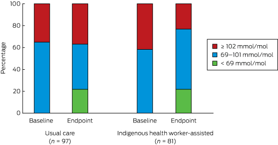
There was a statistically significant reduction in the proportion of participants with extremely poorly controlled HbA1c levels (≥ 102 mmol/mol) in the IHW-S group (from 34 to 19 people, or from 42% to 23%), but a slight increase in the UC group (34 to 36 people, or 35% to 37%; for between-group difference, P = 0.002) (Box 7). If the improvement in the IHW-S group had also been achieved by the UC group, 17 fewer people would have been expected to have had an HbA1c level ≥ 102 mmol/mol.

There were no significant changes in any of the hospitalisation categories. Rates of hospitalisations for all causes (excluding dialysis) and for type 2 diabetes-related diagnoses each increased in both groups; the small differences in favour of the IHW-S group were not statistically significant. Ambulatory care sensitive hospitalisations increased slightly in the UC group but not in the IHW-S group (P = 0.81). The only category in which the change approached statistical significance was for cases in which diabetes was the primary diagnosis: there was a differential net reduction in admission rate of 0.09 per person per year (P = 0.06) (Box 6). The effect size was small, amounting to an estimated eight fewer admissions per year among the 87 IHW participants. This suggests a possible small improvement in morbidity.

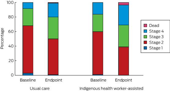
The transition between disease stages from baseline to endpoint is depicted in Box 8. The differences between the IHW-S and UC groups were not statistically significant (Markov transition matrix [Appendix], P = 0.73).

Cost-effectiveness of the intervention

Annual hospitalisation costs are reported in Box 6. There was a small reduction for most categories in the IHW-S group, but the difference only approached significance for type 2 diabetes as the primary diagnosis. Additional expenditure of just over $6700 per participant per year achieved no significant improvement in mean HbA1c levels, rate of disease progression, or quality of life, but realised a statistically sub-significant reduction in hospitalisations for those with type 2 diabetes as the primary diagnosis, yielding an estimated saving of $646 per person per year. The net intervention cost was thus just over $6000 per person per year, or $9000 for the 18-month trial. Taking into account the other significant finding, a reduction in the number of persons with very poorly controlled diabetes, this gives a cost of $42 880 for each person whose HbA1c level was reduced below the critically high level of ≥ 102 mmol/mol.

Discussion

The study examined the costs and outcomes of the GBACC model as implemented in this trial. It is one of few economic analyses of a new model of primary care for addressing poorly controlled diabetes in Indigenous people, building on a high quality cluster RCT design.

The average annual cost of just over $6700 per person for the intensive IHW-S intervention as an adjunct to regular primary care is high relative to the reported costs of primary care in Indigenous communities in Australia. One study estimated the mean annual primary care costs in 21 mainly remote Indigenous communities in north Queensland (including some of the communities involved in this trial) at $1825 per person in 2004–05, equivalent to about $2700 in 2012–13.21 This estimate included IHWs as well as medical, clinic health, nursing, managerial and clerical staff. The Australian Institute of Health and Welfare similarly reported that total primary care expenditure per Indigenous person was $2648 in 2012–13.22 Despite the higher expected primary care costs of a group with poorly controlled diabetes, $6700 per person per year is a considerable additional cost.

In terms of intervention effect, this economic study explored a range of outcomes, including mean HbA1c levels and their distribution, disease progression, quality of life, and hospitalisation. There was evidence of only a modest intervention effect, at best, in any of these measures. The only statistically significant improvement was the reduction in the proportion of patients with HbA1c levels of 102 mmol/mol or more. The difference in the reduction in number of hospitalisations for diabetes (as the primary diagnosis) was close to significant. The study was powered to detect a change in the primary outcome, a mean reduction in HbA1c levels of 12.6 mmol/mol over 18 months, not to detect changes in secondary outcomes such as hospitalisation or quality of life score.9 The effect of the intervention was assessed from the start of the trial, but there may have been a lag between its start and any impact on hospitalisation. A longer follow-up period than 18 months may have found a greater reduction in the hospitalisation rate.

Nonetheless, given the substantial additional resources that were invested, the outcomes were disappointing, with diabetes still poorly controlled in most patients, as indicated by the continued high levels of HbA1c, very high rates of disease progression, and increasing rates of hospitalisation.

There are a number of possible reasons for these outcomes. It is possible that intervening in a group of patients with less advanced disease would have been more successful. It was expected that the employed IHWs would devote 100% of their contracted work time to the trial, but other responsibilities within local health services reduced the capacity of some IHWs to support trial clients. While we adjusted for this in the costing of the trial, it will have diluted the intensity of service delivery. Combined with difficulties in recruiting and retaining staff, this meant that two communities received less than 65% of the intended level of intervention (Box 5). Any change in the IHW position will have disrupted the IHW relationship, a core element of the model. Nonetheless, good community commitment was achieved by ongoing community engagement, with the IHW model building on the Apunipima Cape York Health Council (ACYHC) family-centred approach. ACYHC was a partner in the trial, and author MW, who is a public health medical advisor with ACYHC, was a Chief Investigator in this trial.

Investment in the training and upgrading of qualifications of the IHW, as well as in providing clinical support for them is likely to generate value elsewhere in the health system and over the longer term, a likely benefit not captured by our analysis.

Data quality is a common issue in community trials. For example HbA1c data, which were extracted from participants’ clinical files, included some baseline data gathered well before the trial commenced.

It is also worth reflecting on whether the theory underpinning the trial was correct. In expanding the capacity of IHWs to provide direct and intensive support for Indigenous patients in the community, through both outreach and centre-based care, it was hoped to achieve more effective management of chronic disease because of greater cultural awareness and by improving patient engagement in self-care. While some health gains were identified, the major psychosocial and economic problems that are typical for very disadvantaged populations, and the strong relationship between these factors and chronic disease, mean that it may be necessary to address these factors more directly.23 Most of the IHW-S communities are in the bottom 2% of Queensland communities in terms of socio-economic disadvantage, indicating an extreme level of deprivation, often combined with a range of further serious adverse conditions.24 We did not have data on major life stressors (such as early death of family and friends, involvement with the criminal justice or child protection systems) that affect physical health and, probably, diabetes control; these factors may have affected the intervention and control communities differently.

A separate case study within the GBACC project found that health service providers need to review their systems of care to maximise the value of IHWs as specialist members of the multidisciplinary team.25 IHWs, who participated in regular clinical review sessions, were able to identify examples for improving self-management, which resulted in consistent positive change in HbA1c levels in patients with the poorest control. Further, IHWs could respond to the problem of patient disengagement.

Conclusions

Our results suggest that the costs of delivering the GBACC model were considerable in absolute terms but achieved only a modest effect. This suggests a need to consider how to improve the effectiveness of the program, reduce its costs, and to increase revenue (eg, through Medicare billings).

The training of IHWs and clinical support workers is generally viewed as positive, but translating it into measurable outcomes for people with poorly controlled type 2 diabetes in highly disadvantaged communities remains a challenge. A more holistic cross-agency approach may be required, one that seeks to directly address the psychosocial, pathophysiological and environmental problems that are common in highly disadvantaged populations. While the need to consider social and economic determinants is understood, there are still major gaps in service delivery. The challenge for the public health community is to devise and implement interventions based on broader understanding of the determinants of health and to test the effectiveness of such interventions.

Box 1 –
Project cost calculations for the central team and the Indigenous health workers (IHWs)


The central team

  • The central team consisted of the trial manager and the clinical support team responsible for IHW training, which included:
    • developing training materials, training delivery;
    • enhancing the quality of clinical practice through mentoring, advocacy and reflective practice with IHWs, convening IHW meetings, clinical reference group meetings, team meetings;
    • evaluation as an embedded component (data collection, data entry, conference presentations, workshops), and coordination of project activities, including chief investigator and management group meetings.
  • Costs were extracted from project financial reports for the period 1 January 2011 (commencement of the GBACC project with trial set-up) to 30 September 2013 (trial endpoint). The percentage of time allocated by the manager and the clinical support team to the trial and to the evaluation were determined by the trial manager (BS) after detailed discussion with LS and HN about the type of activities to be classed as intervention and non-intervention (evaluation and trial coordination activities).

Indigenous health workers

  • IHW salaries (including wage on-costs) in the six intervention communities were identified from project records. The proportions of their time allocated to intervention and to non-intervention activities were determined from detailed time logs kept by the IHWs. The IHW cost was calculated from their total wage costs and the percentage of time allocated to the project by each IHW.

Box 2 –
Diabetes vascular severity staging employed in this study, based on reference13


  • Type 2 diabetes with no evidence of microvascular or macrovascular risk factors.
  • Type 2 diabetes with screen-detected microvascular comorbidities and/or risk factors for macrovascular disease.
  • Type 2 diabetes with moderate microvascular or macrovascular complications.
  • Microvascular or macrovascular complications of late stage type 2 diabetes.

Box 3 –
Categorisation of admissions to hospital in this study


  • All hospitalisations.
  • Hospitalisations with principal or other diagnoses related to type 2 diabetes (ICD-10 E11 code in the principal or other diagnoses).
  • Ambulatory care sensitive (ACS) hospitalisations related to chronic disease (used by the Australian Institute of Health and Welfare to estimate ACS hospitalisations for Aboriginal and Torres Strait Islander people).15
  • The top three ACS condition categories (type 2 diabetes as principal diagnosis, cardiovascular diseases, and infections).16

Box 4 –
Baseline characteristics of the study participants

Usual care

Indigenous health worker-supported

P


Number of participants

106

87

Mean HbA1c level (SD), mmol/mol

95 (19)

99 (17)

0.12

Mean age (SD), years

47.6 (8.7)

47.5 (10.6)

0.958*

Sex (female)

70 (66%)

53 (61%)

0.533

Daily smoker

38 (36%)

34 (39%)

0.654

Current drinker

39 (37%)

36 (41%)

0.511

Mean body mass index (SD)

32.6 (6.2)(n = 43)

31.2 (6.3)(n = 44)

0.522*

Obese

28 (65%)

23 (52%)

0.280


SD = standard deviation. * Results of t test for equal means, adjusted for within-group clustering. † Results of χ2 test for equal proportions, adjusted for within-group clustering. ‡ Body mass index ≥ 30.

Box 5 –
Total cost estimates for the Getting Better at Chronic Care (GBACC) project

Total trial expenditure

Time and cost allocated to GBACC intervention


Time*

Expenditure


Central team

Clinical support team

$626 091

57%

$357 353

Management

$234 624

10%

$23 462

Operation

$440 200

23%

$102 791

Sub-total

$1 300 915

37%

$483 606

Indigenous health workers

Community A

$151 551

78%

$118 210

Community B

$151 551

64%

$96 993

Community C

$75 775

44%

$33 341

Community D

$78 028

89%

$69 445

Community E

$156 056

84%

$131 087

Community F

$78 028

94%

$73 346

Sub-total

$690 989

76%

$522 421

Total expenditure

$1 991 904

51%

$1 006 027


Source: Project financial reports. * The allocation of project team time to research and service delivery was determined by the program manager. Allocation of Indigenous health worker time to GBACC was based on time records.

Box 6 –
Summary of the incremental effectiveness analyses (change between baseline and trial end)

Usual care (n = 106)


Indigenous health worker-supported (n = 87)


Difference of differences (95% CI)

P

Baseline

Endpoint

Change*

Baseline

Endpoint

Change*


HbA1c level (SD), mmol/mol

94.7 (19.0)

89.3 (24.1)

–5.4 (n = 97)

99.0 (17.4)

88.8 (25.7)

–10.1 (n = 81)

–4.7 (–11.6 to 2.1)

0.174

AQoL-4D, mean utility score (SD)

0.80 (0.18)

0.79 (0.21)

–0.01

0.75 (0.18)

0.72 (0.28)

–0.03

–0.02 (–0.08 to 0.05)

0.623

Rate of hospitalisation (per person per year; total number of admissions in parentheses)

All causes, excluding dialysis

1.02 (172)

1.24 (176)

0.22

0.98 (135)

1.07 (124)

0.09

–0.13 (–0.68 to 0.41)

0.633

Type 2 diabetes, any diagnosis§

0.53 (88)

0.92 (128)

0.39

0.47 (64)

0.78 (88)

0.31

–0.08 (–0.20 to 0.03)

0.150

Ambulatory care sensitive

All

0.33 (58)

0.44 (60)

0.11

0.31 (45)

0.30 (36)

–0.01

–0.11 (–1.04 to 0.81)

0.811

Type 2 diabetes as principal diagnosis**

0.15 (26)

0.18 (23)

0.03

0.17 (23)

0.11 (13)

–0.06

–0.09 (–0.18 to 0.00)

0.063

Cardiovascular disease††

0.01 (1)

0.08 (12)

0.07

0.02 (3)

0.04 (5)

0.02

–0.05 (–0.13 to 0.02)

0.149

Infections‡‡

0.13 (21)

0.14 (20)

0.02

0.10 (14)

0.09 (11)

–0.01

–0.03 (–0.10 to 0.04)

0.362

Mean hospitalisation cost (per person per year)

All causes

$5438

$7421

$1982

$8010

$9866

$1856

–126 (–5024 to 4771)

0.960

Type 2 diabetes, any diagnosis§

$4248

$6582

$2335

$4921

$8595

$3674

1340 (–2724 to 5404)

0.518

Ambulatory care sensitive

All

$1665

$2132

$467

$2967

$2677

–$290

–757 (–2130 to 616)

0.280

Type 2 diabetes as principal diagnosis**

$907

$1245

$338

$1553

$1245

–$308

–646 (–1348 to 56)

0.071

Cardiovascular disease††

$23

$163

$140

$239

$383

$144

4 (–749 to 757)

0.992

Infections‡‡

$623

$609

–$14

$1040

$451

–$589

–574 (–1490 to 342)

0.219


AQoL-4D = Assessment of Quality of Life 4D score. * Only participants for whom baseline HbA1c levels were measured after 1 January 2009 and endpoint levels after 1 March 2012 were included. † Estimates for incremental difference in outcomes between usual care and IHW groups using linear multi-level models adjusted for within-community clustering. ‡ Two people in the IHW group had dialysis after the intervention commenced (starting July 2012 and March 2013); their dialysis records were excluded. § International Classification of Diseases, revision 10 (ICD-10) code in principal or any other diagnoses starting with E11. ¶ All potentially preventable hospitalisations (ICD code in principal diagnosis: D501, D508, D509, E101–E108, E110–E118, E130–E138, E140–E148, E40–E43, E550, E643, E86, G40, G41, H66, H67, I10, I119, I110, I20, I240, I248, I249, I50, J02, J03, J06, J20, J312, J41–J44, J45, J46, J47, J81, K02–K06, K08, K098, K099, K12, K13, K250–K252, K254, K255, K256, K260–K262, K264–K266, K270–K272, K274–K276, K35–K37, K522, K528, K529, L03, L04, L08, L88, L980, L983, N10–N12, N136, N390, N70, N73, N74, O15, R02 or R56).16 ** ICD-10 code in principal diagnosis starts with E11. †† ICD-10 code in principal diagnosis: I10, I110, I119, I20, I240, I248, I249, J81 or I50. ‡‡ ICD-10 code in principal diagnosis: H66, H67, J02, J03, J06, J312, L03, L04, L08, L980, L88, L983, N10–N12, N136, N390, N70, N73, N74, or R02.

Box 7 –
Distribution of HbA1c level categories at baseline and endpoint*


* Only participants for whom baseline HbA1c levels were measured after 1 January 2009 and endpoint levels after 1 March 2012 were included.

Box 8 –
Distribution of disease stages at baseline and endpoint

Warning over diabetic ketoacidosis after man’s death

GPs are being told to consider diabetic ketoacidosis after a man died less than 24 hours after presenting to his doctor.

According to the Victorian Coroner’s report, a 29-year-old male visited his GP complaining of increased urination, thirst and difficulty sleeping.

He had a fever of 38.6 with high blood pressure and the GP prescribed cephalexin antibiotics for a urinary tract infection.

A urine sample was taken and sent to pathology as well as requests for blood tests including full blood exam, LFT, TSH, urea and electrolytes and fasting blood glucose.

However the patient was found dead in his home at 9pm the following day. A taxi was booked at 5:30am that morning however he didn’t respond to the taxi’s arrival at between 7:40am and 8am.

The pathology report dated the day after testing found ketones and glucose in the urine sample.

Related: MJA – Guidance concerning the use of glycated haemoglobin (HbA1c) for the diagnosis of diabetes mellitus

The coroner found that the patient died from diabetic ketoacidosis. Post mortem toxicological analysis showed a glucose concentration of 45mmol/L, combined with raised acetone levels in the blood and vitreous. The coroner said this indicated that the man had died of undiagnosed diabetes.

The patient had reported considerable weight gain over the previous year, resulting in him weighing 122.9kg when he presented to his doctor. The GP concluded that the patient likely had diabetes and ranked the likelihood as Type 2 ahead of Type 1 diabetes. However he didn’t check the patient’s blood glucose at the time, instead referring him for fasting blood tests the next day.

It is noted in the coroner’s report that the GP was regretful that he didn’t perform a fingerprick test at the time of consultation and that it would have been the appropriate action.

The report stated that the doctor was under the impression that fasting blood tests was the most accurate way to diagnose diabetes. “(The doctor) did not consider that (the patient) was in any immediate danger from diabetes,” the report said.

CPD active learning module: Addressing the challenges in the fast moving field of endocrinology

The coroner recommended the following:

  1. The Royal Australian College of General Practitioners provides a clinical update to GPs to highlight the importance of recognising hyperglycaemia and ketosis in adult diabetic patients, as an uncommon but potentially serious complication of type 2 diabetes, or indication of newly recognised adult-onset type 1 diabetes.
  2. The Royal Australian College of General Practitioners advise GPs that although uncommon in adults and clinically subtle in its earliest states, evolving diabetic ketoacidosis may produce a dangerous metabolic decompensation and require escalation of care to a hospital setting for further assessment and management.

RACGP President Dr Frank R Jones told doctorportal: “The RACGP will certainly review and respond to the coroner’s recommendations regarding diabetic ketoacidosis (DKA) as published in the findings into the death of (the patient) and will communicate a clinical update to members.

“The RACGP does have some existing guidance on DKA in its resource General practice management of type 2 diabetes.”

Latest news:

Vertebroplasty is not a do-not-do treatment

Vertebroplasty has been controversial but remains clinically useful and new evidence awaits publication

Duckett and colleagues have classified vertebroplasty as a do-not-do treatment.1 They referenced two randomised controlled trials (RCTs)2,3 as definitive proof of this. However, the authors failed to heed our clinical opinion published in the MJA that these two trials were “not relevant to the patient group that we treat with vertebroplasty”.4 We have the largest clinical vertebroplasty experience in Australia, yet our published advice was apparently ignored. In the article by Duckett and colleagues, Box 1 illustrated the selection process that the authors used to determine do-not-do procedures. The process supposedly excluded evidence which was “contested” or “which was not supported by consulted clinical experts”. Accordingly, vertebroplasty should have been deleted from the list.

The authors used the United Kingdom National Institute for Health and Care Excellence (NICE) for clinical guidance. Current NICE guidance5 states that “vertebroplasty and kyphoplasty can be considered appropriate interventions for people with recent, unhealed osteoporotic vertebral compression fractures in whom the pain is severe and ongoing despite optimal pain management”.

From 1208 potential treatments, the authors excluded 1200, leaving five apparently incontrovertible do-not-do treatments. The fact that at least one of the five is wrongly included (by the authors’ own criteria) demonstrates the failure of the proposed model and the danger of adopting this kind of formula to influence clinical practice in hospitals.

The evidence for and against vertebroplasty is inconclusive. There is disparity in measured outcomes between blinded RCTs2,3 of vertebroplasty for fractures up to 12 months old and a larger, open-label RCT6 of fractures less than 6 weeks in duration. The blinded trials found no significant benefit of vertebroplasty over placebo, whereas the open-label RCT found significant benefit of vertebroplasty over conservative care. This disparity is well described in the NICE guidance.5

For the past 10 years, my vertebroplasty practice has been confined to treating fractures less than 6 weeks old.7 It is clear to me that the published blinded trials tested a different approach and are not relevant to the patient group that my practice treats with vertebroplasty for two principal reasons: the fractures were mostly non-acute; and the volume of cement used in these trials (2.6 cm3 on average in both trials) would have been insufficient to stabilise an acutely collapsing vertebral fracture.

Attempting to answer the acute fracture conundrum, the authors of the blinded RCTs published a meta-analysis of 52 patients from both trials with fractures less than 6 weeks duration.8 Only outcomes at 2 weeks and 1 month were presented and the evidence is hardly definitive.

The onus was placed on vertebroplasty practitioners to provide high-quality blinded data in this group of patients. For this purpose, my co-investigators and I embarked on the Vertebroplasty for Acute Painful Osteoporotic fractURes (VAPOUR) trial.9 Five years ago, we reconfigured the protocol from the INvestigational Vertebroplasty Efficacy and Safety Trial (INVEST),2,10 the larger of the two blinded vertebroplasty trials. We excluded crossover, which was permitted at 1 month in INVEST. We changed the selection criteria to include only fractures less than 6 weeks duration (average fracture age in the VAPOUR trial was 2.6 weeks compared with 18 weeks in INVEST) with pain scores greater than 7/10 and with either magnetic resonance imaging or single-photon emission computed tomography evidence of acute fracture. In-patients, already hospitalised with acute fractures, comprised 59% of the VAPOUR trial enrolment but were excluded in INVEST. The procedural technique was different in the VAPOUR trial, where we attempted maximum fill of the vertebral body to stabilise the fracture and prevent ongoing collapse. The average cement volume of 7.5 cm3 in the VAPOUR trial was three times that in INVEST. The method of blinding and data collection was similar for the two trials.

Our trial team included four Sydney centres with established vertebroplasty programs. The VAPOUR trial completed enrolment of 120 patients in December 2014 and is the largest RCT and the only acute fracture RCT of vertebroplasty in Australia. Statistical assessments of outcomes are nearing completion and the results of the trial will soon be published.