×

Toxic epidermal necrolysis — an investigation to dye for?

We report the first case in Australia, as confirmed by the Therapeutic Goods Administration, of toxic epidermal necrolysis associated with the iodinated contrast medium iopamidol. It serves as a warning about the use of contrast in imaging and cardiac catheterisation and a reminder of the need for increased awareness of the issue.

Clinical record

A 44-year-old woman presented to the emergency department with a 3-day history of a progressive rash, fever, malaise and mucosal ulceration. She met the diagnosis of toxic epidermal necrolysis (TEN) based on the following criteria: bullae and desquamation affecting about 84% of the body surface (Box 1 and Box 2), buccal and vaginal ulceration, a positive Nikolsky sign (this is a useful sign in bullous skin diseases and can be demonstrated by rubbing the skin surface, which will blister within a few minutes if the sign is positive), fever, tachycardia and mild hypotension. She also had abnormal results of liver function tests: bilirubin level, 69 µmol/L (reference interval [RI], < 20 µmol/L); alkaline phosphatase level, 180 U/L (RI, 25–100 U/L); γ-glutamyl transferase level, 499 U/L (RI, < 30 U/L); alanine aminotransferase level, 1730 U/L (RI, < 30 U/L); and aspartate aminotransferase level, 638 U/L (RI, < 30 U/L). She had a white cell count of 4.3 × 109/L (RI, 4.0–10.0 × 109/L) and a raised C-reactive protein level of 53.1 mg/L (RI, < 5 mg/L).

The patient was immediately transferred to the burns unit and managed with nanocrystalline silver dressings, intravenous immunoglobulin, aggressive fluid and electrolyte balance therapy, analgesia and intravenous antibiotics. She was discharged home on Day 15.

Histopathological examination (Box 3) showed extensive epidermal necrosis and subepidermal clefting with a sparse superficial perivascular infiltrate of lymphocytes, occasional neutrophils and eosinophils, and exocytosis of cells into the epidermis. Results of staining for immunofluorescence were negative. This was consistent with the clinical diagnosis of TEN, and the possibility of pemphigus vulgaris was excluded.

The patient’s only recent exposure to medications included 150–200 µg of thyroxine sodium daily for 19 years and 2.5 mg of indapamide daily for 6 months. The patient underwent a computed tomography (CT) neck scan with the contrast medium iopamidol about 4 weeks before the development of symptoms. The patient had recently (3 weeks before onset of rash) stopped taking a herbal “liver cleanser”. She had been taking this intermittently for 2 months. She had no recent travel history or vaccinations.

The patient recalled having a previous CT scan of the neck before her surgery 17 years ago. Unfortunately, however, any records of this had been destroyed.

The patient’s past medical history was notable for mild stable hypertension, hypothyroidism and a benign mixed salivary gland tumour electively excised 17 years earlier.

Discussion

Toxic epidermal necrolysis (TEN), or Lyell syndrome, is a rare and life-threatening severe systemic condition associated with dramatic cutaneous sloughing of up to 100% of the body surface area. The incidence of TEN is two cases per million person-years.1 It is characterised by necrosis and subsequent detachment of the epidermis from the dermis in more than 30% the body surface. If not treated and managed promptly, the consequences can be fatal; patients are vulnerable to infections and sepsis leading to death. The mortality associated with TEN is high, at 30%–40%.2

At the other end of the spectrum, and more common, are mild-to-moderate skin reactions to contrast media (CM). These include, in increasing severity, lichenoid reaction, erythema multiforme and Stevens–Johnson syndrome.3 Patients at risk of late skin reactions are those with a previous history of CM reactions.3,4

TEN is attributed to medications in 80% of cases.1 The most commonly associated medications include sulfonamides, penicillin and other antibiotics, anticonvulsants, oxicam nonsteroidal anti-inflammatory drugs, allopurinol and corticosteroids.5 TEN commonly occurs 1–3 weeks after the start of therapy. Other triggers include infections, malignancy and vaccination.1

The dermatological reactions caused by CM can be classified as early or late reactions. Early reactions occur soon after injection of the contrast medium, and late reactions occur within a week. The incidence of late adverse reactions is 2%.3,6 They commonly present as maculopapular erythema, angioedema and urticaria. Evidence to date suggests that late reactions are more common with non-ionic CM, in particular dimers,3 despite non-ionic CM being touted as having fewer adverse reactions. Late reaction incidence with non-ionic CM varies between 8% and 71%.3

Iodinated CM can be divided into ionic and non-ionic contrast medium. The ionicity pertains to the osmolality the CM create in blood; ionic CM create higher osmolality leading to CM reaction. The move from the use of ionic CM to non-ionic CM was based on the need for an agent with fewer adverse effects and equal or slightly improved diagnostic efficacy.7 The morbidity and mortality associated with non-ionic CM were less than for ionic CM.8 Although non-ionic CM have these advantages, ionic CM are still in use today. A recent study highlighted that although non-ionic CM are the best tolerated in the early phase, they are associated with a higher level of adverse effects such as late skin reactions.8 Iopamidol is a non-ionic contrast medium.

There have been several cases of TEN caused by CM reported in the literature. Commonly, the cases have involved repeated exposure or sensitisation to the CM in the cardiac catheter laboratory over a period of days to even years.911 TEN occurs with subsequent exposures to the CM administered. In our case report, there was a history of prior CT scan of the neck; however, records of the scan are no longer held by the radiologist to verify the date and contrast medium used.

TEN has also been shown to be caused by gastrointestinal oral CM.12

Only two published cases of TEN have been attributed to the administration of iopamidol. The first case is of a young boy with subsequent exposure to iopamidol.10 The case was not biopsy-proven TEN, but was based on clinical diagnosis. The second case is that of a patient who underwent intravenous urography for investigation of systemic lupus erythematosus with renal involvement.13 The patient died despite intensive care and support. Our case report would be the third reported case of TEN caused by iopamidol.

Although it is difficult to be sure that iopamidol was responsible for the development of TEN in our patient, it is highly likely to be the cause. She had been on indapamide for a period and had been taking the herbal “liver cleanser” intermittently. One recent case of herbal medicines and TEN has been reported.14 However, as noted by the author, it was difficult to determine the causative agent.

It is important to be aware of the risk of CM and to think twice about the necessity of CM in imaging. Although rare, life-threatening adverse effects such as TEN should lead to reconsideration of contrast dyes, as patients may suffer unnecessarily or lose their lives.

1 Bullae and desquamation of back

2 Desquamation of both feet

3 Skin punch biopsy sample showing extensive epidermal necrosis (bracket) and subepidermal clefting (asterisk) with mixed inflammatory infiltrate and negative results of staining for immunofluorescence

A diagnosis that will go down in history

A well described case in the Christmas tradition is that of patient R, reported to have been afflicted with a very shiny nose. We believe his presentation was consistent with one of the cutaneous forms of sarcoidosis: lupus pernio.

A well described case in the Christmas tradition is that of patient R, reported to have been afflicted with a very shiny nose. The lesion was described as having a lustrous, glowing appearance and initially exerting a significant burden on quality of life — the patient’s peers engaged in name-calling and excluded him from social games. It would seem that the case proved a challenge diagnostically, with the disease managed expectantly. Fortuitously, it availed itself to a particularly coveted job prospect which in time eventuated in acceptance by his peers and, notably, this transpired with glee. The case has remained a diagnostic dilemma through the generations. It has been proposed that the cutaneous lesion might be attributable to variations in nasal microcirculation noted in specific genetic populations.1 However, the uniqueness of patient R’s lesion is what has made him a legend and, accordingly, a diagnosis of variations in nasal microcirculation seems unlikely. Rather, a pathological aetiology is more in keeping with his clinical picture. We believe patient R was afflicted with one of the cutaneous forms of sarcoidosis: lupus pernio.

The clinical presentation of lupus pernio is that of violaceous papules, nodules or plaques. The lesions have an indurated, shiny, and somewhat bright character to them and are classically located on the nose, but also the cheeks and ears: in those areas most sensitive to the cold, or “pernio”, that one might experience one foggy Christmas Eve. In lupus pernio, histological findings are remarkable for the presence of sarcoidal granulomas, with birefringent material observed in up to 50% of cases (you could even say they glow).2

An important clinical consideration in patients with lupus pernio is the association with pulmonary sarcoidosis. It has been reported that 74% of patients will have intrathoracic disease and 54% will have upper respiratory tract involvement.3 This form of cutaneous sarcoidosis portends a more aggressive clinical course with a higher likelihood of visceral involvement and recalcitrance to therapy. Cutaneous sarcoidosis is known for its protean clinical presentations, including erythema nodosum, macules, papules and plaques, scar sarcoidosis, ichthyosis, Darier–Roussy lesions, alopecia and onychodystrophy.4 Physicians should be aware of cutaneous signs of sarcoidosis because they lend themselves to biopsy facilitating diagnosis, many have prognostic significance, and they can impose a significant burden on quality of life.5

Based on morphology alone, a wide range of differential diagnoses could account for patient R’s clinical presentation (Box). However, considering the history and examination findings together, lupus pernio represents a unifying diagnosis. This patient’s story represents an instructive case and is one that will go down in history.

Differential diagnoses for Patient R

Aetiology

Diagnoses


Inflammatory

Rosacea (rhinophyma)

Infiltrates


Cellular


Lymphocytic

Tumid lupus, pseudolymphoma, Jessner lymphocytic infiltration

Neutrophilic

Sweet syndrome

Eosinophilic

Angiolymphoid hyperplasia with eosinophilia

Granulomatous

Xanthogranuloma

Mixed

Granuloma faciale

Acellular


Mucin

Cutaneous mucinosis

Amyloid

Cutaneous amyloidosis

Tumours

Basal cell carcinoma, amelanotic melanoma, cutaneous lymphoma, Merkel cell carcinoma, angiosarcoma

Infection

Syphilis (Treponema pallidum), lupus vulgaris (Mycobacterium tuberculosis), leishmaniasis (Leishmania spp)

Vascular malformations

Non-involuting congenital haemangioma, arteriovenous malformation (especially Wyburn-Mason syndrome)

Pseudoxanthoma elasticum

A 31-year-old woman presented with a 20-year history of lesions on the skin of her neck and axillae. Her family history was unremarkable.

Physical examination revealed multiple pinhead-sized yellowish flat-topped papules on the skin of the axillae and lateral aspects of the neck (Figure, A). These lesions made us suspect pseudoxanthoma elasticum, so we performed a fundoscopic examination which showed obvious angioid streaks (Figure, B). A biopsy of the axillary lesions showed a band of degenerated clumps of elastic fibres (Figure, C) and calcium deposits in the upper reticular dermis.

A diagnosis of pseudoxanthoma elasticum (PXE) was made. This condition is caused by mutations in the gene that encodes the ABCC6 protein, an ATP-binding cassette, subfamily C member.1


A: Photograph of the axilla of our patient showing several yellowish waxy papules.


B: Fundus of the left eye showing angioid streaks characteristic of pseudoxanthoma elasticum.


C: Histological examination of the skin biopsy showed fragmentation of elastic fibres in the upper reticular dermis.

Sentinel lymph node biopsy for melanoma: an important risk-stratification tool

This test should be routinely considered for patients with intermediate thickness melanoma

Most patients with a new diagnosis of melanoma present with localised disease without clinical evidence of metastasis and are cured by surgical resection. However, a small proportion will harbour occult metastatic disease in the regional lymph nodes. The presence of nodal involvement is the most significant prognostic factor in patients without clinical evidence of distant metastasis.1 The most sensitive test to identify these patients at presentation is sentinel lymph node biopsy (SLNB).1,2 The sentinel lymph node is the first draining lymph node from the area of skin where the primary tumour arose. The technique was first described over 20 years ago.3 Careful histological examination of the sentinel lymph node by a combination of routine histological and immunohistochemistry tests can accurately identify small-volume metastatic disease.

On the basis of initial reports, SLNB has rapidly become the standard of care in major melanoma centres around the world for patients with melanoma greater than 1 mm in thickness (stage T2 and above; Box 1) and incorporated into the American Joint Committee on Cancer staging system.1 Considerable controversy about the routine use of sentinel node biopsy has focused on the lack of a survival benefit from the procedure.4,5 In this review, we seek to put the rationale for SLNB into context in light of both advances in the management of patients with advanced melanoma and the recent publication of the final report of the Multicenter Selective Lymphadenectomy Trial (MSLT-I).2

The era before sentinel lymph node biopsy

Before the introduction of SLNB, a number of randomised controlled trials had compared elective lymph node dissection (ELND) with nodal observation; none of these trials showed an overall survival benefit for ELND.6 The Intergroup study was the most recent of these trials, and incorporated lymphoscintigraphy to identify the draining nodal basin. This study identified a number of subgroups which appeared to benefit from ELND, including patients with melanoma of Breslow thickness between 1 and 2 mm and patients aged less than 60 years.7 SLNB offered the benefit over ELND of identifying those patients who might benefit from early lymph node dissection while minimising the morbidity to patients without nodal involvement.

Prognostic impact of sentinel lymph node biopsy

The MSLT-I study randomly allocated patients to wide local excision (WLE) with SLNB, and immediate lymphadenectomy for nodal metastases, versus WLE and nodal observation with lymphadenectomy only at nodal relapse (Box 2). The primary end point of the study was disease-specific survival (DSS). The study finished accruing patients in 2002, and the final analysis was recently published.2

The MSLT-I study confirmed that lymph node status is the strongest predictor of DSS for patients with intermediate thickness melanoma (hazard ratio, 2.4; Box 3). Note that intermediate thickness in the MSLT-I study was Breslow thickness 1.2–3.5 mm; this was the thickness of melanomas in the primary analysis. There were too few patients (and events) with thin melanoma (Breslow thickness, < 1.2 mm), so no analysis of this subgroup has been provided to date. Other non-randomised studies have demonstrated the prognostic impact of SLNB in certain patients with thin melanoma (0.75–1.0 mm),9 although SLNB in this group of patients remains controversial.

Therapeutic benefit

The major area of controversy about SLNB is its therapeutic impact on DSS. MSLT-I did not show a significant difference in DSS between the two arms. The rate of sentinel lymph node positivity in the SLNB arm was only 16%, and this is the only population who might benefit from the procedure. MSLT-I was therefore likely underpowered to demonstrate an effect on DSS for the entire group.

A planned post-hoc analysis of node-positive patients was performed, comparing the outcomes of patients with nodal disease diagnosed at SLNB (biopsy-based detection) with the outcomes of those presenting with nodal relapse in the observation group. For patients with intermediate thickness (1.2–3.5 mm) melanoma with nodal involvement, this analysis showed a significant improvement in DSS, with 62.5% 10-year survival in the SLNB arm compared with 41.5% in the observation arm (P = 0.006). This difference remained significant even when patients with a false-negative sentinel node were included in the analysis (intention to treat, P = 0.04). For patients with thick (> 3.5 mm) melanoma, there was no survival benefit from early detection of nodal involvement, which likely reflects the high rate of distant metastatic disease in this less common, high-risk subgroup.

The validity of this post-hoc analysis has been questioned, as critics have suggested that SLNB identifies subclinical micrometastatic disease which is not destined to progress to palpable lymphadenopathy.10 Unlike other tumour types, such as breast cancer and differentiated thyroid cancer, where it is clear that micrometastatic disease can lie dormant without clinical significance, the results of MSLT-I and other non-randomised studies suggest that virtually all patients with microscopic disease will inevitably progress to clinical lymphadenopathy (10-year node-positive rate [mean ± SE], 21.9% ± 1.5% in the SLNB group compared with 19.5% ± 1.9% in the observation group).2 Furthermore, the mean number of lymph nodes harbouring metastatic melanoma in the SLNB arm was 1.4 compared with 3.3 for patients undergoing lymphadenectomy at nodal relapse in the observation arm (P < 0.001), suggesting progression during the period of observation.

Risks of sentinel lymph node biopsy

The major disadvantages of SLNB include cost and morbidity. For some patients, the use of SLNB converts their treatment from one deliverable under local anaesthesia to one requiring a general anaesthetic. Furthermore, SLNB requires preoperative lymphoscintigraphy. However, taking these factors into account, a cost-effectiveness analysis of SLNB in the Australian health care setting showed a significant improvement in quality-adjusted life-years with only a marginal increase in costs.11

Surgical complications from SLNB are seen in 5%–10% of patients, and are mostly minor in severity.8,12 The most common of these are early complications, including seroma and infection, and resolve over time. This is in stark contrast with lymphadenectomy where complication rates are significantly higher, particularly the serious long-term morbidity of lymphoedema.

Some have argued that ultrasound could replace SLNB as a non-invasive means of identifying lymph nodes harbouring metastatic disease.10 However, ultrasound is an operator-dependent technique and published results are variable.13,14 While ultrasound may select patients for immediate lymph node dissection, SLNB in these patients allows a thorough pathological assessment of nodal burden. The volume of disease, as well as the location of the metastatic deposit within the sentinel lymph node, provide independent prognostic information with regard to risk of further lymph node involvement as well as DSS.15,16

What to do with the information

The risk of a positive SLNB is associated with increasing Breslow thickness, presence of ulceration and mitotic count. The current clinical practice guidelines for managing melanoma in Australia and New Zealand recommend that “Patients with a melanoma greater than 1.0 mm in thickness be given the opportunity to discuss sentinel lymph node biopsy to provide staging and prognostic information”.17 At our institution, SLNB is recommended for patients with an expected rate of nodal involvement of greater than 5%. This includes all patients with melanoma greater than 1.0 mm thickness and patients with melanoma between 0.75 mm and 1.0 mm with ulceration or an increased mitotic count. For patients with significant comorbid conditions, the impact of the additional prognostic information provided by SLNB needs to be carefully weighed up against the added morbidity associated with the procedure.

The current standard of care for patients with a positive result on SLNB is completion lymph node dissection (CLND). The question of immediate CLND versus observation for patients with a positive SLNB result will be answered by the MSLT-II trial which will report in the next few years. In the interim, the risk of further lymph node involvement beyond the sentinel lymph node can be estimated from various characteristics of the sentinel lymph node including the size and the location of the tumour deposit within the lymph node.15,16,18 Patients with small deposits, particularly if less than 0.1 mm in size, have a very low risk of further lymph node involvement; this needs to be weighed up against the morbidity of the lymph node dissection as well as the risk of metastatic disease. On the flipside, CLND is associated with lower complication rates than delayed lymphadenectomy at the time of clinical progression,19 so if the risk of further nodal involvement is significant, early intervention may be preferable. Thus, patients with a positive SLNB result require a nuanced discussion in a melanoma multidisciplinary meeting where the individualised pros and cons of CLND are considered.

The past 5 years have seen a dramatic change in the landscape of systemic therapy options for patients with metastatic melanoma. As these therapeutic agents filter into the adjuvant setting, one of the most important roles of SLNB is to select patients for inclusion in these trials. Currently, adjuvant trials for clinically localised melanoma (many of which are recruiting in Australia) are confined to the higher risk subsets of patients, particularly those with a positive SLNB result. Future developments in molecular oncology may identify markers to supersede SLNB. However, currently, SLNB is the best stratification tool available. We strongly recommend that patients with a positive SLNB result should be offered inclusion in these trials with a hope of seeing these new therapeutic agents reach the clinic for routine adjuvant use.

Conclusion

Sentinel lymph node biopsy is the most important prognostic test for patients with intermediate thickness melanoma. It identifies patients at high risk of relapse and allows them to be offered involvement in trials of adjuvant therapy. Data from the MSLT-I study suggest that, for patients with intermediate thickness melanoma with spread to regional lymph nodes, biopsy-based detection of nodal disease using SLNB is associated with a significant improvement in DSS. For these reasons a discussion about SLNB should be standard of care for patients with melanoma greater than 1.0 mm thick.17

1 Primary melanoma staging and corresponding Breslow thicknesses

Melanoma T stage

Breslow thickness*

Description


T1

< 1.0 mm

Thin melanoma

T2

1.0–2.0 mm

Intermediate thickness melanoma

T3

2.0–4.0 mm

Intermediate thickness melanoma

T4

> 4.0 mm

Thick melanoma


* Tumour thickness measured between the upper layer of the epidermis and the deepest point of tumour penetration.

2 Design for the Multicenter Selective Lymphadenectomy Trial (MSLT-I)


Stratification was by Breslow thickness (1.2–1.8 mm v 1.8–3.5 mm) and primary tumour site (limb v other site).8

3 Predictors of disease-specific survival2

Prognostic indicator

Hazard ratio

P


Sentinel node status (positive v negative result)

2.40

< 0.001

Ulceration (present v absent)

1.79

0.002

Breslow thickness* (per 1 mm increase)

1.59

< 0.001

Sex (male v female)

1.22

0.32

Clark level (IV or V v III)

1.07

0.73


* Tumour thickness measured between the upper layer of the epidermis and the deepest point of the tumour; see Box 1. † Measure of tumour penetration into the layers of the skin, defined as follows: Level l, confined to the epidermis; Level II, invasion of the papillary dermis; Level III, filling of the papillary dermis, but no extension in to the reticular dermis; Level IV, invasion of the reticular dermis; Level V, invasion of subcutaneous tissue.

Black bones: minocycline-induced bone pigmentation

An 82-year-old man with bilateral knee osteoarthritis underwent consecutive total knee arthroplasty 5 months apart. During both procedures, he was noted to have black subchondral bone with otherwise normal architecture and normal-coloured cancellous bone. At the time of surgery, bone specimens sent for pathology testing were histologically normal. The patient had been treated with minocycline for rosacea for 7 months before the first procedure. Minocycline is an uncommon cause of skeletal pigmentation and is not known to affect bone quality.1 Discolouration may also be owing to ochronosis, metal deposits, sequestrum and metastatic disease.2

Ipilimumab in pretreated patients with unresectable or metastatic cutaneous, uveal and mucosal melanoma

Australia has the highest incidence of melanoma in the world; melanoma is the fourth most common form of cancer in Australia and the most common cancer in young Australians aged 15–39 years.1 While surgery remains the mainstay of treatment for early stage melanomas, there have been limited treatment options for more advanced disease and, historically, 5-year survival was less than 15%.2

Ipilimumab (Yervoy, Bristol-Myers Squibb) is a fully humanised monoclonal antibody directed against the cytotoxic T lymphocyte antigen 4 and the first treatment to demonstrate a survival benefit in advanced and metastatic disease (see Appendix 1 for a summary of published data). Pivotal studies of ipilimumab at 3 mg/kg in relapsed disease and 10 mg/kg in untreated disease demonstrated significant overall survival (OS) advantage compared with comparative arms (ipilimumab 3 mg/kg v vaccine: median OS, 10.1 v 6.4 months; hazard ratio for death, 0.66; = 0.003;3 and 10 mg/kg plus dacarbazine v dacarbazine alone: median OS, 11.2 v 9.1 months; hazard ratio for death, 0.72; < 0.0014). Ipilimumab has demonstrated clinical activity independent of negative prognostic markers (age, M stage or response to prior systemic therapy), offering a novel treatment to patients with poor prognosis.5 Immune-related adverse events (AEs) are frequently reported in clinical trials with serious events (grade ≥ 3) reported in 10%–56% of patients.3,4,6,7 However, with growing experience, the management of immune-related AEs has improved considerably and established protocols are widely available.8 Incidence of immune-related AEs and changes in lymphocyte count, presumably reflecting an immune response to therapy, have been reported to correlate with improved responses and increased survival.7,9,10

Ipilimumab was registered in Australia by the Therapeutic Goods Administration (TGA) in July 2011 for the treatment (monotherapy at 3 mg/kg) of unresectable or metastatic melanoma in patients who have progressed through or are intolerant to prior therapy.11 The Pharmaceutical Benefits Advisory Committee (PBAC) exhibited caution in recommending ipilimumab for inclusion in the Pharmaceutical Benefits Scheme (PBS) due to uncertainty surrounding clinical benefit and cost-effectiveness in the clinical setting.12 The PBAC public summary document stated that with limited sensitivity analyses conducted on survival data, a small change in the magnitude of any predicted benefit may significantly change the incremental benefit and cost-effectiveness of this already expensive agent; base-case incremental cost per quality-adjusted life-year gained is estimated to be between $45 000–$75 000.12 Thus, the subsequent positive recommendation for PBS listing requires ongoing data collection to determine whether the survival benefit modelled in the PBAC submission (and therefore the cost-effectiveness) can be realised in Australian clinical practice.

Of available published data (see Appendix 1), all except two studies7,9 are from the clinical trial setting. Supportive data from clinical practice settings, and more specifically from within Australia, are lacking. Our primary aim was to provide survival and toxicity outcomes data for Australian melanoma patients treated with ipilimumab in an Australian clinical practice setting. Secondary aims included the investigation of potential relationships between response and melanoma subtype, BRAF mutation status, absolute lymphocyte count (ALC) and immune-related AE.

Methods

Study design

A retrospective review of all patient records over specified time. Ethics approval was obtained from the Peter MacCallum Cancer Centre Divisional Review Panel and the study was conducted according to the National Health and Medical Research Council National Statement on Ethical Conduct in Human Research (http://www.nhmrc.gov.au/guidelines/publications/e72).

Patient population

Patients who commenced ipilimumab therapy between July 2010 (first patient treatment) and April 2012 were identified from hospital pharmacy dispensing records. Patients who received at least one dose of ipilimumab were included in the analysis. Patients were enrolled in an access program that enabled access to ipilimumab before TGA registration for patients, with the following eligibility criteria: a histologically confirmed diagnosis of unresectable (stage III) or metastatic (stage IV) cutaneous, uveal or mucosal melanoma; failure or intolerance to at least one prior treatment; aged ≥ 16 years; and Eastern Cooperative Oncology Group (ECOG) performance status of ≤ 2. According to local protocol, patients received ipilimumab at a dose of 3 mg/kg every 3 weeks for a total of four doses with reinduction offered to patients whose disease had progressed following either stable disease of ≥ 3 months duration or an initial objective response.

Data collection

The medical records of eligible patients were audited to retrieve the following patient- and disease-related factors: melanoma subtype and stage, presence of brain metastasis, lactate dehydrogenase (LDH), BRAF mutation status in exon 15 (BRAF-MT: mutant; BRAF-WT: wild type), past treatments, ECOG performance status, age and sex. Treatment-related information including AEs (serious AE defined as ≥ grade 3), ALC (high ALC defined as > 1 × 109/L), date of progression, and reason for cessation of ipilimumab (progressive disease, AE, death) were also collected. Patients were routinely reviewed every 3 weeks during treatment and every 4–6 weeks during surveillance. Radiological imaging was generally performed 6–8 weeks after induction and at least 8-weekly thereafter; however, a recognised limitation of the retrospective study design was an inability to ensure consistent radiological intervals. At each review, clinicians documented tolerability with standard reference to the United States Common terminology criteria for adverse events.13

Data analysis

Data were analysed using Stata version 12.0 (StataCorp). Survival end points were defined from the time of first ipilimumab dose until death in the case of overall survival; or on retrospective review of radiology reports or clinical notes suggesting progression or death, in the case of progression-free survival (PFS). For patients eligible for reinduction, a second PFS was calculated from the date of first reinduction dose of ipilimumab. Survival curves for OS and PFS were generated using the Kaplan–Meier method and compared using the log-rank test. For all statistical tests, a threshold of P < 0.05 was used to define significance.

Results

Patients and treatment

One hundred and four patients accessed ipilimumab via the named-patient program from July 2010 to April 2012. Patient characteristics are described in Appendix 2. Patients with uveal melanoma typically had more advanced disease, with stage M1c disease present in 100% of patients (compared with 84% of patients with cutaneous and 62% of patients with mucosal melanoma) and elevated LDH in 82% of patients (compared with 41% of patients with cutaneous and 25% of patients with mucosal melanoma). Patients were followed for a median of 7 months (range, 0–30 months). Seventy-three (70%) patients completed induction (four doses), with seven (7%) patients not completing treatment due to AEs and 24 (23%) due to progressive disease. Eighteen patients (17%) received other treatments for melanoma after ipilimumab.

Efficacy

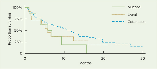
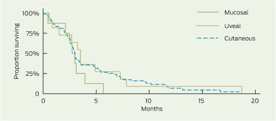
Median OS was 9.6 months (95% CI, 6.6–12.4), and median PFS was 3.0 months (95% CI, 2.7–3.4) (Box 1). The 1- and 2-year survival rates were 42% (95% CI, 32%–52%) and 18% (95% CI, 9%–30%), respectively. There were no non-melanoma-related deaths. Subgroup survival analysis is presented in Box 1. OS was similar for patients with and without brain metastasis (median OS, 9.3 and 10.6 months, respectively; log-rank test, = 0.91), and PFS following reinduction therapy was significantly greater than PFS for the population as a whole (3.6 months: 95% CI, 3.6–undefined [upper limit not yet reached for patients receiving ≥ 2 reinduction ipilimumab doses] v 3 months: 95% CI, 2.7–3.4; = 0.002). Patients with mucosal and uveal melanoma had reduced OS compared with those with cutaneous melanoma (5.8, 5.7 and 11.7 months, respectively), although this was not a statistically significant finding, likely due to small subset numbers (Box 2). PFS was similar across all groups (Box 2). Despite apparent numerical differences in OS by BRAF mutation status and incidence of serious immune-related AEs, small patient numbers resulted in wide confidence intervals and significance was not demonstrated. High ALC measured before or during ipilimumab induction was associated with an increased PFS (≤ 0.005) and numerically superior OS (P > 0.15).

Adverse events

Eighteen (17%) patients experienced a serious AE, with one patient experiencing two serious AEs (Box 1 and Box 3). Seven patients (7%) failed to complete induction due to an AE: diarrhoea/colitis (2), pruritus/rash (2), pituitary failure (1), myasthenia gravis (1) and hepatitis (1). Two patients died due to a treatment-related AE: autoimmune hypophysitis following the fourth dose of ipilimumab (1), and severe diarrhoea/colitis complicated by sepsis following the third dose of ipilimumab.

Discussion

Median OS (9.6 months) and 1-year OS (42%) exceeded proposed benchmark survival targets of 6.5 months for median OS and 25% for 1-year OS from a previous meta-analysis of second-line melanoma treatments.14 Survival outcomes were similar to those reported in the MDX010-20 study (median OS, 10.1 months)3 and in ipilimumab access schemes using a 10 mg/kg dosing regimen (median OS, 7.2–9 months).7,9 Median PFS (3.0 months) was also similar to the 2.9 months and 2.6–4.3 months found in the MDX010-20 and access scheme studies, respectively.3,7,9 With just under half of the study population (44/104) having brain metastasis at baseline, there was no difference in OS compared with patients without brain metastasis, suggesting that ipilimumab is an effective treatment option in both clinical situations. Encouragingly, PFS for patients undergoing reinduction therapy was similar to that seen following induction therapy, affording patients an efficacious subsequent treatment option and correlating with findings from recently published work extending on the MDX010-20 study.15 Competing clinical trials resulting in selection bias with respect to BRAF mutation status resulted in a study population that was not representative of the general melanoma population (BRAF-MT, 12% v 46%, respectively).16 Further, BRAF-MT patients in the study population were those likely to be ineligible for clinical trials or who had progressed following BRAF-inhibitor therapy (9/12 patients). This may be reflected in the poorer survival observed in this patient subgroup.

Although statistical significance is limited by the small number of patients with uveal (n = 11) and mucosal (n = 8) melanomas, there were some observed differences in survival data (Box 1) compared with the more prevalent cutaneous melanoma (n = 79). Trends demonstrated that future investigation of the shorter survival seen in the non-cutaneous subgroups may be warranted. The similar PFS but lower OS may reflect the different behaviour of the disease or the lack of subsequent treatment options compared with patients with cutaneous melanoma. The latter is unlikely in our patient cohort, where most patients (> 80%) with progressive disease following ipilimumab treatment were palliated (regardless of melanoma subtype) and not receiving subsequent therapy. Other explanatory factors may include the greater proportion of patients with uveal melanoma with widespread disease (M1c, 100%), and greater baseline tumour load (elevated LDH, 82%).

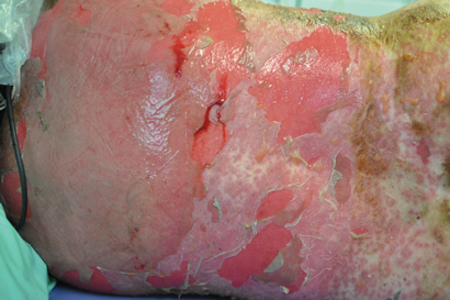
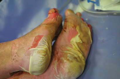
Raised ALC was highly correlated with prolonged PFS; however, although a trend was observed, the association with improved OS as shown in previous studies was not evident.7,9 There was a lower incidence of any immune-related AEs in our study (53/104, 51%) compared with the MDX010-20 study (80/131, 61%).3 However, this is likely to reflect poor documentation of low-grade AEs in the routine clinical setting compared with the highly regulated clinical trial setting with rigorous reporting requirements. When considering only serious immune-related AEs, assumed to be more accurately documented, incidence was more closely aligned: 14% (15/104) of patients in the present study compared with 15% (19/131) in the MDX010-20 study. We report similar incidence of overall skin immune-related AEs (41/104, 39% v 57/131, 44%), grade 3–4 skin immune-related AEs (3/104, 3% v 3/131, 2%) and grade 3–4 diarrhoea/colitis (8/104, 8% v 13/131, 10%). The incidence of treatment-related deaths (grade 5 AE) was comparable, with 2% (2/104) and 3% (4/131) treatment-related mortality rates reported in the current and MDX010-20 studies, respectively.

We reported the clinical details pertaining to the rare and serious ipilimumab AEs experienced by our patients, including two grade 5 events. One patient who had been successfully managed with high-dose steroids for grade 4 diarrhoea/colitis underwent surgical excision of brain metastases. The patient developed colonic microperforation and sepsis, and died, after the steroid dose was tapered postoperatively. The problem of rebound symptoms is well recognised and, consequently, guidelines recommend a prolonged steroid taper over 45–60 days.8 This case highlights the importance of proper communication to patients and between treating teams. Two patients experienced serious autoimmune hypophysitis, with one subsequent death. The death was a result of delayed recognition of symptoms with an initial working diagnosis of lithium toxicity delaying steroid administration. In the second patient, symptoms developed 1 month after induction therapy. Earlier recognition and treatment with oral steroids and hormone replacement resulted in both a symptomatic and biochemical response, and the patient has maintained a sustained melanoma response to therapy. A single case of grade 3 pituitary failure was attributed to disease progression when magnetic resonance imaging scans unexpectedly demonstrated the presence of extensive bone metastases involving the pituitary fossa. One patient experienced myasthenia gravis, a rare (< 1% incidence) immune-related AE associated with ipilimumab. The first two doses were complicated by sweats and lethargy; following the third dose, the patient presented with dysarthria, dysphasia, dysphonia and diplopia. Myasthenia gravis diagnosis was confirmed by elevated levels of antiacetylcholinesterase antibody. Ipilimumab was ceased and symptomatic improvement was achieved using steroids, pyridostigmine and intravenous immunoglobulin-γ (unpublished case report). The frequency and severity of ipilimumab-related AEs (including death), although not dissimilar to rates reported in the clinical trial setting, are notable and warrant the recommendation that ipilimumab treatment should occur only under the supervision of an experienced clinical team.

1 Overall survival (OS) and progression-free survival (PFS), by subgroup


No. of patients

Median OS, months

95% CI

P

Median PFS, months

95% CI

P


All patients

104

9.6

6.6–12.4


3.0

2.7–3.4


Melanoma subtype*

Cutaneous

79

11.7

7.1–13.8

0.11

3.0

2.7–3.4

0.72

Non-cutaneous

19

5.8

2.8–12.4


3.1

2.1–3.9


Uveal

11

5.7

1.5–16.0


3.5

0.9–7.33


Mucosal

8

5.8

1.1–nd


2.7

0.5–3.9


Sex


Male

71

9.6

5.8–13.4

0.90

3.0

2.6–3.5

0.69

Female

33

7.5

5.1–15.7


3.0

2.7–3.5


Age


≤ 65 years

57

10.9

7.1–13.6

0.23

3.1

2.7–3.9

0.83

> 65 years

47

6.7

3.9–16.3


2.7

2.4–3.4


Brain metastases

Yes

44

9.3

4.9–12.4

0.91

3.0

2.8–3.9

0.32

No

60

10.6

5.6–15.7


2.7

2.2–3.5


BRAF mutation*


Positive

12

4.9

1.0–12.4

0.11

2.9

0.4–6.6

0.53

Negative

80

11.7

7.5–16.3


3.0

2.7–3.6


Baseline LDH level > 460 U/L


Yes

56

6.6

4.2–13.6

0.27

2.9

2.4–3.4

0.31

No

43

11.7

7.3–15.7


3.1

2.7–5.7


irAE  grade 3


Yes

17

13.6

5.3–nd

0.41

2.9

2.4–5.5

0.95

No

87

8.3

5.7–12.4


3.0

2.7–3.5


ALC > 1 × 109/L*

Baseline


Yes

65

10.9

5.8–13.8

0.23

3.5

2.9–4.9

0.002

No

35

7.1

2.6–11.9


2.6

2.0–3.0


After ipilimumab dose 1

Yes

70

12.4

9.3–16.3

0.15

3.5

3.0–5.3

< 0.001

No

17

6.8

3.8–19.7


2.6

2.0–3.1


After ipilimumab dose 2

Yes

65

12.4

9.6–17.9

0.23

3.5

3.0–5.3

0.005

No

14

6.8

2.3–nd


2.6

2.0–3.3


ALC = absolute lymphocyte count. CNS = central nervous system. irAE = immune-related adverse event. LDH = lactate dehydrogenase. nd = upper limit of CI not defined (not yet reached for patients receiving ≥ 2 reinduction ipilimumab doses). * Patients with missing or unknown data excluded from analysis. † Before receiving ipilimumab. ‡ Of four planned ipilimumab doses.

2 Overall survival and progression-free survival, by melanoma subtype*

Overall survival

Progression-free survival


* Overall log-rank test for equality: overall survival, P = 0.204; progression-free survival; P = 0.397.

3 Number of patients experiencing adverse events, by grade* (n = 104)


No. of patients with adverse events


Adverse event

Total

Grade 1

Grade 2

Grade 3

Grade 4

Grade 5


Any

70

58

14

15

2

2

Non-immune-related


Any

41

34

7

4

1

Nausea

20

15

3

2

Vomiting

8

4

2

2

Headache

4

2

2

Fatigue

23

20

2

1

Abdominal pain

7

6

1

Uveal irritation

4

2

2

Infection

4

1

2

1

Immune-related


Any

53

48

7

12

2

1

Gastrointestinal

21

13

7

1

Colitis

8

2

5

1

Diarrhoea

19

12

6

1

Hepatic

3

2

1

Hepatitis

1

1

Elevated LFT results

3

2

1

Endocrine

8

3

2

2


1

Hypophysitis

2

1

1

Hypopituitarism

6

3

2

1

Skin

41

35

5

3

Pruritus

30

24

3

3

Rash

34

28

3

3

Other

4

3

1

Myasthenia gravis

1

1


LFT = liver function test.* According to the Common terminology criteria for adverse events.13 † Patients who experienced separate events of different grades and/or of different types were counted separately under each grade and/or type, but only once in the total count. ‡ Any one or more of alkaline phosphatase: reference interval (RI), 30–120 U/L; alanine aminotransferase: RI, 0–50 U/L; bilirubin: RI, 0–17 µmol/L.

Use of unlicensed black salve for cutaneous malignancy

To the Editor: The most frequently diagnosed cancers in Australia are
non-melanoma skin cancers.1 Recommended treatments include surgical excision, radiotherapy and topical chemotherapy for in-situ lesions.2 There has been an increase in sales of lotions and salves that claim to eradicate skin lesions and promote removal of skin cancers. In 2012, the Therapeutic Goods Administration issued a public warning strongly advising patients against the use of red and black salves.3 We report a recent case illustrating the complications resulting from use of these unlicensed products.

A 55-year-old man presented to the emergency department at Princess Alexandra Hospital, Brisbane, with a tissue defect on his right temple (Box). The patient had applied black salve
to a lesion, which he believed to be cancerous, over the previous 4 months.

Before the Therapeutic Goods Administration warning, topical preparations such as black and red salves were being sold online to Australians as an alternative treatment for skin cancer. These products contain sanguinarine, a benzylisoquinolone alkaloid derived from bloodroot (Sanguinaria canadensis).3 This compound is hypothesised to induce tissue necrosis by blocking the sodium–potassium pump, inhibiting nuclear factor-κB or impeding cell cycle progression.4

Zinc chloride has also been identified in salve preparations. Sanguinarine and zinc chloride may cause significant tissue necrosis of both cancerous and healthy tissue, leading to significant scarring and disfigurement.5 In the absence of a biopsy, some patients may commence alternative treatment before attaining a diagnosis of skin cancer,
and a very real risk of recurrence and metastasis remains.4 As a consequence, there may be delays in diagnosis, and it may be difficult to identify the primary site of malignancy.

It is imperative for health professionals to recognise that these unlicensed products may lead to adverse outcomes, and for consumers
to realise that alternative therapies that have been described as natural are not necessarily safe or, by any standard, risk-free.

Tissue defect on the patient’s right temple following use of black salve

Methylisothiazolinone in baby wipes: a rising star among causes of contact dermatitis

To the Editor: Methylisothia-zolinone (MI) is a preservative that has been used alone in cosmetic and personal products since the early 2000s.1 Before that time, MI was combined with methylchloroiso-thiazolinone in the widely used preservative Kathon CG (Dow Chemical Company), in a 1:3 ratio with the concentration of MI limited to 3.75 parts per million. This limit was subsequently increased to 100 parts per million, and MI is now being widely used in consumer products.

MI has been included in our baseline patch test series since 2011, following European reports of an increasing prevalence of MI allergy.1 We have subsequently seen a rapid increase in the number of patients with contact allergy to MI. Our rate
of positive reactions on MI patch tests to November 2013 was 11.3% (40 patients who had relevant reactions
of a total 353), compared with a rate
of 3.5% (15/428) in 2011 and 8.4% (38/454) in 2012. MI is now the most common cause of allergic contact dermatitis in our patient population.

Our Australian experience is reflected overseas. An increase in the frequency of allergic reactions to MI has been reported in Europe2,3 and, in the United States, MI was named the 2013 “Contact Allergen of the Year” by the American Contact Dermatitis Society, which bestows this “award” to highlight important and emerging allergens.

Among our patient population,
the most common source of MI is disposable wet wipes,4 now commonly used in nappy changing. MI is present in many popular
brands of wipes used in Australia. Interestingly, it is parents who use baby wipes on their children who
are presenting with hand dermatitis, although it is likely that allergic contact dermatitis involving the groin in children may not be diagnosed accurately.

Other common consumer sources of MI include make-up removal wipes, shampoos, conditioners, body washes, moisturisers, sunscreens and deodorants. Occupational sources include paints, cooling tower water and cutting oils.

The use of disposable wipes in nappy changing, personal care, cosmetic removal and cleaning has increased rapidly. Preservatives are required to prevent bacterial contamination in moist wipes. Medical practitioners and consumers should be aware of the potential for allergic contact dermatitis to develop to MI from wipes, in particular causing persistent hand dermatitis.

Liability in the context of misdiagnosis of melanoma in Australia

Malignant melanoma is a disease for which misdiagnosis may have very serious ramifications for both patients and clinicians. Given how uncertain and difficult the diagnosis of some melanomas can be, clinicians may well be apprehensive about their potential professional liability arising from claimed misdiagnosis or mismanagement of melanoma. A recent Supreme Court of New South Wales decision1 is one of few Australian cases to directly address this issue specifically in relation to melanoma. Coote v Dr Kelly exists in the context of recent High Court of Australia decisions relating to the common law of professional negligence in Australia. Therefore, it is important to examine the particular facts of the case, how it was decided and why, and whether the court’s decision can reasonably be reconciled with what is understood of melanoma diagnosis clinically and from evidence-based medicine as well as a subsequent appeal which resulted in an order for retrial. We emphasise the importance of early recognition of uncertainty in diagnosis and subsequent escalation, particularly where delayed diagnosis may affect survival. This article provides medical practitioners with a better understanding of the uncertainty inherent in the law regarding certain issues of causation in negligence cases, and gives some guidance on an appropriate standard of care in the diagnosis of melanoma.

Coote v Dr Kelly: the facts

In September 2009, a patient consulted his general practitioner about a lesion on the plantar surface of his foot, which was diagnosed and subsequently treated as a plantar wart. Despite repeated attempts at cryotherapy, paring and topical treatments to the lesion, it continued to enlarge and change in shape and colour over the following 18 months. During this time, the patient was seen by the same doctor and, subsequently, by two other clinicians, all of whom continued to treat the lesion as a plantar wart. By March 2011, the lesion was noted to have substantially increased in size and to have ulcerated. It was then excised and diagnosed histologically as an invasive acral lentiginous melanoma (ALM). The lesion had metastasised, and the plaintiff faced a poor prognosis. Proceedings in negligence against the initial treating GP were brought before the NSW Supreme Court. Despite some uncertainty about the initial appearance of the lesion, the court found that the GP had breached his duty of care by failing to perform a biopsy on the lesion at an earlier stage; had he done so, it may have led to an earlier diagnosis of ALM. However, all the elements for negligence were not established. There was insufficient evidence to show that the breach of duty had caused the ultimate harm that befell the patient and, specifically, that an earlier diagnosis of ALM would have prevented the metastasis and subsequent poor prognosis.

Standard of care and breach of duty

Judicial findings

The court determined that the appropriate clinical standard of care was not met. A breach of duty of care was established, which was held to constitute the GP’s failure to observe a small black mark in the lesion at the initial consultation. The court determined that this ought to have drawn the attention of a reasonably competent practitioner to the need for further investigation. However, the New South Wales Court of Appeal has ordered a retrial (pending) on the basis of both flawed reasoning leading to the original conclusion of breach of duty, and flawed reasoning that such a breach, if it occurred, could nevertheless not be proven on the balance of probabilities to have caused the patient’s loss by using evidence of population-aggregated survival statistics.2

Commentary

Certain melanomas are inherently difficult to diagnose clinically, particularly those not fulfilling the classical ABCD criteria (asymmetry, border irregularity, colour variegation, and diameter > 6 mm).3 In one study, nodular, desmoplastic and ALM subtypes were not only associated with rapid and aggressive tumour growth but were also more likely to be clinically atypical.4 That is, they were more often amelanotic, symmetrical and elevated, with a regular border. Diagnostic features of these atypical melanomas are less effectively taught, and timely and accurate diagnosis presents a major challenge, particularly in the general practice setting.

Given the difficulties inherent in diagnosis, misdiagnosis of an atypical melanoma should not necessarily be considered to be a breach of a reasonable standard of care, especially given that GPs may see very few of these lesions during their careers. Whether a misdiagnosis constitutes a breach of duty of care is determined by a court on the basis of the admissible evidence, including expert peer professional opinion. In this case, the plaintiff’s evidence that there was pigmentation of the lesion at the initial presentation was critical in determining whether a breach of duty had occurred. In the absence of comprehensive clinical notes, it was difficult for the defendant to establish that there was no pigmentation at the initial presentation. All the clinicians involved in this case agreed that if there was pigmentation of the lesion, further investigation would have been warranted, as this might have indicated a diagnosis other than that of a plantar wart. The importance placed by the court on the presence or absence of pigmentation in determining the appropriate response of a reasonably competent practitioner is interesting and emphasised in the appeal judgment.5 It was largely based on the expert opinions provided. While the presence or absence of pigmentation may be an appropriate diagnostic clue, it is only one part of the broader clinical picture. The courts may tend to lend it excessive weight. Pigmentation alone should not dispose of the question of breach of duty of care. A more reliable clue to misdiagnosis may well be failed response to treatments that have been tried.

Repeated failed non-definitive treatments were continued by multiple clinicians, which allowed a significant amount of time to pass without the patient being referred to a clinician with peer-recognised specialist qualifications in the diagnosis and management of skin disease. On the evidence available, we consider that it was not the misdiagnosis per se that amounted to a breach of duty of care, but the lack of recognition of uncertainty and a failure to appropriately refer the patient or conduct further investigations once treatment failure became apparent. Definitive biopsy or escalation of care by referral to a specialist may each have averted the breach in this case.

From a clinical perspective, this reinforces the importance of accurate and precise documentation and personal communication with other clinicians. A change in presentation or a pattern of unsuccessful treatment, and hence uncertainty in diagnosis, can thereby be identified and acted on.

Causation and prognosis

Judicial findings

The court of first instance held that the evidence was insufficient to prove that an improved prognosis was probable, rather than possible, had the patient’s ALM been diagnosed and treated at first presentation. In the absence of proven causation of damage, despite a proven breach of duty of care, the court rejected the claim in negligence.

Commentary

The relationship between delay in diagnosis and poorer prognosis in progressive neoplastic disease may seem intuitive for many clinicians. The court’s decision on this point may therefore seem surprising. Indeed, the Court of Appeal rejected it.

The Breslow thickness of a melanoma at the time of removal is a major predictor of the likelihood of metastasis and therefore of overall prognosis.5 In Australia, the 10-year survival rate is 98% for lesions less than 0.76 mm thick but only 53% for lesions more than 3 mm thick; the outcome for people with distant metastasis is extremely poor (5-year survival rate, < 5%).6

Despite this, any direct relationship between delay in diagnosis and increased melanoma thickness remains controversial.69 It is recognised that melanomas vary widely in their rate of progression, particularly according to subtype, with certain subtypes such as nodular melanoma known to have a rapid vertical growth phase. One explanation for the apparent lack of a demonstrated relationship between diagnostic delay and tumour thickness is that tumour thickness at diagnosis may be more strongly related to the growth rate and biological aggressiveness of the tumour, rather than to the measured delay in diagnosis.9 Considering melanomas together as a homogeneous group, rather than as subtypes with widespread variability in rates of growth, has also been suggested to be a possible confounding factor for any measured association between thickness and delay in diagnosis.5 It therefore becomes difficult to retrospectively draw conclusions to determine the prognostic impact of misdiagnosis.

The court referred to the recent landmark High Court case of Tabet v Gett,10 where a claim of negligence resulting only in a loss of a chance of a better medical outcome was rejected. Tabet v Gett has authoritatively settled the point that the defendant’s negligence must be proven, on the balance of probabilities, to be the cause of the poorer outcome for the patient, compared with the expected outcome had the breach of duty not occurred. A loss of a chance of a better outcome is not of itself sufficient for a claim in negligence to succeed.

In general, in Australia, for there to be factual causation, it is necessary to prove that the harm to the plaintiff would not have occurred without the defendant’s breach. In Coote v Dr Kelly, the breach by the defendant was effectively that of misdiagnosing melanoma and delaying targeted treatment. The key question therefore became whether it could be proven that a difference existed between the prognosis for the patient at the time of the first presentation and the prognosis at the time that the melanoma was eventually diagnosed, and whether this difference was caused by the actions of his GP.

Much of the evidence relied on the interpretation by expert witnesses of epidemiological studies used for determining possible prognosis. The purpose of these studies is not to establish likely prognosis or to determine retrospective prognosis in any particular case. As a consequence, on the balance of probabilities, the first-instance court determined it was not proven that metastasis had not already occurred and, at the time of his initial presentation in 2009, this particular patient may have already had a poor prognosis.

The Court of Appeal expressly rejected the proposition that epidemiological studies cannot provide evidence sufficient to prove causation on the balance of probabilities of loss in an individual case. We respectfully agree with the Court of Appeal and strongly caution the medical profession against relying on an assertion (supported at first instance) that epidemiological evidence is incapable of supporting a legal finding of causation in any individual case. It is so capable. “There is nothing in Tabet v Gett . . . standing in the way of such a conclusion”.11

Melanomas with rapid vertical growth phases and aggressive histological features may have vertical progression rates > 0.5 mm in thickness per month4 and, in these cases, it is even more likely that delay in diagnosis may lead to a significant decline in prognosis. The importance of accurate and timely diagnosis of such lesions, in order to capitalise on a potentially short window of opportunity for improved prognosis, is a clinical imperative granted additional legal force by the recent Court of Appeal findings.

Conclusion

A high index of suspicion is always necessary when considering diagnoses that require early intervention to prevent significant harm to the patient. It may be difficult to diagnose atypical presentations of melanoma. By their very nature, such melanomas will have a higher rate of misdiagnosis.

Failure of initial treatment should trigger a recognition of uncertainty, an awareness of possible serious differential diagnoses and an understanding of the potential significance of error, with consequent escalation to specialist diagnostic and clinical care. Such recognition may be evident on the first consultation, or it may take several consultations before a pattern of uncertainty emerges. Consistency in documentation and communication among colleagues is essential where continuity of care is suboptimal. Recognising uncertainty and the need for escalation when recognised is a principle that applies to all aspects of clinical and histopathological practice. Its importance in the context of melanoma diagnosis, where delay may be a critical factor in the patient’s ultimate survival, is paramount. Establishing causation as a result of delayed diagnosis is often complicated, but it is possible, and epidemiological evidence may be employed to do it. If the litigation is not settled prior to retrial, the authors propose to publish further analysis of this important case as any judgment may become available.

Are bald men more virile than their well thatched contemporaries?

The suggestion that bald men are more virile than their well-thatched contemporaries is probably an old wives’ tale, but it must be conceded that old wives are likely to be unusually authoritative in this matter.1

John Burton and colleagues were the first to examine the contentious view that an association exists between virility and baldness in 1979. Notably Burton himself was balding at this time. However, the hypothesis has never been directly tested. In Burton et al’s study of 48 men aged 35–64 years, surrogate markers of “masculinity” such as hair density on the trunk and limbs, serum testosterone levels, sebum secretion rate, sweat secretion rate, skin thickness, muscle thickness and bone thickness showed no relationship to baldness.1 Virility per se was not assessed. Factors suggestive of a possible association include the lack of balding among eunuchs2 and pseudohermaphrodites,3 indicating that testosterone and its biologically active metabolite dihydrotestosterone are prerequisites for common baldness.4 Furthermore, the principal side effect of treatment of male pattern baldness with finasteride (a compound that prevents the conversion of testosterone to dihydrotestosterone) is loss of libido and erectile dysfunction;5 and oral antiandrogen therapy is not used to treat androgenetic alopecia in men, as it has a profound inhibitory effect on the male libido, evidenced by its use to deliberately reduce sex drive in the treatment of deviant sexual behaviour in men.

Methods

In order to resolve this vexing question, we used data from a study originally designed to examine risk factors for prostate cancer.6 This study was conducted in Australia between 1994 and 1997 in men under 70 years of age. Men with prostate cancer were recruited from cancer registers, and unaffected controls were recruited from electoral registers. All subjects were interviewed in person and were categorised into four patterns of baldness (nil, receding only, vertex only and fully bald) by the interviewer (Box 1). At the end of the interview, in private, subjects completed a questionnaire that elicited not only their history of ejaculations obtained by any means between the ages of 20 and 49 years but also their number of sexual partners.

Ethics approval for the use of the data in this study was obtained from cancer registry human research ethics committees in Victoria, New South Wales and Western Australia.

Results

There were 2836 men who took part in the original study. Information on sexual function and sexual partners was available for 2205 men. We performed a secondary analysis of risk factors for baldness using unconditional logistic regression with baldness as the outcome and adjusting for age. As there were no differences in associations between baldness and virility in men with and without prostate cancer, we combined the data (Box 2).

No significant association was found between baldness (either limited to vertex balding at the crown or being fully bald) and the frequency of ejaculations between age 20 and 49 years (Box 2), but bald men were significantly less likely to have had more than four female sexual partners in their lifetime.

Discussion

For our null findings to be explained by selection bias, we would have to postulate that relatively fewer virile bald men than non-virile bald men participated. Given that virility was not mentioned in any of the study information, and that virile men are likely to be participatory, this seems unlikely. Similarly, for reporting bias to have explained the null finding, would require that the virile bald men underreported the frequency of their ejaculations and non-virile men overreported. At the time the study was conducted, the myth that bald men are more virile was widespread, so we think it more likely that all participants would err on the side of societal expectations and overreport.

In the population we studied, bald men appear to be no more virile than their well thatched contemporaries. On the contrary, they seem to have fewer conquests. Although old wives may not be as authoritative on this matter as was once thought, it may be that bald men have earned this plaudit by being more faithful to them.

1 Examples of androgenetic alopecia patterns in men*


* Categorised in this study as (l to r) “nil”, “receding only”, “vertex only” and “fully bald”.

2 Risk of baldness and men’s average frequency of ejaculations between 20 and 49 years of age and their number of female sexual partners

Baldness


Not bald*

Bald

Odds ratio (95% CI)

P


Weekly average ejaculations

n = 532

n = 1673

0.55

Up to average 2.3 times a week

113 (21%)

398 (24%)

1.00

Average 2.3–3.3 times a week

118 (22%)

330 (20%)

0.86 (0.63–1.16)

Average 3.3–4.7 times a week

110 (21%)

350 (21%)

0.99 (0.73–1.34)

Average 4.7–7 times a week

103 (19%)

341 (20%)

1.08 (0.79–1.48)

More than 7 times a week

88 (17%)

254 (15%)

0.90 (0.65–1.24)

Number of female sexual partners

n = 520

n = 1625

< 0.001

0–1

128 (25%)

549 (34%)

1.00

2–4

58 (11%)

221 (14%)

0.90 (0.63–1.28)

5–14

118 (23%)

318 (20%)

0.71 (0.53–0.95)

15–29

100 (19%)

245 (15%)

0.68 (0.50–0.92)

30+

116 (22%)

292 (18%)

0.69 (0.51–0.92)


* Recorded by the interviewer as either “nil” or “receding only”. Recorded by the interviewer as either “vertex only” or “fully bald”. P < 0.05 significant.