×

Not just a cosmetic problem: facial papules in Birt-Hogg-Dubé syndrome

Clinical record

A 52-year-old man was referred for laser therapy of facial papules for cosmetic reasons. The patient presented with a history of multiple asymptomatic facial lesions that had slowly erupted over a 10-year period. Before this consultation, the patient had been referred to a dermatologist to exclude skin cancer and biopsy of a lesion showed an angiofibroma and no evidence of malignancy. Personal history was remarkable for recurrent bilateral spontaneous pneumothoraces requiring pleurodesis. His family history was positive for recurrent pneumothorax in an adult son and recent onset of similar facial lesions in another son. There was no personal or family history of renal carcinoma.

Physical examination revealed multiple dome-shaped firm whitish papules over the forehead and cheeks (Figure 1 and Figure 2). The clinical presentation of multiple facial fibrous papules occurring on a background of unexplained pneumothoraces was suggestive of the genetic condition, Birt-Hogg-Dubé syndrome (BHD). Repeat punch biopsy of a lesion revealed an infundibulum with perifollicular fibrosis suggestive of a trichodiscoma, supportive of BHD.

The patient was referred to a cancer genetic clinic for counselling, genetic testing and screening. Renal ultrasound revealed a left renal hypoechoic lesion measuring 2.5 × 2.1 × 2.4 cm. The patient underwent robotic left partial nephrectomy, and pathology revealed a hybrid oncocytic/chromophobe tumour. Although this tumour was growing in a benign manner, it displayed low-grade renal cell carcinoma features. However, as the tumour was completely excised, no further treatment was required.

Genetic testing of blood revealed a heterozygous FLCN c.853C>T mutation on chromosome 17. Genetic counselling and radiographic renal screening of family members is planned. As per suggested guidelines, our patient will undergo lifelong medical surveillance by periodic renal ultrasound and/or magnetic resonance imaging.1

BHD is a rare, autosomal dominant, hereditary cancer syndrome due to germline mutations in the folliculin gene (FLCN). The incidence of BHD is about one in 200 000 of the population; however, it is likely underdiagnosed because of low clinical awareness.1,2 Folliculin is a protein expressed on keratinocytes in the basal and spinous layer of the epidermis, dermal fibroblasts, nerve cells, lymphocytes, macrophages and mast cells. Extracutaneously, folliculin is expressed on pulmonary type-1 alveolar epithelial cells and distal nephrons.3 Folliculin is a tumour suppressor protein, important in cell signalling pathways that regulate cell growth. It is proposed that genetic mutation results in loss of a functional protein in cells, resulting in unchecked tumour growth.4 The pattern of folliculin expression correlates with the clinical features of BHD, including skin papules of varying pathologies, basal lung cysts, spontaneous pneumothorax and renal cell carcinomas.

Proposed diagnostic criteria for BHD include the presence of one major or two minor features.2 Major features are at least five adult onset fibrofollliculomas or trichodiscomas, with histological confirmation, or a pathogenic FLCN mutation. Fibrofolliculomas and trichodiscomas present as multiple small flesh-coloured or white dome-shaped papules on the face, neck and upper trunk. Angiofibromas have also been associated with BHD; however, the presence of multiple angiofibromas is more suggestive of tuberous sclerosis.5 Minor features include a history of bilateral basal lung cysts, with or without spontaneous pneumothorax, early onset (age < 50 years) renal carcinoma with multifocal or bilateral carcinomas or a mixed chromophobe/oncocytic histology, and a first-degree relative with BHD.6

Patients with BHD have a 50-fold increased risk of primary spontaneous pneumothorax development and more than 80% of affected individuals have multiple pulmonary cysts visible on a computed tomography scan, with the less common basal rather than apical predilection.6,7 Renal cell carcinomas occur in 27% of affected individuals. A wide range of renal carcinomas have been reported, with the most common pathology being the unusual mixed oncocytic/chromophobe tumour. More than 65% of patients with BHD and renal carcinoma present with multifocal, bilateral or recurrent tumours.

Early recognition of BHD is crucial, given the high risk of renal neoplasm development which, if detected early, is curable. Thus the general practitioner and dermatologist are uniquely placed to consider the diagnosis of BHD when consulted by a patient requesting removal of multiple facial papules. Unexplained spontaneous pneumothorax and basal lung cysts should also raise suspicion of BHD. Referral for genetic testing and multidisciplinary care is essential. Routine renal radiographic screening is recommended for affected individuals > 40 years and, given the likelihood of multiple tumours, nephron-sparing surgery is preferred.8

Treatment of skin lesions associated with BHD is elective. In particular, laser ablation of individual lesions may improve the appearance for cosmetic reasons.

Lessons from practice

  • Birt-Hogg-Dubé syndrome (BHD) is an autosomal dominant condition caused by germline mutations in the folliculin gene.

  • Phenotypical features of this rare syndrome include multiple white dome-shaped facial papules and bilateral basal lung cysts, predisposing to spontaneous pneumothorax.

  • Diagnosing BHD early is important, as the genetic defect is also associated with early onset renal cell carcinomas, of varying pathology.

  • As there are variations in presentation, a combination of multiple facial papules, spontaneous pneumothorax and a personal or family history of renal cell carcinoma should trigger consideration of BHD.

Figure 1 –


Figure 1: Patient’s facial fibrofolliculomas and trichodiscomas presenting as innumerable small white papules on the forehead, cheeks and chin.

Figure 2 –


Figure 2: Close-up image of fibrofolliculomas and trichodiscomas.

Teledermatologists’ management of emergency skin conditions

Traditionally, when patients present to emergency departments or rural hospitals with a complex skin condition, the on-call dermatology registrar will be consulted by telephone. More recently, clinicians have begun using store-and-forward technology to send images directly to the specialist, significantly improving their capacity to accurately diagnose and manage patients remotely. Such teledermatology services also provide valuable teaching opportunities for rural doctors and registrars. While there are numerous benefits, including improved timely access to specialist advice for rural patients and clinicians, and reduced unnecessary investigations and outpatient referrals,1 the issues related to patient privacy and confidentiality must be addressed.

Following a successful pilot study in Queensland in 2008–2009,2 the Skin Emergency Telemedicine Service was implemented at the Princess Alexandra Hospital (PAH) in Brisbane. Despite no active promotion of the service, referrals have almost doubled from 167 cases in 20123 to 318 in 2014. The disease spectrum of referred cases in 2014 was similar to 2012, with 30% diagnosed with dermatitis, 21% with infection of the skin and 15% with drug eruption. Referral sites continue to be dispersed across the state, from Brisbane to Innisfail, 1600 km away. After the cessation of outreach services to Qld towns such as Mt Isa in 2013 and closure of the Dermatology Department at Cairns Hospital in 2014, the PAH Skin Emergency Telemedicine Service has been an essential part of ensuring timely access to a dermatologist (average response time = 4 hours) for patients with acute skin conditions across Qld.

The increased use of smartphones by clinicians to capture images for telemedicine services has raised concerns about patient privacy and consent. The study by Kunde and colleagues of dermatology registrars’ (n = 11) use of digital and smartphone technology for clinical imaging reported widespread use of the technology.4 The vast majority of clinicians had stored over 100 clinical images of patients on their personal phones, and 10 of 11 surveyed clinicians reported they had texted or emailed images to colleagues. Although patient consent was obtained verbally by 10 of the responding clinicians, few reported recording consent in the patient’s medical record (n = 2).

In 2014, the Medical Indemnity Industry Association of Australia and Australian Medical Association developed the Clinical images and the use of personal mobile devices guide for medical students and doctors.5 The guide makes recommendations for the collection, use and disclosure of clinical images as well as storage and security. Clinicians using teledermatology services must familiarise themselves with the relevant guidelines, hospital policies and privacy legislation, and ensure that images and evidence of obtained consent are documented in patients’ medical records.

The addition of store-and-forward technology to the traditional referral system has enabled clinicians to access specialist advice in an efficient and timely manner, potentially improving patient care. Strategies to improve access to specialist advice should be supported, but the relevant ethical and legal issues must also be considered.

[Clinical Picture] DOCK8 primary immunodeficiency syndrome

A 3-year-old boy presented to our dermatology department in July, 2013, with a history of moderate atopic dermatitis, food allergy to cow’s milk protein and hen’s egg, and a 4 week history of very extensive molluscum contagiosum. Initial treatment with topical cantharidin and imiquimod had been unsuccessful. The family had no history of immunodeficiency. Investigations showed lymphopenia with abnormal T-cell subsets (CD4 lymphocyte count of 0·384 × 109 cells per L [normal range 0·5–2·4 × 109 cells per L], CD3 lymphocyte count of 0·462 × 109 cells per L [0·9–4·5 × 109 cells per L], and CD8 lymphocyte count of 0·074 × 109 cells per L [0·3–1·6 × 109 cells per L]), increased IgE (2567 kIU/L [<0·35 kIU/L]), low IgM (210 mg/L [450–1400 mg/L]), and peripheral eosinophilia (1·08 × 109 cells per L [0·04–0·4 × 109 cells per L]).

Tattoo-associated mycobacterial infections: an emerging public health issue

Three men aged 21–24 years presented to our dermatology clinic with a 2-week history of pruritic erythematosquamous papules coalescing into plaques within areas of recent tattooing. The tattoos were done in Thailand 4 weeks before presentation (Box, A–E). The lesions were concentrated in areas of black shading and overlapping colours, and did not involve non-tattooed skin. All patients were afebrile, systemically well with no palpable lymphadenopathy. Investigations, including a full blood count, biochemistry and inflammatory markers, returned results within the normal range. Serological tests for HIV, hepatitis B, hepatitis C and syphilis were negative. Skin biopsies were performed on all three patients. Histopathology showed a suppurative granulomatous reaction with lymphohistiocytic infiltrate in the upper and mid dermis (Box, F–H). Modified Ziehl–Neelsen staining was negative for acid-fast bacilli. However, cultures showed Mycobacterium mucogenicum in Patients 1 and 2, and M. fortuitum in Patient 3. Empiric antibiotic therapy was commenced with oral clarithromycin 500 mg twice a day for 4 weeks. Patients 1 and 2 required 7 days of intravenous amikacin 750 mg daily and cefoxitin 2 g four times a day for failure to respond based on tissue culture and sensitivities.

The surge in interest in tattoo and body art over the past decade has also led to a surge in tattoo-related complications. While the risk of blood-borne disease and secondary bacterial infection is well known, infection with other organisms has received little publicity. However, it remains a significant public health risk,1 with outbreaks of tattoo-associated mycobacterial infection documented in the United States, France and Germany. Common causative organisms include M. chelonae, M. fortuitum and M. abscessus. Infections with Mycobacterium tuberculosis2 and M. leprae3 have also been reported. Mycobacteria are ubiquitous in the environment and species such as M. chelonae are commonly found in water supplies. However, they typically only cause clinical disease in the immunosuppressed host, or when high concentrations of the organism are introduced via surgery, trauma or tattooing. Sources of mycobacteria in tattooing include tattoo inks,4 with the chemical composition of differing pigments possibly promoting or suppressing organism growth.5 Purple inks (containing manganese) may have the potential for inhibiting organism growth, similar to the action of potassium permanganate used in dermatological practice. The dilution of inks with non-sterile water to produce gradations of colour can also introduce mycobacteria. The clinical distribution of papules in the reported cases demonstrates large numbers of papules coalescing at sites of shading and the borders between colour overlaps. This distribution coincides with areas of high puncture density to give desired colour gradations.

Mycobacterial infection is an important consideration in patients with widespread papular eruption in recent tattoos. Diagnosis can only be made on skin biopsy with tissue culture, and definitive antibiotic therapy should be directed by antimicrobial sensitivities.

Box


Erythematous scaled papules on presentation, of Patient 1 (A, B); Patient 2 (C, D); and Patient 3 (E). Representative histopathology from Patient 1 demonstrating multiple granulomas in the upper to mid dermis (F, magnification x 4), with a negative Ziehl–Neelsen stain (G, magnification x 80) and suppurative granuloma formation (H, magnification x 40).

Tinea hidden by a vemurafenib-induced phototoxic reaction in a patient with metastatic melanoma taking dexamethasone

Clinical record

A 41-year-old man with stage IV BRAF-V600E (valine replaced with glutamic acid at amino acid position 600 in the BRAF kinase) metastatic melanoma was started on vemurafenib therapy on a compassionate access program. Before this, he had been on long-term dexamethasone therapy (4 mg daily) for management of brain oedema related to multiple brain metastases. With the exception of his cutaneous melanoma, he had no past history of dermatological conditions, including tinea corporis and photosensitivity.

One week after starting vemurafenib therapy (960 mg twice daily), the patient developed a severe drug-induced photosensitivity reaction, with blistering and erosions on sun-exposed areas of skin. Despite implementing adequate sun avoidance measures and using topical corticosteroids in the acute setting, minimal improvement was seen. As a result, the daily dose of dexamethasone was increased to 8 mg daily for the next 3 weeks and then tapered back down.

Nine months later, the patient was still taking vemurafenib 960 mg twice daily and the dexamethasone dosage had been tapered to 4 mg daily. Although the vemurafenib-induced photosensitivity reaction had ameliorated, persistent blistering, erosions and erythema were noted on the dorsum of both hands (Figure, A). One month later, several new lesions developed on the left forearm, including an annular erythematous plaque with a scaly surface, scattered pustules and ill defined borders. A similar lesion on the left side of the chest was also identified. Skin scrapings were collected and sent for culture. Trichophyton rubrum was identified and the patient was diagnosed with tinea corporis. He was prescribed topical antifungal cream (clotrimazole every 8 hours) for these lesions.

After 7 weeks of clotrimazole therapy, the patient reported a slight reduction in the erythema and scaling of the forearm and chest lesions. However, multiple new pustules had developed on the hands and face in addition to several erythematous nodules over the dorsum of the hands and forearms (Figure, B) suggestive of Majocchi granuloma. A medium-sized inflammatory mass was also identified on the right cheek with multiple pustules, crusts and excoriations and a purulent discharge from follicular orifices (Figure, C), in keeping with facial kerion celsi.

Given the long-term dexamethasone use, previous diagnosis of T. rubrum infection and ongoing progression of the infection despite topical antifungal therapy, the patient was prescribed oral itraconazole, 200 mg daily, for 6 weeks. The kerion celsi was managed with multiple incisions and drainages. Reduced severity of all the skin lesions was subsequently seen over the course of the therapy.

Dermatophytic infections are usually located on the outermost layer of the epidermis.1 However, dermatophytes may affect deeper areas of the skin by invading hair follicles. When this happens, usually through a disruption of the epidermal barrier after the infection of hair follicles,1,2 an inflammatory granulomatous reaction can occur.

Dermatophytic dermal invasion causing inflammatory infiltrates of neutrophils and the development of granulomatous lesions is known as Majocchi granuloma or nodular granulomatous perifolliculitis.2 The dermatophyte that is most commonly involved is T. rubrum;1 the source of the T. rubrum is usually a precedent superficial dermatophytic infection such as the initial tinea corporis in our patient. Clinical examination often shows inflammatory follicular-centred papules, pustules or nodules on hair-bearing skin, which might evolve into larger subcutaneous nodules or abscesses.

While Majocchi granuloma is sometimes found in healthy individuals, development of the granulomatous reaction depends on the effectiveness of the immune system against the pathogen.3 Glucocorticoids affect cell-mediated immunity, impairing the function of macrophages and neutrophils, and reducing T helper 1-mediated immunity, which plays an important role in the complete resolution of fungal infections.4

BRAF inhibitors, such as vemurafenib, are novel drugs that target an important mutation that is present in about 50% of metastatic melanomas. They have secondary effects on skin, such as development of multiple cutaneous squamous cell carcinomas, verrucal keratoses and a variable degree of photosensitivity.58 Since no vemurafenib-related immunosuppressive effects have been reported to date for vemurafenib,9,10 it is unlikely to have played a role in the development of the dermatophytic infection, apart from the severe drug-induced photosensitivity. The immunosuppressive glucocorticoid therapy for the brain metastases and vemurafenib-induced photosensitivity effectively placed the patient at risk of progressive infection from a superficial tinea corporis to a more invasive infection such as Majocchi granuloma. Given the well documented association of vemurafenib and photosensitivity reactions, the diagnosis was initially confounded and further aggravated by the treatment of the ultraviolet A photosensitivity. The introduction of topical antifungals was too late to stop progression of the disease, and the patient eventually developed Majocchi granuloma and a facial kerion for which oral antifungal therapy was needed.

In our patient, multiple cutaneous pathological processes were present concurrently. While the cutaneous secondary effects of vemurafenib are well reported, it is important to consider alternative diagnoses when there is minimal response to therapy. Immunosuppression from corticosteroid therapy (oral or topical) should be taken into consideration in patients taking these medications while also on antineoplastic therapies. Prompt recognition, diagnosis and treatment in this setting could avoid development of more serious complications.

Lessons from practice

  • While vemurafenib induces ultraviolet A photosensitivity, it is important to consider alternative diagnoses when minimal response to therapy is attained.
  • Concurrent administration of topical or systemic corticosteroids can alter the presentation of vemurafenib-induced adverse effects.
  • Trichophyton rubrum can penetrate hair follicles to cause dermal infections that do not respond to topical therapy.

Figure

Acute HIV infection presenting as erythema multiforme in a 45-year-old heterosexual man

Clinical record

A 45-year-old heterosexual man of European descent presented to our hospital with a 3-day history of fever, myalgia, headache and a macular papular rash. The rash originated on his left shoulder and anterior chest wall before extending to chest, back and abdomen. The rash also involved the palmar aspect of his hands and the plantar aspect of his feet.

He had a background history of dyslipidaemia, excessive alcohol consumption, gastro-oesophageal reflux disease, recurrent pancreatitis and gout. He had been taking statin medications, esomeprazole, allopurinol and creon for several years. There had been no new complementary medicines.

On arrival in hospital, his heart rate was 80 beats/min; respiratory rate, 16 breaths/min; blood pressure, 140/88 mmHg; and his temperature was 37.6°C. Cardiovascular, respiratory and abdominal examinations were otherwise unremarkable. Inguinal lymphadenopathy was noted on examination. A skin examination revealed an extensive macular papular rash affecting his face, trunk and limbs, with an erosion noted on his soft palate.

Investigations revealed low levels of haemoglobin (124 g/L; reference interval [RI], 135–175 g/L), white blood cells (2.4 × 109/L; RI, 4.0–11.0 × 109/L) and lymphocytes (0.62 × 109/L (1.50–3.50 × 109/L). His platelet count was normal (192 × 109/L; RI, 150–450 × 109/L), and his C-reactive protein level was slightly elevated (8.1 mg/L; RI, < 8.0 mg/L).

The patient reported having unprotected sex with one new female partner in the previous 3 months. On advice from the hospital’s infectious diseases team, molecular testing for measles and serological testing for syphilis and HIV were performed. The patient was discharged home after 24 hours of observation with investigations pending. The day after discharge, an HIV enzyme immunoassay (EIA) screen was reactive but western blot was negative. The patient was recalled for further HIV testing, which revealed an HIV viral load of 1 060 000 copies/mL.

The patient was reviewed in the hospital’s infectious diseases clinic 4 days later and was found to have a persisting generalised pruritic papular rash of urticarial appearance involving his trunk and proximal limbs, including his elbows and knees (Figure 1). Target lesions typical of erythema multiforme (EM) were noted on the plantar aspects of his feet, appearing red and blue centrally, with tense oedema surrounding the pale area and a well defined erythematous peripheral margin (Figure 2). A dermatologist’s opinion was sought, who agreed on a diagnosis of EM secondary to acute HIV infection.

The patient’s rash was treated with topical steroids and oral antihistamines and abated within 1 week. Follow-up serological testing revealed a rising HIV EIA titre and positive western blot.


Figure 1. Papular rash on the patient’s trunk and arm. Figure 2. Target lesions on the plantar aspects of the feet.

An acute, self-limiting hypersensitivity mucocutaneous reaction pattern, EM is frequently associated with viral infections, most often herpes simplex, and Mycoplasma pneumoniae infection. EM can also arise due to drug reactions, most commonly non-steroidal anti-inflammatory drugs, penicillins, sulfonamides, phenothiazines and anticonvulsants.1

The incidence of EM is unclear, but it is thought to affect less than 1% of the population, with a slight predominance among young women.1 EM is characterised by targetoid lesions distributed in peripheral acral regions, and macular, papular and urticarial patterns peripherally and on the extensor surfaces, and, less frequently, diffusely on the trunk. Oral and genital lesions are generally present. Typical target lesions are most frequently seen in acral locations, especially the dorsal and palmar surfaces of the hands and the dorsal and plantar surfaces of the feet.

Acute HIV infection generally occurs within 4–10 weeks from the time of HIV exposure, with 95% of patients seroconverting within 6 months.24 Acute HIV infection is associated with a maculopapular rash in up to 80% of cases, which presents 48–72 hours after the onset of fever and typically affects the upper trunk and neck. Acute HIV is also commonly associated with headache, lymphadenopathy and myalgia.5,6 Vesicular, pustular exanthematous and enanthematous patterns have also been described.7 However, EM in acute HIV infection is considered extremely rare and we are aware of only three reports.5,8,9

The pathogenesis of EM has largely been derived from studies of herpes simplex virus. The mechanism for mucocutaneous lesions is thought to begin with the release of viral DNA into the blood. DNA fragments are phagocytosed by mononuclear CD34+ cells, then transferred to keratinocytes. Expression of herpes simplex virus genes in the epidermis leads a herpes-specific CD4+ Th1 cell-mediated immune response directed against viral antigens and subsequent epidermal damage.10

If oral or genital erosions are noted, serological testing for syphilis should be performed after taking a detailed sexual history. Clinicians should also consider screening for secondary syphilis among patients presenting with a new rash of unknown cause.

Treatment of EM differs depending on the underlying cause and the severity. If drug aetiology (prescribed or non-prescribed) is considered, the suspected medications should be ceased. In cases where an underlying infection is suspected to be the cause, the pathogen should be identified and treated appropriately. A combination of topical steroids and oral antihistamines can provide symptomatic relief for cutaneous EM. Mucosal involvement may require oral anaesthetic and antiseptic solutions with topical corticosteroids. Ocular involvement should be managed in conjunction with an ophthalmologist.

This case highlights the importance of thorough sexual history-taking and clinical examination. Clinicians should suspect acute HIV infection in any patients who present with a new rash and viral prodrome. EM presenting without the typical new drug introduction, or the more frequently associated herpes simplex or M. pneumoniae infections, mandates us to ensure our clinical review includes a detailed sexual history-taking. When a new diagnosis of HIV is detected, the clinician should consider consulting an infectious diseases specialist for ongoing care.

Lessons from practice

  • HIV should be considered in all patients presenting with a new rash.
  • Acute HIV infection can present as febrile illness associated with any rash, including erythema multiforme.
  • A detailed sexual history should be obtained when a patient presents with a new generalised rash.

From dismal prognosis to rising star: melanoma leads the way with new generation cancer therapies

Attention has now turned to combining new therapies to maximise their value

For decades, the 1-year overall survival for patients with advanced-stage or metastatic melanoma (stage IIIC unresectable or stage IV melanoma) was 30%–35%.1 It is now greater than 70%–80% in clinical trials of targeted and immune drug therapies (Box).210

This is good news for a cancer that continues to rise in incidence around the world and disproportionately affects the young. In Australia, melanoma is the fourth most common cancer and accounts for 10.1% of all new cases and 3.2% of cancer deaths.11 It is also the most common cancer in men aged 25 to 49 years, and women aged 15 to 24 years in New South Wales.12

Although incidence continues to rise in both sexes, mortality and 5-year survival remain stable,11 and the benefits observed with drug therapies in advanced-stage disease are yet to be reflected in population statistics. Most melanomas are cured with surgical excision: efforts are now focused on trialling novel active targeted and immune drug therapies in the 10%–15% of patients with early-stage melanoma that is predicted to recur and cause death.

Drug therapies

Melanoma is the poster child for immune and targeted approaches to cancer drug treatment. It is one of the few cancers where no chemotherapeutic agent has shown a survival benefit over best supportive care or single agent dacarbazine chemotherapy in patients with distant spread. Immunotherapies called checkpoint inhibitors and therapies targeting the mutated BRAF protein in the melanoma have put melanoma centrestage in the battle against cancer.

Immune therapy

Checkpoint inhibitors are a class of immune drugs that activate T cells for tumour-cell killing. Checkpoints are brakes on T cells to protect us from autoimmunity or an overzealous immune response to antigens, but can induce tolerance to cancer if activated. Ipilimumab is a fully human IgG1 monoclonal antibody that blocks the checkpoint called cytotoxic T lymphocyte-associated antigen 4 (CTLA4) and was the first drug to show a survival advantage in advanced-stage melanoma compared with a vaccine or chemotherapy.7,8

The 1-year overall survival for patients treated with ipilimumab was 46% and 47% in each trial, respectively, although the response rates were only 11% and 15% (ie, the proportion of patients with at least a 30% reduction in tumour burden). Five-year survival in the latter study was 18.2% with ipilimumab, compared with 8.8% with chemotherapy.13

And yet, the relatively non-specific activation of the T cell induces autoimmune toxicities in 60% of patients. Although there have been reports of autoimmunity in almost every organ, the most frequent toxicities are dermatitis, inflammatory diarrhoea, endocrinopathies and hepatitis. It is critical that these autoimmune toxicities are identified and managed early to prevent complications.14

Biomarkers in the melanoma or host that enable us to select patients who are more likely to respond have been elusive, although recent research suggests specific neoantigens produced by the melanoma may be predictive.15 Predictive biomarkers would be particularly useful, not only to prevent toxicities in those who will not benefit, but also because responses may not be typical or may occur months after initiation of treatment.16

More recently, inhibitors of the programmed death 1 (PD1) checkpoint on T cells have shown high response rates (30%–40% in Phase I studies) and long survival durations with few autoimmune toxicities.9,17 Nivolumab and pembrolizumab are fully human or humanised IgG4 monoclonal antibodies, respectively, that inhibit the PD1 checkpoint. In the recently reported Phase III study of first-line nivolumab versus dacarbazine chemotherapy, the response rate was 40% and the 1-year overall survival was an impressive 73%.5 Autoimmune toxicities with both pembrolizumab and nivolumab occur in lower frequency and severity than with ipilimumab; most are easily managed, and resolve.

To increase the proportion of patients who benefit from immunotherapies, attention has turned to combining CTLA4 and PD1 checkpoint inhibitors, as each drug acts at a different time point during T-cell activation. In a Phase I study of such a combination, the response rate was over 50% at the recommended phase 3 dose, and the 1-year and 2-year survival rates across all dosing cohorts were estimated to be 85% and 79%, respectively;18 however, 53% of patients experienced at least one grade 3 or 4 autoimmune toxicity.3 Results of Phase III studies of ipilimumab versus nivolumab or pembrolizumab or ipilimumab plus nivolumab will be available in 2015. There are many other checkpoints on the T cell, and drugs targeting these additional points and other aspects of the immune system are in preclinical and clinical development.

Targeted therapy

In contrast, BRAF inhibitors are oral drugs that target the mutated BRAF protein in the melanoma cell. Mutated BRAF occurs in 40% of melanomas, and causes aberrant overactivation of a cell proliferation and survival pathway (the mitogen-activated protein kinase [MAPK] pathway). Treatment with BRAF inhibitors (dabrafenib or vemurafenib) results in rapid and deep shrinkage of metastases, including of brain metastases, in over 50% of patients, and tumour reduction of any amount occurs in over 85% of patients treated with these drugs.2,10

These drugs show a clear survival advantage over chemotherapy, with 1-year and 2-year overall survival rates of 70% and 46%, respectively.6 Adding a MEK inhibitor to a BRAF inhibitor backbone (MEK is the protein below BRAF in the MAPK pathway) results in further improvements in tumour shrinkage, progression-free survival4,19,20 and overall survival,21 and a reduction in the hyperproliferative cutaneous toxicities associated with BRAF inhibitor monotherapy.

How do we maximise response?

Primary resistance to immunotherapy is common; however, in those who respond, the response appears to be durable.9 In contrast, nearly all patients respond initially to targeted therapies, but more than 50% develop acquired resistance within 7 months of treatment with BRAF inhibitor monotherapy,22 and within 11 months of treatment with combined BRAF and MEK inhibitors.23 Compelling data show a brisk infiltration of clonal T cells into melanoma metastases early during treatment with MAPK targeted therapies,24 suggesting that this may be an opportune time to add immunotherapies to enhance the durability of response.

Translational research findings suggest that a cure may be possible for some patients with advanced-stage melanoma, and it may be realised by combining immune and targeted therapies. The pressing matters that now face us are how to sequence and combine these therapies to maximise patient outcomes, and how we recruit to clinical trials that seek to answer these questions as the single agents become available on the Australian Pharmaceutical Benefits Scheme for widespread use. Perhaps even more importantly, we may be able to prevent advanced-stage melanoma by using these therapies in early-stage disease, and trials are underway, although the question remains — how will the Australian community afford these drugs?25

Landmark overall survival rates for patients with advanced-stage melanoma (stage IIIC unresectable or stage IV melanoma) treated with immune or targeted therapies in Phase III clinical trials210


CTLA4 = cytotoxic T lymphocyte-associated antigen 4. BRAF/MEK = proteins in the mitogen-activated protein kinase pathway. When BRAF is mutated, it overactivates the pathway, causing uncontrolled cellular proliferation and survival. PD1 = programmed death 1 checkpoint.

Sentinel lymph node biopsy for melanoma: an important risk-stratification tool

In reply: We thank Zagarella and colleagues for their comments and the opportunity to correct some misconceptions about the Multicenter Selective Lymphadenectomy Trial-1 study.1

It is unclear why they have taken exception to the reporting of disease-specific survival as this is a more robust end point than overall survival and, in this patient cohort (median age, 52 years), the two outcomes are likely equivalent.

The subgroup analysis of patients with lymph node involvement was preplanned, and the novel and robust statistical method (latent subgroup analysis) showed improved outcomes with early intervention for node-positive patients. This finding is intuitive, and Zagarella and colleagues provide no evidence as to why they consider this “unreliable”.

Sentinel lymph node biopsy (SLNB) has repeatedly been shown to be the most significant independent predictor of survival in clinically lymph node-negative patients, including by Mitra et al,2 also cited by Zagarella and colleagues. The use of ultrasound or other clinicopathological algorithms have not been reproducible and cannot be used a surrogate.3

Until a better test is developed, SLNB remains the gold standard staging test for clinically lymph node-negative patients with intermediate thickness melanoma.

Sentinel lymph node biopsy for melanoma: an important risk-stratification tool

To the Editor: The article by Gyorki and Henderson1 is misleading and inaccurate. The stated primary outcome of the Multicenter Selective Lymphadenectomy Trial-1 (MSLT-1) was:

To determine whether wide excision of the primary with intraoperative lymphatic mapping (LM) followed by selective lymphadenectomy will effectively prolong overall survival compared to wide excision of the primary melanoma alone.2

It was prolongation of overall survival, and not disease-specific survival as the authors state.1

The MSLT-1 final report3 is an example of selective reporting bias, and failed to prove its primary outcome. The conclusions based on the post-hoc analysis are unreliable, and should never be used to guide management decisions, yet the MSLT-1 trial reporting focused almost exclusively on these results.

The other claimed benefit by the authors of MSLT-1, disease-free survival, is an illusion and has been rejected on several grounds, lead-time bias being a chief one.4

Lymph node status may not be the strongest predictor of survival for patients with intermediate thickness melanoma. Indeed, combining clinicopathological features (thickness, mitotic count, ulceration, vessel invasion, site, age and sex) better predicts relapse and survival in melanoma than sentinel lymph node biopsy (SLNB) status alone.5

We thus question the prerequisite for SLNB to be performed for patients to enter drug trials.

High-resolution ultrasound can already detect lymph node deposits of 2 mm, and this technique will improve. We reject any assertion that SLNB should be the “standard of care”, as any benefits are unproven.

In conclusion, subjecting patients to a surgical procedure, with the risk of morbidity, for no clearly demonstrated benefit is questionable.

Comparing non-sterile to sterile gloves for minor surgery: a prospective randomised controlled non-inferiority trial

Minor surgery is an important aspect of general practice. This is particularly the case in Australia, where the incidence of skin cancer is reported to be the highest in the world,1 and where general practitioners perform most surgical excisions for skin cancer.2

When the use of gloves for surgery was first implemented by William Stewart Halsted in 1890, it was in an attempt to protect his surgical scrub nurse from dermatitis as a result of contact with mercuric chloride — which was used for sterilisation processes — rather than to prevent infection.3 Nowadays, several guidelines exist in Australia and internationally, which recommend that GPs use sterile gloves for small procedures such as minor surgery in general practice.46 However, these guidelines are based on expert opinion rather than on medical evidence.

Before our study, about half of the participating GPs used non-sterile clean boxed gloves when conducting minor skin excisions in general practice, while the other half used sterile gloves. A comprehensive Medline search found few studies relating to the use of sterile versus non-sterile gloves (Appendix). Randomised trials looking at lacerations in an emergency department,7 wisdom tooth extraction in an outpatient setting8 and Mohs micrographic surgery9 all showed no significant difference between infection rates. However, these studies looked for superiority of the sterile gloves rather than non-inferiority of the non-sterile gloves, resulting in negative trials, and the latter two studies were statistically underpowered. An observational study in a private dermatology setting showed no difference in infection rate for minor procedures; however, sterile gloves were shown to result in a significantly lower infection rate than non-sterile gloves for a subgroup of more complicated reconstructive procedures, which comprised flaps and skin grafts.10 Another observational study of Mohs surgery showed no statistical difference in infection rates.11 The only study conducted in a general practice setting was an audit of 126 patients where non-sterile gloves had been used for minor surgery, which showed an infection rate of 2.4%.12

Prior studies of wound infection after minor surgery involving GPs in Mackay, Queensland, showed overall incidences of wound infection of 8.6% and 8.9%.1316 This incidence was higher than expected based on published results of a similar Australian general practice cohort (1.9%),17 a skin cancer clinic cohort (1.5%)18 and a European dermatology clinic cohort (2%).19 A suggested acceptable rate of infection after clean minor surgery is less than 5%.20 The reason for our high infection rate is unclear, but may be related to the hot, humid environment, or to patient behaviour in our rural setting. A low risk of infection after clean surgery means that studies of more than 1000 procedures (sometimes many more) are required, under normal circumstances, to detect a clinically relevant difference in infection from an intervention with statistical confidence.21 Because of the high incidence of infection in our patient cohort, and the high minor surgery workload,22 we decided to use this capacity to investigate the effect of gloves on infection rates. Our trial sought to establish whether non-sterile clean boxed gloves were non-inferior to sterile gloves with regard to surgical site infection after minor skin excisions.

Methods

Study design

We carried out a randomised controlled single-centre trial with patients presenting for minor skin excisions. The study was approved by the James Cook University Human Research Ethics Committee (approval number H4572). The trial was registered in the Australian New Zealand Clinical Trials Registry ACTRN12612000698875.

Setting and participants

The study was conducted in a single private general practice in Mackay, Queensland between 30 June 2012 and 28 March 2013. Six doctors recruited between one and 100 patients. The GPs and practice were purposively selected as they had previously successfully participated in wound management projects.12,15 Consecutive patients presenting for minor skin excisions were invited to take part in the trial. Practice nurses were responsible for recruiting patients and collecting data. Demographic information was collected from all patients, as well as clinical information about diabetes or any other important pre-existing medical conditions. A body site map was used to define excision site. At the end of the study, practice nurses were asked to re-examine computer records in order to fill in any missing data. Two of us (C H and S S) visited participating GPs and practice nurses to provide training and ensure that recording was standardised.

Eligibility criteria

All patients presenting to a participating GP for “minor skin excision” from any body site were eligible to participate in the study. Two-layer procedures were recorded and included. Patients who were already taking oral antibiotics or immunosuppressive drugs were excluded from the study. Other exclusion criteria were skin flaps, excision of a sebaceous cyst and history of allergy to latex.

Surgical wound management protocol

We conducted a workshop for participating GPs to develop guidelines that would ensure that excisions were managed in a standardised manner. The following excision protocol was agreed on:

  • skin preparation with chlorhexidine solution;
  • usual sterile technique (standard precautions);
  • World Health Organization Hand Hygiene Technique with Soap and Water;23
  • local anaesthesia — subcutaneous injection of excision with 1% lignocaine;
  • excision closure with nylon sutures using simple interrupted sutures;
  • dressing application — application of non-woven polyester fabric with acrylic adhesive and non-woven absorptive pads;
  • no application of antibiotics, either topical or oral. No topical antiseptics such as betadine or alcohol. No antiseptic washes or medicated soaps;
  • patient wound advice — provision of written and verbal advice about wound care and time of return for suture removal; and
  • removal of sutures according to body site: head and neck, 7–10 days; torso, 12–14 days; upper limb, 14 days; lower limbs, 12–16 days.

Recruitment, randomisation and blinding

All patients provided written informed consent before enrolling in the study. After agreeing to participate, patients were randomly allocated to the intervention or control groups using computer-generated random numbers. Allocation information was placed in opaque sealed envelopes. The practice nurse enrolled patients and assigned participants to their groups. Patients were not blinded to their group allocation. The assessing practice nurses and doctors were blinded to the allocation of intervention and control groups. All participating patients received written instructions on postoperative wound care. Both groups were asked to take their dressing off after 24 hours and avoid using antiseptics.

Clinical outcomes

Incidence of wound infection was our primary outcome measure, and incidence of other adverse effects was our secondary outcome measure. Wounds were assessed for infection by the practice nurse or the GP on the agreed day of removal of sutures or sooner if the patient re-presented with a perceived infection. Our definition of wound infection was adapted from standardised surveillance criteria for defining superficial surgical site infections developed by the United States Centers for Disease Control and Prevention’s National Nosocomial Infection Surveillance System (Box 1).24 All participating doctors and nurses were briefed regarding the definition of infection and were also given written information. Practice nurses were asked to swab any discharging infections to investigate any pattern of antimicrobial resistance.

Sample size

Sample size was calculated on the basis of our previous study, which showed an infection rate of 8.6%.14 Based on a projected infection rate of 8%, we decided that an absolute increase in incidence of infection of 7% would be clinically significant. Thus differences in infection rates between non-sterile and sterile gloves of up to 7% were considered clinically unimportant, and based on our anticipated infection rate of 8% for sterile gloves, an infection rate of up to 15% for non-sterile gloves was considered non-inferior. This margin was decided by the investigating GPs, based on what they felt would be relevant to their clinical practice, and this margin was prespecified. To detect this non-inferiority margin of 7% with a power in excess of 80%, and a two-sided 95% confidence interval, a total of 186 patients were required in the intervention group and 186 patients in the control group. Based on our previous results in a similar setting, the design effect of investigating GPs, who were the primary sampling unit and were considered to form “clusters”, was estimated to be 1.21, and the required sample size was adjusted to at least 225 patients per group.16

Statistical analysis

All analyses were based on the intention-to-treat principle. Per-protocol analyses were conducted to cross-validate the intention-to-treat results.25,26 Depending on the distribution, numerical data were described as mean, SD; or median, interquartile range (IQR). Percentages were presented with 95% confidence intervals. A two-sided 95% CI for the difference in infection rate was used to assess non-inferiority. In addition, a per-protocol analysis was conducted, which excluded patients with protocol violations. Further, a sensitivity analysis was performed, including patients lost to follow-up: once as treatment successes (no wound infection) and once as treatment failures (with wound infection). Results were adjusted for their cluster effects. P less than 0.05 were considered statistically significant. Data were analysed using IBM SPSS version 21, Stata version 12.1 (StataCorp) and Power Analysis and Sample Size Software (NCSS).

Results

Practice and study characteristics

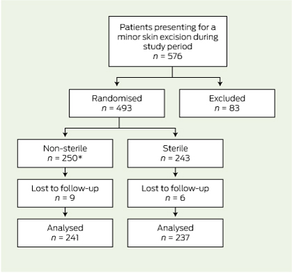
Of the 576 patients who attended for skin excisions during the collection period, 83 were excluded (Box 2).

Of the remaining 493 patients, 250 were randomly assigned to the intervention group (non-sterile gloves) and 243 to the normal treatment control group (sterile gloves). Fifteen patients were eventually lost to follow-up because they had their sutures removed elsewhere (13 patients) or they were not assessed for infection at the time of removal of sutures (two patients). There was one protocol violation where a patient in the intervention group was given an antibiotic for another infection in the follow-up period. This patient did not have a wound infection and was analysed in the intervention group on an intention-to-treat basis. Follow-up was completed in 478 (97.0%) of randomised patients (Box 3).

Comparisons at baseline

There were no large differences at baseline between the intervention and control groups (Box 4).

Incidence of infection

Infection occurred in 43 of the 478 excisions (9.0%). The incidence of infection in the non-sterile gloves group (8.7%; 95% CI, 4.9%–12.6%) was significantly non-inferior compared with the incidence in the control group (9.3%; 95% CI, 7.4%–11.1%). The two-sided 95% CI for the difference in infection rate (− 0.6%) was − 4.0% to 2.9%, and did not reach the predetermined margin of 7%, which was required for non-inferiority.

A further sensitivity analysis was performed on the 15 patients lost to follow-up. If all of these patients were assumed to have an infection, or if all patients were assumed not to have an infection, the results were still significantly non-inferior (Box 5). There were no adverse events.

Discussion

The results of our study suggest that the use of non-sterile clean boxed gloves was not inferior to that of sterile gloves in relation to the incidence of infection. This was both clinically and statistically significant, as the difference in the incidence of infection did not reach our predetermined margin of 7%, considered significant for non-inferiority. The upper limit of our 95% CI was 2.9%, which was well below our predetermined non-inferiority margin of 7.0%.

Comparison with other studies

Our study produced a similar outcome to existing studies.79 This was an adequately powered, positive randomised controlled trial that tested for non-inferiority of the non-sterile gloves rather than for a significant difference in infection rates. We believe this was the first study of its type to be conducted in a general practice setting.

Limitations of study

Our study did have some limitations. Various characteristics influence infections, and although information on as many variables as possible was recorded, it proved difficult to ensure that baseline data were comparable. For example, there were inadequate data recorded on suture size and patient occupation, and consequently, these factors could not be compared. In addition, the prevalence of diabetes and other medically important conditions was probably underrecorded, and power to analyse these subgroups was limited. Surgical training and technique of the GPs involved is a potential confounder that would be difficult to quantify and was not recorded; however, the procedures performed by individual GPs were equally balanced in the baseline data. Our predetermined margin of 7% for non-inferiority may be considered high, and some clinicians may consider a smaller margin to be clinically meaningful. Although our actual difference in infection was − 0.6%, a larger sample size would be required for the study to be adequately powered to detect smaller differences in infection rate.

Although the diagnosis of infection followed guidelines, it is still subjective and there may be inter- and intraobserver variation.27 The definition we used is the most widely implemented standard definition of wound infection.24,27 We have no evidence to support intra- and interpractice reproducibility of measurement and recording procedures.27

Our sterile gloves were powdered, while our non-sterile gloves were non-powdered. However, we have no reason to believe that powder would affect infection rates.

Generalisability

There are some limits to generalising these findings. The population of Mackay is slightly older and has a lower median household income than the general Australian population.28 Mackay is a provincial town in tropical north Queensland. The climate is hot and humid, with the mean daily maximum temperature ranging between 24.2°C and 30°C during the summer months, and a relative humidity of 75% to 79%.29 We have already discussed that our incidence of wound infection is high compared with similar cohorts of patients in temperate climates; however, we have no reason to believe that the effect of sterile gloves would be less non-inferior, that is, any worse, in similar cohorts of patients with lower incidence of surgical site infection.

We did not include skin flaps in our trial, and previous evidence has shown sterile gloves to be superior for more reconstructive dermatological procedures;10 therefore, we do not recommend extrapolating our findings to more complicated procedures such as skin flaps. However, the findings could be extrapolated to less complicated procedures in primary care, such as contraceptive implant insertion and minor procedures involving class 2 wounds such as suturing of lacerations.

Choice of gloves

There are other considerations that might affect doctors’ choice of gloves. Sterile gloves come in several different sizes, while non-sterile gloves are generally only available in small, medium and large. Latex and powder allergy, as well as preference for and availability of powdered or non-powdered gloves, may also affect choice. A recent study showed high bacterial counts on boxed gloves left open for longer than 3 days,30 although the clinical significance of these bacterial counts is unclear. Another study showed no bacterial growth on clean examination gloves after opening a new box.31

Cost saving

There is some cost benefit in the use of non-sterile versus sterile gloves, with about $1 saved per pair of gloves used. We calculated that a single pair of non-sterile gloves costs $0.153 compared with $1.203 for sterile gloves, saving $1.050 per pair of gloves used for each procedure. The cost saving benefit of using non-sterile gloves — without increasing infection rates — may be of particular relevance to developing countries with limited health care resources.

1 Definition of surgical site infection (SSI)

  • infection must be within 30 days of excision;
  • the infection involves ONLY skin or subcutaneous tissue of the incision, AND at least one of the following:
    • purulent discharge;
    • pain or tenderness;
    • localised swelling;
    • redness or heat at site;
    • diagnosis of SSI by general practitioner; and
  • stitch abscess must not be counted as an infection.

2 Reasons for exclusion from study

Reasons for exclusion from study

Patients (n = 83)


Patient declined to participate

38

Patient was taking oral antibiotics

23

Excision of sebaceous cyst

15

Shave biopsy conducted

3

Patient did not plan to return for removal of sutures

2

No sutures required

1

Flap required

1

3 Flowchart of enrolment, randomisation and follow-up of patients


* There was one protocol violation where a patient in the intervention group was given an antibiotic for another infection in the follow-up period. This patient did not have a wound infection and was analysed on an intention-to-treat basis.

4 Baseline comparison of intervention group (non-sterile gloves) and control group (sterile gloves)

Patient characteristics

Intervention group (non-sterile gloves) (n = 241)

Control group (sterile gloves) (n = 237)


Mean age (SD), years

64.9 (15.8)

65.7 (15.3)

Male

58.9%

60.30%

Smoking status


Never smoked

57.7%

52.7%

Ex-smoker

30.7%

35.9%

Current smoker

11.6%

11.4%

Diabetes mellitus

10.0%

12.7%

Other medical conditions*

38.1%

35.9%

Medications


Warfarin

4.1%

5.1%

Clopidogrel or aspirin

28.6%

27.0%

Steroids, oral or inhaled

6.3%

8.1%

Lesion characteristics


Body site


Neck and face

35.3%

31.2%

Upper extremities

26.9%

30.4%

Trunk

19.1%

19.8%

Lower limb above knee

4.6%

1.6%

Lower limb below knee

14.5%

16.9%

Histology


Naevus or seborrhoeic keratosis

15.3%

13.0%

Skin cancer and precursor

66.4%

70.5%

Other

18.3%

16.5%

Skin integrity


Normal

75.9%

74.7%

Ulcerated

19.1%

19.0%

Procedure characteristics


Mean length of excision (SD), mm

20.0 (14.0–27.0)

20.0 (13.5–27.0)

Median number of days until removal of sutures (IQR)

8 (7–10)

9 (7–10)

Two-level procedure

0

0.8%


IQR = interquartile range. * Medical conditions recorded were: chronic obstructive pulmonary disease (n = 18; 3.8%), hypertension (n = 119; 24.9%), ischaemic heart disease (n = 38; 7.9%), peripheral vascular disease (0) and current cancer (n = 7; 1.5%). † Skin cancers were: melanoma, squamous cell carcinoma and basal cell carcinoma. Precursors were: solar keratosis and intra-epithelial carcinoma. ‡ “Other” included: re-excisions of melanoma and basal cell carcinoma, sebaceous cyst, epidermal cyst, wart and dermatitis.

5 Comparisons as intention-to-treat and per-protocol and sensitivity analyses*

Analysis

Intervention group

Control group

Difference
(95% CI)


Intention-to-treat

21/241 (8.7%)

22/237 (9.3%)

− 0.6%
(− 4.0% to 2.9%)

Per-protocol

21/240 (8.8%)

22/237 (9.3%)

− 0.5%
(− 4.0% to 2.9%)

Sensitivity analysis: lost to follow-up; assumed without infection

21/250 (8.4%)

22/243 (9.1%)

− 0.7%
(− 4.0% to 2.7%)

Sensitivity analysis: lost to follow-up; assumed with infection

30/250 (12.0%)

28/243 (11.5%)

0.5%
(− 3.7% to 4.6%)


* Differences between control and intervention groups are presented with two-sided 95% confidence intervals. Results were adjusted for the clustering effects of treating doctors.