×

Prolonged tumour growth after treatment of infantile haemangioma with propranolol

Infantile haemangioma is the most common tumour of infancy, sometimes requiring treatment because of the risk of functional impairment or permanent scarring. Growth usually ceases by 12 months of age, and the tumour gradually regresses. In 2008, a review of all patients previously seen in seven international treatment centres identified only five children in whom any haemangioma growth was seen beyond 2 years of age, and none beyond 4 years.1

Since 2008, the treatment of choice for haemangiomas has been oral propranolol for several months, for up to 18 months with segmental lesions. When commenced early, propranolol usually leads to rapid cessation of growth and tumour involution that is much more rapid than without treatment.2 We have previously reported our experience with outpatient propranolol treatment of 200 infants.3 We now report a subset of 20 children for whom treatment with propranolol was initially effective and had ended, but was followed by progressive regrowth of the tumour, in some cases over many years. It is recognised that some regrowth is possible after stopping propranolol treatment, but it is usually self-limiting, although it may require a few additional months of treatment. Regrowth rarely occurs after 3 years of age.4 In our series, regrowth occurred in children more than 2 years old; six were over 3, and regrowth in two children continued beyond 5 years of age.

One of these children was born at 32 weeks’ gestation and developed a rapidly growing extensive facial haemangioma (Box). She responded superbly to oral propranolol, which was ceased at 10 months of age. Regrowth was slow but progressive over the next 4 years, forming several clumps of recurrent tumour in conjunction with increased numbers of superficial vessels. (Box, D). Some surface clearing was noted before her 7th birthday, but also further regrowth of lip haemangioma that occurred despite oral propranolol (3 mg/kg/day), topical timolol, pulsed dye laser, and topical sirolimus. Different patterns of regrowth were noted in some other children, including recurrence of the tumour mass or a gradual increase in the density of surface telangiectatic vessels.

In our series, propranolol treatment of several children was ceased but then resumed after significant regrowth was seen. This usually, but not always, stopped or reversed regrowth. The oldest children also had a brief period of oral prednisolone treatment during infancy (standard practice in 2008) and some children had had pulsed dye laser therapy. Neither of these treatments has been associated with prolonged haemangioma regrowth as described here. It has been suggested that being female, deep lesions, and early discontinuation of propranolol might increase the risk of regrowth.4 In our series, the mean age at cessation of propranolol was 20 months, later than currently recommended.

Prolonged tumour growth of this nature has not previously been described. Propranolol may act by blocking β-adrenergic receptors on haemangioma stem cells.5 In addition to stopping proliferation and hastening involution of infantile haemangioma, propranolol may have the unwanted side effect in some children of interfering with the natural mechanisms for clearing tumour stem cells.

Box –
A. Patient 1 at 13 days of age, before treatment. B. At 10 months, almost complete clearing after treatment with oral propranolol, together with brief oral prednisolone and pulsed dye laser therapy. C. At 2 years, redevelopment of surface vessels despite continuing propranolol (3 mg/kg/day). D. At 5.5 years, progressive thickening of haemangioma in the lip and several clumps, despite treatment with propranolol, laser, and topical timolol and sirolimus

Using clinical features to treat patients at risk of melanoma

Australian researchers have pinpointed a set of risk factors that could help doctors tailor individuals’ skin examinations and catch melanoma at an early stage.

The study, published online today by JAMA Dermatology, highlights how patients identified as being at high risk of developing melanoma may benefit from tailored surveillance.

The incidence of melanoma that occurs on the skin is increasing in populations with pale complexions, and Australia’s incidence is among the highest in the world.

Caroline Watts from the University of Sydney and co-authors examined clinical features associated with melanomas according to patient risk factors, including many moles, history of previous melanoma and family history of melanoma. The authors used this to improve the identification and treatment of patients at higher risk of melanoma.

The study included 2,727 patients with melanoma from the Melanoma Patterns of Care Study – 1052 (39 per cent) were defined as higher risk because of family history, multiple primary melanomas or many moles. The most common risk factor in this group was having many moles, followed by a personal history and a family history.

The authors reported that the average age at diagnosis was younger for higher-risk patients (62 vs 65 years) compared with those patients at lower risk because they did not have these risk factors.

Related: Cancer success more than skin deep

Additionally, higher-risk patients with many moles were more likely to have melanoma on the trunk, while those with a family history were more likely to have melanomas on the limbs, and those with a personal history were more likely to have melanoma on the head and neck.

Dr Watts wrote that the study suggested personal risk factor status would be useful to improve patient surveillance.

“The results of our study suggest that a person’s risk factor status might be used to tailor their surveillance program in terms of starting age and education about skin self-examination or more intensive surveillance,” she wrote.

“For instance, doctors could encourage people with many moles or with a family history of melanoma to start skin self-examination and monitoring at an earlier age than other people, and discuss the body sites that require particular attention.”

Limitations of the study included risk factors based on physician recall and patient medical records. The authors also did not assess the reliability or validity of the risk factor data.

Latest news

[Clinical Picture] Multiple enchondromas and skin angiomas: Maffucci syndrome

A 52-year-old man presented to our dermatology clinic for evaluation of several bluish nodules of the right hand and wrist (figure) that had gradually increased in number over the previous 31 years. The lesions were asymptomatic but had been causing the patient embarrassment and had started affecting his social interactions. No one in his family had a history of similar lesions. During adolescence he had had surgical excision of several painful enchondromas involving the proximal phalanx of the right fifth finger and three chondrosarcomas of the metacarpo-phalangeal joints of the right hand, but at the time he had no evidence of cutaneous lesions and therefore had been diagnosed as having Ollier disease (enchondromatosis).

New cosmetic surgery guidelines encourage cooling off periods

The Medical Board of Australia has introduced a range of new guidelines in a bid to crackdown on the cosmetic surgery industry.

The guidelines aim to inform medical professionals and the community about the expectations the Board has for doctors who perform cosmetic surgery procedures.

According to Board Chair, Dr Joanna Flynn, “The guidelines will help keep patients safe, without imposing an unreasonable regulatory burden on practitioners.”

Related: Delay implants, women advised

The 6 page guidelines were developed after draft guidelines were circulated in March 2015.

“The Board listened to stakeholder feedback, and responded with a new set of guidelines that will best keep patients safe,” Dr Flynn said.

“The changes prioritise patient safety and reduce some of the regulatory requirements proposed in the previous draft guidelines, when either there was no evidence of improved safety or the costs significantly outweighed the benefits of a proposal,” she said.

Related: Cosmetic clinic under fire over surgeries

9 key points from the guidelines include:

  • There should be a 7 day cooling off period for all adults before any major procedure (anything that involves cutting beneath the skin);
  • Adult patients should be referred to a psychologist, psychiatrist or general practitioner if there are any indications of underlying psychological problems;
  • There should be a 3 month cooling off period for all under 18s before major procedures and a mandatory evaluation from a registered psychiatrist, psychologist or general practitioner;
  • There should be a 7 day cooling off period for all under 18s before minor procedures (cosmetic medical procedures that do not involve cutting beneath the skin but may involve piercing the skin);
  • The treating medical practitioner must take responsibility for any post-operative care;
  • The treating medical practitioner must make sure there are emergency facilities when using sedation, anaesthesia or analgesia;
  • There needs to be a mandatory consultation (either by person or by video conference) before medical practitioner prescribes schedule 4 cosmetic injectables;
  • Medical practioners need to provide detailed, written information to the patient to ensure they are making informed consent. Information should include the range of possible outcomes, complications and recovery times associated with the procedure and the qualifications and experience of the medical practioner;
  • Medical practitioners need to provide patients with detailed written information about costs including any costs for follow-up care or any potential revision surgery or treatment.

The new guidelines will take effect on 1 October 2016. Read the Cosmetic Surgery Guidelines.

Latest news:

Unexpected cause of urticaria

A 30-year-old man presented with acute generalised urticaria (Figure, A) 6 hours after ingestion of raw fish. While most adult food allergies occur within 1–2 hours of ingestion, allergy to the live fish parasite Anisakis simplex which has penetrated the gastrointestinal mucosa manifests 1 hour to a few days after ingestion.1 Emergency upper endoscopy detected the live larva in the stomach (Figure, B) and disinfestation resolved his symptoms. Allergy to A. simplex can be confirmed by testing for allergen-specific IgE or skin prick testing with Anisakis extract. Gastrointestinal anisakidosis can be an unexpected cause of urticaria owing to delay between the ingestion of fish and the appearance of symptoms. While most cases of anisakidosis currently occur in Japan, this may become an increasing problem in Australia due to increasing consumption of sushi and sashimi.2

Figure

Teledermatology and clinical photography: safeguarding patient privacy and mitigating medico-legal risk

Teledermatology is an innovative model of health care that has the potential to deliver significant benefits for both patients and medical practitioners. These include increased access to specialist services and reductions in travel times, waiting times and costs for patients, and reduced professional isolation and improved access to professional education for doctors.

“Store-and-forward” is a popular form of teledermatology. It involves capturing a still clinical image that is forwarded to a specialist, who later responds with an opinion on diagnosis and management. Teledermatology at the Princess Alexandra Hospital is a successful example of this model of care.1,2

As dermatology is a visually oriented specialty, using digital images for diagnosis is a natural fit. Numerous studies have already shown the diagnostic accuracy and reliability of store-and-forward teledermatology. More rapid diagnosis and initiation of treatment coupled with improved patient outcomes have also been demonstrated.1,3,4

The growth of store-and-forward services coincides with the increasing use of email, faster Internet speeds, the development of electronic health records and the advent of smartphones. Importantly, smartphones provide an accessible mode of capturing and transmitting patient images.

To date, at least two studies have surveyed the use of clinical photography in two separate Australian tertiary hospitals.5,6 Both showed that the use of personal smart phones for capturing clinical images was widespread. However, both studies also revealed inadequate privacy practices, including inconsistencies in the consent process, inappropriate disclosure of images to third parties and poor security practices when personal devices were used.

Mitigating the risks

In Australia, medical indemnity insurers have identified privacy as an emerging medico-legal risk for the profession.7 Breaches of privacy may result in legal action against and reputational damage for individuals and institutions.8,9 Individuals also risk being the subject of a complaint to the Australian Health Practitioner Regulation Agency (AHPRA), a health complaints entity, or an internal hospital investigation.7

In 2014, the Australian Medical Association (AMA) in conjunction with the Medical Indemnity Industry Association of Australia (MIIAA) released a guide for medical students and doctors for the use of clinical imaging and personal mobile devices.7 Its main recommendations include:

  • ensuring the patient understands the reasons for taking the image, how it will be used, and with whom it will be shared;

  • obtaining informed consent before taking a clinical image;

  • documenting the consent process in the patient record;

  • having controls on mobile devices to prevent unauthorised access; and

  • deleting clinical images from mobile devices after saving them to patients’ health records.7

These recommendations highlight key privacy practices for practitioners. However, the AMA and MIIAA advise that the guide should always be read in conjunction with any relevant legislation, and hospital policies and contracts related to clinical images and the use of personal mobile devices.7

Mitigating the risk of breaches of patients’ privacy by health practitioners begins with an awareness of their privacy obligations under the law, together with the privacy protocols of their employer organisations and professional indemnity insurers. Apart from explaining to the patients why an image is necessary, how it will be used, and who will see it when obtaining consent, the law also requires that health practitioners take reasonable steps to protect patient information (including images) from loss, disclosure, unauthorised access or misuse.8,9 Consider the following case scenario:

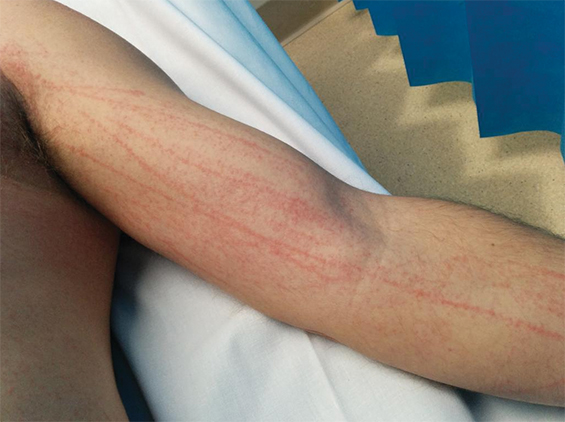
An elderly man presents to the emergency department of a regional base hospital with a new onset rash. The attending resident is not confident to make a diagnosis, but knows of a teledermatology service provided by a tertiary hospital in the nearest capital city. She asks the patient if she can “take some photos of the rash”. The patient agrees. She takes several photos of the patient with her personal smartphone and emails them, from her personal email address, to the on-call dermatology registrar at the tertiary hospital, requesting an opinion. The dermatology registrar reviews the images together with the patient’s history, and replies 2 hours later with a diagnosis of discoid eczema and a recommendation for management. Meanwhile, the dermatology registrar decides to use the patient images for a teaching session for the interns at the tertiary hospital.

Two weeks later the resident’s mobile phone is stolen. The phone had no security features enabled. The thief finds the patient’s images stored on the phone and uploads them to a public website. The thief also peruses the resident’s emails and finds details of the patient, and these are also shared publicly.

In the first instance, the resident should have made it clear to the patient that the purpose of taking the photo was to email the image to a specialist at another hospital to obtain an expert opinion on diagnosis and management. Further, notwithstanding the absence of a direct relationship with the patient, the teledermatology provider does not have consent to use the images for teaching, as the treating doctor did not obtain consent to use the images for this purpose.

Consent may be obtained orally or in writing, although practitioners should follow their institution’s guidelines. When consent is obtained orally, the consent process should be documented in the patient notes. Written consent must still be accompanied by a proper verbal explanation of the procedure.9

After the theft of her mobile phone in this case, the resident would be liable for a breach of privacy and confidentiality. She may also be in breach of the privacy and confidentiality obligations of her employment contract, as well as the conditions of her professional indemnity insurance. As a consequence, the resident could face disciplinary action from her employer or the Medical Board. The hospital may also be vicariously liable for the breach.9 Serious and repeated privacy breaches can attract substantial civil penalties for individuals and organisations under privacy legislation.

Technological precautions

The case above illustrates how easily a simple photograph can result in a costly legal dispute, not to mention untold harm to the patient. This situation could have been avoided if a number of simple precautions had been adopted.

An unlocked phone with patient photographs stored on it is a breach of privacy waiting to happen. Best practice dictates that patient images should be deleted from personal devices as soon as they are added to patients’ health records.7

As best practice may be overlooked during busy periods, security controls on personal smart phone devices, such as passcode locks, should be enabled to prevent unauthorised access. Installing remote locking or data wiping software to personal devices is recommended, as it will allow practitioners to delete data from their devices in the event of theft or loss.

Device settings should be adjusted so that clinical images on devices are not auto-uploaded to social media or back-up sites.7 Once a photo makes its way into the public domain via social media, it is very difficult to limit further sharing of that image.

The process of transmitting a patient image during the teledermatology consultation introduces an additional risk of a breach of privacy. Images should be transmitted via secure methods. Transmitting images through personal email or text messages is not considered secure by some sources, as such methods are typically not encrypted or password protected.10 Despite these concerns, the practice of emailing images is common in many hospitals. Practitioners should be familiar with the policies and systems of their institution or health service in relation to transmitting clinical images.

If a practitioner is required to send images by email or a text message to a colleague or specialist, they should send a test message first to ensure they have the correct email address or phone number for the intended recipient.7,10 Sending patient information or images to incorrect email addresses or phone numbers constitutes an automatic breach of privacy. Once sent, a practitioner has limited control over what the recipient does with that information. Ideally, one should encrypt or password-protect any images before transmitting them, although some may see this extra precaution as an inconvenience.

Like images, email or text messages that were part of the teledermatology consultation should be deleted from personal email accounts and devices after they have been uploaded to the patient record, as these may contain sensitive patient information that is still accessible if a device is lost or stolen. This includes emails saved in the “sent” folder. Automatic forwarding of emails to another email account should be disabled.

Practitioners should also consider the security of other devices that they use for sending emails. Computers and tablets with email management systems that allow email to be accessed on the desktop without logging in are a potential privacy risk. They should avoid sending patient images from an email address that is connected to an email management system. Practitioners should also implement automatic log off on computers and laptops at home and at work that are used to capture and store patient images, and change computer passwords regularly.

Where videoconferencing is being used, the room should be adequately sound-proofed and access should be restricted to those permitted according to the patient’s consent. Any videorecording of the consultation needs to be securely stored.

When consent for using an image for teaching or research purposes is appropriately obtained, best practice dictates that the image should be de-identified where possible, and must comply with relevant research or ethical guidelines.7 Identifying features such as birthmarks, tattoos, metadata, or even the condition itself, if it is rare, should be removed for such purposes.

Practitioner liability

Health care practitioners are personally responsible for patient information they choose to capture and transmit on their personal devices. Practitioners can limit their legal liability in the event of a breach of privacy by demonstrating that reasonable measures were taken to protect patient information. If a privacy breach does occur, clinicians should adopt an open disclosure approach, whereby the breach is notified and acted on. In these circumstances, practitioners should seek advice from hospital management and their medical defence organisations. Once appropriate legal advice has been sought, it may be necessary to inform the patient of the breach, and to explain and apologise.

Conclusions

It remains to be seen how health institutions and policy makers will tackle the issue of patient privacy in the new era of smart phones and teledermatology services. Fear of legal action should not preclude doctors from embracing novel approaches to health care that benefit patients and doctors. Rather, practitioners should take a few sensible precautions to reduce the likelihood of sensitive patient information falling into the hands of malicious third parties.

Before taking that next clinical photograph, individual practitioners should take a moment to review their own privacy practices and make any necessary adjustments. By doing so, practitioners can avoid the financial and emotional cost of a potential lawsuit in the future.

Shiitake dermatitis: the tale of an under-recognised, undercooked fungus

Clinical records

Case 1

A 55-year-old man with no medical history or allergies presented in May 2015 with a widespread, pruritic, whiplash-like rash (Figure 1). On examination, multiple erythematous papules in a linear distribution, corresponding to areas of excoriation, were noted on his trunk, limbs, forehead and scalp. The rash had developed 12 hours after a meal containing fresh shiitake mushrooms. There were no associated systemic symptoms and the patient’s condition did not improve with topical administration of betamethasone dipropionate 0.05%. Based on dietary history and the characteristic appearance of the rash, a diagnosis of shiitake dermatitis was made. The rash resolved in 3 weeks without further treatment. The patient experienced a recurrence 5 months later, after eating another meal containing shiitake mushrooms.

Case 2

A 30-year-old man with no comorbidities or allergies presented in March 2015 with a 2-day history of a striking, linear, erythematous eruption (Figure 2). He was systemically well, but reported associated perioral tingling and pharyngitis, which were unresponsive to 10 mg oral cetirizine. Examination revealed linear groupings of papules on an erythematous urticated base on his forehead and trunk. Despite denying pruritus or excoriations, relative sparing of the central back (out of the patient’s reach) was noted. Further questioning revealed ingestion of lightly cooked shiitake mushrooms the night before the onset of symptoms. No relatives who had eaten the same meal were affected. The patient was diagnosed with shiitake dermatitis, and the rash resolved completely without treatment.

Case 3

A 44-year-old man presented in August 2015 with a 2-day history of a flagellate erythematous rash (Figure 3). His past history included ischaemic heart disease, hypercholesterolaemia and rosacea. He had eaten raw shiitake mushrooms while cooking, and woke the next morning with a rash on his arms. Despite treatment with 25 mg oral prednisolone and topical betamethasone valerate 0.02%, prescribed by his general practitioner, the rash rapidly extended to his trunk, shins and scalp. His full blood count, C-reactive protein level and biochemistry results were unremarkable. A diagnosis of shiitake dermatitis was made. The patient’s wife, with whom he had shared the cooked meal, was asymptomatic, and the patient had previously eaten cooked shiitake without problems. Ten days later, the rash was resolving rapidly, with mild hyperpigmentation.

Shiitake mushrooms (Lentinus edodes) account for 20% of mushroom production worldwide1,2 and are the second most commonly eaten mushroom variety.1,3 Originally, most shiitake were log-grown in Japan. Since the late 1980s, most shiitake have been cultivated on a sawdust-based substrate in China.4,5 Shiitake are used in Asian medicine for their antihypertensive, anticarcinogenic and cholesterol-reducing qualities.1,5 Shiitake spores can cause IgE-mediated illness, such as hypersensitivity pneumonitis, asthma and allergic rhinitis.1,3,6 Shiitake can also cause contact urticaria, protein contact dermatitis and allergic contact dermatitis.1,3,6 In this article, we present the first three published cases of shiitake dermatitis in Australia. Systemic dermatitis caused by ingested allergens has also been reported with other foods, including spices, garlic, onions and root vegetables, metals, drugs, and preservatives.7,8

Shiitake dermatitis is also known as flagellate erythema, flagellate dermatitis and toxicodermia.3 It was first described in Japan in 1977, in Europe in 2006 and in the United States in 2010.1,2 Flagellate erythema occurs with all forms of shiitake — fresh, dried, powdered, boiled, baked, grilled and fried.2,5 It has been reported from adolescence to old age, and is more common in men.2

The highly distinctive rash develops following the ingestion of undercooked shiitake mushrooms by susceptible individuals. Onset usually occurs 12–48 hours after intake, although the reported range is 2 hours to 5 days.1 Intensely pruritic, linear, erythematous papules and petechiae develop on the trunk, limbs, head and neck,2 sparing the oral mucosa. The flagellate morphology is attributed to the Koebner phenomenon.2,3 There may be associated local oedema, fever, malaise, lip tingling, dysphagia and diarrhoea.1,9,10 Up to 47% of patients display ultraviolet A photosensitivity and photo-aggravation.3,9 Differential diagnoses include the flagellate rash associated with bleomycin, dermatomyositis, adult-onset Still disease, and HIV with hypereosinophilic syndrome.11

Lentinan, a thermolabile polysaccharide component of shiitake mushrooms, is the cause of shiitake dermatitis.13,5,9 Lentinan undergoes a conformational change at 130–145°C, so it is recommended that shiitake are cooked at temperatures of 150°C or greater to prevent toxicity.10 The pathogenesis remains unclear, but frequent case reports of single patients who had take part in shared meals and the delayed time course support the likelihood of an allergic reaction,2,10 with a possible role for exacerbating cofactors, such as use of angiotensin-converting enzyme inhibitors or diuretics, and exposure to ultraviolet A light.1,9 A trial of 519 patients in Japan who received intravenous lentinan chemotherapy yielded nine cases of flagellate erythema; extrapolating from these data, up to 2% of the population will be vulnerable to shiitake dermatitis.1,2

A case of shiitake dermatitis following ingestion of log-grown shiitake was recently reported in a patient who had previously tolerated substrate-grown shiitake from China.5 The authors of this report proposed that shiitake dermatitis may occur only with log-grown mushrooms. This hypothesis is supported by the low incidence of shiitake dermatitis in China, where the shiitake are substrate-grown, and the lower rates in Japan since 1992, when local production switched to substrate-grown mushrooms and the Japanese government began importing substrate-grown shiitake from China.4,5 Most cases in Japan were reported before 1998,9 typically during the harvest season for log-grown shiitake mushrooms.2 In Australia, shiitake are produced by both cultivation methods, and some shiitake are imported from China.12

The diagnosis is made clinically, and may be confirmed by oral re-challenge. Histopathological testing of skin biopsy specimens does not provide specific diagnostic information; it generally reveals elongation of rete ridges, spongiosis, degenerating keratinocytes, dermal oedema, and a superficial and perivascular lymphocytic infiltration with eosinophils and neutrophils.1,2 Prick and patch tests conducted by authors of previously published case reports have yielded conflicting results.1,2,10 Positive patch test results have also been obtained in control subjects, further limiting their usefulness in flagellate erythema.2,10

No treatments have been shown to be effective for shiitake dermatitis, although oral antihistamines and topical and oral corticosteroids are often prescribed. Given the high reported rate of ultraviolet A photosensitivity, patients should be given guidance on careful photoprotection.1 The prognosis is excellent, with improvement from 2 days and resolution by 3 weeks.1,2

In Australia, only four confirmed cases and one possible case of shiitake dermatitis were reported in 11 years (Appendix); these were reported to the NSW Poisons Information Centre between January 2004 and April 2015. No cases have been reported to the poisons information centres in Western Australia, Victoria and Queensland. Given our experience of three cases at a tertiary hospital within 12 months, and the prevalence of shiitake consumption in Australia, we believe that shiitake dermatitis is under-recognised. The mushroom industry heavily endorses the health benefits of shiitake as a low kilojoule source of dietary fibre, protein, vitamins, antioxidants and minerals. Considering the availability of shiitake-based health supplements on the internet,1 the popularity of Asian cuisine, and the production of log-grown shiitake in Australia today,12 we anticipate an increasing incidence of shiitake dermatitis. Documenting the preparation temperature, cultivation method and source of the shiitake mushrooms in future cases may help elucidate the pathogenesis.

Lessons from practice

  • Shiitake dermatitis is probably under-recognised and under-reported in Australia

  • The diagnosis is made clinically, based on dietary history and a characteristic erythematous flagellate rash

  • The prognosis is excellent, but patients should be advised to avoid shiitake or to ensure it has been cooked at temperatures of 150°C or greater to prevent further episodes

  • Primary care practitioners, such as general practitioners and emergency physicians, should be aware of the diagnosis to avoid unnecessary investigations and treatments

Figure 1 –


Figure 1: Widespread, characteristic flagellate erythema of shiitake dermatitis in a 55-year-old man

Figure 2 –


Figure 2: Linear grouped papules on an erythematous urticated base in a 30-year-old man with shiitake dermatitis

Figure 3 –


Figure 3: Intensely pruritic, linear, erythematous rash in a 44-year-old man with shiitake dermatitis

The early bird and the worm: a case of cercarial dermatitis

Clinical record

A 36-year-old man presented to our dermatology department with a 5-day history of an intensely pruritic papular eruption on the dorsum of both feet, associated with significant swelling and discomfort. This began the morning after a night-time fishing trip on the central coast of New South Wales at the mouth of the Lake Tuggerah estuary, a system of brackish tidal estuaries.

The patient reported standing ankle-deep in water for 6 hours from dusk (9 pm) until early morning (3 am). He noted that the water was particularly warm after a daytime temperature of 33°C with seaward tidal flows. He awoke the following morning with multiple discrete nodules, oedema and intense pruritus to the dorsa of the feet. He reported previous episodes of “bites” after fishing trips that spontaneously resolved after 24–48 hours.

The next 12 hours saw an increase in pruritus and the size of lesions. The patient presented to a general practitioner, who diagnosed pelican itch with secondary infection and prescribed cephalexin 500 mg four times a day for 5 days. Pain and pruritus continued to increase, resulting in the patient presenting to a metropolitan emergency department where he was diagnosed with bilateral foot cellulitis and administered intravenous cefazolin 1 g twice daily for 5 days. Investigations showed leucocytosis (white cell count, 10.1 × 10⁹/L; reference interval [RI], 4.0–10.0 × 10⁹/L) with eosinophilia (0.8 × 10⁹/L; RI, 0–0.5 × 10⁹/L) and negative blood cultures. On Day 3 of intravenous antibiotics, he was referred to our dermatology department for reassessment of diagnosis.

On presentation, the patient was in significant discomfort with swelling, difficulty ambulating and inability to wear shoes. He did not describe any fever, vomiting, diarrhoea, abdominal cramping or haematuria. Multiple violaceous papules were noted over the dorsum of each foot, with sparing of the plantar surfaces, web spaces of the toes and distal third of the lower limb (Figure, A). Dusky oedema was evident surrounding the papules, with no evidence of lymphangitis. Popliteal and inguinal lymph nodes were palpable.

Histopathological examination (Figure, C and D) revealed epidermal spongiosis and prominent dermal oedema. A heavy inflammatory infiltrate predominately of eosinophils involving all layers of skin down to the subcutis with scattered flame figures was also noted. No parasites were identified. Bacterial, fungal and mycobacterial tissue cultures were negative. This histology supported a markedly pronounced hypersensitivity reaction in a patient clinically suspected to have cercarial dermatitis, and was not compatible with other clinical differential diagnoses such as hypertrophic lichen planus, Sweet syndrome and reticulohistiocytosis.

Our patient was successfully treated with 3 days of oral prednisolone therapy (25 mg daily) with complete resolution of lesions and residual post-inflammatory pigmentation by Day 7 (Figure, B).

Cercarial dermatitis (commonly known as swimmer’s itch, pelican itch or duck itch) conventionally refers to a cutaneous hypersensitivity reaction to non-human trematode larvae (cercariae) of schistasome flukes after penetration of human skin.1,2 In the classic avian form of the disease, the flukes undergo a two-host (digenetic) life cycle with egg development in freshwater snails and release of the larvae into open water (in temperatures above 23°C) to infest migratory aquatic birds (ducks and seagulls, the fluke’s definitive host).2 Human infections with avian cercariae represent a disruption of the natural schistasome life cycle. The fluke was previously thought to die within human skin within 48 hours; however, animal models have shown migration and survival of the trematodes in distant organs weeks after initial infection.1,2

Cercarial dermatitis is a common condition found worldwide1,2 but is under-recognised and under-reported in Australia owing to its self-limiting nature.1 High concentrations of cercariae occur during days of intense sunlight and higher air and sea surface temperatures, with coastal currents concentrating cercariae around shallow estuary outlets.2 Cases have been reported from Queensland,3,4 Terrigal on the central coast of NSW (eponymous for the schistasome Austrobilharzia terrigalensis5), and the Swan River estuary of Western Australia.1,6

The condition manifests as a monomorphic, papular, pruritic dermatitis confined to water-submerged areas that begins to spontaneously regress within 72 hours.1,2 Recurrent exposure can result in sensitisation phenomena precipitating a florid hypersensitivity reaction as seen in our case. Cercarial dermatitis can be differentiated from sea-bather’s eruption, as the latter condition affects areas of skin covered by swimwear, commonly as a swimmer exits the water or after swimwear has dried. It is caused by discharging nematocysts of sea anemones and jellyfish, and can be associated with systemic symptoms of fevers, vomiting and malaise.

Avian schistasomes are homologous to (but not to be confused with) human schistosomiasis, an endemic and potentially fatal tropical disease, manifesting in portal hypertension, bladder carcinoma, granulomatous central nervous system disease, pulmonary fibrosis and glomerulonephritis.7,8 In endemic tropical areas, cercarial dermatitis and schistosomiasis can be difficult to differentiate, although the early cercarial stage of human schistosomiasis is less inflammatory than that of avian schistosomiasis.2 The histopathology of cercarial dermatitis in humans is poorly documented owing to the acute self-limiting nature of the disease.2 Where descriptions are available, they are briefly described as a hypersensitivity reaction with oedema and eosinophils (plus or minus neutrophils). Specificity is provided by finding cercariae in the specimen (located within the epidermis) but they are usually not seen,7 presumably due to the limited life span of the cercariae within non-definitive hosts and the difficulty in finding isolated small (0.75–0.95 mm long) organisms within a disproportionately large area of inflammatory reaction. It is surmised that without identification of the organism, some cases could easily be misclassified as arthropod bite reactions in the absence of awareness of the specific clinical context.

Treatment for mild cercarial dermatitis includes reassurance and symptomatic management with potent topical corticosteroids (betamethasone dipropionate 0.05% ointment twice daily) and emollients. Oedema can be relieved with intermittent cold compress, rest and elevation. Exuberant reactions may require oral prednisolone (0.5 mg/kg up to a maximum dose of 25 mg daily for 3 consecutive days). Some authors recommend routine treatment with oral ivermectin (200 μg/kg up to a maximum of 12 mg per dose)9 until possible systemic complications arising from visceral migration of trematodes in humans are better understood.

It is important for physicians to be aware of this common and under-recognised entity, as well as the potential for schistosomiasis with similar presentations in returning travellers. Although the cutaneous manifestations of this condition spontaneously resolve, further research will clarify the potential for chronic infestation and the role of routine antihelminthic therapy.

Lessons from practice

  • Cercarial dermatitis is a common, under-reported condition caused by a hypersensitivity reaction to the penetration of human skin by the trematode fluke of avian schistosomiasis.

  • It commonly occurs due to submergence of unprotected skin in warm waters of freshwater tidal estuaries.

  • Clinicians should be aware of similar presentations in returning travellers, which may represent the potentially fatal tropical disease of human schistosomiasis.

  • Although most cases spontaneously resolve, topical and systemic corticosteroids are treatment options for exuberant hypersensitivity reactions.

Figure


A: Grouped violaceous papules on the dorsolateral aspect of the foot with associated oedema. B: Significant improvement in foot oedema with healing blisters and erosions after 3 days of oral prednisolone (25 mg). C: Low-power image of upper dermis and epidermis with marked subepidermal oedema and spongiosis responsible for the bullous clinical appearance (pseudobullous change) (objective magnification, ×4; haematoxylin–eosin stain). D: High-power image showing prominent interstitial dermal infiltrate of eosinophils with flame figure (arrow) at top of image (objective magnification, ×20; haematoxylin–eosin stain).

Severe ulcerative herpes zoster

A 78-year-old man presented to a regional emergency department with a severe progressive rash, on a background history of chronic lymphocytic leukaemia, dementia and malnourishment.

The rash was multidermatomal, with patchy areas of ulceration, crusting, excoriation and necrosis (Figure, A and B). Active bleeding, seborrhoeic discharge and occasional vesicles were also noted, extending to the left pelvis. Subsequently, the patient developed concurrent Pseudomonas aeruginosa cellulitis and bacteraemia.

Punch biopsies were non-specific with dermal necrosis, excoriation and possible lichenoid reactivity. However, swabs revealed varicella-zoster virus. The patient was successfully treated with intravenous piperacillin–tazobactam, intravenous acyclovir, normal saline (0.9% sodium chloride) washes, and 50% liquid paraffin with 50% white soft paraffin cream (Figure, C and D). Multifactorial immunodeficiency was deemed to be the aetiology.

Figure

Figure

[Correspondence] Ixekizumab for psoriasis

Christopher Griffiths and colleagues (Aug 8, p 541)1 reported that ixekizumab, a high-affinity antibody targeting interleukin 17A, given to patients with moderate-to-severe psoriasis for 12 weeks had greater efficacy than etanercept and placebo. However, the dermatology life-quality index used in the studies might have been insufficient to assess adverse effects because it only assesses dermatological symptom-associated mental status.