×

Association of delay of urgent or emergency surgery with mortality and use of health care resources: a propensity score-matched observational cohort study [Research]

BACKGROUND:

Delay of surgery for hip fracture is associated with increased risk of morbidity and mortality, but the effects of surgical delays on mortality and resource use in the context of other emergency surgeries is poorly described. Our objective was to measure the independent association between delay of emergency surgery and in-hospital mortality, length of stay and costs.

METHODS:

We identified all adult patients who underwent emergency noncardiac surgery between January 2012 and October 2014 at a single tertiary care centre. Delay of surgery was defined as the time from surgical booking to operating room entry exceeding institutionally defined acceptable wait times, based on a standardized 5-level priority system that accounted for surgery type and indication. Patients with delayed surgery were matched to those without delay using propensity scores derived from variables that accounted for details of admission and the hospital stay, patient characteristics, physiologic instability, and surgical urgency and risk.

RESULTS:

Of 15 160 patients, 2820 (18.6%) experienced a delay. The mortality rates were 4.9% (138/2820) for those with delay and 3.2% (391/12 340) for those without delay (odds ratio [OR] 1.59, 95% confidence interval [CI] 1.30–1.93). Within the propensity-matched cohort, delay was significantly associated with mortality (OR 1.56, 95% CI 1.18–2.06), increased length of stay (incident rate ratio 1.07, 95% CI 1.01–1.11) and higher total costs (incident rate ratio 1.06, 95% CI 1.01–1.11).

INTERPRETATION:

Delayed operating room access for emergency surgery was associated with increased risk of inhospital mortality, longer length of stay and higher costs. System issues appeared to underlie most delays and must be addressed to improve the outcomes of emergency surgery.

Atrial fibrillation screening back in the picture

Population screening for atrial fibrillation is once more up for discussion as a new study shows that people with newly diagnosed ‘silent’ AF actually have a higher risk of stroke than those with a symptomatic condition.

The study, presented this week at the European Heart Rhythm Association meeting in Vienna, included over 6,000 consecutively enrolled patients with non-valvular atrial fibrillation, of whom around two-thirds were asymptomatic or minimally symptomatic at time of diagnosis.

The study found those with asymptomatic AF had more than double the risk of previous stroke, compared to those who were symptomatic (14.7% vs 6.0%). They were also more likely to have permanent atrial fibrillation than those in the symptomatic group (15.8% vs 8.3%).

But both symptomatic and asymptomatic patients in this study had a similar number of stroke risk factors, with an average CHA2DS2-VASc score of 3.3 for each group.

Lead author Dr Steffen Christow, a cardiologist at Hospital Ingolstadt in Germany, said the higher rate of stroke despite the same number of stroke risk factors may be explained by a longer undiagnosed history of AF in those with asymptomatic disease.

“Our study found that in Western Europe, two-thirds of patients newly diagnosed with atrial fibrillation were asymptomatic. Without detection, patients may not receive appropriate preventive therapy and remain at increased risk of stroke.”

He said the results “underline the urgent need for public programs to detect atrial fibrillation in the general population”.

In Australia, RACGP guidelines do not recommend systematic screening for atrial fibrillation, although they say opportunistic screening when taking blood pressure could be cost-effective.

The current study is a sub-analysis of the GLORIA-AF registry, which characterises a population of newly diagnosed patients with non-valvular atrial fibrillation at risk for stroke, studying patterns, predictors and outcomes of different treatment regimens for stroke prevention.

You can access the study abstract here.

Air pollution linked with heart damage

A new report presented by the European Society of Cardiology says that there is strong evidence that particulate matter (PM) emitted mainly from diesel road vehicles is associated with increased risk of heart attack, heart failure, and death.

The lead author Dr Nay Aung, a cardiologist and Wellcome Trust research fellow at the William Harvey Research Institute, Queen Mary University of London, UK, said the cause for the heart damage “appears to be driven by an inflammatory response – inhalation of fine particulate matter (PM2.5) causes localised inflammation of the lungs followed by a more systemic inflammation affecting the whole body.”

Regarding how pollution might have these negative effects on the heart, Dr Aung said PM2.5 causes systemic inflammation, vasoconstriction and raised blood pressure. The combination of these factors can increase the pressure in the heart, which enlarges to cope with the overload. The heart chamber enlargement reduces the contractile efficiency leading to reduction in ejection fraction.

The researchers said they found evidence of harmful effects even when levels of pollution associated with diesel vehicles were less than half the safety limit set by the European Union.

Dr Aung said: “We found that the average exposure to PM2.5 in the UK is about 10 µg/m3 in our study. This is way below the European target of less than 25 µg/m3 and yet we are still seeing these harmful effects. This suggests that the current target level is not safe and should be lowered.”

In the UK, where the study was conducted, the Government recently produced its third attempt at a plan to bring air pollution to within levels considered safe under European Union legislation after judges ruled the previous versions were not effective enough to comply with the law.

Dr Penny Woods, chief executive of the British Lung Foundation, said: “Air pollution (in the UK) is a public health crisis hitting our most vulnerable the hardest – our children, people with a lung condition and the elderly.” 

Dr Woods added that, while progress was being made in high-income countries to reduce deaths from cardiovascular disease and cancer, those caused by lung disease had “remained tragically constant”. 

The World Health Organisation (WHO) estimates that some 3 million deaths a year are linked to exposure to outdoor air pollution. WHO also believes that indoor air pollution can be just as deadly. In 2012, an estimated 6.5 million deaths (11.6 per cent of all global deaths) were associated with indoor and outdoor air pollution together.

Only one in ten people breathe safe air according to WHO guidelines and over 80 per cent of the world’s cities have air pollution levels over what these guidelines deem safe.

The Australian Medical Association has developed a Position Statement on Climate Change and Human Health that acknowledges air pollution is the world’s single largest environmental health risk.

Meredith Horne

[Series] Strategies for long-term preservation of kidney graft function

Kidney transplantation has become a routine procedure in the treatment of patients with kidney failure, and requires collaboration of experts from different disciplines, such as nephrology, surgery, immunology, pathology, infectious disease medicine, cardiology, and oncology. Grafts can be obtained from deceased or living donors, with different logistical requirements and implications for long-term graft patency. 1-year graft survival rates are greater than 95% in many centres but improvement of long-term function remains a challenge.

Reducing cardiovascular disease risk in diabetes: a randomised controlled trial of a quality improvement initiative

The known Managing risk factors for cardiovascular disease (CVD) in patients with diabetes improves their outcomes, but many are not prescribed the recommended treatments. Electronic decision support is a scalable strategy for improving guideline implementation. 

The new The implementation of recommended management of CVD risk factors in people with diabetes is incomplete, but better than for patients without diabetes. An electronic decision support tool achieved modest improvements in CVD risk factor screening and treatment escalation in patients with diabetes. 

The implications While computerised tools may play an important enabling role, broader strategies are needed to close evidence–practice gaps. 

By 2030, diabetes may affect more than 300 million people worldwide.1 Cardiovascular disease (CVD) is the primary cause of mortality and morbidity in patients with type 2 diabetes,2 and large studies have found that managing risk factors for CVD in patients with diabetes reduces both.3,4 According to risk management guidelines, decisions about the need for and the intensity of intervention should be based on the estimated absolute risk.5

A number of guidelines for managing CVD risk in people with diabetes have been published, but studies in Australia6,7 and overseas810 have consistently found that these strategies have been only incompletely implemented. Most Australian studies, however, predate a number of targeted quality improvement (QI) programs, including the National Divisions Diabetes, Australian Primary Care Collaboratives, and National Integrated Diabetes Programs, as well as the introduction of targeted incentive payments to general practitioners and practices, and may therefore not accurately reflect current practice. Whether any of these initiatives improved quality of care is unknown.

In this article we describe the contemporary primary care management in Australia of patients with diabetes participating in a study of CVD risk management in primary health care. Our primary objectives were to assess adherence to CVD risk screening and management guidelines, and to determine the effectiveness of a new QI intervention for improving risk management. The main results of the QI study have been published elsewhere;11 we report here a subgroup analysis comparing the outcomes for patients with and without diabetes.

Methods

The Treatment of Cardiovascular Risk in Primary care using Electronic Decision Support (TORPEDO) study was a parallel arm, cluster randomised, controlled trial involving 60 Australian primary health care services (40 general practices and 20 Aboriginal Community Controlled Health Services [ACCHSs]). It assessed whether a QI intervention combining point-of-care electronic decision support with audit and feedback tools improved CVD risk management. The TORPEDO study methods have been described in detail elsewhere.11

Practice eligibility criteria

Health services were eligible to participate if they exclusively used either of the two most common electronic health record systems in Australia for recording risk factor information, pathology test results and prescribed medications. General practices from the Sydney region were recruited between September 2011 and May 2012 through primary health care networks (previously: Medicare Locals), and ACCHSs through two state representative bodies in New South Wales and Queensland.

Patient eligibility criteria

Eligible patients were Aboriginal and Torres Strait Islander people at least 35 years old and non-Indigenous people at least 45 years old (age ranges based on national CVD risk guidelines5) who had attended a participating service at least three times during the preceding 24 months and at least once during the past 6 months. The presence or absence of diabetes was established by a recorded diagnosis of diabetes (type not specified) or a glycosylated haemoglobin (HbA1c) measurement at baseline of more than 53 mmol/mol. This threshold was chosen as a conservative estimate during the transition period in Australian diagnostic criteria for diabetes, which now recommend a threshold of 48 mmol/mol.

Randomisation

Services were randomised (1:1) to intervention or control groups, stratified at three levels: ACCHSs v mainstream general practices; service size (less than 500 v 500 or more eligible patients); and current participation in a national or state QI program. Permuted block randomisation was performed centrally; outcome analyses were blinded to allocation.

Intervention

Full details of the intervention have been published previously.12 In brief, a screening and management algorithm was developed and validated, based on a synthesis of recommendations from several guidelines (online Appendix 1).13 The algorithm incorporated CVD risk assessment, as well as recommendations for managing CVD, chronic kidney disease, blood pressure (BP) and cholesterol, but not for blood glucose management.

Five-year risk of a cardiovascular event was estimated with the Australian risk calculator, based on the 1991 Anderson Framingham equation.14 High CVD risk is defined in Australian guidelines5 as a calculated 5-year CVD risk of greater than 15%; the presence of diabetes in a person over 60 years old, diabetes together with albuminuria, estimated glomerular filtration rate (eGFR) below 45 mL/min/1.73 m2, systolic BP greater than 180 mmHg, diastolic BP greater than 110 mmHg, or total blood cholesterol level over 7.5 mmol/L; or the presence of CVD, defined as a recorded diagnosis of coronary heart disease, cerebrovascular disease (ischaemic stroke or transient ischaemic attack), or peripheral vascular disease. Risk was based on the most recent available results, whether or not the participant was being treated for that risk factor.

The algorithm interfaced directly with the two eligible electronic records systems. Data from the patient record were automatically prepopulated in the tool, and used to generate point-of-care CVD risk management recommendations. A data extraction tool provided site-specific audits, and feedback performance reports were generated. Clinical staff were trained in the application of the tool and had access to a support desk and bi-monthly webinars. The intervention lasted a minimum of 12 months.

Data collection

De-identified data for all patients who met the eligibility criteria were extracted from the clinical database of each health service with a validated data extraction tool.15 The extracts were uploaded to the study database together with an encrypted identifier code.

Outcomes

The primary outcomes for the randomised trial11 were:

  • The proportion of eligible patients who received appropriate screening for CVD risk factors by the end of the study. This was defined as data for all relevant risk factors having been recorded or updated (smoking status, BP in the past 12 months, total blood cholesterol and high-density lipoprotein (HDL)-cholesterol levels in the past 24 months).

  • The proportion of patients at high CVD risk at baseline who were receiving recommended medication prescriptions at the end of the study (prescription of at least one BP-lowering drug and a statin for people at high risk without CVD; reduction of CVD risk to below 15% by the end of study; prescription of at least one BP-lowering drug together with a statin and an antiplatelet agent for people with established CVD, unless contraindicated by oral anticoagulant use).

Secondary outcomes included:

  • the primary outcomes for individuals at high risk who were undertreated at baseline;

  • measurements of individual CVD risk factors (smoking status, BP, blood lipid levels, body mass index [BMI], eGFR, albuminuria);

  • escalation of drug prescription for patients at high risk of CVD (either newly prescribed or additional antiplatelet, BP-lowering and lipid-lowering agents); and

  • BP and serum lipid levels in people at high risk of CVD.

Sample size

Randomisation of 60 services (30 per arm) would provide 90% power to detect an absolute higher occurrence of 10% for each primary study outcome in the intervention arm, assuming a 10% absolute improvement in the control arm, an average cluster size of 750 patients (30% of whom were at high risk of CVD), baseline risk factor measurement and prescribing rates of 50%, α = 0.05 (two-sided), and an intraclass correlation coefficient of 0.05.

Statistical analysis

Post hoc descriptive analyses of baseline data from the TORPEDO study and of data for the cohort of participants present at both baseline and study end were undertaken. Data are presented as means with standard deviations, medians with interquartile ranges, or proportions. Baseline differences between patients with and without diabetes were tested in generalised estimating equations (GEEs) with an exchangeable correlation structure to account for clustering of patients in services.

To determine the predictors of suboptimal drug therapy at baseline, cross-sectional analyses were conducted in a GEE model with logit link function, including both patient level characteristics and service level data. Associations between risk factors and drug therapy were expressed as unadjusted odds ratios (with 95% confidence intervals [CIs]) for binary outcomes.

Intervention effects were analysed by log-binomial GEE regression. The rate ratios of intervention effect were calculated for the individual outcomes at the end of the study. The effects of the intervention in the subgroup of undertreated participants at baseline were analysed in the same model, stratified by diabetes status. An interaction term was included in all models to assess heterogeneity of effects by diabetes status.

Statistical analyses were conducted in SAS Enterprise Guide 5.1 (SAS Institute).

Ethics approval

The study was approved by the University of Sydney Human Research Ethics Committee (HREC) (reference, 2012/2183) and the Aboriginal Health and Medical Research Council of New South Wales HREC (reference, 778/11). Signed agreements with participating sites were obtained. Individual consent waiver was granted because data were collected from de-identified extracts from the electronic health record system.

Results

Recruitment

Sixty-four services were initially recruited; 31 were randomised to the intervention arm (but one withdrew shortly after randomisation) and 30 to usual care (online Appendix 2). Baseline data were extracted for 53 164 patients, including 8829 with diabetes; a cohort of 38 725 (6909 with diabetes) were followed up for outcome evaluation. The median follow-up time was 17 months.

Sample characteristics at baseline

Of the 8829 patients with diabetes at baseline, most had a recorded diagnosis of diabetes (97%); 3% were defined by HbA1c levels exceeding 53 mmol/mol. The mean age, and the proportions who were men, smokers or Indigenous Australians were higher for people who had diabetes than for those who did not. Their mean systolic BP and blood triglyceride levels were also higher, while their low-density lipoprotein (LDL)- and HDL-cholesterol levels were lower. Albuminuria, renal impairment and an established diagnosis of CVD were more common in people with diabetes (for all differences: P < 0.001; online Appendix 3).

Recording of risk factors and CVD risk at baseline for people with diabetes

Overall, appropriate measurement of CVD risk factors in people with diabetes was greater than for those without diabetes (62.0% v 39.5%; P < 0.001; online Appendix 3), a difference that remained after adjusting for age, sex, and Indigenous status (P < 0.001). BMI was recorded for 81% of people with diabetes, smoking status for 89%, HbA1c levels for 86%, systolic BP for 94%, albuminuria assessment for 59%, and eGFR for 87%. Recording rates for total, LDL- and HDL-cholesterol levels were 87%, 82% and 79% respectively.

More than one-quarter of patients with diabetes (26%) had established CVD; a further 12%, 4%, and 49% had an estimated 5-year CVD risk that was low (< 10%), medium (10–15%), or high (> 15% or clinically high risk condition present) respectively. There was insufficient information for 825 patients (9%) to categorise their risk (online Appendix 3).

Prescribing rates at baseline for people with diabetes at high risk of CVD

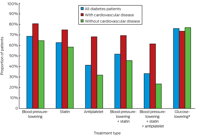
Appropriate prescribing of medications for those identified as being at high risk of CVD was greater among people with diabetes than for those without diabetes (55.5% v 39.6%, P < 0.001; online Appendix 3). Overall, 52.4% of people with diabetes at high risk of CVD but without established CVD and 61.4% of patients with diabetes and established CVD were prescribed recommended medications for averting CVD; the corresponding figures for people without diabetes were lower (22.0% and 49.3% respectively; for each comparison, P < 0.001) (online Appendix 3). The individual medication types prescribed for people with diabetes are shown in Box 1.

Risk factor targets

The HbA1c levels of 57.3% of patients with diabetes exceeded 53 mmol/mol; about one-quarter of these patients were not prescribed glucose-lowering therapy (Box 2). Similarly, the BP and lipid levels of large proportions of patients with diabetes exceeded recommended target levels (online Appendix 4); of the 61.9% of patients who did not meet the LDL-cholesterol target of 2.0 mmol/L, 44.3% were not prescribed a statin (Box 2).

Predictors of drug prescription

People with diabetes who were older (P < 0.001) or Indigenous (P = 0.030), or had a higher HbA1c level (P = 0.030), higher systolic BP (P < 0.001), or albuminuria (P < 0.001), were more likely to be prescribed the recommended combination treatment. Conversely, those with higher total cholesterol levels were less likely to receive optimal combination treatment (P < 0.001). Those who did not have a government-reimbursed health assessment (P = 0.012) or care plan (P < 0.001) were also less likely to be prescribed the recommended medications. Service type (general practice v ACCHS) did not influence drug prescription in univariable or multivariable analyses (online Appendix 5).

Effectiveness of the QI intervention

The baseline characteristics of the cohort used for outcome evaluation were similar for the intervention and control groups (online Appendix 3). The intervention was less effective in improving risk factor screening in patients with diabetes than in those without diabetes (P = 0.01). The intervention was only effective in improving rates of prescribing of recommended medications for undertreated individuals at high risk. and was not influenced by diabetes status (P = 0.28). The intervention was associated with intensification of existing antiplatelet, lipid-lowering, and BP-lowering therapy to a similar extent in people with and without diabetes. The intervention did not affect the prescription of glucose-lowering therapy (Box 3).

Discussion

People with diabetes in a contemporary Australian primary care population were more likely to be screened and prescribed the recommended medications for managing CVD risk factors than those without diabetes. The QI intervention was modestly effective in improving screening and treatment levels, but the evidence–practice gaps remained substantial.

Screening deficits were most marked with regard to cholesterol and albuminuria tests, consistent with both overseas16 and local17,18 reports. A recent French study16 found that only half of a group of patients with diabetes had been screened for proteinuria or albuminuria during the previous 12 months, suggesting that renal function is a poorly assessed CVD risk factor. Underprescribing of recommended treatments was striking when the patients with diabetes in our study were stratified by absolute risk: 39% of those with established CVD and 48% of those at high risk of CVD were not prescribed the recommended treatments; almost half of those with diabetes and LDL-cholesterol levels above 2.0 mmol/L were not receiving statin therapy. Similarly, BP targets were not met by half the patients with diabetes, of whom more than one-quarter were not prescribed antihypertensive therapy. These treatment deficits are consistent with international experience,810,16 and reflect modest improvements when compared with the findings of previous Australian studies of lipid6,7 and BP7,19 management.

About one-half of people with diabetes did not meet the recommended HbA1c goal of 53 mmol/mol or less, similar to the 57% figure in the 2003/2004 assessment of the United States National Health and Nutrition Examination Survey (NHANES) participants.20 It is worrying that about one-quarter of our patients with HbA1c levels over 69 mmol/mol were not prescribed glucose-lowering medication, a proportion substantially larger than the 3% of patients with HbA1c levels of 53 mmol/mol or more not treated in a recent Canadian study.10 Our findings may be partly explained by patient preference for non-pharmacological treatment, and by relaxed glycaemic targets in certain populations (older people, and people with frequent hypoglycaemia or hypoglycaemic unawareness).

Our findings suggest that undertreatment has diminished to some degree since 2002, which may reflect the effect of incentive schemes and quality of care initiatives. The deficits that remain may be explained by the proliferation of guidelines with differing perspectives, and time-pressured consultations with patients presenting with several complaints. Patients with diabetes who had a formal care plan, enabling coordination of their management with other health care providers, were more likely to be treated as recommended. However, causal inferences cannot be made, as numerous factors may confound this association.

The finding that CVD risk screening and management at baseline was better for those with diabetes than for people without diabetes is consistent with other reports,21,22 and may explain why the effect of the intervention was less marked in these patients. Although it was not effective in improving the overall level of new prescriptions for individuals at higher risk of CVD, the intervention was associated with improvements for people who were not receiving recommended treatments at baseline, regardless of their diabetes status. This is important in light of suggestions that therapeutic inertia may be a greater contributor to lost therapeutic benefit in patients with diabetes than lack of treatment.23

There is evidence that patient-directed interventions combined with physician-focused strategies may be more effective than the latter alone.24 Successful elements of collaborative care programs for improving chronic disease management include evidence-based guidelines, systematic screening and monitoring of risk factors, scheduled recall visits, new or adjusted roles for team members, information support for the clinician, enhanced self-management by the patient, effective communication between all members of the care team, and audit information for the practice.25 New policy proposals, such as “Health Care Homes”,26 and a renewed focus on initiatives such as “My Health Record” incorporate some of these elements.

Study limitations

Many of the sites in our study were teaching practices; this may explain why performance was higher than reported in previous studies. However, the recruited services were reasonably representative of Australian general practice with respect to the use of information technology.27 The ACCHSs recruited were geographically diverse and had similar service characteristics to the sector as a whole.28

The National Vascular Disease Prevention Alliance (NVDPA) guidelines5 recommend incorporating pre-treatment risk factor levels when assessing CVD risk. As pre-treatment data were not available, we analysed the patients’ most recent BP and lipid data, regardless of treatment status, and this may have led to underestimating risk for some individuals. However, as 72% of patients with diabetes were at high risk regardless of their risk score (online Appendix 3), this was probably not a major problem.

We regarded 2 years as an appropriate interval between lipid measurements, rather than varying the interval according to risk status as recommended by NVDPA guidelines. This may not have been appropriate for individuals at high risk, for whom more frequent testing is recommended. Conversely, the Royal Australian College of General Practitioners guidelines recommend 5-yearly lipid measurements for people at low risk;29 if doctors are adhering to these recommendations, the frequency of assessment may be adequate, but with a median follow-up period of 17 months we were not able to assess whether this was the case. If biases were introduced by using different lipid measurement intervals, we would expect them to be the same for the intervention and control arms.

Other limitations included the fact that that the type of diabetes was not specified, and that relying on electronic records data precluded assessing the role of clinical judgement in treatment decisions.

Conclusion

Although recommendations for managing CVD risk were more frequently implemented for people with diabetes than for those without diabetes, evidence–practice gaps remain. While the evaluated intervention was moderately effective in improving screening of risk factors, additional strategies are needed if Australia is to meet targets of reducing mortality for CVD and diabetes by 25% over the next 10 years.30

Box 1 –
Rates of prescribing of currently recommended cardiovascular disease risk-factor-specific medications for patients with diabetes


* For patients with HbA1c levels above 53 mmol/mol.

Box 2 –
Patients with diabetes with values above targets at baseline, and number who did not receive the corresponding recommended treatment

Patients with elevated level

Number not treated to reduce level


HbA1c level

> 53 mmol/mol

4329 of 7556 (57.3%)

1038 (24.0%)

> 69 mmol/mol

1822 of 7556 (24.1%)

450 (24.7%)

Blood pressure (BP)

Systolic BP > 130 mmHg or diastolic BP > 80 mmHg

4835 of 8329 (58.1%)

1354 (28.0%)

LDL-cholesterol level

> 2.0 mmol/L

4339 of 7007 (61.9%)

1922 (44.3%)

> 2.5 mmol/L

2769 of 7007 (39.5%)

1412 (51.0%)


HbA1c = glycated haemoglobin; LDL = low-density lipoprotein.

Box 3 –
Effects of the quality improvement intervention in patients with and without diabetes

Intervention

Usual care

Rate ratio

95% CI

P*


Receiving appropriate screening

12 164/19 385 (62.8%)

10 317/19 340 (53.4%)

1.25

(1.04–1.50)

0.01

With diabetes

2738/3617 (75.7%)

2323/3292 (70.6%)

1.14

(1.00–1.30)

Without diabetes

9426/15 768 (59.8%)

7994/16 048 (49.8%)

1.28

(1.04–1.58)

Receiving appropriate screening (undertreated at baseline)

3773/9276 (40.7%)

3532/10 782 (32.8%)

1.38

(1.10–1.73)

< 0.01

With diabetes

559/1160 (48.2%)

507/1151 (44.0%)

1.28

(1.00–1.63)

Without diabetes

3214/8116 (39.6%)

3025/9631 (31.4%)

1.40

(1.11–1.78)

Patients at high risk of cardiovascular disease

Receiving appropriate prescriptions

3030/5335 (56.8%)

2483/4846 (51.2%)

1.11

(0.97–1.27)

0.10

With diabetes

1700/2679 (63.5%)

1458/2495 (58.4%)

1.06

(0.93–1.21)

Without diabetes

1330/2656 (50.1%)

1025/2351 (43.6%)

1.18

(1.03–1.36)

Receiving appropriate prescriptions (undertreated at baseline)

1085/2827 (38.4%)

472/2263 (20.9%)

1.59

(1.19–2.13)

0.28

With diabetes

553/1269 (43.6%)

178/923 (19.3%)

1.63

(1.11–2.38)

Without diabetes

532/1558 (34.2%)

294/1340 (21.9%)

1.53

(1.16–2.01)

Increased antiplatelet therapy

470/2638 (17.8%)

65/2424 (2.7%)

4.79

(2.47–9.29)

0.08

With diabetes

210/908 (23.1%)

20/829 (2.4%)

7.28

(3.34–15.9)

Without diabetes

260/1730 (15.0%)

45/1595 (2.8%)

4.05

(2.03–8.08)

Increased lipid-lowering therapy

1026/5335 (19.2%)

226/4846 (4.7%)

3.22

(1.77–5.88)

0.84

With diabetes

608/2679 (22.7%)

130/2495 (5.2%)

3.32

(1.74–6.33)

Without diabetes

418/2656 (15.7%)

96/2351 (4.1%)

3.22

(1.77–5.86)

Increased blood pressure-lowering therapy

1243/5335 (23.3%)

586/4846 (12.1%)

1.89

(1.09–3.28)

0.54

With diabetes

729/2679 (27.2%)

316/2495 (12.7%)

1.91

(1.09–3.35)

Without diabetes

514/2656 (19.4%)

270/2351 (11.5%)

1.96

(1.10–3.47)

Patients with HbA1clevels > 53 mmol/mol at baseline

Appropriate glucose-lowering drug

1111/1269 (87.6%)

955/1118 (85.4%)

1.02

(0.95–1.11)

Increased glucose-lowering therapy

711/2679 (26.5%)

304/2495 (12.2%)

1.75

(0.95–3.22)


* Patients with diabetes v patients without diabetes. † For patients not meeting recommended targets for corresponding parameters (online Appendix 4).

Expectorate the unexpected

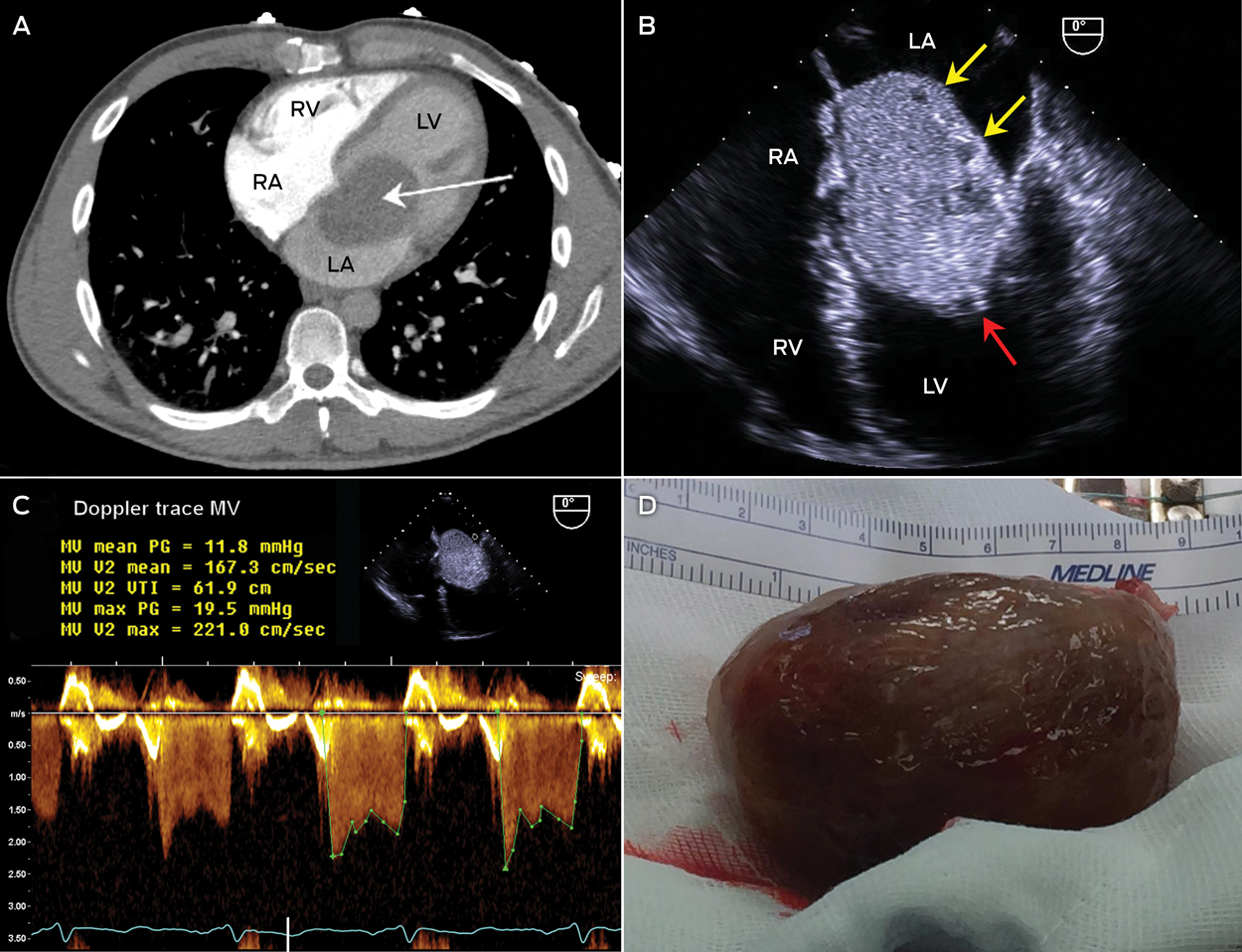
A 43-year-old man underwent a computed tomography pulmonary angiography for suspected pulmonary embolism after he presented with an episode of orthopnoea associated with blood-stained expectorate. The only finding was a left atrial mass (Figure, A, arrow). Further history revealed 6 months of progressive postural presyncope, malaise and weight loss of 3 kg.

Echocardiography showed a circumscribed 7.0 × 4.5 cm heterogeneous mass, with areas of cystic degeneration (Figure, B, yellow arrows) and frond-like extensions (∼ 3 mm; Figure, B, red arrow), consistent with a myxoma. The mass was attached to the interatrial septum via a small stalk and prolapsed through the mitral valve, causing significant mitral inlet obstruction; the mean gradient was approximately 12 mmHg (Figure, C). The mass was successfully removed (Figure, D).

Figure


LA = left atrium. LV = left ventricle. RA = right atrium. RV = right ventricle.

Statins questioned for primary prevention in elderly

People over 65 who start statin therapy for primary prevention may risk hastening their deaths, according to new research published in JAMA Internal Medicine.

The study findings are based on an analysis of a randomised trial of pravastatin that took place from 1994 to 2002. The researchers looked at a trial subset of nearly 3000 people over 65 who had no baseline atherosclerotic cardiovascular disease but had raised cholesterol.

In this group, those randomised to pravastatin had similar rates of cardiovascular heart disease compared with those on usual treatment. Rates of stroke, heart failure and cancer were all about the same across the two groups as well.

But more deaths were reported in the pravastatin group compared with usual treatment – 141 vs 130 in patients aged 65 to 74, and 92 vs 65 in those aged 75 and over.

“No benefit was found when a statin was given for primary prevention to older adults. Treatment recommendations should be individualised for this population,” the authors write.

They suggest that statins in this age group may trigger “untoward effects in the function or health of older adults that could offset any possible cardiovascular benefit”.

They note that some studies have suggested statins can increase fatigue with exertion and may negatively impact on cognition, particularly in people with mild cognitive impairment or dementia.

They also point to observational studies that have had mixed findings regarding the benefit of statins for primary prevention in older people.

But not everyone accepts the finding of the current study.

Professor Jeremy Pearson, who is the Associate Medical Director of the British Heart Foundation, notes that the statin used in the study, pravastatin, is no longer prescribed to lower cholesterol and has since been replaced by stronger statins.

“This could have an impact on the results, so more research is needed to confirm the findings, based on current best practice,” he says.

And Dr Tim Chico, a consultant cardiologist at the University of Sheffield in the UK, said that to study the effectiveness of primary prevention you would need many more than the subset of 3,000 patients examined in the study.

You can read the full paper here.

Coronary occlusion, denial and dissociation

A doctor puts emotions aside in an emergency, but dissociation does not help when the illness is in the family

The speedometer reads 160 km/h. I am driving. My husband Anthony is in the passenger seat. He groans and writhes clutching his chest in that classic closed-fist gesture. We are two rural general practitioners in personal crisis. It is only 45 minutes since he won a bike race in the mountains.

The paramedics know we are coming. We round a bend. There is the ambulance. Both vehicles stop. His blood pressure is 80/40. His pulse is 42. They do an electrocardiogram and hand it to me. “Oh, no ST elevation,” I say, relieved. One. They snatch it back. “There’s elevation everywhere.” I am dismissed.

The paramedic regional manager arrives. He says they’ll thrombolyse here. Anthony is saying, “I want a hot cath.” This is ridiculous; soon the charade will stop. He is not having a heart attack. Two.

Our adult children who followed in another car are here now and standing on the roadside like white posts. I return to the ambulance. The paramedics are injecting tenecteplase intravenously. They have given morphine. Anthony’s groaning continues. They give more morphine, antiemetic, morphine, close the doors and drive the 5 minutes to the hospital where the protocol is completed. His myocardium throws off some cardiac arrhythmias as it reperfuses.

The subsequent urgent coronary angiogram shows only minimal irregularity at the origin of the left anterior descending coronary artery. It seems that this small plaque ruptured and a clot formed during his extreme effort racing to the finish line. No clot can now be seen. Perhaps if he was less fit, if his heart was less used to contracting in a lactic acidotic environment, it might have stopped. He has had an acute myocardial infarction though; even I must admit that. I must.

So there we are, husband and wife, lovers, the parents of four children, co-workers and protagonists in many a clinical argument. He, wired and dripped with tenecteplase bruises spreading like black flowers on his limbs, is forcedly chipper. I sit holding his hand with a strained smile. I ask what I should do about the conference trip to South Korea in 3 days’ time. I am an invited keynote speaker. He says, “Oh of course you should go.” I say, “Yes, of course.” “Grace will be here with me,” he says. “I’ll be fine.” Grace is our 21-year-old daughter. “Oh yes,” I say. We agree that he’s through the worst. Three. I don’t want him to think I am worried about him. I think I mustn’t fuss. Four.

My time in Korea passes in a disconnected blur. Everything I care about is too far away. I can’t even pray. Then I am on the plane heading home. When I arrive we are formal with each other. He might break. I might break. The whole world might break.

One day I look at him and say, “You could have died.” “I knew that,” he replies. What confounds me was that I did not. What use was all my training if I could not see the ST elevation in my beloved husband’s electrocardiogram, if I did not have the wisdom to stay by his bed on day three? I made four critical errors in caring for him. Both of us exhibited impaired clinical reasoning.

On reflection, I think that the training to put our emotions aside and get on with the job in an emergency situation did not serve us well in this personal emergency. My job in this instance was to be there emotionally and practically, but I pushed the emotions away and my judgement was affected. I did not acknowledge how close death had been. A month passed before I cried.

In the weeks following his heart attack, whenever anyone asked, Anthony said he felt 97%. I believed him. Months later he admitted that he felt like crap. He tells me that he coped by rationalising, by talking with chosen experts about the pathology, the prognosis, the pharmaceuticals and their risks. For a while he claimed that he didn’t have an acute myocardial infarction; he had a “coronary occlusion”. As our children say, “Whatever…”. His task was to discover his path through life under this shadow, whatever it is called.

I misrepresented, denied and dissociated from the emotionally loaded information I was attempting to process. When that happens, personal decision making is impaired. Seriously.

The Lighthouse Project

BY AMA PRESIDENT DR MICHAEL GANNON

Last year, when the AMA released its 2016 Report Card on Indigenous Health, it set out a plan for governments to eradicate Rheumatic Heart Disease (RHD) from Australia by 2031. Since the release of this Report Card, the AMA has been a part of growing efforts to reinforce to our political leaders that RHD must be stamped out, and that other cardiovascular health outcomes for Aboriginal and Torres Strait Islander peoples must be improved.

As part of our efforts to improve the cardiovascular health of Aboriginal and Torres Strait Islander people, the AMA has become a founding member of an END RHD Coalition – an alliance of six organisations with a vision to see the end of RHD in Australia, we participated in the inaugural Close the Gap Parliamentary Friendship Group which focussed on the enormous impact of RHD, and we recently met with the Australian Healthcare and Hospitals Association (AHHA) to discuss the Lighthouse Project – a joint initiative of the AHHA and the Heart Foundation to improve outcomes for Aboriginal and Torres Strait Islander peoples experiencing coronary heart disease.

The aim of the Lighthouse Project is to help close the gap in cardiovascular disease between Indigenous and non-Indigenous Australians through the provision of evidence-based, culturally safe care for acute coronary syndrome. With cardiovascular disease being the  leading cause of death among Aboriginal and Torres Strait Islander people, and  a major contributor to the gap in life expectancy between Indigenous and other Australians, it is imperative that the AMA and other health and medical organisations are actively engaged in this area.

It is unacceptable that Aboriginal and Torres Strait Islander people, who represent three per cent of the entire Australian population, are 1.6 times more likely to die from coronary heart disease than their non-Indigenous peers.  It is also unacceptable that Aboriginal and Torres Strait Islander people are less likely to undergo vital coronary tests and procedures once admitted to hospital.

It is clear that the hospital system must better respond to the unique health needs of Aboriginal and Torres Strait Islander patients.  Hospitals have an important role to play in improving access to care and addressing disparities for Aboriginal and Torres Strait Islander peoples. This is where initiatives such as the Lighthouse Project are extremely valuable.

During Phase 1 of the Lighthouse Project, cultural competence, having a skilled workforce, appropriate governance and the use of clinical care pathways were identified as four key areas of best practice for improving care for Aboriginal and Torres Strait Islander peoples with Acute Coronary Syndrome. In Phase 2, a quality improvement toolkit was developed and implemented in eight public hospitals across Australia. 

Through Phase 1 and Phase 2 of the Lighthouse Project, these public hospitals have achieved culturally safe environments and enhanced staff capacity to respond to the unique needs of Aboriginal and Torres Strait Islander patients, and have reported improved relationships with Indigenous patients and communities.

The Lighthouse Project must be seen as a positive example of how gains in health outcomes can be achieved for Aboriginal and Torres Strait Islander people. I am pleased that the work of the Lighthouse Project will continue, with the Commonwealth Government recently announcing that $8 million has been provided to support Phase 3 of the Lighthouse Project, which aims to extend the project to 18 hospitals across the country and allowing it to reach nearly one in every two Indigenous patients admitted to hospital for a cardiac condition.

Eliminating inequities in health service provision to the Aboriginal and Torres Strait Islander population is vital, and it is encouraging to see that the great work of the Lighthouse Project is being recognised. By increasing cardiovascular health outcomes for Aboriginal and Torres Strait Islander peoples, we can reduce mortality rates, increase life expectancy, and help close the unacceptable health gap that exists between Indigenous and non-Indigenous Australians today.


Controversies in diagnosis and management of community-acquired pneumonia

Community-acquired pneumonia (CAP) continues to generate a large amount of interest, both for the clinician and the researcher. It is a very frequent diagnosis and the leading infection-related cause of death in most developed countries.1

Although CAP is a relatively common infection, there are wide disparities in its management, including the class of antibiotics chosen, the duration of therapy and the role of adjunctive therapy such as corticosteroids. In this review, we assess the evidence for the approaches to some of these clinical questions regarding CAP management. We agree with the Australian antibiotic guidelines2 regarding recommended antibiotics. Therefore, we do not specifically consider the question of the most appropriate class of antibiotics for treating patients with CAP — the Box summarises the antibiotics commonly used in Australia.

We used a PubMed search for original and review articles from 2005 to 2017, and reviewed specialist society publications and guidelines from Australia and overseas, to formulate an evidence-based overview of the topic as applied to clinical practice.

Are we overdiagnosing CAP?

Although it may seem self-evident, an essential question in the management of patients with CAP is whether the diagnosis is in fact correct. CAP can present in variable ways, some of which are similar to other conditions such as acute bronchitis, viral respiratory tract infections and cardiac failure. Patients with dementia, who are more likely to develop CAP, may not be able to give a reliable description of symptoms.3 Patients may present with two or more conditions at once, confusing the diagnostic process.3 This may occur as a coincidence or alternatively be due to a cause–effect relationship between them. Examples of the latter include that a chest infection can precipitate either an exacerbation of cardiac failure or an acute coronary syndrome.4 In addition, particularly in the era of the 4-hour National Emergency Access Target, staff members in the emergency department (ED) are under greater pressure to move patients out of the ED and thus may need to change the focus of their assessment to “does this patient need admission?” rather than “what is the correct diagnosis?”.

From clinical studies of CAP performed in Australia, of all the patients screened for inclusion on the basis of being given the label of CAP in the ED, a large proportion are subsequently excluded from the study because their chest x-ray is not consistent with CAP.5,6 This issue is not limited to Australia, with international studies showing that chest x-rays reported by treating clinicians as being consistent with CAP are not confirmed as being so by a radiologist in 20–50% of cases.711

There are several downsides to excessive diagnosis of CAP. The most obvious is the use of unnecessary antibiotics in patients who have conditions that do not require antibiotics such as viral respiratory infections or cardiac failure. This has the potential to add to the problem of antibiotic resistance as well as putting the patient at risk of antibiotic-related complications such as Clostridium difficile-associated diarrhoea. A further issue, particularly when cultures are not performed in patients initially labelled as having CAP, is the potential delay in diagnosis and inappropriate antibiotic therapy of those patients whose true diagnosis is something more serious, such as sepsis, infective endocarditis or pulmonary embolism. Some of these misdiagnosed patients can have their admission prolonged by many days due to the non-performance of blood cultures. We believe that the diagnostic uncertainty for admitted patients initially given the diagnosis of CAP means that recommendations that discourage the performance of blood cultures in CAP patients are inappropriate.1215

Duration of antibiotic therapy

The optimal duration of antimicrobial therapy for CAP is another area of controversy. The tendency in hospitals appears to be to overtreat rather than undertreat, often with a long oral tail.1618 Whether this is a case of believing that “more is better” or due to the disparity between the time to clinical resolution compared with microbiological resolution, the excessive prescription of antibiotics puts the patient at greater risk of side effects and colonisation with resistant organisms, including nasopharyngeal carriage of penicillin-resistant Streptococcus pneumoniae.19,20 Ecologically, the prescription of antibiotics for respiratory infection contributes to a rise in resistance in the community.21

Should the physician turn to national guidelines for advice on duration and choice of antibiotic (Box); the Australian Therapeutic guidelines: antibiotic recommend 7 days of total therapy for moderate and most cases of severe pneumonia,2 as does the British Thoracic Society,22 while the United Kingdom NICE guidelines suggest 5 days for mild CAP and 7–10 days for moderate to severe CAP.23 However, the Infectious Diseases Society of America (IDSA) supports a 5-day treatment for inpatient CAP, provided the patient is afebrile and clinically improving.24 So, with all this variation, which is correct?

There is agreement that a 7-day course of an antibiotic is effective for most cases of CAP, and this is relatively non-controversial, albeit adhered to poorly.25 There is increasing evidence, however, that shorter courses of 5 or even 3 days’ therapy may be just as effective. Overseas literature provides support for short course therapy with azithromycin, including as little as a single dose.26 This likely relates to the high tissue penetration and persistence of adequate tissue levels of this macrolide for some days following administration.27 A multicentre randomised clinical trial evaluating the safety of the IDSA recommendations found that a 5-day course of therapy is safe and effective, although most patients received quinolone antibiotics, a class of antibiotic rarely used in Australia for treating CAP.28 Regarding the β-lactam therapy that would be more likely prescribed in the Australian setting, a 3-day course of intravenous (IV) amoxycillin monotherapy has been shown to be as effective as 3 days of IV amoxycillin followed by 5 days of oral amoxycillin in adult patients who were improving at 72 hours.29 Two previous studies reached a similar conclusion in paediatric populations.30,31

Given the accumulating evidence, we suggest that a 5-day course of antibiotics should be effective in most cases of uncomplicated CAP, even though complete symptom resolution is unlikely to have occurred at this time point. For patients on IV therapy who are clinically improving at 72 hours, a switch to oral therapy is appropriate, but clinicians should keep in mind that the oral antibiotics should complete the 5-day total course and not add another 5 days to what has already been prescribed. If improvement has been rapid in the first 72 hours, it would be reasonable to cease all therapy at 3 days, provided close follow-up is available.

Some international studies have suggested that bundles of care for patients with CAP, which include antibiotic administration within 4 to 8 hours of presentation, may lead to better patient outcomes.3234 However, it is not clear that this would provide benefits in the Australian setting. In relation to the United States studies,33,34 this finding may reflect past differences in the US health system, where antibiotics may not have been given until the patient was seen by their attending physician, potentially leading to delays in therapy. The US recommendations have now changed to recommend commencement of antibiotics while the patient is in the ED.24 This is already the norm in Australia.

Other studies35,36 have suggested that increases in mortality in patients with CAP may be due to an atypical presentation which leads to a delay in diagnosis, rather than being associated with a delay in antibiotic administration. When this was taken into account in one study, the association between a delay in antibiotic administration beyond 4 hours and increased mortality was not statistically significant.35

Potential cardiac side effects of newer macrolide antibiotics

A 2012 study reported an excess of both cardiovascular and all-cause deaths in patients with pneumonia treated with a 5-day course of azithromycin compared with those treated with other antimicrobials, potentially related to its ability to prolong the QT interval.37 As a result, in 2013, the US Food and Drug Administration issued a warning regarding prescription of azithromycin for CAP, even though that study had a number of limitations, including its non-randomised nature and outpatient study population.

However, the case was far from closed, and results from other retrospective studies reached the opposite conclusion. Mortensen and colleagues studied older patients with CAP and found that those treated with macrolides had a lower rate of mortality, in spite of a small rise in rates of myocardial infarction “consistent with a net benefit”.38 This conclusion was shared by Cheng and colleagues in their 2015 meta-analysis.39 In 2016, a Canadian population-based retrospective cohort study involving about one million adults aged over 65 years found no increase in rates of cardiac arrhythmias at 30 days, in addition to lower all-cause mortality, in patients treated with a macrolide antibiotic.40

Given the evidence that the benefit of using macrolide therapy outweighs potential cardiac risk, we support recommendations to use a macrolide in place of doxycycline for atypical cover when the latter cannot be used, and the use of azithromycin in combination therapy for severe hospitalised CAP, such as that requiring management in an intensive care unit (ICU). We also point out the excellent oral bioavailability of oral azithromycin,27 and recommend its use in preference to the IV formulation in patients for whom oral therapy is tolerated and expected to be absorbed.

The link between CAP and cardiovascular disease

In recent years, evidence has emerged regarding the role of inflammatory conditions in the development of cardiovascular disease such as myocardial infarctions and strokes.41 It is postulated that inflammation, especially when persistent, may have an effect on vascular plaques, making them more unstable or prone to acute occlusion.42,43 Various infections including CAP, influenza and human immunodeficiency virus, as well as other sources of chronic inflammation such as rheumatoid arthritis, have all been shown to be associated with higher rates of acute cardiovascular disease and deaths.4,4451

In a large study, in the 30 days following an episode of CAP requiring inpatient care, incidence of worsening heart failure, cardiac arrhythmia and acute myocardial infarction were 21%, 10% and 3% respectively.4 However, it is important to note that the problem does not end after 30 days. There is a measurably higher rate of cardiovascular deaths in the following few years, when patients admitted with CAP are compared with matched cohorts admitted with non-infection-related conditions. The rate increases most in older patients (aged over 40 years) and those with greater number of cardiovascular risk factors.52

The mechanism of this increase in cardiovascular complications during and after the CAP episode appears to be multifactorial. Inflammation is a pro-thrombotic state; myocardial inflammation and damage may occur, potentially in response to NADPH oxidase 2 upregulation; cardiac strain may be present in the setting of increased sympathetic nervous system activity with relative hypoxia caused by the lung consolidation; increased fluid and sodium loading associated with some IV antibiotic may worsen fluid overload problems in some cardiac failure patients; and QT interval prolongation with the use of other antibiotics may contribute to arrhythmic potential.46,47,53

What remains to be seen is whether we can act on this in a useful way. It is notable that the vast majority of patients who die from CAP are very old with multiple comorbidities, for whom death may be an expected terminal event. While acutely addressing cardiac risk factors with, for example, the addition of anti-platelet agents like aspirin or cholesterol-lowering statin therapy has not yet been shown to alter mortality in the acute setting,54 it would appear prudent to assess whether such treatments are indicated in patients admitted with CAP, especially if they are aged over 40 years.52

The role of corticosteroids in the management of CAP

Given that the inflammatory state during and after an episode of CAP appears to have an important role in contributing to both morbidity and mortality,4,4447 there has been interest in the role of inflammatory modulators such as corticosteroids as adjunctive CAP therapy. Levels of cytokines vary with severity of CAP and highest levels of the pro-inflammatory cytokine interleukin (IL)-6 and the anti-inflammatory cytokine IL-10 are associated with higher chance of dying from severe CAP.55 Glucocorticoids reduce the levels of such cytokines,56 and thus are theoretically attractive as a means to reduce CAP mortality.

There have been a number of attempts to address the question about whether this theoretical benefit may be true. Individual studies have varied in terms of the severity of the CAP studied, the choice of corticosteroid used, the route by which it was given, its dose and duration, and the outcomes measured. Results have been mixed, and several attempts at performing meta-analyses on these studies — with all the expected problems associated with attempting to combine such a heterogeneous collection of methodologies — have shown marginal benefits in terms of mortality, particularly in patients with the most severe CAP managed in the ICU, as well as a shorter time to becoming afebrile.5760 These small benefits need to be weighed against the potential downside of high-dose corticosteroids, both in terms of potential side effects like immune suppression and also the fact that outcomes may have been worse in patients whose infection was caused by an influenza virus or Aspergillus.61,62

Thus, the potential role of corticosteroids as adjunctive therapy in CAP appears to be very limited. They could be considered in patients with CAP severe enough to require management in the ICU, but caution should be taken until the aetiology is known, particularly during influenza season. Their use should also be very carefully considered in patients at higher risk from corticosteroid complications, such as the immunocompromised, women who are pregnant, patients with recent gastrointestinal haemorrhages, and patients at greater risk of neuropsychiatric problems.59 The possible shortened time to defervescence is not sufficiently clinically useful to justify the potential harm from such therapy.

Conclusion

In this era of burgeoning antibiotic resistance, the treatment of CAP is an area where we have the potential to reduce antibiotic consumption. We are diagnosing it too often and treating it for too long. Most non-ICU patients with CAP could be treated for 3–5 days in total.

CAP is a common cause of death, both in the short term and also in the subsequent few years, and many of these deaths appear to be cardiovascular related. Although most deaths from CAP occur in very old people with multiple comorbidities — and so may not easily be prevented — the management of a patient with CAP should be seen as an opportunity to address and treat cardiac risk factors when they are present.

Box –
Antibiotics commonly used to treat community-acquired pneumonia (CAP) in Australia2

CAP severity

Antibiotic

Comments

Suggested duration


Mild (treated as outpatient)

Doxycycline

Monotherapy; avoid in pregnancy and young children

3–5 days

Amoxycillin

Monotherapy; side effect profile better than amoxycillin–clavulinate and spectrum of activity more appropriate

3–5 days

Macrolide (eg, clarithromycin, azithromycin or roxithromycin)

Monotherapy; potential option when patient intolerant of doxycycline and amoxycillin

3–5 days

Amoxycillin–clavulinate

Consider in patients from nursing homes or following recent hospital admissions

5 days

Cefuroxime*

Consider in patients with non-hypersensitivity reactions to amoxycillin

3–5 days

Moderate (admitted patients not requiring ICU)

Benzylpenicillin

Use in combination with either doxycycline or a macrolide

Switch to oral therapy when clinical improvement occurs, generally in 1–3 days

Doxycycline

Oral; used in combination with benzylpenicillin

5 days

Macrolide (eg, clarithromycin or azithromycin)

Alternative second agent to doxycycline (oral or IV); used in combination with benzylpenicillin

5 days

Moxifloxacin

Use as monotherapy if hypersensitivity reaction to penicillins; excellent oral bioavailability

5 days

Severe (patients potentially requiring ICU care)

Ceftriaxone plus azithromycin IV

Alternative choices may be appropriate in tropical northern Australia

7 days


ICU = intensive care unit. IV = intravenous. * Cefaclor is not useful owing to poor antibacterial activity and high rate of causing rashes; cephalexin is not ideal given the poor spectrum of activity against respiratory pathogens.