×

Cost and outcomes of assessing patients with chest pain in an Australian emergency department

Patients presenting with chest pain represent a large group of adult emergency department (ED) presentations.1 The most common serious underlying causes for this symptom are acute coronary syndromes (ACS), incorporating acute myocardial infarction and unstable angina pectoris. Over 5.5 million people presented to EDs in the United States in 2007–2008 with a primary complaint of chest pain, yet only 13% of those were diagnosed with an ACS.1 The number of patients presenting to EDs in Australia with a possible ACS is unknown.

Many conditions cause chest pain, yet discriminating between an ACS and alternative and generally less serious aetiologies, such as gastro-oesophageal reflux, is difficult. The 2006 National Heart Foundation and Cardiac Society of Australia and New Zealand (NHF/CSANZ) guidelines on the management of ACS recommended stratifying patients into low-, intermediate- and high-risk categories,2 a strategy that remained unchanged in more recent updates.3 The guidelines recommended that low-risk patients be assessed using serial cardiac biomarkers and electrocardiography. High-risk patients require admission to hospital and intensive management, often including early invasive strategies. The largest group is the intermediate-risk cohort, who require serial testing of biomarkers and electrocardiography. If results of these are negative, further objective testing is required. The most commonly performed objective test in this intermediate-risk group is an exercise stress test (EST). Other more costly tests may include computed tomography coronary angiography, stress echocardiography, myocardial perfusion scanning and invasive angiography.

The costs of applying such guidelines to an undifferentiated population presenting with chest pain to EDs in Australia have not been described. The final diagnoses and 1-year outcomes of patients presenting to the ED with chest pain have also not been described. We aimed to characterise the demographics, length of hospital stay (LOS), final diagnoses, long-term outcome and costs associated with the population who presented to an Australian ED with symptoms of possible ACS.

Methods

Design and participants

We conducted a prospective, single-centre observational study between November 2008 and February 2011. Patients were included if they presented to the ED with at least 5 minutes of chest pain suggestive of an ACS (acute chest, epigastric, neck, jaw, or arm pain; or discomfort or pressure without an apparent non-cardiac source). Data were collected between 08:00 and 17:00.

Patients were excluded if they had a clear non-ACS cause for their symptoms, were unwilling or unable to provide informed consent (eg, dementia), were considered inappropriate for recruitment (eg, terminal illness), were pregnant, were recruited to the study within the past 45 days or were unable or unwilling to be contacted after discharge. Patients transferred to or from another hospital were excluded from the study, as we did not have data on costs or management for these patients. Consecutive eligible cases at the Royal Brisbane and Women’s Hospital’s ED were included. The study protocol was approved by the hospital’s human research ethics committee (no 2008/101 and HREC/11/QRBW/493).

Patients were classified into risk groups according to the Queensland chest pain pathway (Appendix), based on the 2006 NHF/CSANZ guidelines.2 Low-risk and intermediate-risk patients were typically managed in the ED with admission to the ED short-stay unit (Box 1). High-risk patients and patients unable to perform an EST, owing to contraindication or inability, were referred to inpatient cardiology and general medical units for admission and further assessment. A small proportion of patients were managed in the ED (3.9%), while the remainder were transferred to the ED short-stay unit (46.7%) or the inpatient ward (49.4%). Patients requiring urgent cardiac surgery were transferred to another institution after inpatient admission.

Data collection

Research staff collected data using a standardised patient interview as soon as ED clinical assessment was complete. Interviews were cross-checked with patient notes. Blood samples taken on presentation (0 hour) and ≥ 6 hours later were sent to our laboratory for measurement of troponin and analysed using the second-generation AccuTnI (Beckman Coulter) assay. The 0-hour and ≥ 6-hour test results were used for patient care and cardiology end point adjudication. We used the manufacturer’s 99th percentile cut-point to indicate a raised troponin value.

Data on the costs associated with investigation and care of patients during the index admission were extracted from hospital administration records. Inpatient costs were derived from procedure-related Australian refined diagnosis-related group reimbursement codes used for activity-based funding. These cost codes guide federal government payments and are designed to reflect the health care services used during each patient episode.4 To determine appropriate payments to hospitals, the weighted cost combines inputs such as staff time and consumables used for patient care.

ED costs reflect the payments received by the hospital based on triage categories of urgency. Total costs include fixed costs, which make up about 80% of overhead costs, and a variable activity-based component for pathology, imaging, pharmacy, clinical supplies and hotel services.4 Costs from 2008–2010 were adjusted for inflation by 3.4% per year to equate to 2011 costs.5 The 30-day clinical outcome was adjudicated independently by local cardiologists using predefined standardised reporting guidelines, with knowledge of all clinical information collected within a 30-day period.6 A second cardiologist conducted a blind review of all ACS cases and 10% of non-ACS cases. In cases of disagreement, end points were agreed by consensus. This was achieved for all end points.

The 30-day clinical outcomes were grouped into three categories that included cardiac-ACS-related, other cardiac and non-cardiac diagnoses. Cardiac-ACS-related diagnoses included ST-segment elevation myocardial infarction, non-ST-segment elevation myocardial infarction and unstable angina pectoris. These were categorised according to the universal definition.7 An end point of unstable angina pectoris was given for patients with negative serial troponin results, ischaemic symptoms and objective evidence of ischaemia on the EST, stress echocardiography, myocardial perfusion scanning, computed tomography coronary angiography or significant findings on coronary angiography.

Other diagnoses, such as cardiac but non-ACS (cardiac-other), and non-cardiac, were based on all available clinical data including investigations that had occurred within the 30-day period after presentation. A National Death Registry audit was performed in July 2014 to provide mortality data for patients who had consented to longer-term study participation.

Data analysis

Data were analysed using Stata, version 12 (StataCorp). Baseline characteristics of the sample were reported by outcome category using standard descriptive statistics. The baseline characteristics of patients with and without an ACS were compared using the χ2 test for categorical data and t test for continuous data.

Data on diagnosis, LOS and costs were also reported by outcome category. The LOS and cost data were right skewed and were reported in several ways. First, the median and interquartile range were reported to provide a good estimate of the LOS and cost for a typical patient. Second, for economic analysis, mean costs were reported. The mean is the correct estimator because decisionmakers need to understand total costs, which are predicted by the mean and the quantity of services used. Bias-corrected and accelerated bootstrap confidence intervals were calculated using 1000 replications.

One-year mortality was reported for a subset of patients who consented to ongoing participation in the study. Kaplan–Meier survival curves for time to death were generated by diagnostic group, and log-rank tests were used to compare survival curves.

The mean and median LOS and cost per patient were reported by NHF/CSANZ risk category. These data were broken down by ACS and non-ACS outcomes and included all patients within the diagnostic category, irrespective of whether objective testing was performed.

Results

Nine hundred and twenty-six patients were included (566 [61.1%] men; mean age 54.7 years). No patients were lost to 30-day follow up (Box 2) and 693 (74.8%) consented to one-year follow-up. Baseline characteristics are shown in Box 3.

Non-cardiac chest pain was diagnosed in 622 (67.2%) of the 926 patients (Box 3). One hundred and ninety-three patients (20.8%) were diagnosed with other cardiovascular conditions including pericarditis, atrial fibrillation and heart failure. Among those diagnosed with an ACS (103 [11.1%]), the most common condition was non-ST-segment elevation myocardial infarction (51.5% of total ACS group).

Three of the 926 patients died within 30 days; two of these were during the index admission. Two of the deaths were cardiac related. An additional 14 of the 693 patients involved in long-term follow-up died within 12 months (total, 17/693 patients [2.5%]; 95% CI, 1.4%–3.9%) (Box 4).

Kaplan–Meier curves for deaths within the diagnostic categories are shown in Box 5.

The outcomes and costs by NHF/CSANZ risk group are shown in Box 6. ACS events occurred in 0 and 11 (1.9%) of the low-risk and intermediate-risk groups, respectively. Ninety-two (28.0%) of the 329 high-risk patients had an ACS event. Ten patients were transferred for acute cardiac surgery.

Patients with an ACS, high-grade atrioventricular block, heart failure, syncope, pulmonary embolism and respiratory conditions had the longest median LOS (Box 4). Patients with an ACS incurred the highest mean cost per patient ($13 509), followed by other cardiovascular conditions ($7283). Patients with non-cardiac disease had the lowest cost of $3331 per patient.

Three hundred and fifty of the 580 intermediate-risk patients (60.3%) had an EST during the index admission. These patients incurred lower mean costs ($2316; 95% CI, $2126–$2507) than those who did not undergo an EST ($4806; 95% CI, $4094–$5516). Three hundred and six ESTs (87.4%) yielded a negative result. In contrast, 124 (53.9%) of the 230 patients who did not undergo an EST during the index presentation were admitted to an inpatient unit and incurred higher costs. Four hundred and sixty-six (80.3%) of the intermediate-risk patients received some objective testing within 30 days. The total cost for intermediate-risk patients was $1 916 100; if divided across the 11 ACS patients, this equates to $174 191 to identify one ACS event.

In the high-risk group, the mean LOS was 5 days and the mean cost was $8919. The cost of treating the 237 patients in this group who did not have an event was $7075 per patient, while the 92 patients who had an event incurred costs of $13 669. The total cost of investigating high-risk patients in this study was $2 934 317. If this value is divided across the 92 ACS events, this equates to an average of $31 895 spent to identify and treat one ACS event.

Overall, the total ED cost for investigating the 926 patients in this study was $904 221, while inpatient costs totalled $3 977 234. Total ED LOS was 5575.9 hours, making the average cost per hour in ED $162. Total LOS as an inpatient was 59 061.9 hours, making the average inpatient cost $67 per hour.

Discussion

This is the first evaluation of the characteristics, final diagnoses, outcomes and costs for an Australian ED cohort investigated for possible ACS based on the NHF/CSANZ guidelines.2,3 In keeping with other international reports, the final proportion of patients with a diagnosis of ACS was 11.1%, with 20.8% of patients having other cardiovascular causes diagnosed.811

Our study shows that significant resources are consumed in investigating ACSs; however, in the absence of research identifying a “negligible risk” group who do not require objective testing, such resource use is necessary. Alternative strategies to reduce the LOS in a low-risk cohort have been reported,9,1216 and some studies have reported on the implementation and effect of such accelerated protocols.17,18 At the time of this study, such strategies were not in use in our institution. Further research efforts should be directed to identifying patients who could be discharged without requiring additional cardiac investigations.

About one-fifth of intermediate-risk patients did not have objective cardiac testing in hospital or within 30 days of their presentation, against guideline recommendations.2 We did not record the reasons why clinicians deviated from the NHF/CSANZ guidelines; however, it is possible that clinical gestalt, prior recent investigation or known coronary artery disease may have influenced care.19 In addition, decision making may have been influenced by significant debate about the utility of objective testing such as EST for patients thought to be at minimal risk.20,21

No previous study in the Australian setting has examined the cost of assessing patients presenting with chest pain. We were able to show that costs are substantial and varied across the risk categories. The high-risk group incurred the highest cost per patient, but this group had a high rate of ACS. In contrast, the intermediate-risk group was the most resource-intensive, yet these costs were expended to diagnose a very small proportion (1.9%) of patients with ACS. The overall costs per event in the intermediate group were high ($174 191 per ACS event). An ideal accelerated diagnostic protocol would exclude from testing patients who have no risk of ACS. The effect would be to reduce the size of the intermediate-risk group and define them as low risk, saving resources but not having an adverse impact on health outcomes.

Few previous studies have reported the costs related to chest pain assessment in the ED, specifically. A recent publication by KP Health for Queensland Health titled “Report of the evaluation of the clinical services redesign program in Queensland hospitals” recorded an estimated released value of 1 hour of ED cubicle time to be $98 and of an inpatient bed-day to be $779 in the financial year 2013–14 (Sarah Bright, Senior Project Officer, Health Technology Assessment and Evaluation, Queensland Health, personal communication, March 2014). This figure is less than that reported in this study of $162 per hour for an ED bed. The difference is likely to be due to the high triage category assigned to patients presenting with chest pain and the extensive investigations required. These data provide the basis to test accelerated diagnostic protocols and build full cost-effectiveness models that quantify the potential changes to costs and health outcomes from their widespread adoption.

All patients were followed up at 30 days from presentation, and the death registry audit of survival at 1 year was done for patients who had consented; however, overseas deaths may have been missed. This is unlikely to be a significant number. All end points were adjudicated by cardiologists using available information. As such, the subcategories within the non-cardiac end points may not have the same diagnostic rigour as the cardiac end points. Non-cardiac investigations occurring away from the recruitment hospital were not obtained for outcome adjudication.

Patient recruitment was within midweek working hours, owing to the availability of research staff, and the extent of potential selection bias cannot be quantified; however, we have previously reported that there are no statistically significant differences in the demographics or outcomes of the two groups.22 A significant number of patients with ACS may present atypically, or without chest pain, and this cohort was not included in our study.

The cost estimates were derived from activity-based funding cost codes. While our cost estimates do not include microcounts and values for each resource used, they are likely to reflect the resources engendered for care of patients with chest pain. We did not include the surgical costs associated with patients transferred for acute surgical management of ACS.

In conclusion, most ED patients with symptoms of possible ACS do not have a cardiac cause for their presentation. The current guideline-based process of assessment of this cohort is lengthy and requires significant resources. Investigation of strategies to shorten this process or safely reduce the need for objective cardiac testing in patients at intermediate risk according to the NHF/CSANZ guidelines is required.

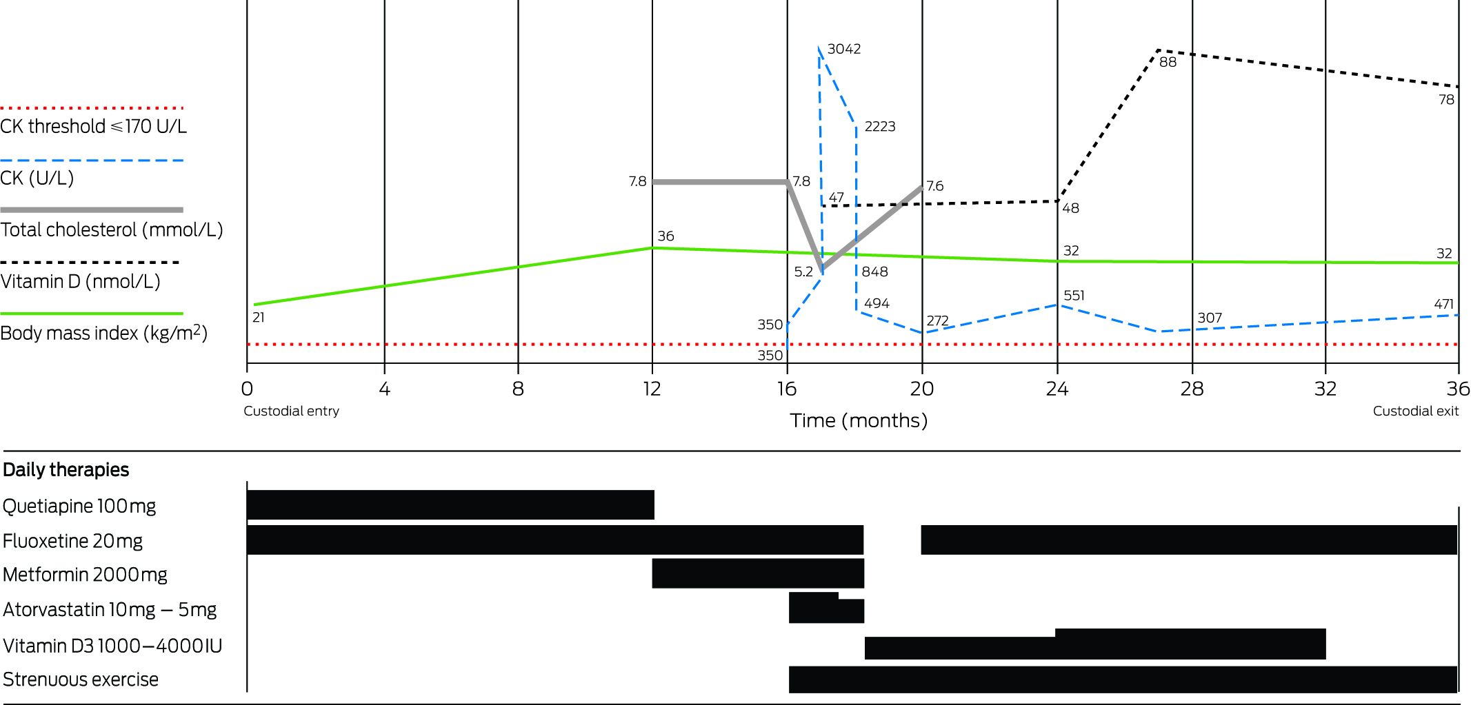
1 Process of care for patients with possible acute coronary syndrome


ECG = electrocardiography. EST =exercise stress test.  ED = emergency department. SSU = short-stay unit. * Risk classifications according to the Queensland chest pathway (Appendix) based on the 2006 NHF/CSANZ guidelines.2

2 Patient flow within the study*


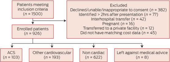
ACS = acute coronary syndromes. * According to Standards for the reporting of diagnostic accuracy studies (see http://www.stard-statement.org).

3 Baseline demographics of patients according to their final diagnosis category*

Baseline characteristics

Cardiac-ACS

Cardiac-other

Non-cardiac

No diagnosis (LAMA)


(n = 103)

(n = 193)

(n = 622)

(n = 8)

Mean age, years (range)

64.50 (33–96)

59.32 (23–97)

51.6 (19–93)

55.13 (37–66)

Women

30 (29.1%)

71 (36.8%)

258 (41.5%)

1

European ancestry

97 (94.2%)

166 (86.0%)

542 (87.1%)

7

Risk factors


Dyslipidaemia

65 (63.1%)

113 (58.5%)

280 (45.0%)

1

Diabetes

24 (23.3%)

36 (18.7%)

68 (10.9%)

1

Hypertension

64 (62.1%)

119 (61.7%)

269 (43.2%)

2

Obesity (BMI > 30)

26 (27.1%)

63 (33.2%)

221 (36.4%)

2

Smoking

33 (32.0%)

52 (26.9%)

188 (30.2%)

6

Medical history


Angina

40 (38.8%)

78 (40.4%)

111 (17.9%)

2

Coronary artery disease

43 (41.7%)

81 (42.0%)

93 (15.0%)

1

AMI

29 (28.2%)

55 (28.5%)

79 (12.7%)

1

Arrhythmia

15 (14.6%)

49 (25.4%)

38 (6.1%)

0

Congestive heart failure

11 (10.7%)

29 (15.0%)

21 (3.4%)

0

CABG surgery

14 (13.6%)

21 (10.9%)

24 (3.9%)

1

Prior angioplasty

19 (18.4%)

37 (19.2%)

51 (8.2%)

0

Stroke

10 (9.7%)

30 (15.5%)

47 (7.6%)

2

Presentation with chest pain in the past year

26 (25.2%)

79 (40.9%)

139 (22.3%)

1


ACS = acute coronary syndromes. AMI = acute myocardial infarction. BMI = body mass index. CABG = coronary artery bypass graft. LAMA = Left against medical advice. * Data are no. (%) unless otherwise specified. † Denominators vary due to missing data: 26/96, 63/190, 221/608.


4 Hospital length of stay, mortality, median and mean costs by diagnostic group

Diagnostic category

No. of patients (% of diagnostic group)

Median hospital length of stay, hours (25th–75th percentile)

Mean hospital length of stay, hours (95% CI)

No. of patients deceased at 1 year/no. in category*

Median costs (25th–75th percentile)

Mean costs (95% CI)


Cardiac-ACS

103 (100.0%)

97.6 (70.7–188.8)

187.0 (125.3–248.8)


$12 002 ($7861–$16 517)

$13 509 ($11 794–$15 223)

STEMI

22 (21.4%)

91.9 (74.1–142.9)

151.5 (95.7–207.2)

0/17

$14 643 ($12 002–$17 323)

$18 297 ($13 487–$23 107)

NSTEMI

53 (51.5%)

101.3 (70.4–173.0)

215.5 (105.0–325.9)

5/35

$11 705 ($8198–$15 196)

$12 829 ($11 028–$14 629)

UAP

28 (27.2%)

96.8 (44.2–191.2)

161.1 (68.0–254.3)

0/23

$8311 ($4728–$17 860)

$11 033 ($8000–$14 067)

Cardiac-other

193 (100.0%)

52.1 (23.3–123.8)

92.2 (75.5–108.8)


$4826 ($2020–$9297)

$7283 ($6152–$8415)

Coronary vasospasm

2 (1.0%)

0/2


Stable CAD

50 (25.9%)

47.2 (22.0–97.1)

81.4 (51.5–111.2)

2/39


Pericarditis

27 (14%)

28.2 (10.8–65.9)

46.5 (29.6–63.4)

0/20


Atrial fibrillation

33 (17.1%)

53.7 (30.5–116.5)

86.0 (53.7–118.2)

2/30


High-grade atrioventricular block

4 (2.1%)

198.0 (138.2–281.4)

209.8 (135.6–284.0)

0/2


Other arrhythmias

16 (8.3%)

38.9 (10.5–155.8)

86.5 (48.3–124.7)

0/12


Heart failure

15 (7.8%)

196.3 (142.4–338.7)

278.6 (161.3–395.9)

1/9


Cardiomyopathy

3 (1.6%)

0/1


Valve disease

7 (3.6%)

30.8 (7.9–76.6)

48.5 (16.4–80.6)

0/4


Hypertension

8 (4.1%)

41.4 (17.5–77.8)

53.5 (22.4–84.6)

0/6


Syncope/presyncope

10 (5.2%)

76.1 (9.3–122.4)

75.0 (36.9–113.2)

0/8


Pulmonary embolism

6 (3.1%)

112.7 (101.0–172.8)

140.0 (65.9–214.0)

0/5


Other cardiac problems

12 (6.2%)

32.0 (10.8–57.9)

39.7 (21.3–58.1)

0/9


Non-cardiac

622 (100.0%)

24.8 (10.0–34.4)

44.2 (36.8–51.6)


$1917 ($1392–$3479)

$3331 ($2976–$3685)

No disease/chest pain not otherwise specified

444 (71.4%)

23.7 (9.8–30.6)

37.7 (29.1–46.3)

2/339


Gastrointestinal


GOR/dyspepsia

35 (5.6%)

25.2 (10.2–48.8)

38.3 (25.9–50.7)

0/24


Other gastrointestinal

12 (1.9%)

33.0 (11.2–157.9)

85.7 (33.1–138.3)

0/8


Liver disease

2 (0.3%)

0/1


Respiratory


Asthma/COAD

5 (0.8%)

30.7 (24.4–193.2)

100.3 (11.8–188.9)

0/2


Respiratory Infection

22 (3.5%)

25.0 (8.8–51.8)

72.6 (27.8–117.4)

1/16


Other respiratory

5 (0.8%)

105.9 (32.1–143.9)

87.0 (35.8–138.3)

1/3


Non-specified musculoskeletal pain

64 (10.3%)

22.7 (9.4–31.0)

35.1 (23.3–47.0)

0/49


Infection (non-respiratory)

8 (1.3%)

74.9 (39.3–133.5)

127.8 (25.1–230.5)

0/8


Neurovascular


Stroke

2 (0.3%)

0/0


Neuropathic pain

8 (1.3%)

43.9 (16.7–126.6)

78.5 (21.03–136.0)

0/7


Cancers

3 (0.5%)

2/2


Other non-cardiac

6 (1.0%)

41.5 (10.5–58.0)

58.8 (7.9–109.8)

1/5


Anxiety/depression

6 (1.0%)

20.4 (8.6–49.7)

36.2 (5.5–66.8)

0/4


Left against medical advice

8 (100.0%)

14.1 (6.0–24.0)

15.0 (8.5–21.5)

0/3

$1366 ($1007–$2027)

$1585 ($1128–$2042)


ACS = acute coronary syndromes. CAD = coronary artery disease. COAD = chronic obstructive airways disease. GOR = gastro-oesophageal reflux. NSTEMI = non- ST-segment elevation myocardial infarction. STEMI = ST-segment elevation myocardial infarction. UAP = unstable angina pectoris. * Mortality data are for the 693 patients who consented to 1-year follow-up.


5 Kaplan–Meier survival curves*


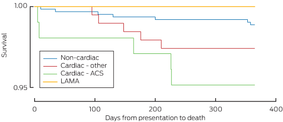
ACS = acute coronary syndromes. LAMA = left against medical advice. * The omnibus log-rank test provided some support for differences in the survival function across diagnostic groups (P = 0.05). The rate of death was slightly higher for patients with cardiac-ACS diagnoses compared to those in the non-cardiac group (P = 0.01) but did not differ in the cardiac-ACS versus cardiac-other (P = 0.31) or non-cardiac versus cardiac-other groups (P = 0.14).

6 Median and mean cost and length of stay for ACS events and non-ACS-events among patients presenting to the emergency department with chest pain, stratified by NHF/CSANZ risk group

Category and event type

No. of patients (%)

Median cost (25th–75th percentile)

Mean cost per patient (95% CI)

Median length of stay, hours (25th–75th percentile)

Mean length of stay, hours (95% CI)


All categories

926 (100.0%)

$2443 ($1458–$6778)

$5272 ($4835–$5708)

27.8 (10.5–75.0)

69.8 (59.9–79.7)

ACS

103 (11.1%)

$12 003 ($7861–$16 517)

$13 509 ($11 794–$15 223)

97.6 (70.7–188.8)

187.0 (125.3–248.8)

Non-ACS

823 (88.9%)

$2127 ($1406–$5027)

$4241 ($3843–$4638)

26.4 (10.2–52.5)

55.13 (48.0–62.3)

Low risk

9 (1.0%)

$1530 ($1298–$3050)

$2040 ($1306–$2774)

11.5 (11.3–29.6)

20.4 (11.2–29.7)

ACS

0 (0)

$0 ($0)

$0 ($0)

0.0 (0.0)

0.0 (0.0)

Non-ACS

9 (1.0%)

$1530 ($1298–$3050)

$2040 ($1306–$2774)

11.5 (11.3–29.6)

20.4 (11.2–29.7)

Intermediate risk

580 (62.6%)

$1849 ($1376–$3570)

$3304 ($2963–$3644)

24.5 (9.9–34.6)

42.4 (34.6–50.2)

ACS

11 (1.9%)

$8082 ($7174–$18 554)

$12 169 ($6803–$17 536)

99.1 (51.5–222.0)

148.4 (66.1–230.8.8)

Non-ACS

569 (61.4%)

$1831 ($1372–$3338)

$3132 ($2844–$3420)

23.9 (9.9–32.8)

40.3 (33.2–47.5)

High risk

329 (35.5%)

$6452 ($2650–$11 829)

$8919 ($7971–$9867)

72.3 (27.5–142.4)

120.8 (97.8–142.9)

ACS

92 (9.9%)

$12 357 ($8216–$16 353)

$13 669 ($11 857–$15 481)

97.2 (71.0–180.9)

191.6 (116.7–266.6)

Non-ACS

237 (25.6%)

$4380 ($2151–$8812)

$7075 ($6013–$8137)

50.4 (22.9–125.0)

93.3 (77.7–108.9)

Left against medical advice

8 (0.9%)

$1366 ($1007–$2027)

$1585 ($1128–$2042)

14.1 (6.0–24.0)

15.0 (8.5–21.5)

ACS

0 (0)

$0 ($0)

$0 ($0)

0.0 (0.0)

0 (0.0)

Non-ACS

8 (0.9%)

$1366 ($1007–$2027)

$1585 ($1128–$2042)

14.1 (6.0–24.0)

15.0 (8.5–21.5)


ACS = acute coronary syndromes. NHF = National Heart Foundation. CSANZ = Cardiac Society of Australia and New Zealand. * This group was added to the original NHF risk groups for completion.


Severe carbon monoxide poisoning from waterpipe smoking: a public health concern

We believe this is the first Australian report of severe carbon monoxide poisoning caused by waterpipe use. Carbon monoxide poisoning causes neurological dysfunction and myocardial toxicity, effects that can be irreversible. Despite a widespread misconception that waterpipes are safer than cigarettes, the recognised risks of tobacco products also apply to waterpipe use.

Clinical record

A 20-year-old woman was brought by ambulance to the emergency department of a district hospital after an episode of presyncope. She described symptoms of severe light-headedness, mild headache and nausea, but denied experiencing weakness or sensory disturbances. Seizure activity was not reported. She had used a waterpipe for 1 hour before the onset of symptoms. Although this was her first hospital presentation with these symptoms, she had frequently experienced queasiness and light-headedness after using a waterpipe, which she did on most days of the week, each session lasting about 45 to 60 minutes. She denied depression, suicidal ideation, and recent alcohol or drug use.

The patient appeared lethargic. Her vital signs were within normal limits: blood pressure, 115/70 mmHg; pulse rate, 75 beats/min; temperature, 36.7°C; Spo2, 98%; and Glasgow Coma Scale score, 15. Her pupils were dilated (5 mm), equal and reactive to light. Her cranial nerve, upper limb and lower limb functions were normal. There was no nystagmus, nor did she have any cerebellar symptoms. Her chest was clear and her heart sounds were normal, with no murmurs. Full blood count and biochemistry and urinalysis data were normal. Severe carbon monoxide (CO) poisoning was diagnosed on the basis of the initial venous and arterial blood gas levels on room air (Box 1). The results of a urinary drug screen and blood alcohol testing were negative.

High-flow oxygen was administered to the patient, and she was transferred to our institution. The results of serial troponin T assessment 3 and 14 hours after presentation were negative (< 3 ng/L). Her initial electrocardiogram (ECG) showed a normal sinus rhythm. T wave inversion was noted in the anteroseptal leads 5 hours after presentation, but had normalised 9 hours after presentation (Appendix 2). Transthoracic echocardiography showed that left and right ventricular size and systolic function were normal, with normal left ventricular wall thickness, no regional wall motion abnormalities, and normal valve function. Computed tomography coronary angiography was performed because of dynamic T wave changes referable to the left anterior descending artery, but showed that the coronary arteries were normal. The patient was observed for 24 hours and discharged the next day, and was advised to abstain from waterpipe use in the future.

Discussion

We believe this is the first report in Australia of severe CO poisoning caused by waterpipe use. A waterpipe, also known as a hookah, narghile, shisha or goza, is an apparatus for smoking organic material (often flavoured or non-flavoured tobacco, although non-tobacco herbal preparations are also available). Smoke formed by the heating of organic material is siphoned through water before being inhaled. Variants of the waterpipe have been used in the Middle East, Africa and Asia since the 16th century, and were also popular in Victorian England. Alice encountered the Caterpillar smoking a hookah on a mushroom in Alice’s adventures in Wonderland (Box 2).1 The portrayal by Lewis Carroll of the lethargic, irritable and forgetful Caterpillar in this classic children’s tale afforded an astonishingly accurate depiction of the dangerous physiological effects of waterpipe use.

Although longstanding public health initiatives have reduced overall rates of cigarette smoking, waterpipe use, especially by young adults, poses an important public health threat. Despite concerns about the rapid increase in waterpipe use across the world (prevalence of 6%–34% among Middle Eastern adolescents and 5%–17% among American adolescents),2 its prevalence in Australia is unclear. The only available study, a survey of 1102 Arabic-speaking residents in south-west Sydney, found that 11.4% reported current use.3

Two main factors have contributed to its popularity: the pleasant experience of social pipe smoking, and the ongoing misconception that the passage of smoke through water before inhalation filters out most toxic substances. This erroneous perception of reduced harm when compared with cigarette smoking is reinforced by the availability of herbal (tobacco-free) preparations. Waterpipe users, however, typically inhale greater amounts of smoke than cigarette smokers. One session of waterpipe use exposes the user to the same amount of smoke as 50–100 cigarettes.4,5 As a result, waterpipe users are subject to similar risks of cancer, heart disease, respiratory illness, pregnancy complications and other smoking-related health problems.

CO poisoning is also a genuine risk for users, with CO exposure during waterpipe use being almost nine times higher than for cigarette smoking, and peak carboxyhaemoglobin levels three times higher.5 Passive exposure to CO also occurs in hookah lounges, and should be of concern to pregnant women, as fetuses are particularly sensitive to low levels of CO. During waterpipe use, CO is formed from incomplete combustion of charcoal and organic material owing to the reduced oxygen content within the waterpipe apparatus and the relatively low heating temperature. CO binds to haemoglobin with high affinity (more than 200 times that of oxygen) to form carboxyhaemoglobin, which shifts the oxyhaemoglobin dissociation curve markedly to the left, impairing oxygen delivery throughout the body.6 Due to the high affinity of CO for haemoglobin, significant carboxyhaemoglobinaemia can develop during even relatively low-level CO exposure. This reduced oxygen-carrying capacity is not apparent, however, in the reported values for oxygen saturation of haemoglobin (Appendix 1, as they are based on the haemoglobin fraction that can carry oxygen; the carboxyhaemoglobin fraction is thus excluded from the calculation of oxygen saturation.

CO poisoning causes myocardial toxicity. Transient ECG abnormalities, suggestive of myocardial ischaemia, are relatively common, and are evident in 30% of patients with moderate to severe CO poisoning.7 This occurs even in the absence of significant coronary artery disease, and probably reflects impaired cellular respiration. CO also acts as an important signalling molecule, capable of altering the function of various cellular ion channels important to the cardiac action potential, as well as of proteins that regulate intracellular calcium levels in cardiomyocytes.8 CO binding to myoglobin may also account for impaired left ventricular systolic function,9 with the degree of dysfunction correlated with carboxyhaemoglobin levels and duration of CO exposure.10 Left ventricular dysfunction is usually transient, with recovery occurring within 24 hours of CO poisoning. This may explain the normal left ventricular function seen in this patient’s ECG, performed 19 hours after presentation. Myocardial fibrosis can develop, however, after a single episode of severe CO poisoning.11 Neurological symptoms of CO toxicity include headache, dizziness, weakness, nausea, confusion and anterograde amnesia. In the longer term, CO poisoning can also cause lesions in the basal ganglia. Treatment of CO poisoning is largely supportive and involves administration of high-flow oxygen to hasten the elimination of CO. In severe cases, and where facilities are available, hyperbaric oxygen therapy can be considered.

The World Health Organization Framework Convention on Tobacco Control (2005) recommended better regulation of waterpipe use, including health warnings on packaging, prohibiting claims of harm reduction and safety, and banning it in public places, similar to measures currently in place for cigarette smoking.4 In many countries, including Australia, waterpipe smoking has previously escaped the strict regulation imposed on other tobacco products, particularly as tobacco is not always the major constituent of hookah preparations. Although harm minimisation techniques, such as improving ventilation in hookah lounges and limiting the duration of smoking sessions, may reduce the risk of severe CO poisoning, public health measures should focus on encouraging changes to existing legislation, and on discouraging waterpipe use, especially among young adults, by highlighting the health risks.

1 Initial venous and arterial blood gas levels of the patient while breathing room air after using a waterpipe for 1 hour*


Venous blood

(at presentation)


Arterial blood

(20 minutes after presentation)


Parameter

Level

RI

Level

RI


pH

7.37

7.32–7.43

7.43

7.35–7.45

po2 (mmHg)

19

25–40

82.3

75–105

pco2 (mmHg)

53

41–50

43

32–34

Oxyhaemoglobin

35.3%

25%–40%

73.6%

95%–99%

Carboxyhaemoglobin

25.4%

0–1.5%

23.9%

0–1.5%


RI = reference interval. * Further details in Appendix 1.

2 Alice meets the Caterpillar


From Alice’s adventures in Wonderland by Lewis Carroll. Illustration by Sir John Tenniel, 1865.

A systematic approach to chronic heart failure care: a consensus statement

To the Editor: We commend Page and colleagues for their comprehensive statement on chronic heart failure (CHF) care.1 However, we wish to highlight an important omission to the discussion regarding telemonitoring.

There is Level 1 evidence for the use of telemonitoring as part of CHF management.2 Currently, telehealth is a major strategic item on the agenda of the federal Department of Health.3 Telehealth and telemonitoring can be the best options for Australians with CHF who do not have access to multidisciplinary or specialist heart failure care for reasons of carer responsibility, geography, socioeconomics, cultural and linguistic diversity, frailty, immobility or complexity of illness. To provide truly consumer-focused CHF care, options need to be available to all patients equally.4

We believe an opportunity has been missed in this consensus statement, particularly in regard to the poor access to multidisciplinary and specialist CHF care services in Australia.5 Although the evidence base for the use of telehealth in CHF care and management is still evolving, we now have a significant body of evidence demonstrating effectiveness in improving CHF outcomes.2

We agree with the authors that future research should consider the role of telehealth. However, we recommend that this research should be in the form of translation, implementation and integration of telehealth-based CHF care within the Australian health care system, to overcome current unresolved inequities.

A systematic approach to chronic heart failure care: a consensus statement

In reply: I and my coauthors on the consensus statement1 support these comments. At the time of writing, we acknowledged that further research to build the evidence base on the benefits and application of telehealth for patients with chronic heart failure would be welcomed. This is particularly so, given its potential benefit in reducing cardiovascular health inequities.

Statin-associated myotoxicity in an incarcerated Indigenous youth — the perfect storm

Clinical record

A previously healthy 18-year-old dark-skinned Indigenous man was incarcerated in a juvenile detention centre in New South Wales for 3.5 years from 2010 to 2013. Juvenile detention limits outdoor activity and, consequently, exposure to sunlight. Young people are confined indoors for schooling and other programs, with additional periods of cell lockdowns to accommodate detainee movements and staff handovers. Periods outdoors involve bursts of strenuous physical activity, mostly team sports or swimming. Further, detention centre policy requires young people to wear T-shirts and hats, and to use sun protection factor 30+ sunscreen when outdoors.

On entering custody, the patient’s weight was 65 kg, with a healthy body mass index (BMI) of 21 kg/m2 (reference interval [RI], 18.5–24.9 kg/m2). Full blood count, urea, electrolyte and creatinine levels and liver function test results were normal, and a blood-borne virus screen returned a negative result. He had a strong family history of type 2 diabetes in his mother and maternal grandmother, and, reportedly, of hypercholesterolaemia and early cardiovascular death in his father and paternal grandfather.

Seven months after incarceration, the man developed auditory and visual hallucinations and was noted to be withdrawn and depressed, with long periods spent resting in his cell owing to fatigue. He was commenced on the antipsychotic quetiapine 150 mg at night and the antidepressant fluoxetine 20 mg in the morning. At commencement of these medications, his weight was 89 kg (BMI, 29 kg/m2). Baseline pathology tests were not repeated at this time.

Six months after commencement of psychotropic medications, his weight had increased a further 25 kg to 114 kg and he was morbidly obese (BMI, 36 kg/m2), with phenotypes of metabolic syndrome including central obesity (waist circumference, 108 cm [RI, < 94 cm]), hyperlipidaemia (total cholesterol, 7.8 mmol/L [RI, < 5.5 mmol/L]; low-density lipoprotein cholesterol, 4.9 mmol/L [RI, < 4.0 mmol/L]; high-density lipoprotein cholesterol, 0.8 mmol/L [RI, > 1.0 mmol/L]), elevated triglyceride level (2.28 mmol/L [RI, < 2.0 mmol/L]), and fatty liver disease (γ-glutamyl transferase, 83 U/L [RI, 0–60 U/L]; alkaline phosphatase, 208 U/L [RI, 30–110 U/L]; alanine transaminase, 72 U/L [RI, 0–55 U/L]; aspartate transaminase, 46 U/L [RI, 0–45 U/L]) (Figure). Blood pressure and thyroid-stimulating hormone levels were within normal limits. With concerns about his obesity and metabolic derangements, quetiapine was ceased. He received counselling for dietary restriction (portion control, low saturated fat diet, reduction of energy-dense snacks) and, in particular, was encouraged to avoid the additional bread, butter and sugary drinks that are available to supplement meals. Increased physical activity was encouraged. An off-label trial of metformin was commenced, given the evidence for weight benefits in antipsychotic recipients,1 and increased to 1 g twice daily over the following 4 weeks.

Three months later, the patient’s fasting lipid levels remained similarly elevated despite lifestyle changes, and he agreed to trial atorvastatin 10 mg daily. He was also permitted to take recreational leave from the centre and commenced thrice-weekly training with the local football club.

Three weeks after commencing atorvastatin, the patient complained of worsening fatigue but denied having muscle tenderness, myalgia or cramping. Creatine kinase (CK) levels were normal at atorvastatin commencement, but had risen to 350 U/L (RI, < 170 U/L). Atorvastatin dosage was reduced to 5 mg in the morning, metformin was continued and fluoxetine was ceased.

Serial changes in the patient’s CK levels are shown in the Figure. Five weeks after atorvastatin commencement, lipid levels had improved but CK levels continued to rise and all medications were ceased. There were concerns regarding rhabdomyolysis, but urinalysis results, estimated glomerular filtration rate and renal function remained normal. His physical symptoms remained unchanged. The patient was encouraged to rest and drink plenty of water. He continued to play competition football. CK levels continued to rise, peaking at 3042 U/L.

Serum 25-hydroxyvitamin D levels were found to be low, and he was treated with cholecalciferol (vitamin D3) 1000 IU daily, increasing temporarily to 4000 IU daily after endocrinologist consultation. Serial CK and 25-hydroxyvitamin D levels showed slow improvement initially, with substantial improvements contemporaneous with aggressive vitamin D supplementation (Figure). The patient continued with lifestyle strategies and (in concert with cessation of psychotropic medications) lost 10 kg in weight, but lipid levels remained elevated. Fluoxetine was recommenced by the treating psychiatrist at 20 months because of concerns regarding the patient’s mood.

At the conclusion of his sentence, the patient was released from custody and referred to the local Aboriginal Medical Service for continuing management of his hypercholesterolaemia, myositis, and metabolic and mental health problems.

Indigenous Australians have a reduced life expectancy of up to 20 years compared with non-Indigenous Australians and, by 40 years of age, are 10 times more likely to suffer premature cardiac-related death.2 In recognition of this, the Indigenous Chronic Disease Package (through Closing the Gap initiatives) encourages the use of statins, recommending treatment at lower lipid thresholds.3

There is evidence that Indigenous populations may be at higher risk of statin-related myopathy owing to a higher risk of vitamin D deficiency,4 higher rates of human T-cell lymphotropic virus type 1 infections causing polymyositis5 and, possibly, genetic susceptibility to statin-associated myotoxic effects (the SLCO1B1 gene prevalent in other indigenous populations6). Other risk factors predisposing our patient to statin-related myopathy were his age, strenuous exercise, mild hepatic dysfunction and concomitant use of fluoxetine (a CYP3A4 inhibitor).4,7,8 As CK elevation persisted after atorvastatin cessation, the differential diagnosis was necrotising autoimmune myopathy, previously described in indigenous patients with persistent myopathy.9 The recommencement of fluoxetine at 20 months may also have perpetuated the elevated CK level.

Almost 12 000 people are incarcerated in NSW, with a quarter being Indigenous Australians and at greatest risk of vitamin D deficiency.10 This is the first report of statin-related myopathy in an Indigenous adolescent or an incarcerated person. It is worth noting by other clinicians who work with Indigenous and incarcerated groups that the risk factors for this patient’s “perfect storm” were not unusual — metabolic syndrome, vitamin D deficiency, and use of statins in the context of mental illness and concomitant psychotropic medication use.1,2

This report highlights the need for monitoring of vitamin D levels and supplementation (with an argument for easier access to injectable vitamin D in this group), with pre-statin counselling, particularly for those at high risk of statin-related myopathy — Indigenous Australians, youths, females, and those serving lengthy custodial sentences.

This case also highlights the detrimental effects of antipsychotics on weight and metabolic risk. An international declaration supporting young people with psychosis11 has delineated the obligations of health care providers to prevent weight gain and metabolic complications that contribute to the 25-year shortfall in life expectancy in people with severe mental illness.

In addition to these learning points, there are the obvious problems of the unmet health needs and human tragedy in this vulnerable patient group: a baseline high metabolic risk associated with Aboriginality and family medical history, the constraints of incarceration exacerbating the risk of vitamin D deficiency, and a doubling of weight resulting in rapid-onset obesity secondary to antipsychotic use. Co-prescription of lifestyle interventions at the time of commencing antipsychotic therapy is essential. In addition, metformin has proven efficacy in abrogating weight gain following antipsychotic commencement, with its use encouraged in patients who make clinically significant weight gains.1

Lessons from practice

  • Indigenous Australians, young people and those serving lengthy custodial sentences are at risk of low vitamin D levels and statin-associated myopathy.
  • Aggressive vitamin D supplementation may be required to normalise levels before commencing statin therapy.
  • For all people receiving antipsychotics, lifestyle intervention should be co-prescribed, and weight gain should be monitored and actively prevented.
  • Metformin has proven benefit for weight loss in patients who significantly gain weight on antipsychotic treatment.

Creatine kinase (CK) and other markers according to time and medications.

Jack

Highly commended — Practising and retired doctors category

When I first met him, Jack was in a hospital bed, tired with cool feet. A small hand-held AM radio was at his bedside softly playing golden oldies, though the radio itself did not appear old. Even in bed, his heavy frame, built to last, showed how mighty a force he had been in his prime. His chief complaint was progressively worsening fatigue, which he blamed on his age. But his physical examination suggested otherwise: the soft, whining systolic murmur; the left ventricular heave; the tell-tale carotid upstroke that felt as though it was grunting against the dead weight of an old refrigerator. All these signs bore evidence of his diagnosis: calcific aortic stenosis. His was a stolid, stubborn valve with hinges rusted quiet, though he was, as he said, old.

Still, he was mentally clear. He spoke about symptoms present or absent, about his medical history, his life and his priorities. His wife of 63 years had dementia and he was her primary carer. He described his daily routine in detail, attending her needs and the needs of the house he had built with his bare hands, making every single brick himself when he was still young and strong. He said the brick-making helped him get on with his life after the war; helped him pick up the pieces, to emotionally repair and nurse wounds without much fuss.

“Was it worth the trouble?” I asked. “Trouble?” he asked dryly, “what trouble?”

My job for him was brief: I confirmed the diagnosis and spoke to the interventional cardiologist, who was happy to help. The risks of the procedure were made clear, as well as the benefits — this was purely to make him feel better and would not add a single day to his life. Jack listened carefully, thanking everyone for their bother and accepted the stated risks in order to continue helping his wife. He had no other reasons. With no further need for me, I signed off on his case.

The endovascular repair of his valve, an expensive exchange for quality of days, went off without any technical glitch; but, of course, things are never that simple. There are always risks not included in the brochure. Neutropenia developed, a complication of a pre-existing problem, followed by nosocomial pneumonia. A month later, I slipped past him in the hallway working with a physiotherapist. He looked frail, feeble and destined to fail. I thought he was finished.

The next and last time I heard Jack speak was at the ANZAC Day service 6 months later, a service to pay respects to war veterans alive and dead. Australian and New Zealand Army Corps. Courage, loyalty, trust and mateship defined his era of soldier, qualities that linger long after a war is over.

The service was attended by more people than should be available in a town of only two hundred: old people who know the unromantic sounds of war — sounds base and arbitrary — as well as young and middle-aged people who do not. The eulogy was read as wind blew through the maroon and burnt orange leaves falling from the pear trees along the main street. The crowd of a hundred people circled the dark grey polished granite memorial and listened to the reading as sparrows flitted playfully and a crow called rudely in the distance in tapering tones. In the breeze, I thought I could hear the mortars drill through the air and explode without malice in no-man’s land; the machine guns firing warning shots at Gallipoli; bomber engines droning over the North Sea. Just faintly, the mundane sounds of war mingled with those of the day. On the obelisk were the names of the fallen, engraved and painted with gold, as well as those who had returned; written across the bottom “For Our Clarendon Boys who died in the Great War 1914–1918”, with addenda as other wars and other names were added. And they were, indeed, boys’ names, mostly.

I recognised Jack in the crowd only moments before he spoke. He was much thinner than when I had last seen him in the hospital corridor. He sat on the seat function of his four-wheeled walker with brakes set, elbows resting on the handlebars and eyes staring downward without care. He blinked slowly, listened carefully, and waited. When his turn came, he stood with assistance from his son under one arm. Without reading, he spoke the words with the slow, crackling voice of a 93-year-old man, but deliberately as a man who knew his time was short:

They shall not grow old as we that are left grow old;
Age shall not weary them, nor the years condemn.
At the going down of the sun and in the morning
We will remember them.

He spoke the words on that clear autumn day as birds played in the trees, the same sparrows that gave men hope when things fell quiet on the Western Front.

He spoke the words without haste, as an old man can speak without tears about things that break hearts and make mothers weep.

He spoke the words by heart, from the same stubborn loyalty to remember so often felt during the quiet times of a day when a person is alone, such as when laying the bricks of a house, or driving to work on a rainy day, or sweeping the kitchen floor after kids are safely in bed at night. A loyalty to remember mates and the sound of their laughter, though the sounds of war always intruded without invitation, a drizzling rain of quiet grief from wounds that never fully heal. And wounds Jack would not discuss.

Those words, the last I heard Jack speak, served as the benediction of the memorial service and the backdrop for the raising of the Australian flag. An honoured boy hoisted it to the top, the flag blowing and popping in the wind, while the squeaky pulleys turned. The rope hit the flagpole irregularly with a mechanical baritone pitch, “thunk, thu-thunk”, the same irregular cadence of a malfunctioning machine of war, hobbled by enemy fire. A lone bugler played the “Last Post”. There was no other sound but the birds. A few other official words were spoken, awkwardly juxtaposed with the sacred, then everyone adjourned across the street to the town hall for morning tea.

Jack was tired and was taken back to the brick home he had built and to the care of his wife. A few weeks later, he had a fall and died.

Was his endovascular valve repair worth the trouble? The medical economist would say no, label it a “low-value intervention” or even a waste, and state that the expense of this procedure could have been spent elsewhere — vaccines; education to prevent teenage pregnancy; or public transportation safety ads. This calculation would not, however, factor in the inspiring words that filled the gathering with reverence that ANZAC Day when Jack spoke, just as the pneumonia, deconditioning and the prolonged hospital stay could only be factored in with hindsight.

Was it worth the trouble? Would I have paid cash out of my own pocket to fix that crusty, stubborn valve of his just to see Jack stand unconquered and hear him say those last words against the backdrop of a fluttering flag and a lone bugler? Words about men fallen many years prior, words charging me not to forget them, or what they had done, ever. And words that linger still.

Was it worth the trouble?” I asked him.

“Trouble?” he asked dryly, “what trouble?”


The MJA Dr Eric Dark Creative Writing Competition was judged by Leah Kaminsky, MJA Deputy Editor, Poetry and Fiction.

An unusual case of implantable cardioverter-defibrillator inhibition

Clinical record

A 34-year-old woman with an implantable cardioverter-defibrillator (ICD) for mitral valve prolapse and non-sustained ventricular tachycardia presented for clinical and device review on a background of intermittent palpitations associated with syncope since the age of 22 years.

At that time, an echocardiogram showed mild left ventricular dysfunction and moderate mitral regurgitation. After careful consideration of the risks and benefits, the patient underwent ICD implantation for primary prevention. A Maximo VR 7232 single chamber ICD (Medtronic) was implanted with a Sprint Quattro Secure 6947 active fixation lead (Medtronic) positioned in the right ventricular apex, and defibrillation threshold testing performed after implantation was successful. Device programming was set to ventricular pacing and sensing mode (VVI mode) with a lower rate of 30 beats/min. Therapy settings were programmed with a ventricular tachycardia monitor zone of 200–250 beats/min and a ventricular fibrillation zone of > 250 beats/min with 35 J shock therapy. On follow-up, device function proved to be normal.

At review, the patient noted that an alarm sound was being emitted from her ICD. She had been well otherwise, with no record of arrhythmic events seen on device interrogation, and no device alerts or alarms. She described a continuous alarm sound (suggesting to us that the device had switched into magnet mode with all tachyarrhythmia detections and therapies turned off) that usually occurred when she was at home. She denied being situated close to any electrical devices or placing her mobile phone near her ICD. Activation of the alarm mainly occurred at night and disappeared when she was having a shower or bath. Further questioning revealed she had recently purchased a new maternity bra, which she predominantly wore at home — in particular, when she was sleeping. She was asked to bring the bra for us to look at and to avoid wearing it in the meantime.

On close examination of the bra, we noted a small magnetic clip (about 1 cm x 1 cm) located on each side of the anterior-facing straps, for ease of breastfeeding (Figure). When the patient wore the bra, one clip was located directly over the ICD in the left subclavicular region and did not vary when she moved. As the magnetic clip was positioned over the ICD, the alarm sounded. The alarm ceased when the clip was moved from this position.

The patient avoided using this bra again, and no further episodes of alarm activation were noted.

Electromagnetic interference is becoming a more frequent hazard for patients with an ICD.13 This potential threat usually arises from a relatively obvious or clear source.4,5

The case we report particularly highlights the danger of small powerful magnets that are now used in a wide variety of objects, including clothing.6 It illustrates the inherent danger when a magnet in clothing or underclothing is held firmly in place over an ICD, which should be a cause for concern for the many manufacturers of clothing items that have these for decorative or functional purposes.

Magnetic fields of > 10 gauss appear to be sufficient to inhibit the device.4 Our tests showed that the magnet in question exhibited field strengths of up to 800 gauss (Appendix 1, Appendix 2).

Of particular concern is the lack of manufacturer warnings about magnets in clothing for people who have an implanted cardiac device. Both the underwear manufacturer and Medtronic have been alerted to the potential hazard of permanent electromagnetic interference from such a magnetic clip. Discussion is underway regarding the addition of warning labels to their products.

Lessons from practice

  • Electromagnetic interference (EMI) inhibits programmed delivery of life-saving therapy by implantable cardioverter-defibrillators (ICDs). This potential threat usually arises from a relatively obvious or clear source.4,5
  • Our report highlights the dangers of using magnets in clothing, as they are a hidden source of EMI.
  • Patients with ICDs and their treating medical practitioners need to remain vigilant for less obvious sources of EMI.

The magnetic clip on the patient’s maternity bra (size comparison with an Australian $1 coin).

Better prevention and management of heart failure in Aboriginal Australians

Correction

Incorrect label in graph: In Better prevention and management of heart failure in Aboriginal Australians” in the 16 February 2015 issue of the Journal (Med J Aust 2015; 202: 116-117), there was an error in the graph in the Box (page 117). The y-axis label should read “Rate/100 000 person-years”.

[Comment] Coronary intervention: radial artery access comes of age

It has taken a quarter of a century for the level of evidence supporting radial artery access, rather than femoral access, for cardiac intervention to reach that of common pharmaceutical interventions, such as antiplatelet therapy in acute coronary syndrome.1,2 Historically, small trials and meta-analyses seemed to suggest that radial compared with femoral access reduced bleeding complications related to access site.3 However, early uptake of the radial technique (ie, during 1993–2003) among interventional cardiologists was slow for several reasons.